Embed Size (px)
Citation preview

Hindawi Publishing CorporationCase Reports in Dermatological MedicineVolume 2013, Article ID 489618, 3 pageshttp://dx.doi.org/10.1155/2013/489618
Case ReportSuccessful Treatment of Localized Pemphigus Foliaceus withTopical Pimecrolimus
G. Tyros,1 K. Kalapothakou,1 E. Christofidou,1,2 A. Kanelleas,1 and P. G. Stavropoulos1
1 1st Department of Dermatology and Venereology, “A. Sygros” Hospital for Skin and Venereal Diseases,University of Athens School of Medicine, 5 Dragoumi Street, 16121 Athens, Greece
2Histopathology Department, “A. Sygros” Hospital for Skin and Venereal Diseases, 5 Dragoumi Street,16121 Athens, Greece
Correspondence should be addressed to G. Tyros; [email protected]
Received 24 June 2013; Accepted 17 August 2013
Academic Editors: T. Berger and M. Ramos-e-Silva
Copyright © 2013 G. Tyros et al.This is an open access article distributed under the Creative Commons Attribution License, whichpermits unrestricted use, distribution, and reproduction in any medium, provided the original work is properly cited.
We report the case of successful treatment of a 79-year-old male patient with recurrent pemphigus foliaceus with pimecrolimuscream 1% once daily for 40 days. The patient initially presented with localized lesions on the scalp and nose area and was treatedwith systemic corticosteroids. At his fourth relapse within a period of 16 months, he refused any systemic treatment. Pimecrolimuscream was suggested to him as an alternative option.
1. Introduction
Pemphigus foliaceus (PF) is a chronic autoimmune blisteringdisease mainly affecting the cornified skin of the face andupper torso, such as the presternal and interscapular regions,rather than the lower torso or the scalp. PF, as well as pemphi-gus vulgaris (PV), is characterized by the loss of subcornealkeratinocyte cell adhesion. The latter is clinically expressedby the formation of fragile vesicles which rupture easily,leaving behind erosions. In PF, pathogenic immunoglobulinG (IgG) targets the desmosome cadherin desmoglein1, a160KDa, calcium-dependent, transmembrane glycoproteinthat plays an important role in cell-to-cell adhesion of themost differentiated epidermal epithelia.
Most cases of PF are treated with systemic glucocor-ticosteroids with or without immunosuppressive therapy,although some mild cases can respond well to topical glu-cocorticosteroids alone [1]. We report a case of PF withrecurrent localized lesions at the face and scalp area, whichwas successfully treated with topical pimecrolimus.
2. Case Report
We reviewed a 79-year-old patient in the outpatient depart-ment of our hospital. He presented with scalp erosions which
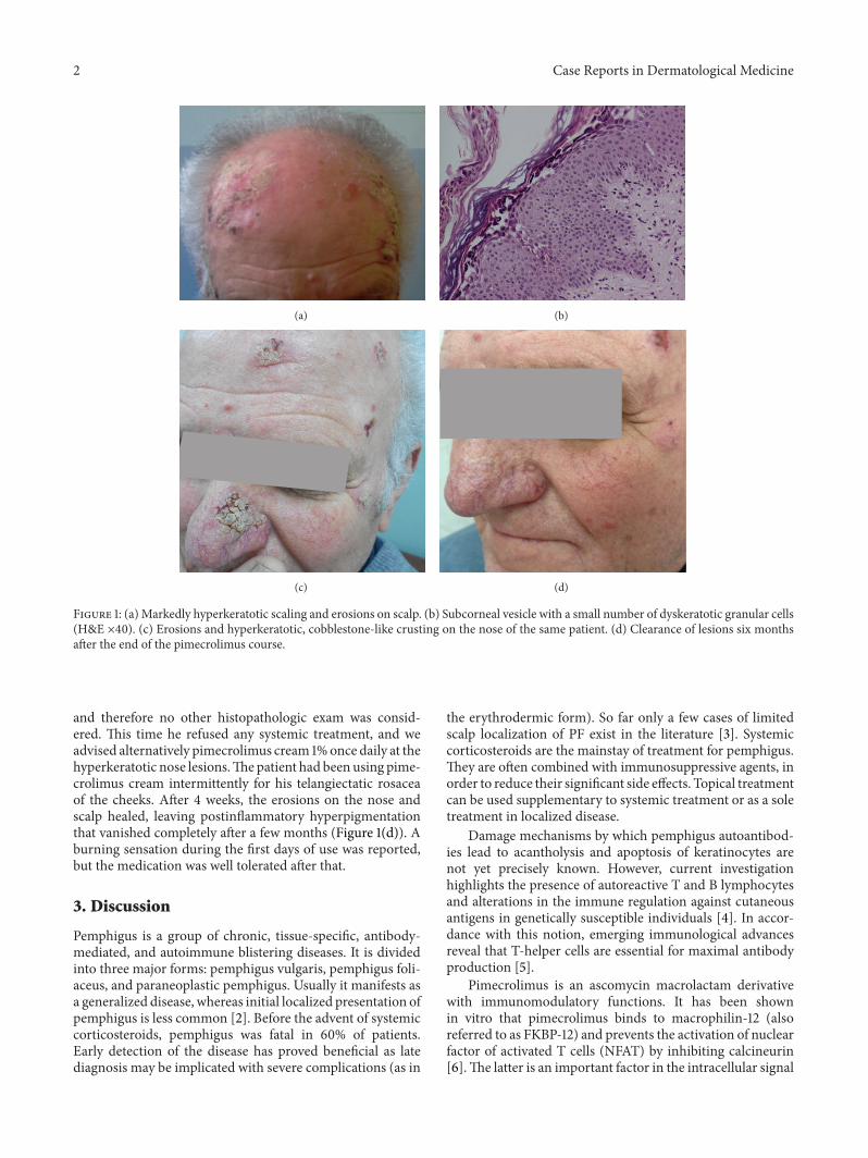
had been covered with markedly hyperkeratotic scalingfor the last 4 months (Figure 1(a)). He also reported mildpruritus. No previous trauma, surgery, irradiation, or anytopical treatment preceded the appearance of these lesions.The lesions had been treated unsuccessfully in the past withcryotherapy. From his previous medical history, he reportedrosacea of the cheeks under treatment.
A skin biopsy from the scalp was performed (Figure 1(a)).The histology report revealed features of pemphigus foli-aceus, such as formation of superficial bullae with acan-tholytic cells, parakeratosis, acanthosis, and slight spongiosis(Figure 1(b)).Thediagnosiswas confirmedbydirect and indi-rect immunofluorescence (ELISA and immunoblot), whiletests for antinuclear antibodies (ANA) were negative.
He was treated with topical betamethasone 0.05% twicedaily and prednisolone 0.7mg/kg daily for 20 days taperedto 5mg daily over the course of the following four weeks.There was significant improvement of the lesions withcomplete clearance maintained for more than ten months.Subsequently, there were two relapses treated similarly asabove, and our patient relapsed again sixteenmonths after theoriginal diagnosis, with development of novel hyperkeratoticlesions on the nose and the scalp. The clinical features ofthe lesions (Figure 1(c)) were similar to the one biopsied,
2 Case Reports in Dermatological Medicine
(a) (b)
(c) (d)
Figure 1: (a) Markedly hyperkeratotic scaling and erosions on scalp. (b) Subcorneal vesicle with a small number of dyskeratotic granular cells(H&E ×40). (c) Erosions and hyperkeratotic, cobblestone-like crusting on the nose of the same patient. (d) Clearance of lesions six monthsafter the end of the pimecrolimus course.
and therefore no other histopathologic exam was consid-ered. This time he refused any systemic treatment, and weadvised alternatively pimecrolimus cream 1%once daily at thehyperkeratotic nose lesions.Thepatient had been using pime-crolimus cream intermittently for his telangiectatic rosaceaof the cheeks. After 4 weeks, the erosions on the nose andscalp healed, leaving postinflammatory hyperpigmentationthat vanished completely after a few months (Figure 1(d)). Aburning sensation during the first days of use was reported,but the medication was well tolerated after that.
3. Discussion
Pemphigus is a group of chronic, tissue-specific, antibody-mediated, and autoimmune blistering diseases. It is dividedinto three major forms: pemphigus vulgaris, pemphigus foli-aceus, and paraneoplastic pemphigus. Usually it manifests asa generalized disease, whereas initial localized presentation ofpemphigus is less common [2]. Before the advent of systemiccorticosteroids, pemphigus was fatal in 60% of patients.Early detection of the disease has proved beneficial as latediagnosis may be implicated with severe complications (as in
the erythrodermic form). So far only a few cases of limitedscalp localization of PF exist in the literature [3]. Systemiccorticosteroids are the mainstay of treatment for pemphigus.They are often combined with immunosuppressive agents, inorder to reduce their significant side effects. Topical treatmentcan be used supplementary to systemic treatment or as a soletreatment in localized disease.
Damage mechanisms by which pemphigus autoantibod-ies lead to acantholysis and apoptosis of keratinocytes arenot yet precisely known. However, current investigationhighlights the presence of autoreactive T and B lymphocytesand alterations in the immune regulation against cutaneousantigens in genetically susceptible individuals [4]. In accor-dance with this notion, emerging immunological advancesreveal that T-helper cells are essential for maximal antibodyproduction [5].
Pimecrolimus is an ascomycin macrolactam derivativewith immunomodulatory functions. It has been shownin vitro that pimecrolimus binds to macrophilin-12 (alsoreferred to as FKBP-12) and prevents the activation of nuclearfactor of activated T cells (NFAT) by inhibiting calcineurin[6].The latter is an important factor in the intracellular signal

Case Reports in Dermatological Medicine 3
transduction pathway resulting in suppression of T cells,inhibition of the production, and release of inflammatorycytokines such as IL-2, IL-3, IL-4, granulocyte-macrophagecolony-stimulating factor (GM CSF), tumor necrosis factor𝛼 (TNF𝛼), and interferon-𝛾 (IFN𝛾). Pimecrolimus also pre-vents the release of inflammatory cytokines and mediatorsfrom mast cells but not from Langerhans cells as doesTacrolimus [6, 7]. However, some of these cytokines have alsobeen implicated to directly increase keratinocyte fragility inthe aetiology of pemphigus vulgaris lesions [8]. We speculatethat the same T lymphocyte-dependent processes might beimplicated in PF, and it is through the inhibition of these thatpimecrolimus seems to have benefited our patient.
Topical calcineurin inhibitors have already been estab-lished in the treatment of atopic dermatitis, and they are beingused in an increasingly wide range of other dermatologicconditions. There are limited reports in the literature oftacrolimus topical use in the treatment of antibody-mediatedautoimmune diseases such as PV [9] or PF [10]. However,we have found no report of pimecrolimus topical use as PFtreatment.
4. Conclusion
Pimecrolimus appeared to be a safe and successful alternativetreatment to topical corticosteroids for our patient withlocalized PF. Topical calcineurin inhibitors have been usedsporadically for this indication in the past. Further workis needed to determine the place of topical calcineurininhibitors in the management of PF.
Conflict of Interests
The authors declare that they have no conflict of interests.
References
[1] V. Dumas, J. C. Roujeau, P. Wolkenstein, J. Revuz, and A.Cosnes, “The treatment of mild pemphigus vulgaris and pem-phigus foliaceus with a topical corticosteroid,” British Journal ofDermatology, vol. 140, no. 6, pp. 1127–1129, 1999.
[2] I. Danopoulou, P. Stavropoulos, A. Stratigos et al., “Pemphigusvegetans confined to the scalp,” International Journal of Derma-tology, vol. 45, no. 8, pp. 1008–1009, 2006.
[3] M.Kishibe,M.Kinouchi, A. Ishida-Yamamoto,K.Koike, andH.Iizuka, “Pemphigus foliaceus localized to the nose,”Clinical andExperimental Dermatology, vol. 28, no. 5, pp. 560–562, 2003.
[4] M. Hertl, R. Eming, and C. Veldman, “T cell control in autoim-mune bullous skin disorders,” Journal of Clinical Investigation,vol. 116, no. 5, pp. 1159–1166, 2006.
[5] G. vanKooten and J. Banchereau, “CD40-CD40 ligand,” Journalof Leukocyte Biology, vol. 67, no. 1, pp. 2–17, 2000.
[6] A. K. Gupta and M. Chow, “Pimecrolimus: a review,” Journal ofthe European Academy of Dermatology and Venereology, vol. 17,no. 5, pp. 493–503, 2003.
[7] A. Panhans-Groß, N. Novak, S. Kraft, and T. Bieber, “Humanepidermal Langerhans’ cells are targets for the immunosup-pressive macrolide tacrolimus (FK506),” Journal of Allergy andClinical Immunology, vol. 107, no. 2, pp. 345–352, 2001.
[8] C. Feliciani, P. Toto, P. Amerio et al., “In vitro and in vivo expres-sion of interleukin-1𝛼 and tumor necrosis factor-𝛼 mRNAin pemphigus vulgaris: Interleukin-1𝛼 and tumor necrosisfactor-𝛼 are involved in acantholysis,” Journal of InvestigativeDermatology, vol. 114, no. 1, pp. 71–77, 2000.
[9] J. E. Gach and A. Ilchyshyn, “Beneficial effects of topicaltacrolimus on recalcitrant erosions of pemphigus vulgaris,”Clinical and Experimental Dermatology, vol. 29, no. 3, pp. 271–272, 2004.
[10] C. C. Termeer, K. Technau, M. Augustin, and J. C. Simon,“Topical tacrolimus (protopic) for the treatment of a localizedpemphigus foliaceus,” Journal of the European Academy ofDermatology and Venereology, vol. 18, no. 5, pp. 636–637, 2004.

Submit your manuscripts athttp://www.hindawi.com
Stem CellsInternational
Hindawi Publishing Corporationhttp://www.hindawi.com Volume 2014
Hindawi Publishing Corporationhttp://www.hindawi.com Volume 2014
MEDIATORSINFLAMMATION
of
Hindawi Publishing Corporationhttp://www.hindawi.com Volume 2014
Behavioural Neurology
EndocrinologyInternational Journal of
Hindawi Publishing Corporationhttp://www.hindawi.com Volume 2014
Hindawi Publishing Corporationhttp://www.hindawi.com Volume 2014
Disease Markers
Hindawi Publishing Corporationhttp://www.hindawi.com Volume 2014
BioMed Research International
OncologyJournal of
Hindawi Publishing Corporationhttp://www.hindawi.com Volume 2014
Hindawi Publishing Corporationhttp://www.hindawi.com Volume 2014
Oxidative Medicine and Cellular Longevity
Hindawi Publishing Corporationhttp://www.hindawi.com Volume 2014
PPAR Research
The Scientific World JournalHindawi Publishing Corporation http://www.hindawi.com Volume 2014
Immunology ResearchHindawi Publishing Corporationhttp://www.hindawi.com Volume 2014
Journal of
ObesityJournal of
Hindawi Publishing Corporationhttp://www.hindawi.com Volume 2014
Hindawi Publishing Corporationhttp://www.hindawi.com Volume 2014
Computational and Mathematical Methods in Medicine
OphthalmologyJournal of
Hindawi Publishing Corporationhttp://www.hindawi.com Volume 2014
Diabetes ResearchJournal of
Hindawi Publishing Corporationhttp://www.hindawi.com Volume 2014
Hindawi Publishing Corporationhttp://www.hindawi.com Volume 2014
Research and TreatmentAIDS
Hindawi Publishing Corporationhttp://www.hindawi.com Volume 2014
Gastroenterology Research and Practice
Hindawi Publishing Corporationhttp://www.hindawi.com Volume 2014
Parkinson’s Disease
Evidence-Based Complementary and Alternative Medicine
Volume 2014Hindawi Publishing Corporationhttp://www.hindawi.com

![Oral Manifestations of Pemphigus Vulgaris: Clinical ... · bullous pemphigus, and paraneoplastic pemphigus [4]. The differential diagnosis includes other dermatological diseases with](https://img.pdfslide.net/doc/110x75/5cbb138688c9930c5f8bb27d/oral-manifestations-of-pemphigus-vulgaris-clinical-bullous-pemphigus-and.jpg)

![Manifestations buccales du pemphigus paranéoplasique · 2013-02-08 · différencier le pemphigus paranéoplasique du pemphigus vulgaire [41, 63, 75, 83, 93, 100]. Par contre, la](https://img.pdfslide.net/doc/110x75/5f49b405f3d6f653f74e2428/manifestations-buccales-du-pemphigus-paranoplasique-2013-02-08-diffrencier.jpg)