Embed Size (px)
Citation preview

About the AuthorsWasim Mansoor, Shaan Chugh, and Rodrigo B. Cavalcanti are with the Department of Medicine, Division of General InternalMedicine, University of Toronto. Carlo Hojilla is with the Department of Pathology and Laboratory Medicine, Sinai Health Systemand the Department of Laboratory Medicine and Molecular Diagnostics, Sunnybrook Health Sciences Centre.RashmiGoswami is with the Department of Hematology and Transfusion Medicine, University Health Network and the Department ofLaboratory Medicine and Pathobiology, University of Toronto.Corresponding Author: [email protected]: August 10, 2017; Accepted: January 22, 2018. Published: June 25, 2018.
The Recurrent Flush: A Case and Review of Systemic MastocytosisWasim Mansoor MD, Shaan Chugh MD, FRCPC, Carlo V. Hojilla MD PhD FRCPC, Rashmi S. Goswami MD PhD FRCPC FCAP,Rodrigo B. Cavalcanti MD MSc FRCPC
ABSTRACTSystemic Mastocytosis (SM) is a hematologic neoplasm characterized by an abnormal proliferation of mast cells, which have the potential to infiltrate one or more visceral organs. Patients can present with a wide constellation of symptoms making it a challenging diagnosis for clinicians. Non-specific symptoms such as fatigue, headache, and weight loss may predominate; however, some patients may present with acute onset of urticaria, flushing, and diarrhea. Due to its rarity, clinicians often face a challenge in evaluating, diagnosing and effectively treating SM. Identification during the indolent phase is important as SM can progress to aggressive leukemias or myeloproliferative disorders. In this article, we present a case of SM, and discuss current practices in diagnosis, evaluation and management. We conclude with future directions for treatments and diagnosis.
RESUMELa mastocytose systémique (SM) est une néoplasie hématologique caractérisée par une prolifération anormale de mastocytes, qui ont le potentiel d’infiltrer un ou plusieurs organes viscéraux. Les patients peuvent présenter une vaste gamme de symptômes, ce qui en fait un diagnostic difficile pour les cliniciens. Les symptômes non spécifiques tels que la fatigue, les maux de tête et la perte de poids peuvent prédominer. Cependant, certains patients peuvent présenter un début aigu d’urticaire, de rougeur et de diarrhée. En raison de sa rareté, les cliniciens sont souvent confrontés à un défi dans l’évaluation, le diagnostic et le traitement efficace de la SM. L’identification au cours de la phase indolente est importante car la SM peut évoluer vers des leucémies agressives ou des troubles myéloprolifératifs. Dans cet article, nous présentons un cas de SM, et discutons des pratiques actuelles en matière de diagnostic, d’évaluation et de gestion. Nous concluons avec les orientations futures pour les traitements et le diagnostic.
Case Presentation
Clinical HistoryA 48-year-old woman presented with syncope and hypotension with a blood pressure of 50/27 mm Hg. The patient was
not known to have any chronic medical illness and was on no routine medications at home. She was aggressively fluid resuscitated followed by development of pulmonary edema. She was intubated and transferred to the ICU where she required ongoing vasopressor therapy. Over the next few days, she was
C a n a d i a n J o u r n a l o f G e n e r a l I n t e r n a l M e d i c i n e V o l u m e 1 3 , I s s u e 2 , 2 0 1 8 33
C a s e R e p o r t
DOI: 10.22374/cjgim.v13i2.234
hemodynamically stabilized and returned to her usual state of health. The patient was discharged after 3 days and asked to follow-up with an allergy and immunologist specialist; however, allergy testing was negative. Over the next few months, the patient continued to have episodic feelings of fatigue, chest heaviness, increased warmth, and associated dyspnea. She also
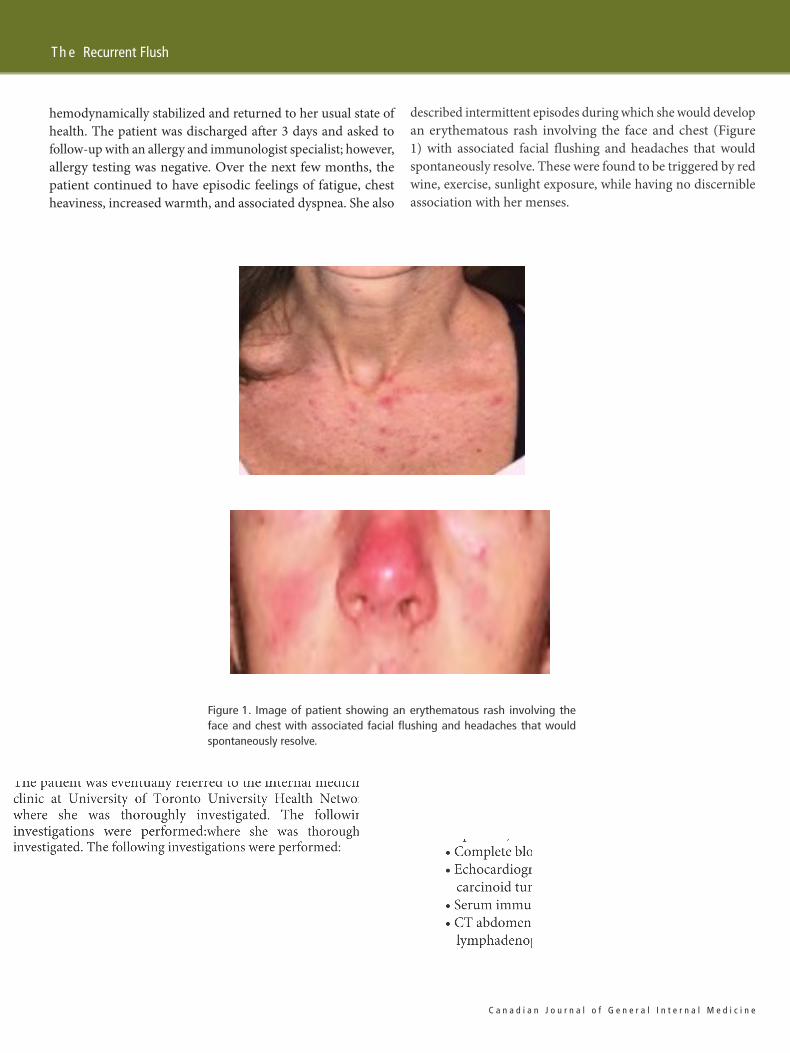
described intermittent episodes during which she would developan erythematous rash involving the face and chest (Figure1) with associated facial flushing and headaches that wouldspontaneously resolve. These were found to be triggered by redwine, exercise, sunlight exposure, while having no discernibleassociation with her menses.
Figure 1. Image of patient showing an erythematous rash involving theface and chest with associated facial flushing and headaches that wouldspontaneously resolve.
C a n a d i a n J o u r n a l o f G e n e r a l I n t e r n a l M e d i c i n e34 V o l u m e 1 3 , I s s u e 2 , 2 0 1 8
Th e Recurrent Flush
Despite the absence of elevated serum tryptase levels, thepatient’s constellation of symptoms was concerning for anunderlying diagnosis of SM. Recently published guidelinessuggest serum KIT mutation analysis if there is a suspicion ofSM despite normal tryptase levels (30); however, this was alsofound to be within normal range.
The patient was conservatively managed with daily Zantacand Claritin; however, episodes continued to get more frequentand debilitating, prompting a bone marrow biopsy to excludeSM. Biopsy and aspiration results revealed SM with presenceof a KIT D816V mutation.
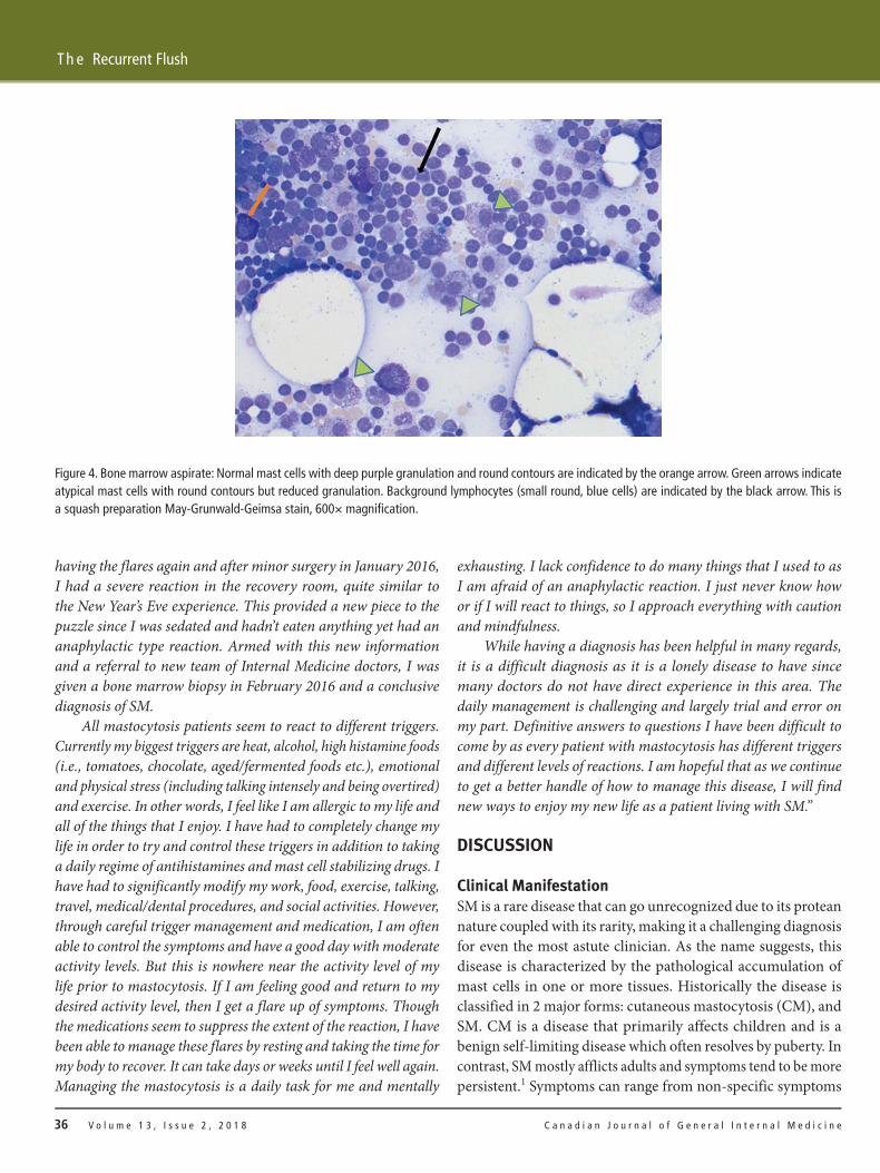
Histopathological reports showed that the bone marrow showed 20% hypocellularity, and immunohistochemicalstaining for tryptase was positive in mast cells (Table 1; Figure 4).Management and Therapy
A total skeletal survey was performed to rule out bonylesions. She was started on vitamin D and calcium supplementsfor maintaining bone health. Due to the continued recurrenceof her symptoms she was started on ketotifen and Aspirin whichresulted in symptomatic improvement. Hematology consultationsuggested no systemic therapy was needed, given the relativelylow levels of mast cells demonstrated on histopathology.
Patient Experience“New Years Eve 2014 was my first major incident. I ended up in theER in anaphylactic shock after a home cooked meal and some wine.Subsequent allergy testing was negative so the event was deemedidiopathic anaphylaxis and I was told to carry Epi-pens. For thefollowing three months I kept having allergic type reactions, oftenafter a full day of activities and dinner. The reactions consisted ofmajor facial flushing, nasal congestion, hives across chest, increasedheart rate with sometimes chest pains/palpitations., resulting infrequent trips to the ER. After a significant episode, typically Iwould have the shakes and a severe headache followed by daysor weeks of extreme exhaustion, diarrhea, and brain fog. I spentmany days in bed just too tired to get up, with a very heavy feelingin my body like my muscles were lead weights. In hindsight now Ialso recall breaking out in red spots across my abdomen and thighsafter spending time in a hot tub or hot bath. My family doctorreferred me to several different allergists/immunologists and anendocrinologist. Many tests were run but all were normal. By April2015 I started to feel better so we decided it was an unexplainedevent that cleared on its own. Then in December 2015, I started
Table 1. Histopathological Report for the Patient
Histology:Histological evaluation of the specimen provided shows a hypocellular (20%) bone marrow with readily apparent mast cells on the aspirate, which aremostly rounded in morphology with a few spindled and many (>25%) degranulated forms present. Lymphoid infiltrates are readily identified on thesquash preparation and the trephine biopsy, associated with linear groups of spindled mast cells in a perivascular distribution.
Immunohistochemical Staining:Tryptase: Positive in mast cells; subset show spindling and are found in a linear distribution (>15 mast cells)CD2, CD25 and CD117: Positive in atypical mast cells
Flow CytometryBone marrow aspirate demonstrates a normal proportion of CD34+ blasts, comprised of myeloid blasts and hematogones, showing normal maturationpatterns. In addition, there is an aberrant population of CD117+ mast cells that are positive for CD2 and CD25.
Molecular DiagnosticsKIT D816V mutation detected
C a n a d i a n J o u r n a l o f G e n e r a l I n t e r n a l M e d i c i n e V o l u m e 1 3 , I s s u e 2 , 2 0 1 8 35
M a n s o o r e t a l
having the flares again and after minor surgery in January 2016, I had a severe reaction in the recovery room, quite similar to the New Year’s Eve experience. This provided a new piece to the puzzle since I was sedated and hadn’t eaten anything yet had an anaphylactic type reaction. Armed with this new information and a referral to new team of Internal Medicine doctors, I was given a bone marrow biopsy in February 2016 and a conclusive diagnosis of SM.
All mastocytosis patients seem to react to different triggers. Currently my biggest triggers are heat, alcohol, high histamine foods (i.e., tomatoes, chocolate, aged/fermented foods etc.), emotional and physical stress (including talking intensely and being overtired) and exercise. In other words, I feel like I am allergic to my life and all of the things that I enjoy. I have had to completely change my life in order to try and control these triggers in addition to taking a daily regime of antihistamines and mast cell stabilizing drugs. I have had to significantly modify my work, food, exercise, talking, travel, medical/dental procedures, and social activities. However, through careful trigger management and medication, I am often able to control the symptoms and have a good day with moderate activity levels. But this is nowhere near the activity level of my life prior to mastocytosis. If I am feeling good and return to my desired activity level, then I get a flare up of symptoms. Though the medications seem to suppress the extent of the reaction, I have been able to manage these flares by resting and taking the time for my body to recover. It can take days or weeks until I feel well again. Managing the mastocytosis is a daily task for me and mentally
exhausting. I lack confidence to do many things that I used to as I am afraid of an anaphylactic reaction. I just never know how or if I will react to things, so I approach everything with caution and mindfulness.
While having a diagnosis has been helpful in many regards, it is a difficult diagnosis as it is a lonely disease to have since many doctors do not have direct experience in this area. The daily management is challenging and largely trial and error on my part. Definitive answers to questions I have been difficult to come by as every patient with mastocytosis has different triggers and different levels of reactions. I am hopeful that as we continue to get a better handle of how to manage this disease, I will find new ways to enjoy my new life as a patient living with SM.”
DISCUSSION
Clinical ManifestationSM is a rare disease that can go unrecognized due to its protean nature coupled with its rarity, making it a challenging diagnosis for even the most astute clinician. As the name suggests, this disease is characterized by the pathological accumulation of mast cells in one or more tissues. Historically the disease is classified in 2 major forms: cutaneous mastocytosis (CM), and SM. CM is a disease that primarily affects children and is a benign self-limiting disease which often resolves by puberty. In contrast, SM mostly afflicts adults and symptoms tend to be more persistent.1 Symptoms can range from non-specific symptoms
Figure 4. Bone marrow aspirate: Normal mast cells with deep purple granulation and round contours are indicated by the orange arrow. Green arrows indicateatypical mast cells with round contours but reduced granulation. Background lymphocytes (small round, blue cells) are indicated by the black arrow. This is a squash preparation May-Grunwald-Geimsa stain, 600× magnification.
C a n a d i a n J o u r n a l o f G e n e r a l I n t e r n a l M e d i c i n e36 V o l u m e 1 3 , I s s u e 2 , 2 0 1 8
Th e Recurrent Flush
study, four of nine patients with SM had peptic ulcer disease on upper GI endoscopy.6 Neurological manifestations include depression, anxiety, and cognitive impairment and are often under-recognized. The proposed mechanism of these manifestations include MC degranulation leading to central nervous system (CNS) neuroinflammation7 Constitutional symptoms are rare in ISM, unless the disease has progressed to advanced SM (ASM), or mast cell leukemia (MCL). Extracutaneous MC infiltration is the hallmark of SM when compared to CM. Commonly affected organs include the bone marrow, liver, lymph nodes, and spleen. Due to bone marrow involvement, hematological abnormalities are common. Anemia is present in 50% of patients with SM, and is more prevalent in aggressive forms of SM when compared to ISM. Eosinophilia is seen in only roughly 15% of patients.7,8
Increased MC degranulation can also have effects on the MSK system. Patients with SM can experience widespread body aches and myalgias which can often be misdiagnosed as fibromyalgia. A study reported that roughly 31% of SM patients may experience similar symptoms.8 Furthermore, it was noted in a cohort study that 50% of patients with SM had evidence of bone involvement on imaging, with osteoporosis being the most common finding.10 The risk of developing osteoporosis-related fractures, although higher in aggressive forms of SM, were also found in 37% of patients with ISM.11
Diagnosis and ClassificationAs with many orphan diseases, the incidence and prevalence of SM is unknown due to a lack of adequate epidemiological studies. The only population-based study was a retrospective cohort conducted in Denmark that examined 548 patients with SM between 1997-2010 . Incidence of SM was estimated to be 0.89 per 100,000 people, with a cumulative incidence of 12.46 for every 100,000 people, and a 14-year prevalence of 9.59 per 100,000.2 The most common subtype identified in the study was indolent systemic mastocytosis (ISM) including urticaria pigmentosa, followed by SM with subtype unknown.2
As understanding of this has disease evolved, the World Health Organization released consensus diagnostic criteria and a classification system for the diagnosis of SM and revised them in 2016 (Tables 3 and 4).12,13
Once a diagnosis of SM is made, the disease is classified based on severity to determine prognosis and the appropriate treatment. According to the revised 2016 WHO classification there are 5 major subtypes of SM: Indolent SM (ISM), Smoldering SM (SSM), SM with associated hematologic neoplasm (SM-AHN), Aggressive SM (ASM), and MC Leukemia (MCL). The classification is based on bone marrow findings coupled with the presence or absence of B and C Findings (see Table 4).12
Table 2. Tissue Involvement and Clinical Symptoms Associated with SM
Skin pruritis, flushing, urticaria pigmentosa
Gastrointestinalabdominal pain, diarrhea, duodenal ulcers (PUD), protein-losing enteropathy
Cardiovascularpalpitations, syncope, pre-syncope, anaphylaxis (hypotension, angioedema)
Neurologicaldepression, anxiety, cognitive impairment, peripheral neuropathy
Musculoskeletalosteopenia, secondary osteoporosis, fractures, chronic bone pain
Constitutional night sweats, fatigue, anorexia, weight loss, fever
Organ Infiltrationbone marrow (pancytopenia), lymphadenoapthy, hepatosplenomegaly
such as weight loss, diarrhea, and fatigue, to specific symptoms consisting of pancytopenia, osteoporosis, and gastrointestinal symptoms. In severe cases, a second hematological process can concomitantly occur resulting in a secondary myeloproliferative disorder, myelodysplastic syndrome, myelofibrosis or acute leukemia. Table 2 contains a comprehensive list of symptoms patients with SM may report.
As noted in Table 2, symptoms associated with SM are broad and non-specific. Skin manifestations are the hallmark of CM but are also a common presentation seen in SM, especially in indolent systemic mastocytosis (ISM). Urticaria pigmentosa is one of the many skin manifestations which presents itself as brown papules and are indicative of systemic disease in 10–70% of cases of SM.2–4 Mast cell (MC) infiltration in the dermis can also lead to flushing, pruritis, and urticaria, thought to be secondary to spontaneous release of MC mediators. Interestingly, some patients may be able to reproduce cutaneous manifestations when experiencing surface friction, a phenomenon known as Darier Sign.5
Anorexia is also common and is thought to be caused by persistent diarrhea and enteropathy and can often be mistaken for irritable bowel syndrome. Additionally, due histamine’s role in acid production, patients are also at risk for developing duodenal ulcers and may need to be prophylactically treated with a proton pump inhibitor. It is estimated that GI bleeding occurs in 11% of SM. Contrary to previously reports, it is estimated that peptic ulcer disease is under-diagnosed. In a recent prospective
C a n a d i a n J o u r n a l o f G e n e r a l I n t e r n a l M e d i c i n e V o l u m e 1 3 , I s s u e 2 , 2 0 1 8 37
M a n s o o r e t a l
Table 3. Diagnostic Criteria
A. Criteria(a)
1. Major Criteria: Multifocal, dense infiltrates of mast cells (>15 MCs in aggregates) detected in sections of BM and/or other extracutaneousorgans
2. Minor Crtieria:a. In biopsy sections of BM or other extracutaneous organs, > 25% of MCs in infiltrate are spindle shaped or have atypical morphology or, of all
MCs in BM aspirate smears, > 25% are immature or atypical.b. Detection of activating point mutation at codon 816 of KIT in BM, blood or other extracutaneous organ.c. MCs in BM, blood or other extracutaneous organs express CD2 and/or CD25 in addition to normal MC markers (CD117).d. Serum total tryptase persistently exceeds 20 ng/mL (unless there is associated clonal myeloid disorder, in which case this parameter is not
valid).
Abbreviations: ANC = Absolute neutrophil count, BM = Bone marrow, MC = mast cellaDiagnosis: 1 major + 1 minor or 3 minor
Table 4. WHO Classification of Systemic Mastocytosis
B. Findingsa. BM biopsy shows > 30% infiltration by MCs (focal, dense aggregates) and/or serum total tryptase level > 200 ng/mL.b. Signs of dysplasia or myeloproliferation in non-MC lineage, but insufficient criteria for definite diagnosis of hematopoietic neoplasm (AHN) with
normal or slightly abnormal blood counts.c. Hepatomegaly without impairment of liver function, and/or palpable splenomegaly without hypersplenism, and/or lymphadenopathy on palpation
or imaging.C. Findings
a. BM dysfunction manifested by 1 or more cytopenias (ANC < 1.0 × 10⁹/L, hemoglobin < 10 g/dL or platelets < 100 x 109/L) but no obvious non-MC hematopoietic malignancy.
b. Palpable hepatomegaly with impairment of liver function, ascites and/or portal hypertension.c. Palpable splenomegaly with hypersplenism.d. Malabsorption with weight loss due to gastrointestinal MC infiltrates.
Indolent Systemic Mastocytosis (ISM)Meets criteria for systemic mastocytosis (SM). No C findings.
Smoldering Systemic Mastocytosis (SSM)Meets criteria for SM, but with 2 or more B findings and no C findings.
SM With Associated Clonal Hematologic Neoplasm (SM-AHN)Meets criteria for SM and criteria for AHN as distinct entity per WHO classification.
Aggressive Systemic Mastocytosis (ASM)Meets criteria for SM. One or more C-findings. No evidence of MC leukemia.
Mast Cell Leukemia (MCL)Meets criteria for SM. Bone marrow biopsy shows a diffuse infiltration, usually compact, by atypical, immature MCs. Bone marrow aspirate smearsshow >20% or more MCs. In typical MCL, MCs account for >10% or more of peripheral blood white cells.
Rare VariantAleukemic mast cell leukemia, as above, but < 10% of white blood cells are MCs.
ANC = absolute neutrophil count; BM = bone marrow; MC = mast cell; AHNMD = associated hematological neoplasm
AHN. Any non-mast cell hematological malignancy that is occuring concomitantly with SM with myeloproliferative neoplasms occuring in >75% of cases. It is a multigene mutated disease
in which SM occurs as a late event.14
C a n a d i a n J o u r n a l o f G e n e r a l I n t e r n a l M e d i c i n e38 V o l u m e 1 3 , I s s u e 2 , 2 0 1 8
Th e Recurrent Flush
It is evident that diagnosis and classification of SM ischallenging, and as such the above-mentioned diagnostic andclassification algorithm was created to aid clinicians to properlydiagnose and prognosticate patients with SM. However, someclinicians have proposed removing MCL and SM-AHN andplacing them under myeloproliferative disorder due to similaritiesin morphology and prognosis. This proposal, however, has notbeen formally adopted as of yet.14
EvaluationWhen suspecting SM, it is necessary to take a thorough historyand physical exam. Patients should be assessed for B-findingssuch as hepatomegaly, splenomegaly, lymphadenopathy orC-findings such as ascites, cachexia, and pallor. Laboratoryexamination includes complete blood count looking for anemia,thrombocytopenia or pancytopenia. In more aggressive forms ofthe disease, liver enzymes may be elevated. If there is suspicionfor bone involvement, serum calcium levels may be elevated.Appropriate imaging should be obtained based on clinicalsymptoms (i.e., X-rays and bone densitometry to rule outpathological fracture or osteoporosis and an abdominal CT toevaluate for organomegaly).
Identification of potential triggers is important as somepatients can present with persistent, or episodic symptoms.Additionally, a serum tryptase level should be obtained to evaluatefor MC degranulation. Elevations above 20ng/mL in suspectedpatients clinches a diagnosis of SM. Therefore, tryptase levelscan be used a screening tool in diagnosing more aggressiveforms of SM. However, many patients with ISM have normaltryptase levels and thus serum tryptase screening may providefalse negative results.
Although not part of the WHO diagnostic criteria, other surrogatemarkers of MC degranuation can also be useful in evaluatingpatients for SM. These include 24 hour urine N-methylhistamine(NMH) and 11ß-prostaglandin F2α (BPG), which has shown tohave a sensitivity of 71% and 53% respectively.15 Additionally,testing peripheral blood for presence of KIT mutations should beperformed (minor criteria) as it correlates to disease severity.16,17
However, due to low sensitivity a negative result does not excludedisease if clinical suspicion is high.
Once appropriate data has been gathered and if there is ahigh pretest likelihood for disease, then the next step is bonemarrow biopsy and aspirate. Biopsy and aspirate should be sentfor total MC% (stain for the receptor tyrosine kinase c-Kit (also known as CD117) and/or tryptase), CD25 and CD2 expression, and KIT mutation analysis to fullfill the diagnostic criteria for SM. The aspirate should also be examined by flow cytometry for CD30 and CD117. In patients with leukocytosis and/or eosinophilia, it is necessary
to examine the bone marrow cells for the JAK2 V617F mutation as well as for BCR/ABL1 and FIPL1/PDGRA rearrangements.1
Once a diagnosis of SM is made, patients should be classifiedin one of the subcategories based on the presence or absence ofB and C findings (see Table 4). Treatment is then tailored basedon the subtype of SM. Refer to Table 5 for diagnostic algorithmfor suspected SM patients.
Treatment and PrognosisTreatment of SM is based on limited clinical data due to paucityof evidence in the literature. We aim to briefly discuss standardtherapies for various subtypes of SM, however the discussionregarding treatment will focus on treatments to he consideredby a general internist.
Indolent Systemic MastocytosisPatients with Indolent Systemic Mastocytosis (ISM) have a normallife expectancy, and thus their treatment is primarily aimed atsymptom management.18 Although rare, progression can occurin 1 to 5% of individuals. In one study, it was noted that patientswith elevated serum B2-microglobulin and KIT mutation inmyeloid and lymphoid cells were independent predictors ofprogression.19 It is important for these patients to keep a diaryto identify any potential triggers of MC degranulation. Commontriggers include: hymenoptera stings, alcohol (red wine mostcommonly), and medications (NSAID and Aspirin) have beenreported in the past.20 Additionally, patients should be counseledto carry an epinephrine pen in case of an acute emergency.
First-line therapy revolves around mediator-relatedsymptomatic control. Antihistamines can be used to easeflushing and pruritis, and may also relieve GI symptoms. Patientsrefractory to antihistamines may also benefit from Aspirin tocontrol flushing as seen in our patient.21 It is important to notehowever that NSAIDs and ASA may also trigger symptomsin certain patients and thus careful evaluation must be takenwhen administering these drugs.20 Cromolyn sodium has beenassessed in limited randomized control trials of patients with SMwhich showed improvement in GI symptoms.22 Omalizumab,a humanized IgG antibody against IgE has been demonstratedin limited case studies to reduce frequency of anaphylaxis inSM. As such, omalizimab can be used in patients who presentwith daily symptoms and whose disease has been refractory tomaximum doses of antihistamines.23
When above-mentioned symptomatic control therapy fails,cytoreductive therapy can be considered. Interferon (IFN-alpha)and cladribine have shown good responses.24
Patients suspected of SM should also receive a skeletal surveyand bone densitometry and be started on appropriate vitamin Dand calcium supplements if they are unable to meet their daily
C a n a d i a n J o u r n a l o f G e n e r a l I n t e r n a l M e d i c i n e V o l u m e 1 3 , I s s u e 2 , 2 0 1 8 39
M a n s o o r e t a l

Clin
ical P
rese
ntat
ion
and
Exam
inat
ion
Find
ings
Pr
uritu
s
Sync
ope
Bo
ny p
ain
Fl
ushi
ng
Pr
esyn
cope
Nigh
t sw
eats
Urtic
aria
pig
men
tosa
Hypo
tens
ion
W
eigh
t los
s
Diar
rhea
Pallo
r
Feve
r and
chill
s
Naus
ea a
nd v
omiti
ng
De
pres
sion
Br
onch
ospa
sm
Dy
spep
sia
Ne
urop
athy
URI s
ympt
oms
He
pato
/spl
enom
egal
y
Head
ache
s
Ascit
es
Ly
mph
aden
opat
hy
An
xiety
Darie
r sig
n*
Labo
rato
ry In
vest
igat
ions
and
Rad
iolo
gica
l Fin
ding
s An
emia
(Hb
<12)
CXR
dem
onst
ratin
g ly
mph
aden
opat
hy
Th
rom
bocy
tope
nia
(Pla
tele
t <10
0)
Le
ukop
enia
or l
euko
cyto
sis
Ab
norm
al b
one
dens
itom
etry
Seru
m tr
ypta
se >
20ng
/mL
Urin
ary
NMH
or B
PG e
leva
ted*
Seru
m K
ITD8
1V6
mut
atio
n po
sitiv
e
CT S
can
show
ing
hepa
tom
egal
y or
sple
nom
egal
yEl
evat
ed tr
ansa
min
ases
Frac
ture
(s) o
n sk
elet
al
surv
ey
El
evat
ed se
rum
B2-
Micr
oglo
bulin
TABL
E 5:
Dia
gnos
tic A
lgor
ithm
for S
yste
mic
Mas
tocy
tosis
If hi
gh cl
inica
l sus
picio
n fo
r SM
, the
n pr
ocee
d to
bon
e m
arro
w b
iops
y
Rule
out
oth
er ca
uses
for f
lush
ing:
1)
Phe
ochr
omoc
ytom
a 2)
Car
cinoi
d sy
ndro
me
3) A
llerg
ic re
actio
n 4)
Ana
phyl
axis
5) R
osac
ea
Rule
out
oth
er ca
uses
for G
I sym
ptom
s 1)
Infla
mm
ator
y bo
wel
dise
ase
2) Ir
ritab
le b
owel
synd
rom
e 3)
Mal
abso
rptio
n sy
ndro
me
4) Z
ollin
ger E
lliso
n sy
ndro
me
5) H
epat
itis a
nd/o
r cirr
hosis
1.
Maj
or: M
ultif
ocal
, den
se in
filtr
ates
of m
ast c
ells
(>15
MCs
in a
ggre
gate
s) d
etec
ted
in se
ctio
ns o
f BM
and
/or o
ther
ext
racu
tane
ous o
rgan
s 2.
M
inor
: a)
In
bio
psy
sect
ions
of B
M o
r oth
er e
xtra
cuta
neou
s org
ans,
> 25
% o
f MCs
in in
filtr
ate
are
spin
dle
shap
ed o
r hav
e at
ypica
l mor
phol
ogy
or, o
f all
MCs
in B
M a
spira
te sm
ears
, > 2
5% a
re
imm
atur
e of
aty
pica
l. b)
De
tect
ion
of a
ctiv
atin
g po
int m
utat
ion
at co
don
816
of K
IT in
BM
, blo
od o
r oth
er
extr
acut
aneo
us o
rgan
. c)
M
Cs in
BM
, blo
od o
r oth
er e
xtra
cuta
neou
s org
ans e
xpre
ss C
D2 a
nd o
r CD2
5 in
add
ition
to
norm
al M
C m
arke
rs (C
D117
). d)
Se
rum
tota
l try
ptas
e pe
rsist
ently
exc
eeds
20
ng/m
L (un
less
ther
e is
asso
ciate
d clo
nal
mye
loid
diso
rder
, in
whi
ch ca
se th
is pa
ram
eter
is n
ot v
alid
).
Diag
nosis
of S
M m
ade
if 1
maj
or a
nd 1
min
or O
R 3
Min
or cr
iteria
met
1
3
4
If Di
agno
sis o
f SM
m
ade,
clas
sify
into
su
btyp
es
Indo
lent
Sys
tem
ic M
asto
cyto
sis (I
SM):
Mee
ts cr
iteria
for
syst
emic
mas
tocy
tosis
(SM
). No
C fi
ndin
gs*.
No
evid
ence
of
asso
ciate
d he
mat
olog
ic ne
opla
sm (A
HN) o
n bo
ne m
arro
w
biop
sy.
SM W
ith A
ssoc
iate
d He
mat
olog
ic Ne
opla
sm (S
M-A
HN):
Mee
ts
crite
ria fo
r SM
and
crite
ria fo
r AHN
as d
istin
ct e
ntity
per
WHO
cla
ssifi
catio
n.
Aggr
essiv
e Sy
stem
ic M
asto
cyto
sis (A
SM):
Mee
ts cr
iteria
for
SM. O
ne o
r mor
e C
findi
ngs*
. No
evid
ence
of M
C le
ukem
ia.
Mas
t Cel
l Leu
kem
ia (M
CL):
Mee
ts cr
iteria
for S
M. B
one
mar
row
bio
psy
show
s a d
iffus
e in
filtr
atio
n, u
sual
ly co
mpa
ct, b
y at
ypica
l, im
mat
ure
MCs
. Bon
e m
arro
w a
spira
te sm
ears
show
>2
0% o
r mor
e M
Cs. I
n ty
pica
l MCL
, MCs
acc
ount
for >
10%
or
mor
e of
per
iphe
ral b
lood
whi
te ce
lls.
*C Fi
ndin
gs
1.
BM d
ysfu
nctio
n m
anife
sted
by
1 or
mor
e cy
tope
nias
(ANC
< 1
.0 x
109 /
L, he
mog
lobi
n <
10 g
/dL o
r pla
tele
ts <
100
x 1
09 /L)
but
no
obvi
ous n
on-M
C he
mat
opoi
etic
mal
igna
ncy.
2.
Pa
lpab
le h
epat
omeg
aly
with
impa
irmen
t of l
iver
func
tion,
asc
ites a
nd/o
r po
rtal
hyp
erte
nsio
n.
3.
Palp
able
sple
nom
egal
y w
ith h
yper
sple
nism
. 4.
M
alab
sorp
tion
with
wei
ght l
oss d
ue to
gas
troi
ntes
tinal
MC
infil
trat
es
(end
osco
pic
biop
sy re
quire
d).
*Dar
ier S
ign:
Ski
n be
com
es sw
olle
n, it
chy
and
red
whe
n st
roki
ng th
e sk
in o
f the
pat
ient
Ab
brev
: NM
H =
N-m
ethy
lhist
amin
e, B
PG =
11ß
-pro
stag
land
in F
2α, C
T =
Com
pute
d To
mog
raph
y, C
XR =
Che
st X
-ray
Abbr
ev: M
C =
Mas
t Cel
l, BM
= B
one
Mar
row
Abbr
ev: M
C =
Mas
t Cel
l, BM
= B
one
Mar
row
, SM
= S
yste
mic
Mas
tocy
tosis
,
C a n a d i a n J o u r n a l o f G e n e r a l I n t e r n a l M e d i c i n e40 V o l u m e 1 3 , I s s u e 2 , 2 0 1 8
Th e Recurrent Flush
requirements through diet alone. Patients who are at high riskfor osteoporosis should be evaluated on a case by case basis forconsideration of bisphosphanates.
Associated Hematologic Neoplasm, AggressiveSM, and MC LeukemiaPatients with Associated Hematologic Neoplasm (AHN), AggressiveSM (ASM), and MC Leukemia (MCL) carry a poor prognosiscompared to ISM due to their aggressive disease course. Themedian survival for aggressive SM is 41 months and for MCLit is less than 6 months.25 The prognosis for AHN is dependenton the concomitant hemotalogical malignancy present.
Hematopoetic stem cell transplantation (HSCT) is the mostdefinitive treatment option that confers the potential for curein appropriate patients. Based on a large retrospective study itwas noted that patients with SM-AHN conferred a 74% 3-yearsurvival benefit with HSCT followed by 43 and 17% for ASMand MCL patients, respectively.25–27 The strongest risk factor forworse overall survival was a diagnosis of MCL. As such, due tothe paucity of treatment options for ASM and MCL, clinical trialdrugs are often first line of therapy in patients. This continues tobe an area of further research as the efficacy of new cytoreductivemodalities are currently being investigated.
ConclusionSM remains a rare disease with protean manifestations. Epigeneticstudies are creating a better understanding of systemic mastocytosis,and tyrosine kinase inihibitors are currently being evaluated toaddress gain of function mutations of the c-Kit receptor in manyvariants of SM. The results of these studies are promising andcould represent future avenues for treatment of ASM and MCL.More efforts need to be placed in creating standardized screeningtools for early diagnosis. One promising approach is outlinedin Table 5. This could be followed by categorizing symptomsusing a scoring system. This may help in establishing a pretestlikelihood for the disease, to guide clinicians on pursuing bonemarrow biopsy for early prognostication and treatment.
REFERENCES1. Valent P, Sperr WR, Schwartz LB, et al. Diagnosis and classification of mast
cell proliferative disorders: delineation from immunologic diseases and non-mast cell hematopoietic neoplasms. J Allergy Clin Immunol 2004;114:3–11.
2. Cohen SS, Skovbo S, Vestergaard H, et al. Epidemiology of systemicmastocytosis in Denmark. Br J Haematol 2014;166:521–8.
3. Soter NA. Mastocytosis and the skin. Hematol Oncol Clin North Am2000;14:537-55, vi.
4. Moura DS. Neuropsychological features of adult mastocytosis. ImmunolAllergy Clin North Am 2014;34:407–22.
5. Soter NA. Mastocytosis and the skin. Hematol Oncol Clin North Am2000;14:537-55, vi.
6. Georgin-Lavialle S, Lhermitte L, Dubreuil P, et al. Mast cell leukemia. Blood2013; 121:1285-95.
7. Lim KH, Tefferi A, Lasho TL, et al. Systemic mastocytosis in 342 consecutive adults: survival studies and prognostic factors. Blood 2009;113:5727–36.
8. Horny HP, Ruck M, Wehrmann M, et al. Blood findings in generalized mastocytosis: evidence of frequent simultaneous occurrence of myeloproliferative disorders. Br J Haematol 1990;76:186–93.
9. Lim KH, Tefferi A, Lasho TL, et al. Systemic mastocytosis in 342 consecutive adults: survival studies and prognostic factors. Blood 2009;113:5727–36.
10. Barete S, Assous N, de Gennes C, et al. Systemic mastocytosis and bone involvement in a cohort of 75 patients. Ann Rheum Dis 2010;69:1838–41.
11. Van Der Veer E, Van Der Goot W, De Monchy JGR, et al. High prevalence of fractures and osteoporosis in patients with indolent systemic mastocytosis. Allergy 2012;67:431–8.
12. Arber DA, Orazi A, Hasserjian R, et al. The 2016 revision to the World Health Organization (WHO) classification of myeloid neoplasms and acute leukemia. Blood 2016;127:2391–5.
13. Horny H, Metcalfe D, Bennet J, et al. Mastocytosis. In: Swerdlow S, Campo E, Harris N, et al, eds. WHO Classification of Tumours of Haematopoietic and Lymphoid Tissues. 4th ed. Lyon, France: World Health Organization; 2008.
14. Wang SA, Hutchinson L, Tang G, et al. Systemic mastocytosis with associated clonal hematological non-mast cell lineage disease: clinical significance and comparison of chromosomal abnormalities in SM and AHNMD components. Am J Hematol 2013;88:219–24.
15. Valent P, Arock M, Akin C, et al. The classification of systemic mastocytosis should include mast cell leukemia (MCL) and systemic mastocytosis with a clonal hematologic non-mast cell lineage disease (SM-AHNMD). Blood 2010;116:850–1.
16. Akin C, Metcalfe DD. Surrogate markers of disease in mastocytosis. Int Arch Allergy Immunol 2002;127:133–6.
17. Valent P, Horny HP, Escribano L, et al. Diagnostic criteria and classification of mastocytosis: a consensus proposal. Leuk Res 2001;25:603–25.
18. Valent P, Sotlar K, Sperr WR, et al. Refined diagnostic criteria and classification of mast cell leukemia (MCL) and myelomastocytic leukemia (MML): a consensus proposal. Ann Oncol 2014;25:1691–700.
19. Escribano L, Álvarez-Twose I, Sánchez-Muñoz L, et al. Prognosis in adult indolent systemic mastocytosis: a long-term study of the Spanish Network on Mastocytosis in a series of 145 patients. J Allergy Clin Immunol 2009;124:514–21.
20. Pardanani A, Lim KH, Lasho TL, et al. WHO subvariants of indolent mastocytosis: clinical details and prognostic evaluation in 159 consecutive adults. Blood 2010;115:150–1.
21. Brockow K, Jofer C, Behrendt H, et al. Anaphylaxis in patients with mastocytosis: a study on history, clinical features and risk factors in 120 patients. Allergy 2008;63:226–32.
22. Butterfield JH, Weiler CR. Prevention of mast cell activation disorder associated clinical sequelae of excessive prostaglandin D2 production. Int Arch Allergy Immunol 2008;147:338–43.
23. Horan RF, Sheffer AL, Austen KF. Cromolyn sodium in the management of systemic mastocytosis. J Allergy Clin Immunol 1990;85:852–5.
24. Carter MC, Robyn JA, Bressler PB, et al. Omalizumab for the treatment of unprovoked anaphylaxis in patients with systemic mastocytosis. J Allergy Clin Immunol 2007;119:1550–1.
25. Lim KH, Pardanani A, Butterfield JH, et al. Cytoreductive therapy in 108 adults with systemic mastocytosis: outcome analysis and response prediction during treatment with interferon-alpha, hydroxyurea, imatinib mesylate or 2-chlorodeoxyadenosine. Am J Hematol 2009;84:790–4.
26. Ustun C, Reiter A, Scott BL, et al. Hematopoietic stem-cell transplantation for advanced systemic mastocytosis. J Clin Oncol 2014; 32:3264-74.
27. Paul C, Sans B, Suarez F, et al. Masitinib for the treatment of systemic and cutaneous mastocytosis with handicap: a phase 2a study. Am J Hematol 2010; 85:921-5.
28. Ustun C, Reiter A, Scott BL, et al. Hematopoietic stem-cell transplantation for advanced systemic mastocytosis. J Clin Oncol 2014; 32:3264-74.
29. Georgin-Lavialle S, Lhermitte L, Dubreuil P, et al. Mast cell leukemia. Blood 2013; 121:1285-95.
C a n a d i a n J o u r n a l o f G e n e r a l I n t e r n a l M e d i c i n e V o l u m e 1 3 , I s s u e 2 , 2 0 1 8 41
M a n s o o r e t a l
30. Cherner JA, Jensen RT, Dubois A, O’Dorisio TM, Gardner JD, Metcalfe DD. Gastrointestinal dysfunction in systemic mastocytosis. A prospective study. Gastroenterology 1988; 95:657–667.
31. Lim KH, Tefferi A, Lasho TL, et al; Systemic mastocytosis in 342 consecutive adults: survival studies and prognostic factors. Blood. 2009;113(23):5727-36.
32. Theoharides TC, Valent P, Akin C. Mast cells, mastocytosis, and related disorders. N Engl J Med 2015;373:163-72
C a n a d i a n J o u r n a l o f G e n e r a l I n t e r n a l M e d i c i n e42 V o l u m e 1 3 , I s s u e 2 , 2 0 1 8
Th e M a n y Fa c e s o f S y s t e m i c M a s t o c y t o s i s
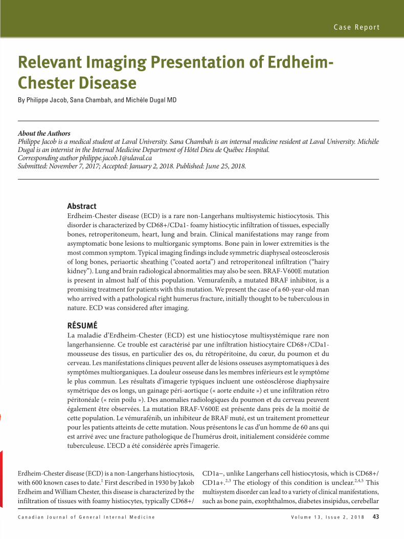
AbstractErdheim-Chester disease (ECD) is a rare non-Langerhans multisystemic histiocytosis. This disorder is characterized by CD68+/CDa1- foamy histiocytic infiltration of tissues, especially bones, retroperitoneum, heart, lung and brain. Clinical manifestations may range from asymptomatic bone lesions to multiorganic symptoms. Bone pain in lower extremities is the most common symptom. Typical imaging findings include symmetric diaphyseal osteosclerosis of long bones, periaortic sheathing (“coated aorta”) and retroperitoneal infiltration (“hairy kidney”). Lung and brain radiological abnormalities may also be seen. BRAF-V600E mutation is present in almost half of this population. Vemurafenib, a mutated BRAF inhibitor, is a promising treatment for patients with this mutation. We present the case of a 60-year-old man who arrived with a pathological right humerus fracture, initially thought to be tuberculous in nature. ECD was considered after imaging.
RÉSUMÉLa maladie d’Erdheim-Chester (ECD) est une histiocytose multisystémique rare non langerhansienne. Ce trouble est caractérisé par une infiltration histiocytaire CD68+/CDa1-mousseuse des tissus, en particulier des os, du rétropéritoine, du cœur, du poumon et du cerveau. Les manifestations cliniques peuvent aller de lésions osseuses asymptomatiques à des symptômes multiorganiques. La douleur osseuse dans les membres inférieurs est le symptôme le plus commun. Les résultats d’imagerie typiques incluent une ostéosclérose diaphysaire symétrique des os longs, un gainage péri-aortique (« aorte enduite ») et une infiltration rétro péritonéale (« rein poilu »). Des anomalies radiologiques du poumon et du cerveau peuvent également être observées. La mutation BRAF-V600E est présente dans près de la moitié de cette population. Le vémurafénib, un inhibiteur de BRAF muté, est un traitement prometteur pour les patients atteints de cette mutation. Nous présentons le cas d’un homme de 60 ans qui est arrivé avec une fracture pathologique de l’humérus droit, initialement considérée comme tuberculeuse. L’ECD a été considérée après l’imagerie.
About the AuthorsPhilippe Jacob is a medical student at Laval University. Sana Chambah is an internal medicine resident at Laval University. MichèleDugal is an internist in the Internal Medicine Department of Hôtel Dieu de Québec Hospital.Corresponding author [email protected]: November 7, 2017; Accepted: January 2, 2018. Published: June 25, 2018.
Relevant Imaging Presentation of Erdheim-Chester DiseaseBy Philippe Jacob, Sana Chambah, and Michèle Dugal MD
Erdheim-Chester disease (ECD) is a non-Langerhans histiocytosis, with 600 known cases to date.1 First described in 1930 by Jakob Erdheim and William Chester, this disease is characterized by the infiltration of tissues with foamy histiocytes, typically CD68+/
CD1a−, unlike Langerhans cell histiocytosis, which is CD68+/CD1a+.2,3 The etiology of this condition is unclear.2,4,5 This multisystem disorder can lead to a variety of clinical manifestations, such as bone pain, exophthalmos, diabetes insipidus, cerebellar
C a n a d i a n J o u r n a l o f G e n e r a l I n t e r n a l M e d i c i n e V o l u m e 1 3 , I s s u e 2 , 2 0 1 8 43
C a s e R e p o r t
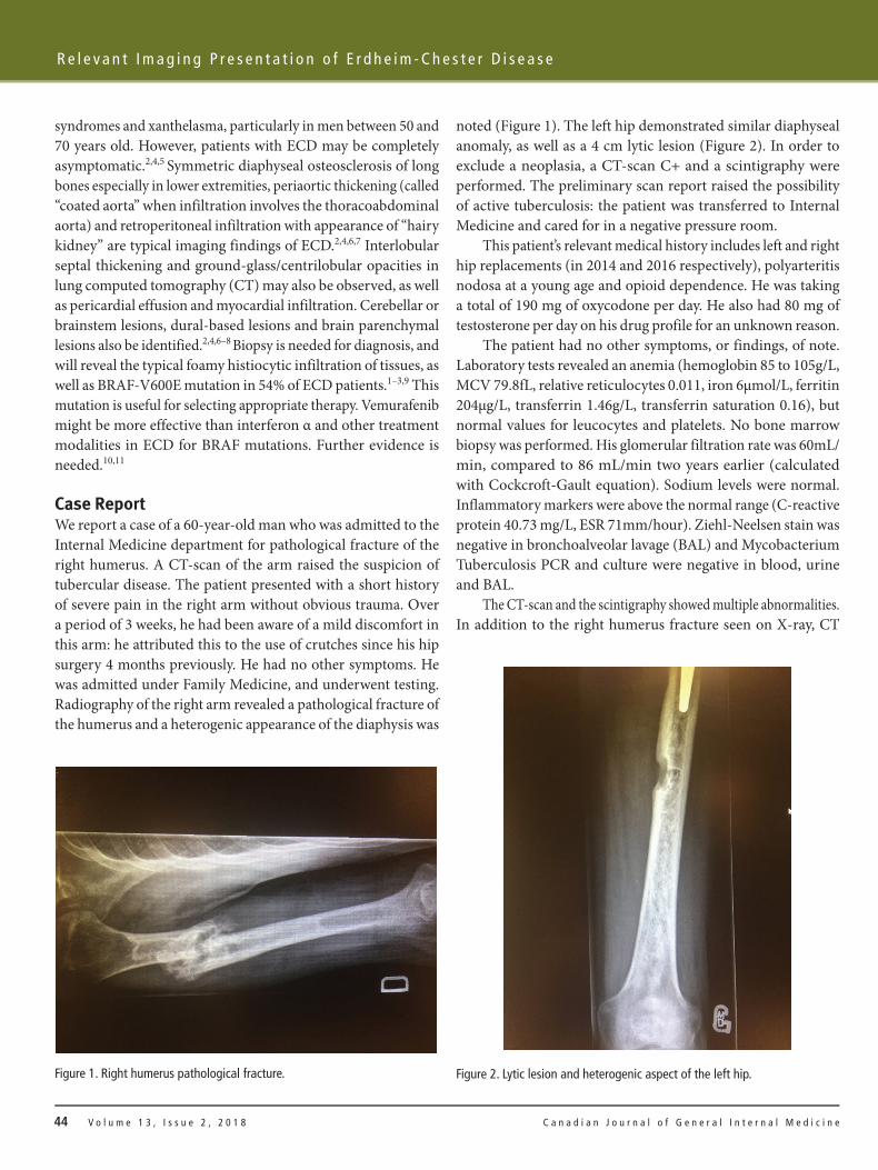
noted (Figure 1). The left hip demonstrated similar diaphyseal anomaly, as well as a 4 cm lytic lesion (Figure 2). In order to exclude a neoplasia, a CT-scan C+ and a scintigraphy were performed. The preliminary scan report raised the possibility of active tuberculosis: the patient was transferred to Internal Medicine and cared for in a negative pressure room.
This patient’s relevant medical history includes left and right hip replacements (in 2014 and 2016 respectively), polyarteritis nodosa at a young age and opioid dependence. He was taking a total of 190 mg of oxycodone per day. He also had 80 mg of testosterone per day on his drug profile for an unknown reason.
The patient had no other symptoms, or findings, of note. Laboratory tests revealed an anemia (hemoglobin 85 to 105g/L, MCV 79.8fL, relative reticulocytes 0.011, iron 6μmol/L, ferritin 204μg/L, transferrin 1.46g/L, transferrin saturation 0.16), but normal values for leucocytes and platelets. No bone marrow biopsy was performed. His glomerular filtration rate was 60mL/min, compared to 86 mL/min two years earlier (calculated with Cockcroft-Gault equation). Sodium levels were normal. Inflammatory markers were above the normal range (C-reactive protein 40.73 mg/L, ESR 71mm/hour). Ziehl-Neelsen stain was negative in bronchoalveolar lavage (BAL) and Mycobacterium Tuberculosis PCR and culture were negative in blood, urine and BAL.
The CT-scan and the scintigraphy showed multiple abnormalities. In addition to the right humerus fracture seen on X-ray, CT
syndromes and xanthelasma, particularly in men between 50 and 70 years old. However, patients with ECD may be completely asymptomatic.2,4,5 Symmetric diaphyseal osteosclerosis of long bones especially in lower extremities, periaortic thickening (called “coated aorta” when infiltration involves the thoracoabdominal aorta) and retroperitoneal infiltration with appearance of “hairy kidney” are typical imaging findings of ECD.2,4,6,7 Interlobular septal thickening and ground-glass/centrilobular opacities in lung computed tomography (CT) may also be observed, as well as pericardial effusion and myocardial infiltration. Cerebellar or brainstem lesions, dural-based lesions and brain parenchymal lesions also be identified.2,4,6–8 Biopsy is needed for diagnosis, and will reveal the typical foamy histiocytic infiltration of tissues, as well as BRAF-V600E mutation in 54% of ECD patients.1–3,9 This mutation is useful for selecting appropriate therapy. Vemurafenib might be more effective than interferon α and other treatment modalities in ECD for BRAF mutations. Further evidence is needed.10,11
Case ReportWe report a case of a 60-year-old man who was admitted to the Internal Medicine department for pathological fracture of the right humerus. A CT-scan of the arm raised the suspicion of tubercular disease. The patient presented with a short history of severe pain in the right arm without obvious trauma. Over a period of 3 weeks, he had been aware of a mild discomfort in this arm: he attributed this to the use of crutches since his hip surgery 4 months previously. He had no other symptoms. He was admitted under Family Medicine, and underwent testing. Radiography of the right arm revealed a pathological fracture of the humerus and a heterogenic appearance of the diaphysis was
Figure 1. Right humerus pathological fracture. Figure 2. Lytic lesion and heterogenic aspect of the left hip.
C a n a d i a n J o u r n a l o f G e n e r a l I n t e r n a l M e d i c i n e44 V o l u m e 1 3 , I s s u e 2 , 2 0 1 8
R e l e v a n t I m a g i n g P r e s e n t a t i o n o f E r d h e i m - C h e s t e r D i s e a s e
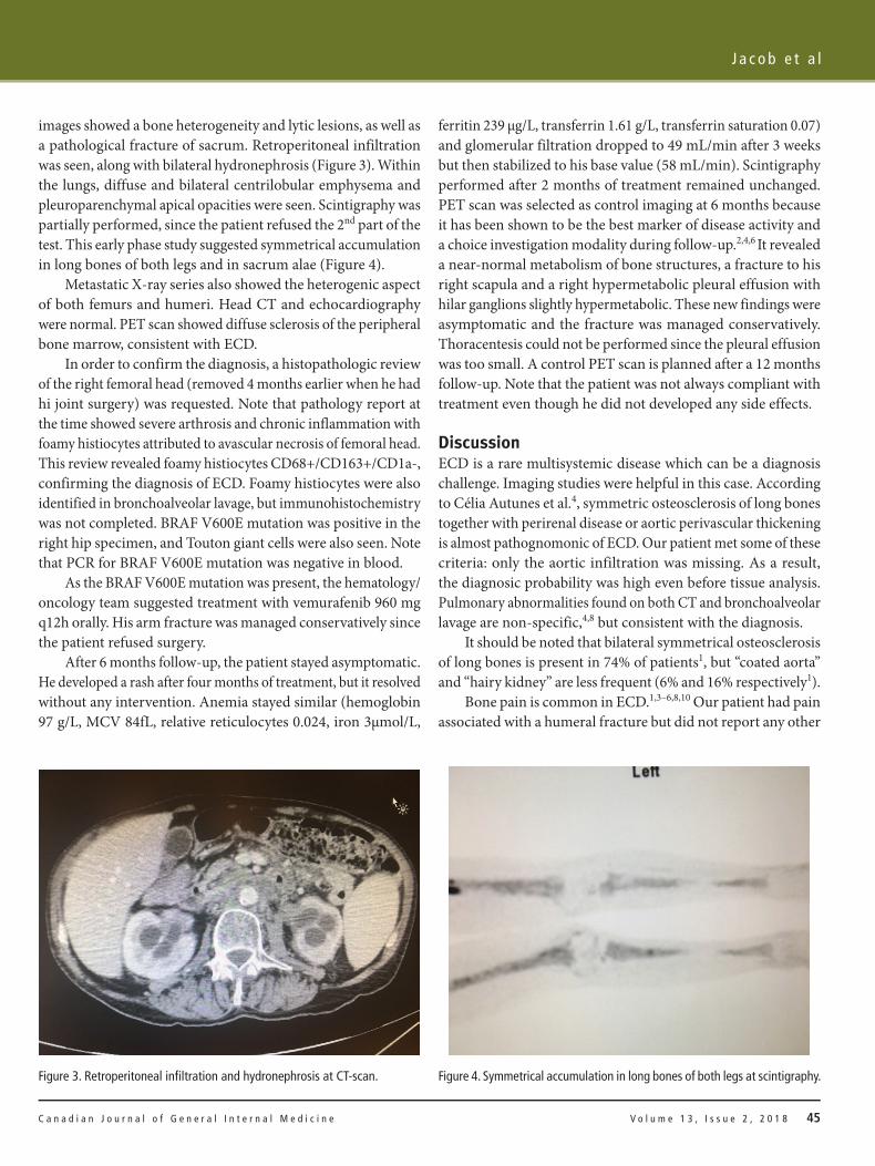
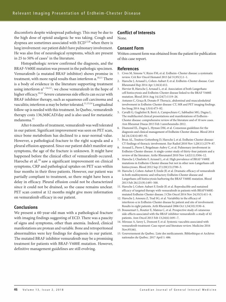
images showed a bone heterogeneity and lytic lesions, as well as a pathological fracture of sacrum. Retroperitoneal infiltration was seen, along with bilateral hydronephrosis (Figure 3). Within the lungs, diffuse and bilateral centrilobular emphysema and pleuroparenchymal apical opacities were seen. Scintigraphy was partially performed, since the patient refused the 2nd part of the test. This early phase study suggested symmetrical accumulation in long bones of both legs and in sacrum alae (Figure 4).
Metastatic X-ray series also showed the heterogenic aspect of both femurs and humeri. Head CT and echocardiography were normal. PET scan showed diffuse sclerosis of the peripheral bone marrow, consistent with ECD.
In order to confirm the diagnosis, a histopathologic review of the right femoral head (removed 4 months earlier when he had hi joint surgery) was requested. Note that pathology report at the time showed severe arthrosis and chronic inflammation with foamy histiocytes attributed to avascular necrosis of femoral head. This review revealed foamy histiocytes CD68+/CD163+/CD1a-, confirming the diagnosis of ECD. Foamy histiocytes were also identified in bronchoalveolar lavage, but immunohistochemistry was not completed. BRAF V600E mutation was positive in the right hip specimen, and Touton giant cells were also seen. Note that PCR for BRAF V600E mutation was negative in blood.
As the BRAF V600E mutation was present, the hematology/oncology team suggested treatment with vemurafenib 960 mg q12h orally. His arm fracture was managed conservatively since the patient refused surgery.
After 6 months follow-up, the patient stayed asymptomatic. He developed a rash after four months of treatment, but it resolved without any intervention. Anemia stayed similar (hemoglobin 97 g/L, MCV 84fL, relative reticulocytes 0.024, iron 3μmol/L,
ferritin 239 μg/L, transferrin 1.61 g/L, transferrin saturation 0.07) and glomerular filtration dropped to 49 mL/min after 3 weeks but then stabilized to his base value (58 mL/min). Scintigraphy performed after 2 months of treatment remained unchanged. PET scan was selected as control imaging at 6 months because it has been shown to be the best marker of disease activity and a choice investigation modality during follow-up.2,4,6 It revealed a near-normal metabolism of bone structures, a fracture to his right scapula and a right hypermetabolic pleural effusion with hilar ganglions slightly hypermetabolic. These new findings were asymptomatic and the fracture was managed conservatively. Thoracentesis could not be performed since the pleural effusion was too small. A control PET scan is planned after a 12 months follow-up. Note that the patient was not always compliant with treatment even though he did not developed any side effects.
DiscussionECD is a rare multisystemic disease which can be a diagnosis challenge. Imaging studies were helpful in this case. According to Célia Autunes et al.4, symmetric osteosclerosis of long bones together with perirenal disease or aortic perivascular thickening is almost pathognomonic of ECD. Our patient met some of these criteria: only the aortic infiltration was missing. As a result, the diagnosic probability was high even before tissue analysis. Pulmonary abnormalities found on both CT and bronchoalveolar lavage are non-specific,4,8 but consistent with the diagnosis.
It should be noted that bilateral symmetrical osteosclerosis of long bones is present in 74% of patients1, but “coated aorta” and “hairy kidney” are less frequent (6% and 16% respectively1).
Bone pain is common in ECD.1,3–6,8,10 Our patient had pain associated with a humeral fracture but did not report any other
Figure 3. Retroperitoneal infiltration and hydronephrosis at CT-scan. Figure 4. Symmetrical accumulation in long bones of both legs at scintigraphy.
C a n a d i a n J o u r n a l o f G e n e r a l I n t e r n a l M e d i c i n e V o l u m e 1 3 , I s s u e 2 , 2 0 1 8 45
J a c o b e t a l
discomforts despite widespread pathology. This may be due to the high dose of opioid analgesic he was taking. Cough and dyspnea are sometimes associated with ECD1,5,8 when there is lung involvement: our patient didn’t have pulmonary involvement. He was also free of neurological symptoms, which are present in 25 to 50% of cases1 in the literature.
Histopathologic review confirmed the diagnosis, and the BRAF-V600E mutation was present in the pathologic specimen. Vemurafenib (a mutated BRAF inhibitor) shows promise in treatment, with more rapid results than interferon α.10,11 There is a body of evidence in the literature supporting treatment using interferon α1–3,6,12 : we chose vemurafenib in the hope of higher efficacy.10,11 Severe cutaneous side effects can occur with BRAF inhibitor therapy, such as squamous cell carcinoma and vasculitis; interferon α may be better tolerated.11,13,14 Longitudinal follow-up is needed with this treatment. In Quebec, vemurafenib therapy costs 136,56$CAD/day and is also used for metastatic melanoma.15
After 6 months of treatment, vemurafenib was well tolerated in our patient. Significant improvement was seen on PET scan, since bone metabolism has declined to a near-normal value. However, a pathological fracture to the right scapula and a pleural effusion appeared. Since our patient didn’t manifest any symptoms, the age of the fracture is unknown. It might have happened before the clinical effect of vemurafenib occured. Haroche et al.10 saw a significant improvement on clinical symptoms, CRP and pathological uptakes on PET scan within four months in their three patients. However, our patient was partially compliant to treatment, so there might have been a delay in efficacy. Pleural effusion could not be characterized since it could not be drained, so the cause remains unclear. PET scan control at 12 months might give more information on vemurafenib efficacy in our patient.
ConclusionsWe present a 60-year-old man with a pathological fracture with imaging findings suggesting of ECD. There was a paucity of signs and symptoms, other than anemia. Indeed, clinical manifestations are protean and variable. Bone and retroperitoneal abnormalities were key findings for diagnosis in our patient. The mutated BRAF inhibitor vemurafenib may be a promising treatment for patients with BRAF-V600E mutation. However, definitive management guidelines are still evolving.
Conflict of InterestsNone.
Consent FormWritten consent form was obtained from the patient for publication of this case report.
References1. Cives M, Simone V, Rizzo FM, et al. Erdheim–Chester disease: a systematic
review. Crit Rev Oncol Hematol 2015 Jul 31;95(1):1–1.2. Haroche J, Arnaud L, Cohen-Aubart F, et al. Erdheim–Chester disease. Curr
Rheumatol Rep 2014 Apr 1;16(4):412.3. Hervier B, Haroche J, Arnaud L, et al. Association of both Langerhans
cell histiocytosis and Erdheim-Chester disease linked to the BRAF V600E mutation. Blood 2014 Aug 14;124(7):1119–26.
4. Antunes C, Graça B, Donato P. Thoracic, abdominal and musculoskeletal involvement in Erdheim-Chester disease: CT, MR and PET imaging findings. Ins Imag 2014 Aug 1;5(4):473–82.
5. Cavalli G, Guglielmi B, Berti A, Campochiaro C, Sabbadini MG, Dagna L. The multifaceted clinical presentations and manifestations of Erdheim–Chester disease: comprehensive review of the literature and of 10 new cases. Ann Rheumat Diseas 2013 Feb 1:annrheumdis-2012.
6. Diamond EL, Dagna L, Hyman DM, et al. Consensus guidelines for the diagnosis and clinical management of Erdheim-Chester disease. Blood 2014 Jul 24;124(4):483–92.
7. Brun AL, Touitou-Gottenberg D, Haroche J, et al. Erdheim-Chester disease: CT findings of thoracic involvement. Eur Radiol 2010 Nov 1;20(11):2579–87.
8. Arnaud L, Pierre I, Beigelman‐Aubry C, et al. Pulmonary involvement in Erdheim‐Chester disease: A single‐center study of thirty‐four patients and a review of the literature. Arthr Rheumatol 2010 Nov 1;62(11):3504–12.
9. Haroche J, Charlotte F, Arnaud L, et al. High prevalence of BRAF V600E mutations in Erdheim-Chester disease but not in other non-Langerhans cell histiocytoses. Blood 2012 Sep 27;120(13):2700–3.
10. Haroche J, Cohen-Aubart F, Emile JF, et al. Dramatic efficacy of vemurafenib in both multisystemic and refractory Erdheim-Chester disease and Langerhans cell histiocytosis harboring the BRAF V600E mutation. Blood 2013 Feb 28;121(9):1495–500.
11. Haroche J, Cohen-Aubart F, Emile JF, et al. Reproducible and sustained efficacy of targeted therapy with vemurafenib in patients with BRAFV600E-mutated Erdheim-Chester disease. J Clin Oncol 2014 Nov 24;33(5):411–8.
12. Haroche J, Amoura Z, Trad SG, et al. Variability in the efficacy of interferon‐α in Erdheim‐Chester disease by patient and site of involvement: Results in eight patients. Arth Rheumatol 2006 Oct 1;54(10):3330–6.
13. Boussemart L, Routier E, Mateus C, et al. Prospective study of cutaneous side-effects associated with the BRAF inhibitor vemurafenib: a study of 42 patients. Ann Oncol 2013 Feb 13;24(6):1691–7.
14. Mirouse A, Savey L, Domont F, et al. Systemic vasculitis associated with vemurafenib treatment: Case report and literature review. Medicine 2016 Nov;95(46).
15. Gouvernement du Québec. Liste des médicaments. Bibliothèque et Archives nationales du Québec. 2017 April 1: 486.
C a n a d i a n J o u r n a l o f G e n e r a l I n t e r n a l M e d i c i n e46 V o l u m e 1 3 , I s s u e 2 , 2 0 1 8
R e l e v a n t I m a g i n g P r e s e n t a t i o n o f E r d h e i m - C h e s t e r D i s e a s e