Embed Size (px)
Citation preview

CASE REPORT Open Access
A case report of recurrentmembranoproliferative glomerulonephritisafter kidney transplantation due toventriculoatrial shunt infectionLinus A. Völker1 , Katharina Burkert1, Niklas Scholten1, Franziska Grundmann1, Christine Kurschat1,Thomas Benzing1, Jürgen Hampl2, Jan Ulrich Becker3 and Roman-Ulrich Müller1*
Abstract
Background: Transplant failure requires the consideration of numerous potential causes including rejection, acutetubular necrosis, infection, and recurrence of the original kidney disease. Kidney biopsy is generally required toapproach these differential diagnoses. However, the histopathological findings on their own do not always lead toa definite diagnosis. Consequently, it is crucial to integrate them with clinical findings and patient historywhen discussing histopathological patterns of injury. The histopathologic finding of a membranoproliferativeglomerulonephritis (MPGN) is one of the most challenging constellations since it does not refer to a specificdisease entity but rather reflects a pattern of injury that is the result of many different causes. Whilst MPGN isoccasionally classified as immune complex mediated, careful evaluation usually reveals an underlying disordersuch as chronic infection, plasma cell dyscrasia, complement disorders, and autoimmune disease.
Case presentation: We describe the case of a 43-year-old woman who was referred to us because of a slowly risingserum creatinine 4 years after kidney transplantation. As in the native kidney, the biopsy revealed an MPGNpattern of injury. The cause of this finding had not been established prior to transplantation leading to aclassification as idiopathic MPGN in the past. Further workup at the time of presentation and allograft failurerevealed chronic infection of a ventriculoatrial shunt as the most probable cause.
Conclusion: This case underlines the fact that MPGN is not a disease but a histopathological description.Consequently, the causative disorder needs to be identified to avoid kidney failure and recurrence aftertransplantation.
Keywords: Shunt nephritis, Membranoproliferative glomerulonephritis, Kidney transplantation, Recurrence
BackgroundAllograft failure can be caused by numerous differentialdiagnoses and usually requires kidney biopsy to confirmthe actual pathology. Since one of these is recurrence ofthe disease leading to failure of the native kidneys, a lackof knowledge regarding this point adds to the challengefor nephrologists and nephropathologists. Even in pa-tients in whom the native kidneys were biopsied, the
histopathological diagnosis does not automatically leadto a clinical diagnosis. Most commonly, this is the caseif the histopathological pattern of injury can be causedby numerous different clinical entities and if the under-lying disease is generally rare. MPGN is such a findingwith a number of different disorders – most of whichare very rare – that can cause the same histopathologicalpicture (Fig. 1) [1]. With chronic infections being one ofthe possible causes, shunt-nephritis may not be the firstthought of many clinicians due to its low incidence. Thisholds true especially in the setting of transplant failure.Shunt-nephritis refers to a form of glomerulonephritis
© The Author(s). 2019 Open Access This article is distributed under the terms of the Creative Commons Attribution 4.0International License (http://creativecommons.org/licenses/by/4.0/), which permits unrestricted use, distribution, andreproduction in any medium, provided you give appropriate credit to the original author(s) and the source, provide a link tothe Creative Commons license, and indicate if changes were made. The Creative Commons Public Domain Dedication waiver(http://creativecommons.org/publicdomain/zero/1.0/) applies to the data made available in this article, unless otherwise stated.
* Correspondence: [email protected] II of Internal Medicine and Center for Molecular MedicineCologne University of Cologne, Faculty of Medicine and University Hospitalof Cologne, Uniklinik Köln, Kerpener Str. 62, 50937 Cologne, GermanyFull list of author information is available at the end of the article
Völker et al. BMC Nephrology (2019) 20:296 https://doi.org/10.1186/s12882-019-1472-1
that is often characterized by an MPGN pattern on thehistological level associated with polyclonal immunecomplexesin subacute and chronic courses while acutecourses may present as endocapillary or extracapillarydiffuse-proliferative, mesangiocapillary, and mesangiallesions [2–5].In the case presented here, MPGN is the conse-
quence of immune-complex deposition induced bychronic infection. Chronic infections of a ventriculoa-trial or –peritoneal shunts are most commonly causedby colonization with slowly-growing bacteria of theskin flora, e.g. staphylococci [3, 4]. Many cases ofthese infections are characterized by a subacute tochronic clinical course (such as recurrent fever, hepa-tosplenomegaly, and cerebral symptoms). However,very slow chronic and oligosymptomatic courses havealso been described [5, 6]. Apart from the unspecificclinical picture, problems in cultivating slowly grow-ing bacteria from blood and liquor cultures make boththe diagnosis and a targeted anti-infective therapy difficult.Consequently, these cases are prone to be misinterpretedand diagnosed late when immunological complications such
as shunt nephritis have already manifested. However, if re-current MPGN threatens graft survival, identification of thisrare cause is crucial since this problem can be addressedspecifically by removing the shunt material and treating withantibiotics according to the resistogram. Finding a way to re-move all artificial materials and solving the problem causinghydrocephalus in such a patient without reimplantation of anew shunt are of utmost importance. Here, we present theexemplary case of a young woman illustrating both the dif-ferential diagnosis of diseases underlying recurrent MPGNin kidney transplants including the algorithm to identifyingshunt nephritis as the causative disorder and the interdiscip-linary approach to solving the problem.
Timeline
Year Event
1986 Diagnosis of hydrocephalusPlacement of ventriculoperitoneal shunt
1992 Placement of ventriculoatrial shunt
2004 Diagnosis of membranoproliferative glomerulonephritis
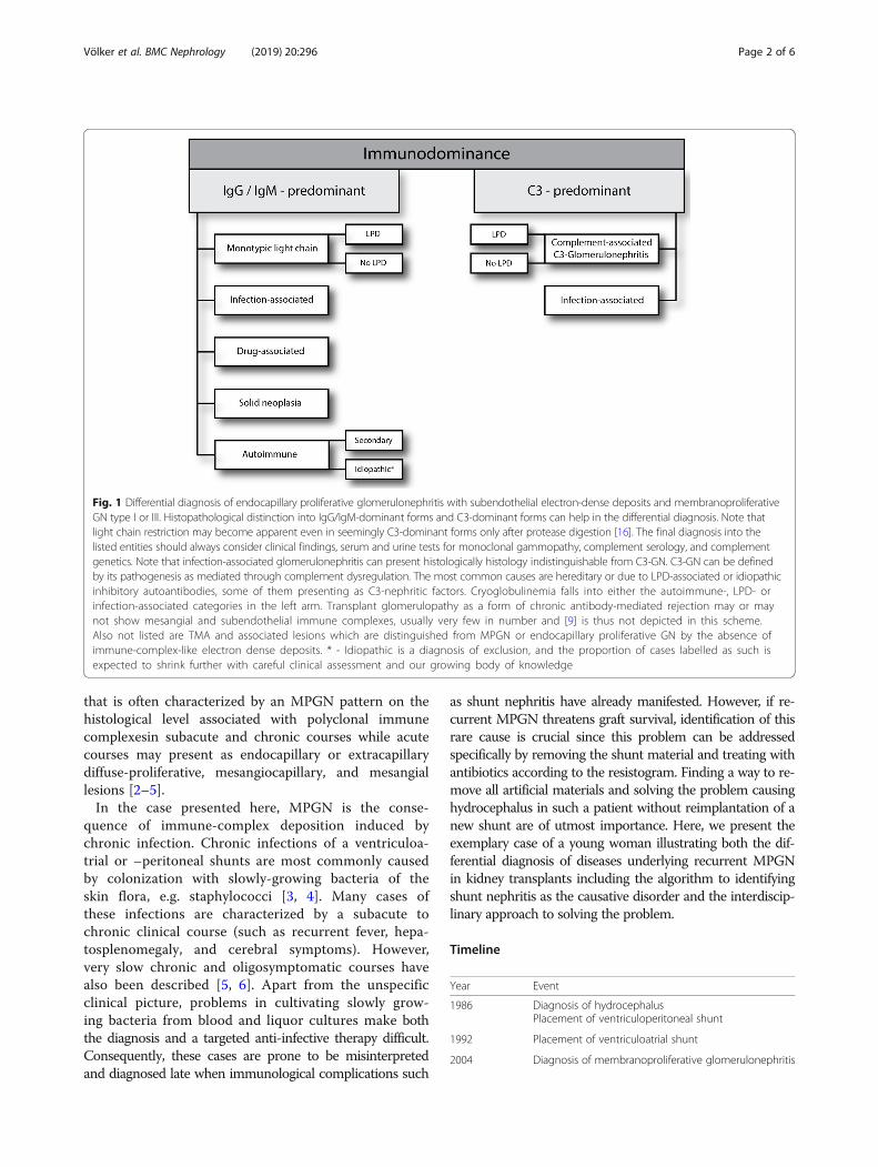
Fig. 1 Differential diagnosis of endocapillary proliferative glomerulonephritis with subendothelial electron-dense deposits and membranoproliferativeGN type I or III. Histopathological distinction into IgG/IgM-dominant forms and C3-dominant forms can help in the differential diagnosis. Note thatlight chain restriction may become apparent even in seemingly C3-dominant forms only after protease digestion [16]. The final diagnosis into thelisted entities should always consider clinical findings, serum and urine tests for monoclonal gammopathy, complement serology, and complementgenetics. Note that infection-associated glomerulonephritis can present histologically histology indistinguishable from C3-GN. C3-GN can be definedby its pathogenesis as mediated through complement dysregulation. The most common causes are hereditary or due to LPD-associated or idiopathicinhibitory autoantibodies, some of them presenting as C3-nephritic factors. Cryoglobulinemia falls into either the autoimmune-, LPD- orinfection-associated categories in the left arm. Transplant glomerulopathy as a form of chronic antibody-mediated rejection may or maynot show mesangial and subendothelial immune complexes, usually very few in number and [9] is thus not depicted in this scheme.Also not listed are TMA and associated lesions which are distinguished from MPGN or endocapillary proliferative GN by the absence ofimmune-complex-like electron dense deposits. * - Idiopathic is a diagnosis of exclusion, and the proportion of cases labelled as such isexpected to shrink further with careful clinical assessment and our growing body of knowledge
Völker et al. BMC Nephrology (2019) 20:296 Page 2 of 6
Timeline (Continued)
Year Event
2004–2008 Immunosuppressive therapy withcyclosporine and steroids
2008 Placement of a AV fistula as dialysis access
2009–2012 Hemodialysis
Feb 2012 Kidney transplantation (cadaveric donor)
Mar 2012 Allograft biopsy: No sign of rejection
Jun 2012 Allograft biopsy: Borderline T cell-mediatedrejection, steroid pulse
Jan 2016 Allograft biopsy: Recurrence of MPGNVP−/VA-shunt-removalEndoscopic ventriculostomy
Feb 2016 Surgical removal of remaining shunt fragmentsTranscutaneous-endovascular retrievalof VA-shunt fragments
Oct 2018 Improved allograft function, complete resolutionof hematuria and proteinuria
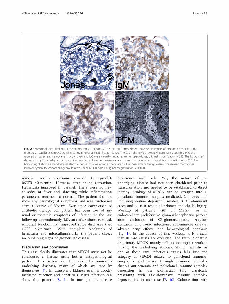
Case presentationA 43-year-old Caucasian woman, who had undergonecadaveric kidney transplantation 4 years earlier, was referredto us due to a creeping rise in serum creatinine. Hermedication included cyclosporine, mycophenolate-mofetil,prednisone, omeprazole, and oral sodium bicarbonate. Shehad been on hemodialysis for 3 years before receiving theallograft. To determine the diagnosis leading to end-stagerenal disease, a native kidney biopsy had been performed 12years ago showing MPGN in histopathology. According toher file, a further diagnostic workup back then had notrevealed any signs of an underlying condition leading to theclassification as idiopathic MPGN. However, due to the longtime passed, no data was available regarding the details ofthe exams that had been performed. Upon presentation, thepatient reported malaise, elevated body temperature andshivering when combing her hair or working over-head. Shehad been experiencing occasional milder episodes of unclearinfection during the last decades. Aside from an elevatedarterial pressure of 165/85mmHg, the physical examinationwas unremarkable. We conducted a complete laboratoryblood analysis, which showed a serum creatinine of183 μmol/L (eGFR CKD-EPI 24ml/min; best creatinineafter transplantation: 92.3 μmol/L, eGFR 56.1ml/min), amildly elevated c-reactive protein of 275 nmol/L (28.9mg/L), a significantly increased procalcitonin (11.1 μg/L), lowC3 complement (0.78 g/L), and cyclosporine levels withinthe target range. The initial urine analysis revealedmicrohematuria with mild albuminuria of 49mg/gcreatinine. Repeated blood and urine microbial culturesremained sterile. Ultrasound examination of the transplantkidney revealed a normal size without parenchymal orvascular abnormalities. Kidney biopsy showed an activeendocapillary proliferative glomerulonephritis - consistentwith an early stage of recurrent membranoproliferative
glomerulonephritis type I (Fig. 2) with electron densesubendothelial deposits lacking significant glomerularbasement membrane splitting. Deposits were positive forIgM, C1q and C4d, with IgA, IgG and C3c virtuallynegative, excluding a C3-glomerulopathy. Serological studiesfor human immunodeficiency virus, hepatitis virus,Epstein-Barr virus, cytomegalovirus, parvovirus B19 showedno signs of active disease. Monoclonal gammopathy wasexcluded by serum and urine immuneelectrophoresisand a serum free light-chain assay. Transesophagealechocardiography demonstrated absence of endocarditicvegetations. Other infectious foci were excluded bysonography and clinical examination. DsDNA-antibodies,anti-neutrophil antibodies, and donor-specific antibodieswere not detected. We found anti-nuclear antibodies(subtype SS-A / Ro-60) with a titer of 1:320. Apart from thetransplant dysfunction there were no clinical signs ofspecific autoimmune diseases. The patient did not reportrecent travels to foreign countries. A ventriculoperitonealshunt (VP) had been placed at the age of 14 due tocongenital aqueductal stenosis and hydrocephalus. At theage of 21 the VP shunt had been switched to aventriculoatrial shunt (VA) due to recurrent abdominalpain. It had not been possible to recover remnants of theVP-shunt, which had remained in situ.Shivering on working over-head, recurrent laboratory
signs of inflammation, and excluding other causes ofMPGN were the clinical cues that led us to suspect achronically infected VA-shunt as trigger of transplantfailure.To assess the feasibility of shunt removal, we conducted
an MRI of the cerebrospinal fluid circulation andinterspaces. Imaging revealed a triventricular hydrocephaluswith aqueductal stenosis but patent interventricularforamina. Thus, the patient was subjected to endoscopicventriculocisternostomy and placement of an externalliquor drainage, which was removed as soon as theventriculocisternostomy had proven to drain liquorefficiently. VP- and VA-shunt material was removedsurgically. Due to extensive calcification of the VA-shunt-fragment in the superior VC, a 5 cm fragment could not bemobilized manually and had to be removed interventionallyvia femoral vein access, thus obviating open cardiothoracicsurgery. The calcifications remained in place. Despitenegative blood and cerebrospinal fluid (CSF)-cultures,microbial analysis detected Staphylococcus hominis andepidermidis on the VP-shunt and Staphylococcusepidermidis on the atrial catheter fragment. All recoveredintracranial material remained culture-negative. Inaccordance with the resistogram, flucloxacilline (3 g IVQID) was administered for 14 days intravenously; antibiotictherapy was continued for another ten days usingamoxicillin/clavulanic acid (500/125mg PO TID). Kidneyfunction began to improve immediately after shunt
Völker et al. BMC Nephrology (2019) 20:296 Page 3 of 6
removal, serum creatinine reached 119.8 μmol/L(eGFR 40 ml/min) 10 weeks after shunt extraction.Hematuria improved in parallel. There were no newepisodes of fever and shivering while inflammationparameters returned to normal. The patient did notshow any neurological symptoms and was dischargedafter a course of 39 days. Ever since completion ofantibiotic therapy our patient has been free of anyrenal or systemic symptoms of infection at the lastfollow-up approximately 1.5 years after shunt removal.Allograft function has improved since discharge (lasteGFR 46 ml/min). With complete resolution ofhematuria and microalbuminuria, the patient showsno remaining signs of glomerular disease.
Discussion and conclusionThis case clearly illustrates that MPGN must not beconsidered a disease entity but a histopathologicalpattern. This pattern can be caused by numerousunderlying diseases, many of which are rare bythemselves [7]. In transplant kidneys even antibody-mediated rejection and hepatitis C-virus infection canshow this pattern [8, 9]. In our patient, disease
recurrence was likely. Yet, the nature of theunderlying disease had not been elucidated prior totransplantation and needed to be established to directtherapy. Etiology of MPGN can be grouped into 1.polyclonal immune-complex mediated, 2. monoclonalimmunoglobuline deposition related, 3. C3-dominantcases and 4. as a result of primary endothelial injury.Workup of patients with an MPGN (or anendocapillary proliferative glomerulonephritis) patternafter exclusion of C3-glomerulopathy requiresexclusion of chronic infections, autoimmune disease,adverse drug effects, and hematological neoplasia(Fig. 1). In the course of this workup, it is crucialthat all rare causes are excluded. The term idiopathicor primary MPGN mainly reflects incomplete workupmissing the underlying etiology. Shunt nephritis asone of these rare infectious causes falls into thecategory of MPGN related to polyclonal immune-complexes and arises through immune complexchronic antigenemia and polyclonal immune complexdeposition in the glomerular tuft, classicallypresenting with IgM-dominant immune complexdeposits like in our case [7, 10]. Colonization with
Fig. 2 Histopathological findings in the kidney transplant biopsy. The top left (Jones) shows increased numbers of mononuclear cells in theglomerular capillaries (arrows). Jones silver stain, original magnification × 400. The top right (IgM) shows IgM dominant deposits along theglomerular basement membrane in brown. IgA and IgG were virtually negative. Immunoperoxidase, original magnification × 630. The bottom leftshows strong C1q co-deposition along the glomerular basement membrane in brown. Immunoperoxidase, original magnification × 630. Thebottom right shows subendothelial electron dense immune complex deposits on the inner side of the glomerular basement membranes(arrows), typical for endocapillary proliferative GN or MPGN type I. Original magnification × 10,000
Völker et al. BMC Nephrology (2019) 20:296 Page 4 of 6
Staphylococcus epidermidis is believed to be the mostfrequent cause of shunt nephritis although multipleother pathogens have been identified [6, 11–14].Similar processes can take place in case of bacterialendocarditis. Shunt nephritis can manifest as diffuseendocapillary proliferative glomerulonephritis similarto poststreptococcal GN, as mesangioproliferative GN,or as MPGN [3]. The severity of GN predominantlydepends on the duration of antigenemia and chronicinfection. Most cases have a subacute course withneurological symptoms and rapid decline of kidneyfunction so that early shunt removal is critical. Ifcomplete shunt removal is possible, renal prognosis isfavorable [15].The case presented clearly shows the importance of
being aware of shunt nephritis as a differential diagnosis ofdiseases underlying MPGN or endocapillary proliferativeGN. In hindsight, it is highly likely that the MPGN inthe patient’s native kidneys was in fact shuntnephritis. However, due to an equivocal clinicalpicture and a lack of alternative therapeutic optionsat that time, the shunt had remained in place.Differently from most cases reported so far, ourpatient presented with a rather chronic course of thedisease - as indicated by persistently negative bloodand CSF-cultures and lack of neurological symptoms– which may be the consequence of slowly growingbacteria with low pathogenicity.The remaining calcifications in the superior VC may
still harbor pathogens causing persistent antigenemiapotentially leading to immune complex formation anddeposition. However, in contrast to artificial shuntmaterial, which is predisposed to staphylococcal biofilmformation, calcifications undergo constant turnover sothat innate host immune mechanisms may clearremaining pathogens. The optimal duration ofantibiotic therapy after shunt removal remains to bedetermined.
AbbreviationsANCA: Anti-neutrophil cytoplasmic antibody; CMV: Cytomegalovirus;CSF: Cerebrospinal fluid; dsDNA: Double stranded desoxyribonucleic acid;EBV: Epstein-Barr virus; GN: Glomerulonephritis;MPGN: Membranoproliferative glomerulonephritis; MRI: Magnetic resonanceimaging; TMA: Thrombotic microangiopathy; VA: Ventriculoatrial;VP: Ventriculoperitoneal
AcknowledgementsNot applicable.
Authors’ contributionsRUM and LV wrote the main part of the manuscript. RUM, LV and JBdesigned the Figures. JB, KB, NS, FG, CK, TB and JH contributed to writingthe manuscript. All authors read and approved the final manuscript.
FundingRUM receives funding from the German Research Foundation (DFG) and theMinistry of Science Northrine-Westfalia (MIWF). JUB receives funding from
the DFG. No external funding source was involved in the design and writingof this case report.
Availability of data and materialsThe data set used in this study has been reported in the case presentation.Additional clinical data and laboratory values are available upon requestfrom the corresponding author.
Ethics approval and consent to participateEthics approval not applicable since the manuscript is a case report. Writtenconsent was obtained and a copy of written consent is available uponrequest.
Consent for publicationWritten informed consent was obtained from the participant for publicationof this case report and any accompanying tables/images. A copy of thewritten consent is available for review by the Editor of this journal.
Competing interestsThe authors declare that they have are no competing interest.
Author details1Department II of Internal Medicine and Center for Molecular MedicineCologne University of Cologne, Faculty of Medicine and University Hospitalof Cologne, Uniklinik Köln, Kerpener Str. 62, 50937 Cologne, Germany.2Department of Neurosurgery, University Hospital Cologne, Kerpener Str. 62,50937 Cologne, Germany. 3Institute of Pathology, University of Cologne,Cologne, Germany.
Received: 22 April 2018 Accepted: 19 July 2019
References1. Sethi S, Fervenza FC. Membranoproliferative glomerulonephritis — a new
look at an old entity. N Engl J Med. 2012;366(12):1119–31.2. Ohara S, Kawasaki Y, Takano K, Isome M, Nozawa R, Suzuki H, et al.
Glomerulonephritis associated with chronic infection from long-term centralvenous catheterization. Pediatr Nephrol Berl Ger. 2006;21(3):427–9.
3. Arze RS, Rashid H, Morley R, Ward MK, Kerr DN. Shunt nephritis: report oftwo cases and review of the literature. Clin Nephrol. 1983 Jan;19(1):48–53.
4. Haffner D, Schindera F, Aschoff A, Matthias S, Waldherr R, Schärer K. Theclinical spectrum of shunt nephritis. Nephrol Dial Transplant Off Publ EurDial Transpl Assoc - Eur Ren Assoc. 1997;12(6):1143–8.
5. Kiryluk K, Preddie D, D’Agati VD, Isom R. A young man withPropionibacterium acnes-induced shunt nephritis. Kidney Int. 2008;73(12):1434–40.
6. Groeneveld AB, Nommensen FE, Mullink H, Ooms EC, Bode WA. Shuntnephritis associated with Propionibacterium acnes with demonstration ofthe antigen in the glomeruli. Nephron. 1982;32(4):365–9.
7. Cook HT, Pickering MC. Histopathology of MPGN and C3 glomerulopathies.Nat Rev Nephrol. 2015;11(1):14–22.
8. Kamar N, Izopet J, Alric L, Guilbeaud-Frugier C, Rostaing L. Hepatitis C virus-related kidney disease: an overview. Clin Nephrol. 2008;69(3):149–60.
9. Grau V, Zeuschner P, Immenschuh S, Bockmeyer CL, Zell S, Wittig J, et al.Immune complex-type deposits in the Fischer-344 to Lewis rat model ofrenal transplantation and a subset of human transplant Glomerulopathy.Transplantation. 2016;100(5):1004–14.
10. Dobrin RS, Day NK, Quie PG, Moore HL, Vernier HL, Michael AF, et al. Therole of complement, immunoglobulin and bacterial antigen in coagulase-negative staphylococcal shunt nephritis. Am J Med. 1975;59(5):660–73.
11. Black JA, Challacombe DN, Ockenden BG. Nephrotic syndrome associatedwith bacteraemia after shunt operations for hydrocephalus. Lancet LondEngl. 1965;2(7419):921–4.
12. Schoenbaum SC, Gardner P, Shillito J. Infections of cerebrospinal fluidshunts: epidemiology, clinical manifestations, and therapy. J Infect Dis.1975;131(5):543–52.
13. Stickler GB, Shin MH, Burke EC, Holley KE, Miller RH, Segar WE. Diffuseglomerulonephritis associated with infected ventriculoatrial shunt. N Engl JMed. 1968;279(20):1077–82.
Völker et al. BMC Nephrology (2019) 20:296 Page 5 of 6

14. Strife CF, McDonald BM, Ruley EJ, McAdams AJ, West CD. Shunt nephritis:the nature of the serum cryoglobulins and their relation to the complementprofile. J Pediatr. 1976;88(3):403–13.
15. Wakabayashi Y, Kobayashi Y, Shigematsu H. Shunt nephritis: histologicaldynamics following removal of the shunt. Case report and review of theliterature. Nephron. 1985;40(1):111–7.
16. Larsen CP, Messias NC, Walker PD, Fidler ME, Cornell LD, Hernandez LH, etal. Membranoproliferative glomerulonephritis with masked monotypicimmunoglobulin deposits. Kidney Int. 2015;88(4):867–73.
Publisher’s NoteSpringer Nature remains neutral with regard to jurisdictional claims inpublished maps and institutional affiliations.
Völker et al. BMC Nephrology (2019) 20:296 Page 6 of 6