Embed Size (px)
Citation preview
Case ReportTwo Case Reports on Thalamic and Basal Ganglia Involvementin Children with Dengue Fever
Guwani Liyanage,1 Lihini Adhikari,2 Saraji Wijesekera,1
Maheshaka Wijayawardena,2 and Suchithra Chandrasiri2
1Department of Paediatrics, Faculty of Medical Sciences, University of Sri Jayewardenepura, Sri Soratha Mawatha,10250 Nugegoda, Sri Lanka2Professorial Paediatric Unit, Colombo South Teaching Hospital, Kalubowila, 10350 Dehiwala, Sri Lanka
Correspondence should be addressed to Guwani Liyanage; [email protected]
Received 17 March 2016; Revised 11 June 2016; Accepted 19 June 2016
Academic Editor: Larry M. Bush
Copyright © 2016 Guwani Liyanage et al. This is an open access article distributed under the Creative Commons AttributionLicense, which permits unrestricted use, distribution, and reproduction in any medium, provided the original work is properlycited.
There have been increasing numbers of case reports of dengue infection with unusual manifestations. Such unusual manifestationsincluding acute liver failure and encephalopathy could be manifested even in the absence of significant plasma leakage. Further,severe organ involvement including nervous system involvement indicates severe dengue infection. However, neurologicalmanifestations of dengue fever are rare.This is the first case report of dengue infection with thalamic and basal ganglia involvementin Sri Lanka.
1. Introduction
The clinical manifestations of dengue fever form a broadspectrum including uncomplicated dengue fever, denguehemorrhagic fever, and dengue shock syndrome according tothe traditional classification. However, in 2009, World healthOrganization (WHO) has introduced a new classification andit categorizes the disease into dengue without warning signs,dengue with warning signs, and severe dengue [1]. Impor-tantly, encephalopathy, encephalitis, and any other unusualneurological involvement are recognized as manifestations ofsever dengue. However, neurological manifestations are rarein dengue infection. There are few case reports of thalamicand basal ganglia involvement in dengue fever [2–4]. Mostof these patients had full recovery with transient thalamicinvolvement on neuroimaging. However, this is the firstreport of dengue infection complicated by thalamic and basalganglia involvement in children in Sri Lanka.
2. Case Reports
Patient 1 was a 10-year-old girl admitted with 2 days of fever,retroorbital headache, vomiting, and myalgia. Her white
blood cell (WBC) count on the 3rd daywas 2.2× 10³ with 65%neutrophils and platelet count of 117,000. Alanine transam-inase (ALT) was 17.8 and aspartate transaminase (AST) was55.2. Treatment was commenced as dengue fever in febrilephase. By the 4th day, she became sleepy and lethargic andthe level of consciousness gradually deteriorated. She wasmoved to high dependency care for further management.On neurological examination, she had increased tone in allfour limbs with cogwheel rigidity. The lowest Glasgow comascale documented was 10/15. Her vital parameters and serialpacked cell volume readings remained stable without signs offluid leakage.
Lowest WBC count (1.9 × 10³) was reported on day5 of the illness and lowest platelet count was 50,000/mm3on day 6. Liver enzymes deteriorated (ALT: 357, AST: 478)and serum albumin was 38 g/dL and INR was 1.17 on day6. Tender liver was palpable 2 cm below the costal margin.Renal functions (sodium: 142mEq/L, potassium: 4.2mEq/L,blood urea: 7.6mmol/L, and serum creatinine: 52 𝜇mol/L)remainednormal. Serumcalcium remained between 2.08 and2.69mmol/L. Primary dengue infection was suggested by thepresence of IgM antibodies and absence of IgG antibodies todengue virus. Dengue NS-1 antigen and the viral load were
Hindawi Publishing CorporationCase Reports in Infectious DiseasesVolume 2016, Article ID 7961368, 3 pageshttp://dx.doi.org/10.1155/2016/7961368
2 Case Reports in Infectious Diseases
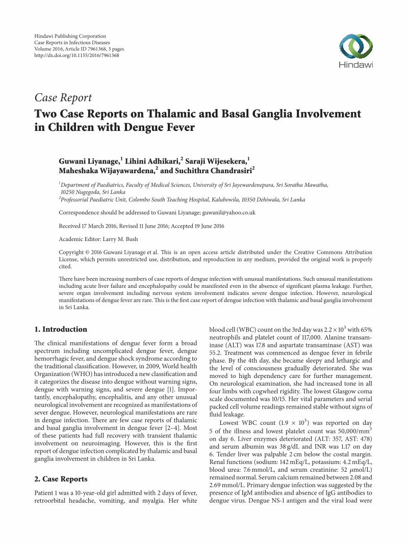
Figure 1: Axial brain CT image (with contrast) showing hypodenseareas in the region of thalamus and basal ganglia.
not performed due to lack of laboratory support. A CT scanrevealed hypodense areas in bilateral thalamic and basal gan-glia regions (Figure 1). Cerebrospinal fluid examination didnot reveal any cells and had normal protein levels (42mg/dL).Over the next 2 weeks following the acute illness, she showedcomplete recovery with no residual neurological disability.
Patient 2 was a 3-year-old girl who presented with 3days of continuous high fever, intermittent abdominal pain,vomiting, and retroorbital headache. There had been oneepisode of gum bleeding on the first day of illness. She hadbeen apparently a well child without any significant illnessesin the past. WBC count done on day 2, on request of thegeneral practitioner, was 8.15 × 10³ with 85% neutrophils andplatelet count was 120,000mm3. She had right hypochondrialtenderness with just palpable liver on day 5. ALT and ASTwere 16mEq/L and 69.7mEq/L, respectively, on day 3 andthey increased to 29.7mEq/L and 122.2mEq/L on day 5.Initial C-reactive protein was 6mg/dL and remained normal.She was monitored for vital signs including urine output andpacked cell volume (PCV). Baseline PCV was 32 and thehighest noted was 35. The lowest platelet count documentedwas 67,000/mm3 and the lowest WBC count was 2.27 × 10³.She did not show signs of fluid leakage. However, there was asecond episode of gumbleeding onday 5with normal clottingprofile (INR: 1.1 and APTT: 35). She was positive for IgM andnegative for IgG antibodies for dengue virus.
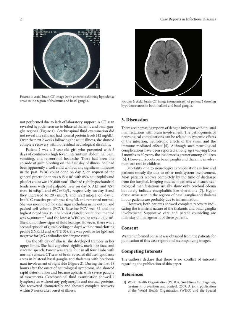
On the 5th day of illness, she developed tremors in herupper limbs. She had cogwheel rigidity, mask like face, andstaccato speech. Power was grade four in all four limbs withnormal reflexes. CT scan of brain revealed diffuse hypodenseareas in bilateral basal ganglia and thalamus with predomi-nant involvement of right side (Figure 2). During the first 48hours after the onset of neurological symptoms, she showedrapid deterioration and became aphasic with severe paucityof movements. Cerebrospinal fluid examination showed 2lymphocytes without any polymorphs and normal proteins.She recovered dramatically and showed complete recoverywithin 3 weeks after onset of illness.
Figure 2: Axial brain CT image (noncontrast) of patient 2 showinghypodense areas in both thalami and basal ganglia.
3. Discussion
There are increasing reports of dengue infectionwith unusualmanifestations with brain involvement. The pathogenesis ofneurological complications can be related to systemic effectsof the infection, neurotropic effects of the virus, and theimmune mediated effects [5]. Although such neurologicalcomplications have been reported among ages varying from3months to 60 years, the incidence is greater among children[6]. However, reports on basal ganglia and thalamic involve-ment are rare in children.
Mortality due to neurological complications is low andpatients mostly die due to other multisystem involvement.Most patients recover completely by the time of dischargefrom the hospital. Imaging studies of patients with such neu-rological manifestations usually show only cerebral edemabut rarely indicate encephalitis like alterations [7]. Hypo-dense areas seen in the regions of basal ganglia and thalamiin our patients are probably due to inflammation.
However, both patients showed complete recovery indi-cating the transient nature of the thalamic and basal gangliainvolvement. Supportive care and parent counseling aremainstay of management of these patients.
Consent
Written informed consent was obtained from the patients forpublication of this case report and accompanying images.
Competing Interests
The authors declare that there is no conflict of interestsregarding the publication of this paper.
References
[1] World Health Organization (WHO), Guidelines for diagnosis,treatment, prevention and control. 2009. A joint publicationof the World Health Organization (WHO) and the Special
Case Reports in Infectious Diseases 3
Programme for Research and Training in Tropical Diseases(TDR), http://www.who.int/tdr/publications/documents/den-gue-diagnosis.pdf.
[2] A. K. Mallick, R. Purkait, and T. K. Sinhamahapatra, “Denguefever with unusual thalamic involvement,” Journal of the IndianMedical Association, vol. 110, no. 1, pp. 48–49, 2012.
[3] S. K. Bhoi, S. Naik, S. Kumar, R. V. Phadke, J. Kalita, and U.K. Misra, “Cranial imaging findings in dengue virus infection,”Journal of the Neurological Sciences, vol. 342, no. 1-2, pp. 36–41,2014.
[4] R. Kamble, J. N. Peruvamba, J. Kovoor, S. Ravishankar, and B.S. Kolar, “Bilateral thalamic involvement in dengue infection,”Neurology India, vol. 55, no. 4, pp. 418–419, 2007.
[5] M. Hemungkorn, U. Thisyakorn, and C. Thisyakorn, “Dengueinfection: a growing global health threat,”BioScience Trends, vol.1, no. 2, pp. 90–96, 2007.
[6] J. Thakare, B. Walhekar, and K. Banerjee, “Hemorrhagic man-ifestations and encephalopathy in cases of dengue in India,”Southeast Asian Journal of Tropical Medicine and Public Health,vol. 27, no. 3, pp. 471–475, 1996.
[7] M. Wasay, R. Channa, M. Jumani, G. Shabbir, M. Azeemuddin,and A. Zafar, “Encephalitis and myelitis associated with dengueviral infection. Clinical and neuroimaging features,” ClinicalNeurology and Neurosurgery, vol. 110, no. 6, pp. 635–640, 2008.

Submit your manuscripts athttp://www.hindawi.com
Stem CellsInternational
Hindawi Publishing Corporationhttp://www.hindawi.com Volume 2014
Hindawi Publishing Corporationhttp://www.hindawi.com Volume 2014
MEDIATORSINFLAMMATION
of
Hindawi Publishing Corporationhttp://www.hindawi.com Volume 2014
Behavioural Neurology
EndocrinologyInternational Journal of
Hindawi Publishing Corporationhttp://www.hindawi.com Volume 2014
Hindawi Publishing Corporationhttp://www.hindawi.com Volume 2014
Disease Markers
Hindawi Publishing Corporationhttp://www.hindawi.com Volume 2014
BioMed Research International
OncologyJournal of
Hindawi Publishing Corporationhttp://www.hindawi.com Volume 2014
Hindawi Publishing Corporationhttp://www.hindawi.com Volume 2014
Oxidative Medicine and Cellular Longevity
Hindawi Publishing Corporationhttp://www.hindawi.com Volume 2014
PPAR Research
The Scientific World JournalHindawi Publishing Corporation http://www.hindawi.com Volume 2014
Immunology ResearchHindawi Publishing Corporationhttp://www.hindawi.com Volume 2014
Journal of
ObesityJournal of
Hindawi Publishing Corporationhttp://www.hindawi.com Volume 2014
Hindawi Publishing Corporationhttp://www.hindawi.com Volume 2014
Computational and Mathematical Methods in Medicine
OphthalmologyJournal of
Hindawi Publishing Corporationhttp://www.hindawi.com Volume 2014
Diabetes ResearchJournal of
Hindawi Publishing Corporationhttp://www.hindawi.com Volume 2014
Hindawi Publishing Corporationhttp://www.hindawi.com Volume 2014
Research and TreatmentAIDS
Hindawi Publishing Corporationhttp://www.hindawi.com Volume 2014
Gastroenterology Research and Practice
Hindawi Publishing Corporationhttp://www.hindawi.com Volume 2014
Parkinson’s Disease
Evidence-Based Complementary and Alternative Medicine
Volume 2014Hindawi Publishing Corporationhttp://www.hindawi.com


![RESEARCH ARTICLE Open Access Abnormalities of cortical ......campus [18], and basal ganglia [19]. These abnormal brain regions are predominantly located in the limbic-cortical-striatal-pallidal-thalamic](https://img.pdfslide.net/doc/110x75/60c2168295781709ed2436d2/research-article-open-access-abnormalities-of-cortical-campus-18-and.jpg)