Embed Size (px)
Citation preview
Abstract. Transformed cells are subject to intercellularinduction of apoptosis by neighbouring nontransformed cellsand to autocrine apoptotic self-destruction. Both processesdepend on extracellular superoxide anion generation by thetransformed cells and on the release of peroxidase from bothnontransformed and transformed cells. This concerted actionresults in HOCl synthesis, HOCl-superoxide anion interactionand generation of apoptosis-inducing hydroxyl radicals. Incontrast to transformed cells, ex vivo tumor cells are resistantagainst intercellular induction of apoptosis and autocrineapoptotic self-destruction. Resistance of tumor cells againstintercellular ROS signaling depends on interference throughcatalase expression on the membrane. Intercellular ROSsignaling of tumor cells can be restored when i) exogenousHOCl is added; ii) exogenous hydrogen peroxide is supplied,or iii) catalase is inhibited. These findings define thebiochemical basis for specific apoptosis induction in tumorcells through re-establishment of intercellular ROS signaling,a potential novel approach in tumor prevention and therapy.
Multistep oncogenesis is characterized by distinct andinterconnected steps such as abrogation of senescence control,oncogene activation, tumor suppressor gene inactivation (1-3), independence of exogenous proliferation signals throughautocrine mechanisms (4-6), independence of control byneighbouring cells (7-10), acquisition of new defensemechanisms by the tumor cells (11-13), escape from immunesurveillance (2), resistance against hypoxia-induced p53-mediated cell death (14, 15), tumor angiogenesis and others(reviewed in 16-20).
Generation of extracellular superoxide anions through amembrane-associated NADPH oxidase (Nox1) is associatedwith oncogene activation and seems to represent one of thehallmarks of the transformed state (21-28). Ras and rac playcentral roles for the activation of Nox1 (21, 29). The activatedNADPH oxidase seems to be required for the control ofproliferation and the maintenance of the transformed state(21, 23, 24, 28, 30), changes in the cytoskeleton oftransformed cells (31) and induction of the angiogenic switch(32). Nox1 activity also seems to be relevant fortumorigenesis in vivo (28, 33, 34).
On the flip side of the coin, extracellular superoxide anionsgenerated by transformed cells drive both the efficiency andselectivity of intercellular induction of apoptosis, a hithertounrecognized potential control step during multistageoncogenesis (17-20, 25, 26, 35). During intercellular inductionof apoptosis, transformed cells are selectively induced to dieby apoptosis after a concerted action of transformed cell-derived reactive oxygen species (ROS) and signalingcomponents released by surrounding nontransformed cells(17-19, 25, 26). Four signaling pathways have been elucidatedso far: i) the HOCl signaling pathway (25), ii) the nitric oxide(NO)/peroxynitrite signaling pathway (25, 27), iii) the nitrylchloride signaling pathway (36) and iv) the metal ioncatalyzed Haber Weiss reaction (37). In many transformed cellsystems studied by our group, the HOCl pathway representedthe major signaling pathway of intercellular induction ofapoptosis. Therefore, this pathway is the focus of the studyreported here.
The HOCl signaling pathway depends on the generation ofsuperoxide anions by the transformed target cells (25). Theirdismutation product hydrogen peroxide is utilized by a novelperoxidase for the synthesis of HOCl. HOCl in themicromolar concentration range does not affect cells directly(38, 39). However, the interaction of HOCl with superoxideanions (40-42) leads to the generation of hydroxyl radicals,which have a very limited free diffusion path length and havethe ability to trigger the onset of apoptosis through lipidperoxidation. As superoxide anions and hydroxyl radicalshave relatively short free diffusion path lengths (43, 44),apoptosis induction is selectively directed against the
4541
*Present address: Harvard Medical School, BIDMC, Departmentof Matrix Biology, Boston, MA 02215, U.S.A.
Correspondence to: Georg Bauer, Abteilung Virologie, Institut fürMedizinische Mikrobiologie und Hygiene, Hermann-Herder Strasse11, D-79104 Freiburg, Germany. e-mail: [email protected]
Key Words: HOCl signaling pathway, hydrogen peroxide,hypochlorous acid, hydroxyl radical.
ANTICANCER RESEARCH 29: 4541-4558 (2009)
Catalase Protects Tumor Cells from Apoptosis Induction by Intercellular ROS Signaling
WIBKE BECHTEL* and GEORG BAUER
Abteilung Virologie, Institut für Medizinische Mikrobiologie und Hygiene, Universität Freiburg, D-79104 Freiburg, Germany
0250-7005/2009 $2.00+.40
transformed target cells. As nontransformed cells generatefewer extracellular superoxide anions than their transformedcounterparts (25-27, 38), they are not affected by HOCl in themicromolar concentration range.
Ongoing work has demonstrated that intercellular inductionof apoptosis does not necessarily require the presence ofnontransformed cells, as peroxidase is also released bytransformed cells themselves (Bauer, unpublished result).Therefore, the presence of transformed cells at high densityand in high numbers allows the establishment of autocrineapoptotic ROS-mediated self-destruction of transformed cells.This process depends on the same signaling chemistry as theinteraction between nontransformed and transformed cells(Bauer, unpublished result). Likewise, autocrine apoptoticself-destruction is highly selective for transformed cells.Again, this selectivity is based on extracellular superoxideanion generation by the transformed cells. During autocrineself-destruction of transformed cells, sufficient local densityof the cells ensures optimal generation of hydrogen peroxidethrough dismutation of superoxide anions and using asufficient total number of cells per assay ensures an optimalsupply of peroxidase that is released into the medium.
Autocrine apoptotic self-destruction can be tested in twoways. When clumps of transformed cells (2,000 cells, 300cells/mm2) are surrounded by 15,000 dispersely seededeffector cells (40 cells/mm2), the transformed cells areinduced to die by apoptosis, but the effector cells are not,even if they are transformed. This is due to insufficienthydrogen peroxide generation in the dispersely seeded cellsand sufficient hydrogen peroxide generation in the denselyseeded target cells, followed by the interaction of target cell-derived hydrogen peroxide with effector cell-derivedperoxidase. If a clump of target cells is seeded alone, itsperoxidase is diluted and therefore apoptosis is not induceddespite sufficient hydrogen peroxide. Dispersely seededeffector cells generate sufficient peroxidase but, even if theyare transformed, do not generate sufficient hydrogen peroxidedue to suboptimal density. Alternatively, autocrine apoptoticself-destruction can be determined when homogeneouspopulations of transformed cells are seeded at optimal celldensity, cell number and volume of overlaying medium (fordetails please see the Methods section).
Whereas the term ‘intercellular induction of apoptosis’ hasbeen used for the interaction between nontransformed andtransformed cells, the term ‘autocrine apoptotic self-destruction’ is restricted to the interaction of transformedcells. We suggest using the term ‘intercellular ROS signaling’to describe the signaling chemistry of both processes, as it isidentical except for the source of effector molecules such asperoxidase or NO. We also suggest to use the operationalterms ‘target cells’ for the superoxide anion generating cellsthat are subject to apoptosis induction by intercellular ROSsignaling and ‘effector cells’ for the cells that supply
intercellular ROS signaling with free peroxidase and NO. The‘target cell function’ is strictly dependent on the transformedstate of the cells, whereas the ‘effector cell function’ is not.Whereas cells transformed in vitro show sensitivity againstintercellular induction of aptoptosis and autocrine self-destruction, independently of the origin of tissue and thetransforming principle, bona fide tumor cells established fromtumors showed resistance against intercellular induction ofapoptosis (45, 46). This resistance might be caused by amultitude of different biochemical effects: tumor cells mighthave defects in their apoptosis machinery, lack superoxideanion generation or generate insufficient concentrations ofsuperoxide anions, have established strong intracellulardefense mechanisms against apoptosis-inducing signals orinterfere with intercellular ROS signaling through expressionof antioxidative enzymes. If the latter scenario were true, itshould be possible to resensitize tumor cells for ROS-mediated apoptosis induction through enhancement ofsignaling components or through the inhibition of theinterfering enzyme(s). Thus the aim of the present study wasto define the exact biochemical mechanism of tumor cellresistance against intercellular ROS signaling.
Materials and Methods
Materials. 4-(2-Aminoethyl-benzenesulfonyl fluoride (AEBSF), aspecific inhibitor of NADPH oxidases (47), was obtained fromSigma-Aldrich (Schnelldorf, Germany) and stored as a stock solutionof 10 mM in phosphate-buffered saline (PBS) at –20˚C. 4-Aminobenzoyl hydrazide (ABH), a mechanism-based inhibitor ofMPO (48, 49), obtained from Acros Organics (Geel, Belgium) wasdissolved in dimethyl sulfoxide (DMSO) at a concentration of 1 M. Itwas then diluted with medium to a concentration of 1 mM (stocksolution). The stock solution was kept at –20˚C. The catalaseinhibitior 3-aminotriazole (3-AT) [for review of its action see (50)]was obtained from Sigma - Aldrich. The stock solution (2 M in sterilePBS) was stored at –20˚C. Dimethylthiourea (DMTU), a hydroxylradical-specific scavenger, was obtained from Sigma Aldrich and keptas a stock solution of 1M in PBS at –20˚C. NaOCl was obtained fromSigma, Schnelldorf, Germany. The stock solution of 860 mM waskept at 4˚C in the dark. As the pKa of OCl– is 7.64, the majority ofthe species are present as HOCl at neutral pH. For simplicity, the term‘HOCl’ is used through out this paper. HOCl was diluted in cold,sterile PBS and then added to the assays as a single aliquot (10-20 μlper 100 μl assay). Care was taken to add HOCl at a similar speed andfrom the same distance above the medium in order to avoiddifferences in local concentration of HOCl immediately after addition.Mannitol, a hydroxyl radical-specific scavenger (51), was obtainedfrom Sigma Aldrich and kept as a stock solution of 1 M in PBS at–20˚C. Taurine (Sigma Aldrich), a HOCl-specific scavenger (52), waskept as a stock solution of 500 mM in sterile PBS at –20˚C and wasused at a concentration of 50 mM in our assays. Glucose oxidase(GOX, from Aspergillus niger) generates hydrogen peroxide usingglucose as substrate. (Glucose is present in abundance in Eagle’sminimal essential medium (EMEM) and RPMI-1640 medium.) GOXwas obtained from Sigma Aldrich (Schnelldorf, Germany) and keptas 6,000 U/ml stock solution at 4˚C. Myeloperoxidase (MPO, from
ANTICANCER RESEARCH 29: 4541-4558 (2009)
4542
human leukocytes) was obtained from Sigma Aldrich. Stock solution(5 U/ml) in EMEM with 5% fetal bovine serum (FBS) was kept at–20˚C and only used once per aliquot. MPO catalyzes the generationof HOCl from H2O2 and chloride (53). Manganese-containingsuperoxide dismutase (Mn-SOD) from Escherichia coli (SigmaAldrich) (stock solutions 30,000 Units/ml in sterile PBS) were kept at–20˚C and only used once per aliquot. Mn-SOD is an efficientscavenger of superoxide anions, in a two step reaction. Mn-SOD isnot cellpermeable (54, 55) and therefore allows the functional role ofextracellular superoxide anions to be demonstrated. Mn-SOD doesnot exhibit the sharp bell-shaped inhibition curve that is characteristicfor copper-containing SOD and therefore is superior to Cu-SOD ininhibition studies. Transforming growth factor β-1 (TGF-β-1) waspurified from human platelets (56) and kept as a stock solution of 1.5μg/ml in EMEM plus 5% FBS at –20˚C. Caspase-3 inhibitior (Z-DEVD-FMK) and caspase-9 inhibitor (Z-LEHD-FMK) were obtainedfrom R&D Systems (Wiesbaden-Nordenstadt, Germany). Theinhibitors were first dissolved in DMSO to reach a concentration of60 mM and were then diluted with ethanol to a final concentration of20 mM. These stock solutions were kept at –20˚C and used withinthe subsequent weeks. Caspase-3 inhibitor was applied at a finalconcentration of 50 μM, caspase-9 inhibitior at a final concentrationof 25 μM. The residual DMSO concentration was below the criticalconcentration which affects ROS signaling.
Media for cell culture. Cells were either kept in EMEM, containing5% FBS or in RPMI-1640 medium, containing 10% FBS, asindicated for the respective cell lines. FBS (Biochrom, Berlin,Germany) had been heated for 30 minutes at 56˚C prior to use. Bothmedia were supplemented with penicillin (40 U/ml), streptomycin(50 μg/ml), neomycin (10 μg/ml), moronal (10 U/ml) and glutamine(280 μg/ml). Cell culture was performed in plastic tissue cultureflasks. Cells were passaged once or twice weekly.
Cells. ‘Nontransformed cells’ (208F) are normal rat fibroblasts thatdo not show criss cross morphology, colony formation in soft agarand are not tumorigenic. They do not show sufficient extracellularsuperoxide anion generation to be the target of intercellular ROSsignaling (25-27). Nontransformed 208F cells exhibit effectorfunction in vitro, i.e. the release of a novel peroxidase and, to alesser extent, nitric oxide (25) which both establish apoptosis-inducing ROS signaling in transformed cells. ‘Transformed cells’(208Fsrc3, FE-8, fgr413, fms41, raf55) are derived from 208F cellsthat have been transformed in vitro and are defined in the context ofthis article as having the potential for tumorigenesis without havingyet been confronted by the natural antitumor mechanisms of anorganism and the resultant selection processes. Transformed cellsshow criss cross morphology in monolayer, colony formation in softagar and extracellular superoxide anion generation that drives boththe efficiency and selectivity of intercellular ROS signaling (25-27).‘Tumor cells’ of murine (L929, CMS-5, SSK, CCL-107) or humanorigin (BG-1, MKN-45, SIHA) are defined in this article as havingbeen isolated from an in vivo tumor. L929, CMS-5, SSK and CCL-107 are fibrosarcomas, BG-1 has been isolated from an ovarialcarcinoma, MKN-45 from a gastric carcinoma and SIHA from acervical carcinoma. They form colonies in soft agar. Despiteextracellular superoxide anion generation, they are resistant tointercellular ROS signaling. The resistance mechanism andstrategies to resensitize tumor cells for intercellular ROS signalingare the focus of this paper. Nontransformed rat fibroblasts 208F and
their derivatives transformed through constitutive expression ofv-src (208Fsrc3), HRAS (FE-8), v-fgr (fgr 413), v-fms (fms41) andv-raf (raf-55) were established by and a generous, valuable gift byDr. C. Sers and Dr. R. Schäfer, Berlin, Germany. The transformedcell lines FE-8, fgr413, fms41 and raf-55 show similarcharacteristics to 208Fsrc3 cells. 208F cells and their transformedderivatives were cultured in EMEM, 5% FBS and supplemented asindicated above. The murine fibrosarcoma cell line L929 wasobtained from Dr. Adam, Kiel, Germany and was cultured inEMEM, 5% FBS and supplements. The murine fibrosarcoma celllines CMS-5 and SSK and the rat glioblastoma line CCL-107 (C6)have been recently described (46). They were cultured in EMEM,5% FBS and supplements. The human ovarial carcinoma cell lineBG-1 was obtained from Dr. T. Bauknecht, Freiburg, Germany; thecervix carcinoma line SIHA from Dr. L. Gissmann, DKFZHeidelberg, Germany. The cell lines were cultivated in EMEM, 5%FBS and supplements. The gastric carcinoma cell line MKN-45was purchased from DSMZ, Braunschweig, Germany. The cellsgrowing in suspension with some cells attaching to the plastic werecultured in RPMI-1640, 10% FBS and supplements. Care wastaken to avoid cell densities below 300,000/ml and above 106/ml.Under optimal conditions, the percentage of spontaneous apoptosisinduction was below 1%.
Apoptosis induction through intercellular ROS signaling. A. Tissue culture insert system (25, 35). For co-cultivation of cellswithout cell-to-cell contact, a combination of Falcon 6-well tissueculture clusters with tissue culture inserts (TCI) was used (pore-sizeof inserts 0.4 μm, distance between cell layers approximately 2 mm,Becton Dickinson, Heidelberg, Germany). Effector cells (i.e. thecells to be tested for support of intercellular ROS signaling throughrelease of peroxidase and NO) were seeded into the inserts (4×10⁴
cells per insert or as indicated in the respective figure legends).After the cells were attached, they were treated with 20 ng/ml TGF-βfor two days (37˚C, 5% CO2) or not, as indicated in the respectivefigure legends. Medium was then removed, the inserts were washedwith medium and placed above target cells (i.e. cells to be testedfor their apoptotic response to intercellular ROS signaling, based ontheir superoxide anion generation) in 6-well plates. Target cells wereseeded dispersely (40,000 cells per assay or number as indicated).Tissue culture inserts were placed above target cells within less thana day after the seeding of the latter. After the indicated time of co-culture, the assays were checked for the classical morphologicalsigns for apoptotic cells (membrane blebbing, chromatincondensation and fragmentation) using phase-contrast microscopyas described elsewhere (35, 57). We have recently confirmed (27,57) that chromatin condensation/fragmentation was paralled byDNA strand breaks, detectable by the TUNEL reaction, followingthe method described by Gorcyca et al. (58).
The percentage of apoptotic cells was determined from at least200 cells categorized per assay. Care was taken to differentiateapoptotic cells from nonapoptotic rounded cells with intact nuclei.
B. Coculture of clumps of target cells with overlaid dispersedeffector cells. Alternatively to the tissue culture insert system,apoptosis induction through intercellular ROS signaling can bemeasured when target cells are seeded as two clumps (2,000 cellsin 5 μl medium) in 12-well tissue culture clusters. After the cellsattached, the clumps were overlaid with 1 ml of medium and15,000 effector cells. TGF-β and inhibitors were added or not. Asessential control, clumps of target cells were cultivated in medium
Bechtel and Bauer: Tumor Cell Catalase and ROS Signaling
4543

in the absence of effector cells. As further control, disperselyseeded effector cells were cultivated in the absence of target cells.Apoptosis induction in the target cell clumps was determined asdescribed above for the tissue culture insert system. The principleof this assay is based on the support of peroxidase and NO by thedispersely seeded effector cells (which have high number but lowlocal density) to the target cells that are low in total cell numberbut high in local density. This high local density is required forefficient hydrogen peroxide formation through dismutation of targetcell-derived superoxide anions. Therefore, the combination ofdispersely seeded effector cells and target cells in clumps leads toapoptosis specifically in the target cells. This assay can be used forthe measurement of intercellular induction of apoptosis (whentransformed target cells are surrounded by nontransformed effectorcells) or autocrine apoptotic self-destruction (when transformedtarget cells are surrounded by dispersely seeded transformedeffector cells).
C. Direct measurement of autocrine apoptotic self-destruction.Cells to be tested were seeded at a density of 25,000 cells in 48 welltissue culture clusters (overlaied by 200 μl of complete medium)(Figures 1 B and 9) or 12,500 cells in 96 well tissue culture clusters(100 μl of complete medium) (Figures 4-8). After attachment of thecells, TGF-β as well as inhibitors were added or not, as indicatedin the respective figure legends. The percentage of apoptotic cellswas determined at the indicated times according to the criteriadescribed under A.
D. Treatment of tumor cells with specific compounds. Treatmentof tumor cells with HOCl, GOX, MPO, 3-AT in the absence orpresence of specific scavengers (AEBSF, Mn-SOD, taurine,mannitol, DMTU, ABH) or caspase-3 and caspase-9 inhibitors wasperformed under the conditions of direct measurement of autocrineapoptotic self-destruction (12,500 cells/100 μl complete medium in96-well tissue culture clusters). The concentrations of thecompounds added and the time of measurement are given in therespective figure legends.
Statistics. In all experiments, assays were performed in duplicate.The empirical standard deviation was calculated and is shown in thefigures. Absence of standard deviation bars for certain pointsindicates that the standard deviation was too small to be reportedby the graphic program, i.e. that results obtained in parallel werenearly identical. Empirical standard deviations were calculatedmerely to demonstrate how close the results were obtained inparallel assays within the same experiment and not with theintention of statistical analysis of variance, which would requirelarger numbers of parallel assays. Standard deviations were notcalculated between different experiments, due to the usual variationin kinetics of complex biological systems in vitro. The Yatescontinuity corrected chi-square test was used for the statisticaldetermination of significances.
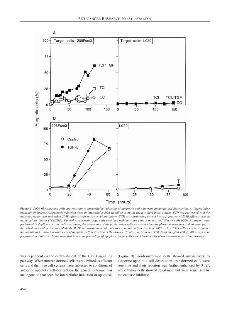
ResultsIn order to gain insight into the mechanism of the recentlydescribed resistance of tumor cells against intercellularinduction of apoptosis (45, 46) and to test whether thisfinding extends to autocrine apoptotic self-destruction, srconcogene-transformed fibroblasts (208Fsrc3) and the murinefibrosarcoma cell line L929 were subjected to conditions ofintercellular induction of apoptosis by nontransformed
murine fibroblasts and to autocrine self-destruction. As canbe seen in Figure 1, the transformed cells readily underwentintercellular induction of apoptosis and autocrine self-destruction, whereas the tumor cells were resistant againstboth effects. When nontransformed cells were used as targetcells, they showed no sensitivity for apoptosis induction,confirming the selectivity of the process with respect to thetransformed state of the target cells (data not shown).
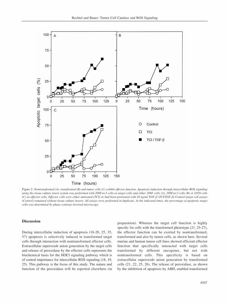
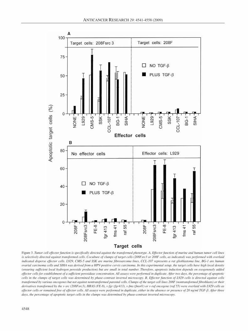
For a direct measurement of the cellular effector function,tissue culture inserts containing either transformed,nontransformed or tumor cells were placed above transformedtarget cells. Cell-containing inserts had been pretreated withTGF-β or not. As can be seen in Figure 2, all three cellsystems (nontransformed, transformed, tumor cells) exhibiteda strong apoptosis-inducing effect on transformed target cellswhen they had been pretreated with TGF-β, and a delayedresponse without preceding TGF-β pretreatment. In theabsence of exogenous TGF-β pretreatment, the tumor cellsshowed the strongest effect amongst the three cell systemstested. This result demonstrates that nontransformed,transformed and tumor cells show comparable effectorfunction. Therefore, the effector cell function (in contrast tothe target cell function, which is restricted to cells with thetransformed phenotype) is not specific for a distinct stage ofcells during multistage oncogenesis. In addition, the lack ofautocrine self-destruction of tumor cells seems not to be due totheir lacking effector function. Figure 3 shows that a variety ofmurine and human tumor cells tested uniformly showedeffector function specifically against transformed target cells,while nontransformed target cells remained unaffected. Whennontransformed 208F cells and several of its derivativestransformed by different oncogenes were tested as target cellschallenged by the tumor cell L929 as effector cells, thenontransformed parental cell 208F showed no apoptoticresponse, whereas all transformed lines, independent of theoncogene responsible for their transformation, were found tobe sensitive to apoptosis induction.
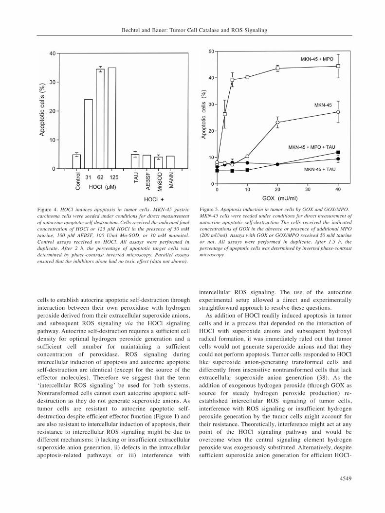
The experiments shown so far indicate that the tumor cellsstill possess the effector function which is necessary forautocrine self-destruction. Their lacking an apoptoticresponse in the effector cell-driven and the autocrine assaymight therefore be due to a lack of extracellular superoxideanion generation, a defect in intracellular apoptoticpathways, or resistance against intercellular ROS signaling.In order to test for superoxide anion production and afunctional apoptotic response, the human gastric tumor cellline MKN-45 was treated with increasing concentrations ofexogenous HOCl, which represents the major player in theHOCl signaling pathway (25, 18) and that requiresinteraction with target cell-derived superoxide anions toallow generation of apoptosis-inducing hydroxyl radicals. Asshown in Figure 4, HOCl induced apoptosis in the tumorcells rapidly and in a concentration-dependent manner.
ANTICANCER RESEARCH 29: 4541-4558 (2009)
4544
Apoptosis induction was blocked by the HOCl scavengertaurine, verifying that HOCl was indeed the apoptosis-mediating agent. Apoptosis induction by HOCl was inhibitedby Mn-SOD (a scavenger of superoxide anions), AEBSF (aninhibitor of the superoxide anion generating NADPHoxidase) and the hydroxyl radical-scavenger mannitol.
First of all, this finding confirms that HOCl does notinduce apoptosis directly, but rather that it acts through itsreaction with superoxide anions, leading to the formation ofhydroxyl radicals. These seem to represent the ultimateapoptosis inducers. Secondly and very importantly, theapoptosis-inducing effect of HOCl and the inhibitor datademonstrate that i) the tumor cells generate sufficientsuperoxide anions for the interaction with HOCl, ii) NADPHoxidase seems to be the source of superoxide anions, andthat iii) the intracellular apoptosis pathways that are inducedby intercellular ROS signaling are functional. Therefore,interference with extracellular ROS signaling remains onevery reasonable explanation for the resistance of tumor cellsto intercellular ROS signaling.
In order to clarify the basis of a potential interferencemechanism, the tumor cells were kept under conditions thatwould allow autocrine self-destruction in sensitivetransformed cells. Two major signaling components wereadded to the tumor cells: GOX to establish continuoushydrogen peroxide generation, MPO, and a combination ofboth. As can be seen in Figure 5, the tumor cells showed aremarkable insensitivity against hydrogen peroxide generatedby GOX. Only relatively high concentrations of GOXinduced apoptosis. This apoptosis-inducing effect was,however, not due to the direct apoptosis-inducing potentialof hydrogen peroxide (39), as the process was completelyinhibited by the HOCl scavenger taurine. Addition of 200mU/ml of MPO alone had no significant direct effect onapoptosis induction. However, in combination with hydrogenperoxide-generating GOX, MPO exhibited an impressivesynergistic effect. This synergistic effect also seemed to bedue to the formation and action of HOCl, as it wascompletely inhibited by taurine.
For a detailed analysis of apoptosis induction in tumorcells after addition of GOX alone or in combination withMPO, specific inhibitors of intercellular ROS signaling aswell as caspase inhibitors were added to the system and theeffects were measured. As can be seen in Figure 6, both theeffect of GOX given alone and its synergistic effect withMPO were inhibited by the superoxide anion scavenger MN-SOD, the NADPH oxidase inhibitor AEBSF, the peroxidaseinhibitor ABH, the HOCl scavenger taurine, the hydroxylradical-scavenger mannitol, as well as by caspase-3 andcaspase-9 inhibitors. These findings indicate that the additionof high concentrations of glucose oxidase or the combinationof glucose oxidase with MPO restores the HOCl signalingpathway. The inhibition of the process by caspase-3 and -9
inhibitors demonstrates that the cells die by caspase-dependent apoptosis. The strong effect of the caspase-9inhibitor thereby indicates that the mitochondrial apoptosissignaling pathway is used (59).
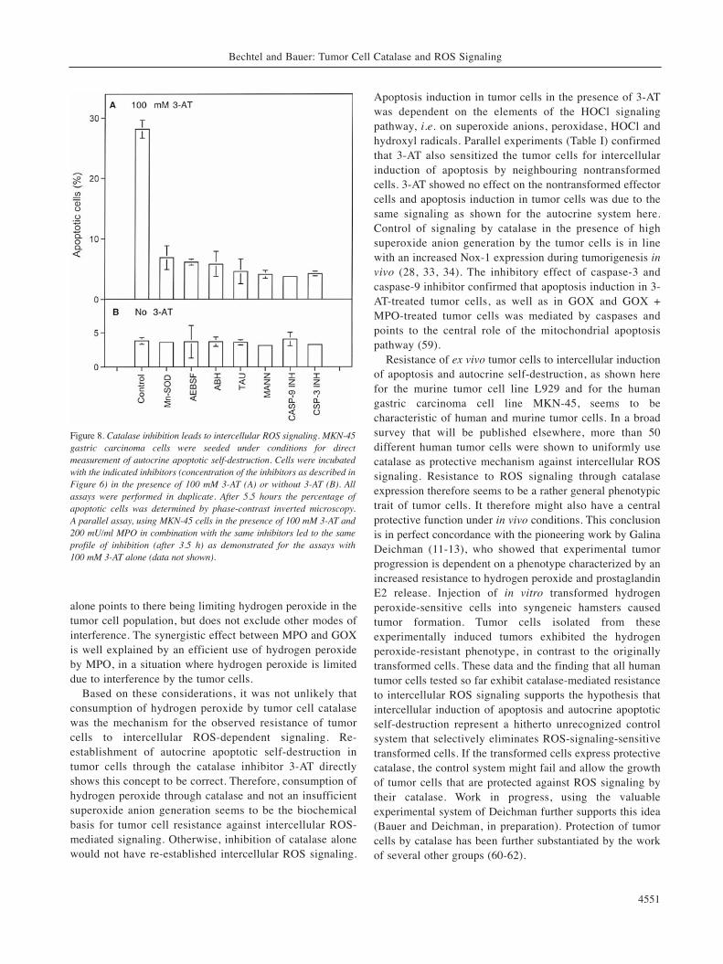
The data shown so far indicate that tumor cells can beresensitized for intercellular ROS-induces apoptosis whenthe HOCl signaling pathway is re-established by theaddition of an exogenous hydrogen peroxide source. Thebest explanation for this finding is a protective role of tumorcell catalase against intercellular ROS signaling. If thisassumption were correct, the addition of the specificcatalase inhibitor 3-AT should restore autocrine ROS-mediated apoptosis in tumor cells. In analogy to theexperiments shown in Figures 5 and 6, this process ofresensitization should be further enhanced by the additionof MPO. To address this question, human tumor cells weretreated with increasing concentrations of 3-AT in theabsence or presence of additional MPO. As can be seen inFigure 7, 3-AT caused a significant increase in apoptosisinduction in tumor cells. At 3.5 hours, apoptosis inductionby 3-AT alone showed the characteristics of an optimumcurve. As expected from our previous findings, MPO addedto the cells alone showed no significant effect, but itexhibited a strong synergistic effect with that of 3-AT. Theexperiments shown in Figure 8 demonstrate that thesynergistic effect between MPO and 3-AT, as well as theeffect of 3-AT alone, were dependent on intercellular ROSsignaling by the HOCl pathway, as scavenging of each oneof the components of this signaling pathway caused stronginhibition of apoptosis. As in the previous controls, tumorcells alone or in the presence of MPO but without 3-ATshowed no apoptosis induction above background levels.The strong effect of the catalase inhibitor 3-AT indicates theprotective role of tumor cell catalase against intercellularROS signaling. It also points to the potential of catalaseinhibition for resensitization of tumor cells. The stronginhibitory effect of caspase-3 and caspase-9 inhibitordemonstrates that tumor cells use the mitochondrialapoptosis pathway when their intercellular ROS signaling isre-established after catalase inhibition.
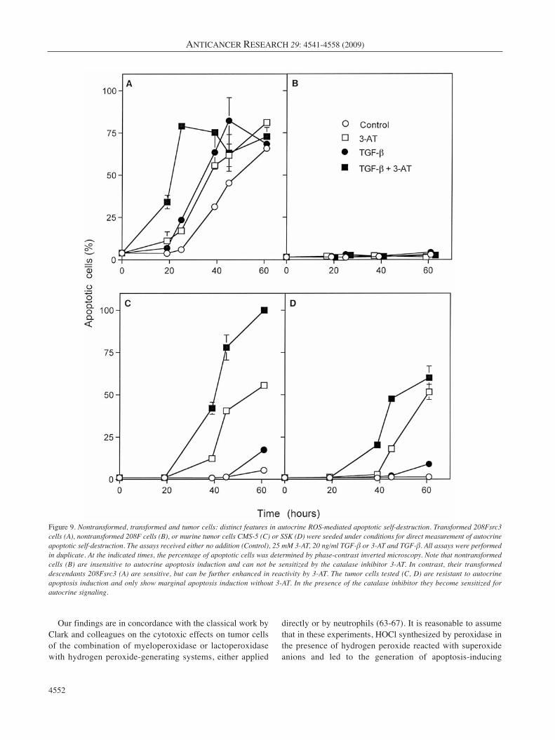
The final experiments (Table I and Figure 9) define thedifferential modes of intercellular ROS signaling in cells thatrepresent three consecutive stages of tumor development, i.e.nontransformed, transformed and tumor cells. Table Idemonstrates that nontransformed 208F cells are insensitiveto intercellular induction of apoptosis by nontransformedeffector cells both in the absence and presence of the catalaseinhibitior 3-AT. Transformed 208Fsrc3 cells responded readilyto intercellular induction of apoptosis by nontransformedeffector cells. Their reaction was further enhanced by 3-AT.As expected from the preceding experiments, tumor cellsshowed resistance to intercellular induction of apoptosis butwere resensitized by 3-AT. The resultant apoptosis induction
Bechtel and Bauer: Tumor Cell Catalase and ROS Signaling
4545
was dependent on the establishment of the HOCl signalingpathway. When nontransformed cells were omitted as effectorcells and the three cell systems were subjected to conditions ofautocrine apoptotic self-destruction, the general outcome wasanalogous to that seen for intercellular induction of apoptosis
(Figure 9): nontransformed cells showed insensitivity toautocrine apoptotic self-destruction, transformed cells weresensitive and their reaction was further enhanced by 3-AT,while tumor cells showed resistance, but were sensitized bythe catalase inhibitor.
ANTICANCER RESEARCH 29: 4541-4558 (2009)
4546
Figure 1. L929 fibrosarcoma cells are resistant to intercellular induction of apoptosis and autocrine apoptotic self-destruction. A, Intercellularinduction of apoptosis. Apoptosis induction through intercellular ROS signaling using the tissue culture insert system (TCI) was performed with theindicated target cells and either 208F effector cells in tissue culture inserts (TCI) or transforming growth factor-β-pretreated 208F effector cells intissue culture inserts (TCI/TGF). Control assays with target cells remained without tissue culture inserts and effector cells (CO). All assays wereperformed in duplicate. At the indicated times, the percentage of apoptotic target cells was determined by phase-contrast inverted microscopy, asdescribed under Materials and Methods. B, Direct measurement of autocrine apoptotic self-destruction. 208Fsrc3 or L929 cells were tested underthe conditions for direct measurement of apoptotic self-destruction in the absence (Control) or presence (TGF-β) of 20 ng/ml TGF-β. All assays wereperformed in duplicate. At the indicated times, the percentage of apoptotic target cells was determined by phase-contrast inverted microscopy.
Discussion
During intercellular induction of apoptosis (18-20, 25, 35,57) apoptosis is selectively induced in transformed targetcells through interaction with nontransformed effector cells.Extracellular superoxide anion generation by the target cellsand release of peroxidase by the effector cells represents thebiochemical basis for the HOCl signaling pathway which isof central importance for intercellular ROS signaling (18, 19,25). This pathway is the focus of this study. The nature andfunction of the peroxidase will be reported elsewhere (in
preparation). Whereas the target cell function is highlyspecific for cells with the transformed phenotype (21, 25-27),the effector function can be exerted by nontransformed,transformed and also by tumor cells, as shown here. Severalmurine and human tumor cell lines showed efficient effectorfunction that specifically interacted with target cellstransformed by different oncogenes, but not withnontransformed cells. This specificity is based onextracellular superoxide anion generation by transformedcells (21, 22, 25, 26). The release of peroxidase, as shownby the inhibition of apoptosis by ABH, enabled transformed
Bechtel and Bauer: Tumor Cell Catalase and ROS Signaling
4547
Figure 2. Nontransformed (A), transformed (B) and tumor cells (C) exhibit effector function. Apoptosis induction through intercellular ROS signalingusing the tissue culture insert system was performed with 208Fsrc3 cells as target cells and either 208F cells (A), 208Fsrc3 cells (B) or L929 cells(C) as effector cells. Effector cells were either untreated (TCI) or had been pretreated with 20 ng/ml TGF-β (TCI/TGF-β) Control target cell assays(Control) remained without tissue culture inserts. All assays were performed in duplicate. At the indicated times, the percentage of apoptotic targetcells was determined by phase-contrast inverted microscopy.
ANTICANCER RESEARCH 29: 4541-4558 (2009)
4548
Figure 3. Tumor cell effector function is specifically directed against the transformed phenotype. A, Effector function of murine and human tumor cell linesis selectively directed against transformed cells. Coculture of clumps of target cells (208Fsrc3 or 208F cells, as indicated) was performed with overlaidindicated disperse effector cells. L929, CMS-5 and SSK are murine fibrosarcoma lines, CCL-107 represents a rat glioblastoma line, BG-1 are humanovarial carcinoma cells and SIHA was derived from a HPV-positive cervix carcinoma. In this experimental setup, the target cells have high local density(ensuring sufficient local hydrogen peroxide production) but are small in total number. Therefore, apoptosis induction depends on exogenously addedeffector cells for establishment of a sufficient peroxidase concentration. All assays were performed in duplicate. After two days, the percentage of apoptoticcells in the clumps of target cells was determined by phase-contrast inverted microscopy. B, Effector function of L929 cells is directed against cellstransformed by various oncogenes but not against nontransformed parental cells. Clumps of the target cell lines 208F (nontransformed fibroblasts) or theirderivatives transformed by the v-src (208Fsrc3), HRAS (FE-8), v-fgr (fgr413), v-fms (fms41) or v-raf oncogene (raf 55) were overlaid with L929 cells aseffector cells or remained free of effector cells. All assays were performed in duplicate, either in the absence or presence of 20 ng/ml TGF-β. After threedays, the percentage of apoptotic target cells in the clumps was determined by phase-contrast inverted microscopy.
cells to establish autocrine apoptotic self-destruction throughinteraction between their own peroxidase with hydrogenperoxide derived from their extracellular superoxide anions,and subsequent ROS signaling via the HOCl signalingpathway. Autocrine self-destruction requires a sufficient celldensity for optimal hydrogen peroxide generation and asufficient cell number for maintaining a sufficientconcentration of peroxidase. ROS signaling duringintercellular induction of apoptosis and autocrine apoptoticself-destruction are identical (except for the source of theeffector molecules). Therefore we suggest that the term‘intercellular ROS signaling’ be used for both systems.Nontransformed cells cannot exert autocrine apoptotic self-destruction as they do not generate superoxide anions. Astumor cells are resistant to autocrine apoptotic self-destruction despite efficient effector function (Figure 1) andare also resistant to intercellular induction of apoptosis, theirresistance to intercellular ROS signaling might be due todifferent mechanisms: i) lacking or insufficient extracellularsuperoxide anion generation, ii) defects in the intracellularapoptosis-related pathways or iii) interference with
intercellular ROS signaling. The use of the autocrineexperimental setup allowed a direct and experimentallystraightforward approach to resolve these questions.
As addition of HOCl readily induced apoptosis in tumorcells and in a process that depended on the interaction ofHOCl with superoxide anions and subsequent hydroxylradical formation, it was immediately ruled out that tumorcells would not generate superoxide anions and that theycould not perform apoptosis. Tumor cells responded to HOCllike superoxide anion-generating transformed cells anddifferently from insensitive nontransformed cells that lackextracellular superoxide anion generation (38). As theaddition of exogenous hydrogen peroxide (through GOX assource for steady hydrogen peroxide production) re-established intercellular ROS signaling of tumor cells,interference with ROS signaling or insufficient hydrogenperoxide generation by the tumor cells might account fortheir resistance. Theoretically, interference might act at anypoint of the HOCl signaling pathway and would beovercome when the central signaling element hydrogenperoxide was exogenously substituted. Alternatively, despitesufficient superoxide anion generation for efficient HOCl-
Bechtel and Bauer: Tumor Cell Catalase and ROS Signaling
4549
Figure 4. HOCl induces apoptosis in tumor cells. MKN-45 gastriccarcinoma cells were seeded under conditions for direct measurementof autocrine apoptotic self-destruction. Cells received the indicated finalconcentration of HOCl or 125 μM HOCl in the presence of 50 mMtaurine, 100 μM AEBSF, 100 U/ml Mn-SOD, or 10 mM mannitol.Control assays received no HOCl. All assays were performed induplicate. After 2 h, the percentage of apoptotic target cells wasdetermined by phase-contrast inverted microscopy. Parallel assaysensured that the inhibitors alone had no toxic effect (data not shown).
Figure 5. Apoptosis induction in tumor cells by GOX and GOX/MPO.MKN-45 cells were seeded under conditions for direct measurement ofautocrine apoptotic self-destruction The cells received the indicatedconcentrations of GOX in the absence or presence of additional MPO(200 mU/ml). Assays with GOX or GOX/MPO received 50 mM taurineor not. All assays were performed in duplicate. After 1.5 h, thepercentage of apoptotic cells was determined by inverted phase-contrastmicroscopy.

superoxide anion interaction, the concentration of superoxideanions generated by the tumor cells might be too low todrive an optimal hydrogen peroxide generation through thedismutation reaction. The inhibitory effects of taurine,AEBSF, Mn-SOD, mannitol and ABH proved that apoptosisinduction after GOX addition was not due to the directapoptosis-inducing effect of hydrogen peroxide (which is notselective with respect to the transformed state) (39), but mustbe due to the specific re-establishment of the HOCl pathway.The lack of apoptosis induction by exogenous MPO given
ANTICANCER RESEARCH 29: 4541-4558 (2009)
4550
Figure 6. Apoptosis induction by GOX or GOX / MPO is mediated byintercellular ROS signaling. MKN-45 gastric carcinoma cells wereseeded under conditions for direct measurement of autocrine apoptoticself-destruction. A, The cells received the indicated concentrations ofGOX, as well as 20 mU/ml GOX in the presence of 100 μM AEBSF(NADPH oxidase inhibitor), 120 U/ml Mn-SOD, 150 μM ABH(mechanism-based peroxidase inhibitor), 50 mM taurine (HOClscavenger), 10 mM mannitol (hydroxyl radical scavenger), 25 μMcaspase-9 inhibitor and 50 μM caspase-3 inhibitor. All assays wereperformed in duplicate. After 2 h, the percentage of apoptotic cells wasdetermined by inverted phase-contrast microscopy. Control assaysensured that the inhibitors alone had not significant effect on apoptosisinduction (data not shown). B, The experiment was performed asdescribed under A, except that the cells received the indicatedconcentrations of GOX in combination with 200 mU/ml MPO. Theinhibitor studies were performed with 5 mU/ml GOX plus 200 mU/mlMPO in the presence of the same inhibitors as described under A. Allassays were performed in duplicate. After 2 h, the percentage ofapoptotic cells was determined as described under A.
Figure 7. Apoptosis induction in tumor cells after catalase inhibition by3-AT. MKN-45 gastric carcinoma cells were seeded under conditionsfor direct measurement of autocrine apoptotic self-destruction and weretreated with the indicated concentrations of 3-AT in the absence orpresence of 200 mU/ml MPO. All assays were performed in duplicate.After 1.5 (A) and 3.5 h (B) the percentages of apoptotic cells weredetermined by phase contrast inverted microscopy.
alone points to there being limiting hydrogen peroxide in thetumor cell population, but does not exclude other modes ofinterference. The synergistic effect between MPO and GOXis well explained by an efficient use of hydrogen peroxideby MPO, in a situation where hydrogen peroxide is limiteddue to interference by the tumor cells.
Based on these considerations, it was not unlikely thatconsumption of hydrogen peroxide by tumor cell catalasewas the mechanism for the observed resistance of tumorcells to intercellular ROS-dependent signaling. Re-establishment of autocrine apoptotic self-destruction intumor cells through the catalase inhibitor 3-AT directlyshows this concept to be correct. Therefore, consumption ofhydrogen peroxide through catalase and not an insufficientsuperoxide anion generation seems to be the biochemicalbasis for tumor cell resistance against intercellular ROS-mediated signaling. Otherwise, inhibition of catalase alonewould not have re-established intercellular ROS signaling.
Apoptosis induction in tumor cells in the presence of 3-ATwas dependent on the elements of the HOCl signalingpathway, i.e. on superoxide anions, peroxidase, HOCl andhydroxyl radicals. Parallel experiments (Table I) confirmedthat 3-AT also sensitized the tumor cells for intercellularinduction of apoptosis by neighbouring nontransformedcells. 3-AT showed no effect on the nontransformed effectorcells and apoptosis induction in tumor cells was due to thesame signaling as shown for the autocrine system here.Control of signaling by catalase in the presence of highsuperoxide anion generation by the tumor cells is in linewith an increased Nox-1 expression during tumorigenesis invivo (28, 33, 34). The inhibitory effect of caspase-3 andcaspase-9 inhibitor confirmed that apoptosis induction in 3-AT-treated tumor cells, as well as in GOX and GOX +MPO-treated tumor cells was mediated by caspases andpoints to the central role of the mitochondrial apoptosispathway (59).
Resistance of ex vivo tumor cells to intercellular inductionof apoptosis and autocrine self-destruction, as shown herefor the murine tumor cell line L929 and for the humangastric carcinoma cell line MKN-45, seems to becharacteristic of human and murine tumor cells. In a broadsurvey that will be published elsewhere, more than 50different human tumor cells were shown to uniformly usecatalase as protective mechanism against intercellular ROSsignaling. Resistance to ROS signaling through catalaseexpression therefore seems to be a rather general phenotypictrait of tumor cells. It therefore might also have a centralprotective function under in vivo conditions. This conclusionis in perfect concordance with the pioneering work by GalinaDeichman (11-13), who showed that experimental tumorprogression is dependent on a phenotype characterized by anincreased resistance to hydrogen peroxide and prostaglandinE2 release. Injection of in vitro transformed hydrogenperoxide-sensitive cells into syngeneic hamsters causedtumor formation. Tumor cells isolated from theseexperimentally induced tumors exhibited the hydrogenperoxide-resistant phenotype, in contrast to the originallytransformed cells. These data and the finding that all humantumor cells tested so far exhibit catalase-mediated resistanceto intercellular ROS signaling supports the hypothesis thatintercellular induction of apoptosis and autocrine apoptoticself-destruction represent a hitherto unrecognized controlsystem that selectively eliminates ROS-signaling-sensitivetransformed cells. If the transformed cells express protectivecatalase, the control system might fail and allow the growthof tumor cells that are protected against ROS signaling bytheir catalase. Work in progress, using the valuableexperimental system of Deichman further supports this idea(Bauer and Deichman, in preparation). Protection of tumorcells by catalase has been further substantiated by the workof several other groups (60-62).
Bechtel and Bauer: Tumor Cell Catalase and ROS Signaling
4551
Figure 8. Catalase inhibition leads to intercellular ROS signaling. MKN-45gastric carcinoma cells were seeded under conditions for directmeasurement of autocrine apoptotic self-destruction. Cells were incubatedwith the indicated inhibitors (concentration of the inhibitors as described inFigure 6) in the presence of 100 mM 3-AT (A) or without 3-AT (B). Allassays were performed in duplicate. After 5.5 hours the percentage ofapoptotic cells was determined by phase-contrast inverted microscopy. A parallel assay, using MKN-45 cells in the presence of 100 mM 3-AT and200 mU/ml MPO in combination with the same inhibitors led to the sameprofile of inhibition (after 3.5 h) as demonstrated for the assays with 100 mM 3-AT alone (data not shown).
Our findings are in concordance with the classical work byClark and colleagues on the cytotoxic effects on tumor cellsof the combination of myeloperoxidase or lactoperoxidasewith hydrogen peroxide-generating systems, either applied
directly or by neutrophils (63-67). It is reasonable to assumethat in these experiments, HOCl synthesized by peroxidase inthe presence of hydrogen peroxide reacted with superoxideanions and led to the generation of apoptosis-inducing
ANTICANCER RESEARCH 29: 4541-4558 (2009)
4552
Figure 9. Nontransformed, transformed and tumor cells: distinct features in autocrine ROS-mediated apoptotic self-destruction. Transformed 208Fsrc3cells (A), nontransformed 208F cells (B), or murine tumor cells CMS-5 (C) or SSK (D) were seeded under conditions for direct measurement of autocrineapoptotic self-destruction. The assays received either no addition (Control), 25 mM 3-AT, 20 ng/ml TGF-β or 3-AT and TGF-β. All assays were performedin duplicate. At the indicated times, the percentage of apoptotic cells was determined by phase-contrast inverted microscopy. Note that nontransformedcells (B) are insensitive to autocrine apoptosis induction and can not be sensitized by the catalase inhibitor 3-AT. In contrast, their transformeddescendants 208Fsrc3 (A) are sensitive, but can be further enhanced in reactivity by 3-AT. The tumor cells tested (C, D) are resistant to autocrineapoptosis induction and only show marginal apoptosis induction without 3-AT. In the presence of the catalase inhibitor they become sensitized forautocrine signaling.
hydroxyl radicals. At the time of these classic studies, it wasnot known that tumor cells generate extracellular superoxideanions and thus can specifically contribute to ROS signaling.As MPO and hydrogen peroxide generation had to be presentto obtain the cytotoxic effect, the experimental situation inthese earlier papers seems to resemble the synergistic effectbetween MPO and GOX shown in Figure 5 of the workpresented here. Our own work does not support the findingby Weiss and Slivaka (68) on the independence of HOCl-mediated cytotoxic effects from hydroxyl radical formation.Furthermore, our work is in direct contradiction to thefindings by Wagner et al. (69) on the role of chloramines forHOCl-mediated cytotoxic effects. Chloramines do not seem
to play a detectable role in HOCl-mediated apoptosis in ourexperiments, as the HOCl effect was inhibited by taurine,despite the interaction of HOCl and taurine resulting in theformation of taurine chloramine.
As large numbers of resistant tumor cells can act aseffector cells that establish apoptosis induction in clumps ofneighbouring sensitive transformed target cells at high localdensity (Figures 2 and 3), catalase does not seem to bereleased by the tumor cells, but rather seems to be adherent tothem in a stable mode. Otherwise the effector function of thetumor cells would have been masked by interference of tumorcell-derived catalase with ROS signaling of the transformedtarget cells. This argument is further strengthened through the
Bechtel and Bauer: Tumor Cell Catalase and ROS Signaling
4553
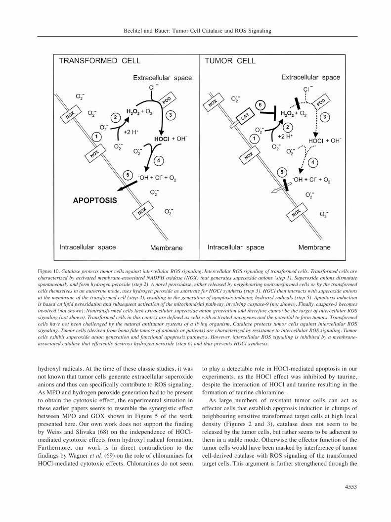
Figure 10. Catalase protects tumor cells against intercellular ROS signaling. Intercellular ROS signaling of transformed cells. Transformed cells arecharacterized by activated membrane-associated NADPH oxidase (NOX) that generates superoxide anions (step 1). Superoxide anions dismutatespontaneously and form hydrogen peroxide (step 2). A novel peroxidase, either released by neighbouring nontransformed cells or by the transformedcells themselves in an autocrine mode, uses hydrogen peroxide as substrate for HOCl synthesis (step 3). HOCl then interacts with superoxide anionsat the membrane of the transformed cell (step 4), resulting in the generation of apoptosis-inducing hydroxyl radicals (step 5). Apoptosis inductionis based on lipid peroxidation and subsequent activation of the mitochondrial pathway, involving caspase-9 (not shown). Finally, caspase-3 becomesinvolved (not shown). Nontransformed cells lack extracellular superoxide anion generation and therefore cannot be the target of intercellular ROSsignaling (not shown). Transformed cells in this context are defined as cells with activated oncogenes and the potential to form tumors. Transformedcells have not been challenged by the natural antitumor systems of a living organism. Catalase protects tumor cells against intercellular ROSsignaling. Tumor cells (derived from bona fide tumors of animals or patients) are characterized by resistance to intercellular ROS signaling. Tumorcells exhibit superoxide anion generation and functional apoptosis pathways. However, intercellular ROS signaling is inhibited by a membrane-associated catalase that efficiently destroys hydrogen peroxide (step 6) and thus prevents HOCl synthesis.
finding that tumor cells retain their high resistance toexogenous hydrogen peroxide even if they are centrifuged,washed and challenged immediately (data not shown).
When individualized transformed cells were mixed with anexcess of tumor cells, ROS signaling of the transformed cellswas abrogated as catalase located on neighbouring tumor cellsseemed to destroy hydrogen peroxide generated bytransformed cells (data not shown). In a reverse experiment,a small number of individualized tumor cells mixed with anexcess of transformed cells showed sensitivity to intercellularsignaling, as the bound catalase was unable to interfere withROS signaling of the neighbouring transformed cells presentin excess (data not shown). Thus, it was shown that HOClgenerated by the transformed cells reaches the tumor cells andinduces apoptosis after interaction with superoxide anions and
generation of hydroxyl radicals. Catalase responsible forinterference with intercellular and autocrine ROS signalingseems to be located on the outside of the tumor cellmembrane, as i) its activity can be blocked by monoclonalantibodies against catalase and ROS signaling of tumor cells isthen restored (Bauer et al., in preparation), ii) it can beinactivated by extracellular singlet oxygen (Riethmüller andBauer, in preparation), iii) it can be detected by indirectimmunofluorescence and FACS analysis on intact cells (datanot shown). Protective catalase of tumor cells seems to belocated specifically at the outside of the cell membrane, inaddition to classical intracellular catalase. Although thelocations are different, both enzyme activities are otherwiseindistinguishable, as indicated by siRNA interferenceexperiments (work in progress). The localization of catalase
ANTICANCER RESEARCH 29: 4541-4558 (2009)
4554
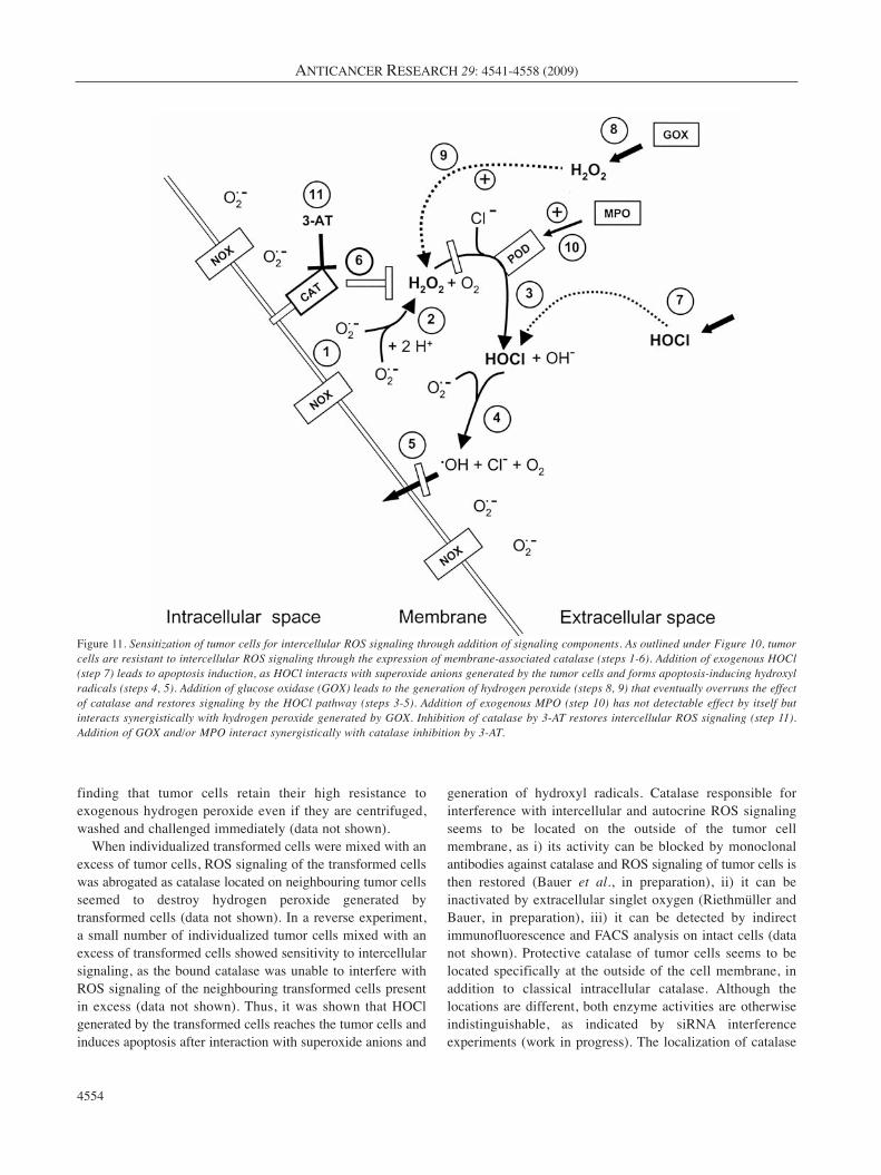
Figure 11. Sensitization of tumor cells for intercellular ROS signaling through addition of signaling components. As outlined under Figure 10, tumorcells are resistant to intercellular ROS signaling through the expression of membrane-associated catalase (steps 1-6). Addition of exogenous HOCl(step 7) leads to apoptosis induction, as HOCl interacts with superoxide anions generated by the tumor cells and forms apoptosis-inducing hydroxylradicals (steps 4, 5). Addition of glucose oxidase (GOX) leads to the generation of hydrogen peroxide (steps 8, 9) that eventually overruns the effectof catalase and restores signaling by the HOCl pathway (steps 3-5). Addition of exogenous MPO (step 10) has not detectable effect by itself butinteracts synergistically with hydrogen peroxide generated by GOX. Inhibition of catalase by 3-AT restores intercellular ROS signaling (step 11).Addition of GOX and/or MPO interact synergistically with catalase inhibition by 3-AT.
at the cell membrane of tumor cells may be of advantage, asthis leads to a high local catalase concentration at the site to beprotected. Ottaviano et al. (70) suggested that intracellularcatalase would efficiently counteract extracellular hydrogenperoxide due to the rapid diffusion of hydrogen peroxidethrough membranes. However, based on our data, it seems thatthe extracellular location of catalase is favorable in the caseof tumor cell protection against extracellular ROS signaling.In line with our data, catalase at the surface of tumor cells hasbeen directly demonstrated by proteomic analysis (71). Thesefindings contrast with but not contradicted by the findings ofGupta et al. (72) and Finch et al. (73) who demonstrated thatan increase in total cellular catalase attenuates or even reversestumorigenicity. A lower intracellular catalase activity has alsobeen described in lung cancer (74). It will be important todifferentiate between the effects of intracellular and cellmembrane-associated tumor cell catalase in the future, as theseactivities are distinct and seem to influence tumorigenesis inopposite ways: a high level of extracellular membrane-associated catalase protects against extracellular ROSsignaling, whereas a low level of intracellular catalase allowsefficient intracellular signaling by hydrogen peroxide.
Figure 10 summarizes our findings on the protective roleof tumor cell catalase against intercellular ROS signaling.The focus thereby is on the HOCl signaling pathway. Workin progress indicates that catalase also protects tumor cellsagainst apoptosis induction by the NO-peroxynitrite and thenitryl chloride signaling pathway, as well as against themetal-catalyzed Haber Weiss reaction (Heinzelmann andBauer, in preparation). Figure 11 summarizes theexperimental approaches taken in this paper to elucidate themechanism of tumor cell resistance to intercellular ROSsignaling. Although already rather complex, this focuses onthe major reactions only. Work in progress is elucidating acomplicated network of secondary reactions arising from thebasic scheme (Bauer, in preparation).
The knowledge of there being protective catalase on themembrane of tumor cells in combination with the potential toexert powerful apoptosis-inducing ROS signaling after catalaseinhibition or destruction should allow novel and specific formsof antitumor therapy to be established and enlighten ourunderstanding of tumor prevention. This approach is especiallyintriguing as the extracellular ROS generation of tumor cells, aspecific trait which is linked to their transformed state, drivestheir selective apoptosis induction. Membrane-associatedcatalase thereby seems to represent the critical control element.Inhibition or destruction of membrane-associated catalase orprevention of its expression through siRNA might becomeuseful and specific tools to resensitize tumor cells to apoptosis-inducing ROS signaling. Work along these lines may hopefullystimulate novel approaches in tumor prevention, drugdevelopment and cancer therapy.
Acknowledgements
This work was supported by a grant from EuroTransBio (ETB10315012B) and from RiscRad. We appreciate the gift of cells by D.Adam (Kiel) and Drs. C. Sers and R. Schäfer (Berlin). We are gratefulfor intellectual support by the COST consortium ‘ChemBioRadical’(COST Action CM0603). This work would not have been possiblewithout the pioneering work of the late Manfred Saran (Munich) andthe concepts of Galina Deichman (Moscow) on the role of thehydrogen peroxide-resistant phenotype during tumor progression.
References
1 Weinberg RA: Oncogenes, Anti-oncogenes and the molecular basisof multistep carcinogenesis. Cancer Res 49: 3713-3721, 1989.
2 Hanahan D and Weinberg RA: The hallmarks of cancer. Cell100: 57-70, 2000.
3 Vogelstein B and Kinzler KW: Cancer genes and the pathwaysthey control. Nature Medicine 10: 789-799, 2004.
4 Temin HM: Control by factors in serum of multiplication ofuninfected and cells infected and converted by avian sarcomaviruses. In: Growth Regulatory Substances for Animal Cells inCulture. Vol. 7. Defendi V and Stoker M (eds.). Philadelphia,The Wistar Symposium Monograph, Wistar Institute Press, pp.103-116, 1967.
Bechtel and Bauer: Tumor Cell Catalase and ROS Signaling
4555
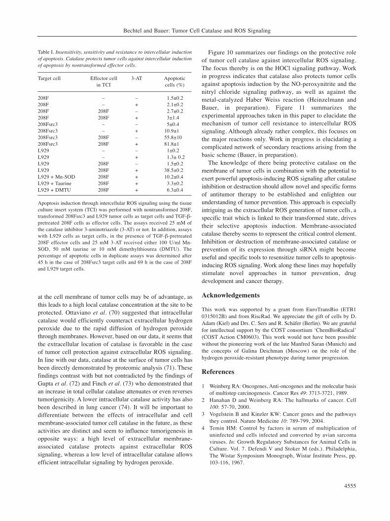
Table I. Insensitivity, sensitivity and resistance to intercellular inductionof apoptosis. Catalase protects tumor cells against intercellular inductionof apoptosis by nontransformed effector cells.
Target cell Effector cell 3-AT Apoptotic in TCI cells (%)
208F – – 1.5±0.2208F – + 2.1±0.2208F 208F – 2.7±0.2208F 208F + 3±1.4208Fsrc3 – – 5±0.4208Fsrc3 – + 10.9±1208Fsrc3 208F – 55.8±10208Fsrc3 208F + 81.8±1L929 – – 1±0.2L929 – + 1.3± 0.2L929 208F – 1.5±0.2L929 208F + 38.5±0.2L929 + Mn-SOD 208F + 10.2±0.4L929 + Taurine 208F + 3.3±0.2L929 + DMTU 208F + 6.3±0.4
Apoptosis induction through intercellular ROS signaling using the tissueculture insert system (TCI) was performed with nontransformed 208F,transformed 208Fsrc3 and L929 tumor cells as target cells and TGF-β-pretreated 208F cells as effector cells. The assays received 25 mM ofthe catalase inhibitor 3-aminotriazole (3-AT) or not. In addition, assayswith L929 cells as target cells, in the presence of TGF-β-pretreated208F effector cells and 25 mM 3-AT received either 100 U/ml Mn-SOD, 50 mM taurine or 10 mM dimethylthiourea (DMTU). Thepercentage of apoptotic cells in duplicate assays was determined after45 h in the case of 208Fsrc3 target cells and 69 h in the case of 208Fand L929 target cells.

5 Sporn MB and Todaro GJ: Autocrine secretion and malignanttransformation. New Engl J Med 303: 878-880, 1980.
6 Heldin CH and Westermark B: Growth factors as transformingproteins. Eur J Biochem 184: 487-496, 1989.
7 Stoker MGP, Shearer M and O’Neill C: Growth inhibition ofpolyoma-transformed cells by contact with static normalfibroblasts. J Cell Sci 1: 297-310, 1966.
8 Delinassios JG: Fibroblasts against cancer cells in vitro.Anticancer Res 7: 1005-1010, 1987.
9 Trosko JE, Chang CC, Madhukar BV and Klaunig JE: Chemical,oncogene and growth factor inhibition of gap junctionalintercellular communication: an integrative hypothesis ofcarcinogenesis. Pathobiology 58: 265-278, 1990.
10 Barcellos-Hoff MH: It takes a tissue to make a tumor.Epigenetics, cancer and microenvironment. J Mammary GlandBiol Neoplasia 6: 213-221, 2001.
11 Deichman G, Matveeva VA, Kashkina LM, Dyakova NA,Uvarova EN, Nikiforov MA and Gudkov AV: Cell transforminggenes and tumor progression: in vivo unified secondaryphenotypic cell changes. Int J Cancer 75: 277-283, 1998.
12 Deichman G: Natural selection and early changes of phenotypeof tumor cells in vivo: Acquisition of new defense mechanisms.Biochem (Mosc) 65: 78-94, 2000.
13 Deichman G: Early phenotypic changes of in vitro transformedcells during in vivo progression: possible role of the host innateimmunity. Sem Cancer Biol 12: 317-326, 2002.
14 Graeber TG, Osmanian C, Jacks T, Housman DE, Koch CJ,Lowe SW and Giaccia AJ: Hypoxia-mediated selection of cellswith diminished apoptotic potential in solid tumours. Nature379: 88-91, 1996.
15 Kinzler KW and Vogelstein B: Life (and death) in a malignanttumour. Nature 379: 19-20, 1996.
16 Bauer G: Resistance to TGF-β-induced elimination oftransformed cells is required during tumor progression. Int JOncol 6: 1227-1229, 1995.
17 Bauer G: Elimination of transformed cells by normal cells: anovel concept for the control of carcinogenesis. HistolHistopathol 11: 237-255, 1996.
18 Bauer G; Reactive oxygen and nitrogen species: efficient,selective and interactive signals during intercellular induction ofapoptosis. Anticancer Res 20: 4115-4140, 2000.
19 Bauer G: Signaling and proapoptotic functions of transformedcell-derived reactive oxygen species. Prostagl Leukotri EssentFatty Acid 66: 41-56, 2002.
20 Bauer G: Low dose radiation and intercellular induction ofapoptosis: potential implications for the control of oncogenesis.Int J Radiation Biol 83: 887-902, 2007.
21 Irani K, Xia Y, Zweier JL, Sollott SJ, Der CJ, Fearon ER,Sundaresan M, Finkel T and Goldschmidt-Clermont PJ:Mitogenic signalling by oxidants in Ras-transformed fibroblasts.Science 275: 1649-1652, 1997.
22 Irani K and Goldschmidt-Clermont PJ: Ras, superoxide andsignal transduction. Biochem Pharmacol 55: 1339-1346, 1998.
23 Suh Y-A, Arnold RS, Lassegue B, Shi J, Xu X, Sorescu D,Chung AB, Griendling KK and Lambeth JD: Cell transformationby the superoxide-generating oxidase Mox1. Nature 401: 79-82,1999.
24 Yang JQ, Li S, Domann FE, Buettner G and Oberley LW :Superoxide generation in v-Ha-ras-transduced human keratinocyteHaCaT cells. Mol. Carcinogenesis 26: 180-188, 1999.
25 Herdener M, Heigold S, Saran M and Bauer G: Target cell-derived superoxide anions cause efficiency and selectivity ofintercellular induction of apoptosis. Free Radical Biol Med 29:1260-1271, 2000.
26 Schwieger A, Bauer L, Hanusch J, Sers C, Schäfer R and BauerG: Ras oncogene expression determines sensitivity for intercellularinduction of apoptosis. Carcinogenesis 22: 1385-1392, 2001.
27 Heigold S, Sers C, Bechtel W, Ivanovas B, Schäfer R and BauerG: Nitric oxide mediates apoptosis induction selectively intransformed fibroblasts compared to nontransformed fibroblasts.Carcinogenesis 23: 929-941, 2002.
28 Mitsushita J, Lambeth JD and Kamata T: The superoxide-generating oxidase Nox1 is functionally required for Rasoncogenic transformation. Cancer Res 64: 3580-3585, 2004.
29 Cheng G, Diebold BA, Hughes Y and Lambeth JD: Nox1-dependent reactive oxygen generation is regulated by Rac1. J Biol Chem 281: 17718-17726, 2006.
30 Arnold RS, Shi J, Murad E, Whalen AM, Sun CQ, PalavarapuR, Parthasarathy S, Petros JA and Lambeth JD: Hydrogenperoxide mediates the cell growth and transformation caused bythe mitogenic oxidase Nox1. Proc Natl Acad Sci USA 98: 5550-5555, 2001.
31 Shinohara M, Shang W-H, Kubodera M, Hanada S, MitsushitaJ, Kato M, Miyazaki H, Suminoto H and Kamata T: Nox1 redoxsignaling mediates oncogenic Ras-induced disruption of stressfibers and focal adhesions by down-regulating Rho. J Biol Chem282: 17640-17648, 2007.
32 Arbiser JL, Petros J, Klafter R, Govindajaran B, McLaughlin ER,Brown LF, Cohen C, Moses M, Kilroy S, Arnold RS and LambethJD: Reactive oxygen generated by Nox1 triggers the angiogenicswitch. Proc Acad Natl Acad Sci USA 99: 715-720, 2002.
33 Tominaga K, Kawahara T, Sano t, Toida K, Kuwano Y, SasakiH and Kawai T: Evidence for cancer-associated expression ofNADPH oxidase 1 (Nox1)-base oxidase system in the humanstomach. Free Radical Biol Med 43: 1627-1638, 2007.
34 Laurent E, McCoy JW, Maccina RA, Liu W, Cheng GJ, RobineS, Papkoff J and Lambeth JD: Nox1 is overexpressed in humancolon cancers and correlates with activating mutations in K-Ras.Int J Cancer 123: 100-107, 2008.
35 Jürgensmeier J, Schmitt CP, Viesel E, Höfler P and Bauer G:TGF-β-treated normal fibroblasts eliminate transformedfibroblasts by induction of apoptosis. Cancer Res 54: 393-398,1994.
36 Steinebach C and Bauer G: An alternative signalling pathwaybased on nitryl chloride during intercellular induction ofapoptosis. In vitro and applied molecular toxicology 14: 107-120, 2001.
37 Schimmel M and Bauer G: Proapoptotic and redox state-relatedsignalling of reactive oxygen species generated by transformedfibroblasts. Oncogene 21: 5886-5896, 2002.
38 Engelmann I, Dormann S, Saran M and Bauer G: Transformedtarget cell-derived superoxide anions drive apoptosis inductionby myeloperoxidase. Redox Report 5: 207-214, 2000.
39 Ivanovas B and Bauer G: Selective and nonselective apoptosisinduction in transformed and nontransformed fibroblasts byexogenous reactive oxygen and nitrogen species. Anticancer Res22: 841-856, 2002.
40 Long CA and Bielski BH: Rate of reaction of superoxide radicalwith chloride-containing species. J Phys Chem 84: 555-557,1980.
ANTICANCER RESEARCH 29: 4541-4558 (2009)
4556
41 Candeias LP, Patel KB, Stratford MRL and Wardmann P: Freehydroxyl radicals are formed on reaction between the neutrophil-derived species superoxide anion and hypochlorous acid. FEBS333: 151-153, 1993.
42 Folkes LK, Candeias LP and Wardman P: Kinetics andmechanisms of hypochlorous acid reactions. Arch BiochemBiophys 323: 120-126, 1995.
43 Saran M and Bors W: Signalling by O2– and NO: how far caneither radical, or any specific reaction product, transmit a messageunder in vivo conditions? Che Biol Inter 90: 35-45, 1994.
44 Saran M, Michel C and Bors W: Radical functions in vivo: acritical review of current concepts and hypotheses. Zeitschriftfür Naturforschung 53 c: 210-227, 1998.
45 Engelmann I and Bauer G: How can tumor cells escapeintercellular induction of apoptosis? Anticancer Res 20: 2297-2306, 2000.
46 Engelmann I, Eichholtz-Wirth H and Bauer G: Ex vivo tumorcell lines are resistant to intercellular induction of apoptosis andindependent of exogenous survival factors. Anticancer Res 20:2361-2370, 2000.
47 Diatchuk V, Lotan O, Koshkin V, Wikstroem P and Pick E:Inhibition of NADPH oxidase activation by 4-(2-aminoethyl-)benzenesulfonyl fluoride and related compounds. J Biol Chem272: 13292-13301, 1997.
48 Kettle AJ, Gedye CA, Hampton MB and Winterbourn CC:Inhibition of myeloperoxidase by benzoic acid hydrazides.Biochem J 308: 559-563, 1995.
49 Kettle AJ, Gedye CA and Winterbourn CC: Mechanisms ofinactivation of myeloperoxidase by 4-aminobenzoic acidhydrazide. Biochem J 321: 503-508, 1997.
50 Putnam CD, Arvai AS and Bourne Y: Active and inhibitedhuman catalase structures: Ligand and NADPH binding andcatalytic mechanism. J Mol Biol 296: 295-309, 2000.
51 Goldstein S and Czapski G: Mannitol as an OH. scavenger inaqueous solutions and in biological systems. Int J Rad Biol 46:725-729, 1984.
52 Aruoma OI, Halliwell B, Hoey BM and Butler J: Theantioxidant action of taurine, hypotaurine and their metabolicprecursors. Biochem J 256: 251-256, 1988.
53 Klebanoff SJ: Myeloperoxidase: friend and foe. J Leucocyte Biol77: 1-28, 2005.
54 Konorev EA, Kennedy MC and Kalyanaraman B: Cell-permeable superoxide dismutase and glutathione peroxidasemimetics afford superior protection against doxorubicin-induced cardiotoxicity: The role of reactive oxygen andnitrogen intermediates. Arch Biochem Biophys 368: 421-428,1999.
55 Estevez AG, Sampson JB, Zhuang YX, Spear N, Richardson GJ,Crow JP, Tarpey MM, Barbeito L and Beckman JS: Liposome-delivered superoxide dismutase prevents nitric oxide-dependentmotor neuron death induced by trophic factor withdrawal. FreeRadical Biol Med 28: 437-446, 2000.
56 Bauer G, Höfler P and Simon M: Epstein-Barr virus inductionby a serum factor II. Purification of a highmolecular weightprotein that is responsible for induction. J Biol Chem 257:11405-11410, 1982.
57 Beck E, Schäfer R and Bauer G: Sensitivity of transformedfibroblasts for intercellular induction of apoptosis isdetermined by their transformed phenotype. Exp Cell Res 234:47-56, 1997.
58 Gorcyca W, Gong J and Darzynkiewicz Z: Detection of DNAstrand breaks in individual apoptotic cells by the in situ terminaldeoxynucleotidyl transferase and nick translation assays. CancerRes 53: 1945-1951, 1993.
59 Kroemer G, Zamzami N and Susin SA: Mitochondrial control ofapoptosis. Immunol Today 18: 44-51, 1997.
60 Sandstrom PA and Buttke TM: Autocrine production ofextracellular catalase prevents apoptosis of the human CEM T-cell line in serum-free medium. Proc Natl Acad Sci USA 90:4708-4712, 1993.
61 Tome ME, Baker AF, Powis G, Payne CM and Briehl MM:Catalase-overexpressing thymocytes are resistant toglucocorticoid-induced apoptosis and exhibit increased net tumorgrowth. Cancer Res 61: 2766-2773, 2001.
62 Moran EC, Kamiguti AS, Cawley JC and Pettitt AR: Cytoprotectiveantioxidant activity of serum albumin and autocrine catalase inchronic lymphatic leukaemia. Br J Haematol 116: 316-328, 2002.
63 Clark RA, Klebanoff SJ, Einstein AB and Fefer A: Peroxidase-H2O2-halide system: cytotoxic effect on mammalian tumor cells.Blood 45: 161-170, 1975.
64 Clark RA and Klebanoff SJ: Neutrophil-mediated tumor cellcytotxicity – role of peroxidase system. J Exp Med 141: 1442-1447, 1975.
65 Clark R A and Klebanoff S J: Role of the myeloperoxidase-H2O2-halide system in concanavalin A-induced tumor cellkilling by human neutrophils. J Immunol 122: 2605-2610, 1979.
66 Clark RA and Szot S: The myeloperoxidase–hydrogen peroxide–halide system as effector of neutrophil-mediated tumor-cellcytotxicity. J Immunol 126: 1295-1301, 1981.
67 Okajima T, Onishi M, Hayama E, Motoji N and Momose Y:Cytolysis of B-16 melanoma tumor cells mediated by themyeloperoxidase and lactoperoxidase systems. Biol Chem 377:689-693, 1996.
68 Weiss SJ and Slivka A: Monocyte and granulocyte-mediatedtumor cell destruction. A role for the hydrogen peroxidase–myeloperoxidase–chloride system. J Clin Invest 69: 255-262,1982.
69 Wagner BA, Britigan BE, ReszkaKJ, McCormick ML and BurnsCP: Hydrogen peroxide-induced apoptosis of HL-60 humanleukemia cells is mediated by the oxidants hypochlorous acidand chloramines. Arch Biochem Biophys 401: 223-234, 2002.
70 Ottaviano FG, Handy DE and Loscalzo J: Redox regulation inthe extracellular environment. Cir J 72: 1-16, 2008.
71 Jang JH and Hanah S: Profiling of the cell surface proteome.Proteomics 3: 1947-1954, 2003.
72 Gupta A, Butts B, Kwei KA, Dvorakova K, Stratton SP, BriehlMM and Bowden GT: Attenuation of catalase activity in themalignant phenotype plays a functional role in an in vitro modelfor tumor progression. Cancer Lett 173: 115-125, 2001.
73 Finch JS, Tome ME, Kwei KA and Bowden GT: Catalase reversestumorigenicity in a malignant cell line by an epidermal growthfactor receptor pathway. Free Radical Biol Med 40: 863-875, 2006.
74 Ho JCM, Zheng S, Comhair SAA, Farver C and Erzurum SC:Differential expression of manganese superoxide dismutase andcatalase in lung cancer. Cancer Res 61: 8578-8585, 2001.
Received July 2, 2009Revised September 9, 2009
Accepted September 23, 2009
Bechtel and Bauer: Tumor Cell Catalase and ROS Signaling
4557