Embed Size (px)
Citation preview
CD30-NegativeCutaneous T-Cell
Lymphomas Other than MycosisFungoides Rein Willemze, MDKEYWORDS
� Cutaneous T-cell lymphoma � Subcutaneous panniculitis–like T-cell lymphoma� Extranodal NK/T-cell lymphoma, nasal type � Adult T-cell leukemia/lymphoma� Primary cutaneous peripheral T-cell lymphoma, not otherwise specified� Primary cutaneous aggressive epidermotropic CD81 cytotoxic T-cell lymphoma� Primary cutaneous gamma-delta T-cell lymphoma� Primary cutaneous CD4-positive small-medium pleomorphic T-cell lymphoma
KEY POINTS
� Histopathologic criteria are insufficient to differentiate between different types of cutaneous T-celllymphoma (CTCL). Correct classification requires integration of histopathologic findings with clinicaldata and with the results of immunophenotypic and often genetic data.
� In every patient with a diagnosis of primary cutaneous peripheral T-cell lymphoma, not otherwisespecified (PTCL, NOS), the diagnosis of mycosis fungoides (MF) should be ruled out by an accurateclinical history, complete clinical examination, and biopsy of any concurrent eczematous skinlesions.
� The category of non-MFCD30-negative CTCL includes both indolent (subcutaneous panniculitis–likeT-cell lymphoma [SPTCL] and primary cutaneous CD4-positive small-medium pleomorphic T-celllymphoma [PCSM-TCL]) andaggressive entities (extranodal natural killer [NK]/T-cell lymphoma, nasaltype; PTCL, NOS; primary cutaneous aggressive epidermotropic CD81 cytotoxic T-cell lymphoma[CD81 AECTCL]; and primary cutaneous gamma-delta T-cell lymphoma [PCGD-TCL]).
ABSTRACT
m
C utaneous T-cell lymphomas (CTCLs), otherthan mycosis fungoides/Sezary syndromeand the group of cutaneous CD301 lympho-
proliferative disorders, are rare. These includesubcutaneous panniculitis–like T-cell lymphoma(SPTCL); extranodal natural killer/T-cell lym-phoma, nasal type; primary cutaneous peripheralT-cell lymphoma, not otherwise specified (PTCL,NOS); and rare subtypes of PTCL, NOS. Apartfrom SPTCL and primary cutaneous CD4-positive small-medium pleomorphic T-cell
Conflict of Interest: None declared.Department of Dermatology, Leiden University MedicaThe NetherlandsE-mail address: [email protected]
Surgical Pathology - (2014) -–-http://dx.doi.org/10.1016/j.path.2014.02.0061875-9181/14/$ – see front matter � 2014 Elsevier Inc. All
lymphoma, these lymphomas have in commonaggressive clinical behavior and poor prognosis.Differentiation between these different types ofCTCL may be difficult and requires integration ofhistopathologic findings with clinical data and theresults of phenotypic and often molecular geneticstudies.
OVERVIEW
The term, CTCL, refers to T-cell lymphomas thatpresent in the skin with no evidence of
l Center, B1-Q-93, PO Box 9600, Leiden 2300 RC,
rights reserved. surgpath.th
eclinics.co
Table 1Relative frequency of different types of CTCL inthe Western world
WHO-EORTC ClassificationFrequency(%)a
MF 48
MF variants
Folliculotropic MF 9
Pagetoid reticulosis <1
Granulomatous slack skin <1
Sezary syndrome 3
Adult T-cell leukemia/lymphoma <1
Primary cutaneous CD301 LPDs
C-ALCL 12
LyP 20
SPTCL 1
Extranodal NK/T-cell lymphoma,nasal type
<1
Primary cutaneous peripheralT-cell lymphoma, rare subtypes
PCGD-TCL <1
CD81 AECTCL (provisional) <1
PCSM-TCL (provisional) 3
PTCL, NOS 3
a Based on 2010 patients with a CTCL included in theDutch Cutaneous Lymphoma Registry between 1986 and2013.
Willemze2
extracutaneous disease at the time of diagnosis. Inthe World Health Organization–European Organi-zation for Research and Treatment of Cancer(WHO-EORTC) classification for primary cuta-neous lymphomas and WHO 2008 classification,3 major categories of CTCL can be distinguished:(1) the group of classical CTCLs, includingMF, var-iants of MF, and Sezary syndrome; (2) the group ofprimary cutaneous CD301 lymphoproliferative dis-orders (LPDs), including cutaneous anaplasticlarge cell lymphoma (C-ALCL) and lymphomatoidpapulosis (LyP); and (3) a group of rare, oftenaggressive, cutaneous T/NK-cell lymphomas,including SPTCL; extranodal NK/T-cell lymphoma,nasal type; and PTCL, NOS.1,2 Because of theircharacteristic clinicopathologic, phenotypical,and prognostic features, 3 subtypes of PTCL,NOS have been delineated and included as provi-sional entities in the WHO-EORTC classification.These include CD81 AECTCL, PCGD-TCL, andPCSM-TCL. In the WHO 2008 classification, these3 lymphomas have been included in a separatecategory of primary cutaneous peripheral T-celllymphoma, rare subtypes, with CD81 AECTCLand PCSM-TCL as provisional entities. For casesthat do not fit into any of these 3 subgroups, thedesignation, primary cutaneous PTCL, NOS, ismaintained.In theWestern world, the first 2 major categories
account for approximately 90% of CTCLs. The re-maining CTCLs, discussed in this article, accountfor less than 10% (Table 1). Different distributionshave been observed, however, in other parts of theworld.3,4
Because of overlapping clinicopathologic fea-tures, differentiation between these different en-tities may be difficult. Moreover, distinction fromMF may be impossible if detailed clinical informa-tion is lacking. Correct diagnosis and classificationis only possible if histologic findings are combinedwith clinical data and the results of extensivephenotypical analysis and molecular geneticstudies.The characteristic clinicopathologic features
and differential diagnosis of these uncommontypes of CTCL are described.
SUBCUTANEOUS PANNICULITIS–LIKE T-CELL
LYMPHOMA
OVERVIEW
SPTCL was originally defined as a cytotoxic T-celllymphoma, which preferentially infiltrates the sub-cutaneous tissue, is often complicated by a hemo-phagocytic syndrome (HPS), has an aggressiveclinical course, and should therefore be treated
with aggressive multiagent chemotherapy.5,6 Inpast classification schemes, SPTCL includedcases of an ab T-cell phenotype (75%) and casesof a gd T-cell phenotype (25%).6,7 More recentstudies, however, showed clinical, histologic, andimmunophenotypical differences between SPTCLwith an ab T-cell phenotype and SPTCL with agd T-cell phenotype, suggesting that these mayrepresent different entities (Table 2).8,9 In recentclassifications, the term, SPTCL, is used only forcases with an ab T-cell phenotype, whereas casesof expression of gd T-cell receptor (TCR) are nowreclassified as PCGD-TCL.1,2
CLINICAL FEATURES
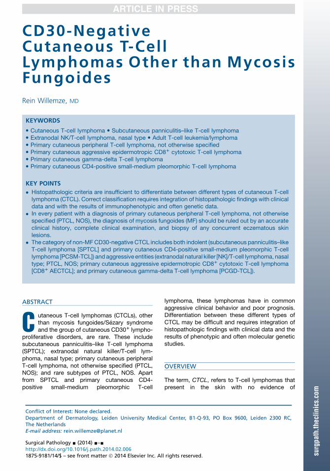
SPTCL occurs in adults as well as young children.Patients generally present with solitary or multiplenodules or deeply seated plaques with a diametervarying between 1 and 20 cm. The skin lesionsmainly involve the legs, the arms, the trunk, andless commonly the face andmay leave areas of lip-oatrophy after disappearance (Fig. 1). Ulceration is
Table 2Distinguishing features between subcutaneous panniculitis–like T-cell lymphoma and primarycutaneous gamma-delta T-cell lymphoma
SPTCL PCGD-TCL
Immunophenotype TCRb1, CD4�, CD81, CD56� TCRg1, CD4�, CD8�/1, CD561/�
Architecture Subcutaneous Subcutaneous and/orepidermal/dermal
Clinical features Nodules and plaques; rarely ulceration;association with autoimmunedisorders (20%)
Nodules and plaquesUlceration common
HPS Uncommon (17%) Common (50%)
5-Year survival (%) 82a 11
Treatment Systemic steroids Systemic chemotherapy
a 5-Year survival: 91% in patients with HPS, 46% in patients with HPS.9
Non-MF CD30-Negative CTCL 3
uncommon. Systemic symptoms, such as fever,fatigue, and weight loss, and laboratory abnormal-ities, including cytopenias and elevated liver func-tion tests, are common, but a frank HPS isobserved in only 15% of patients.5,9 Disseminationto extracutaneous sites is rare. Hepatosplenome-galy may be seen but is generally not due tolymphomatous involvement. Up to 20%of patientsmay have associated autoimmune disease, mostcommonly systemic lupus erythematosus.9
DIAGNOSIS: MICROSCOPIC FEATURES
Histologically, SPTCL reveals subcutaneous infil-trates simulating a lobular panniculitis showingsmall, medium-sized, or sometimes large pleo-morphic T cells with hyperchromatic nuclei and
Fig. 1. SPTCL. Subcutane-ous nodules on the leftarm. Ulceration is notpresent.
often many macrophages (Fig. 2A). The overlyingepidermis and dermis are typically uninvolved.Rimming of individual fat cells by neoplastic T cellsis a helpful, although not completely specific, diag-nostic feature (see Fig. 2B). Necrosis, karyor-rhexis, cytophagocytosis, and fat necrosis arecommon findings.10 In the early stages, theneoplastic infiltrates may lack significant atypiaand a heavy inflammatory infiltrate maypredominate.11,12
DIAGNOSIS: ANCILLARY STUDIES
The neoplastic cells have a mature CD31, CD4�,CD81 T-cell phenotype, with expression of cyto-toxic proteins (see Fig. 2C).8–10,13 The neoplasticT cells express bF1 but not TCRg and are negative
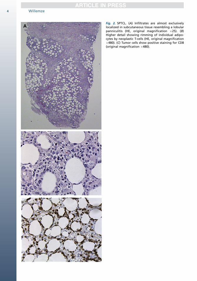
Fig. 2. SPTCL. (A) Infiltrates are almost exclusivelylocalized in subcutaneous tissue resembling a lobularpanniculitis (HE, original magnification �25). (B)Higher detail showing rimming of individual adipo-cytes by neoplastic T-cells (HE, original magnification�480). (C) Tumor cells show positive staining for CD8(original magnification �480).
Willemze4
Non-MF CD30-Negative CTCL 5
for CD56, facilitating differentiation from cuta-neous gamma-delta T-cell lymphoma.9,14 CD30is rarely, if ever, expressed. The neoplastic T cellsshow clonal TCR gene rearrangements. Epstein-Barr virus (EBV) is absent.1
DIFFERENTIAL DIAGNOSIS
The differential diagnosis of SPTCL includesother types of CTCL with subcutaneous involve-ment, in particular PCGD-TCL, and lupus panni-culitis. In contrast to SPTCL, PCGD-TCL withpanniculitis-like features commonly involves notonly the subcutis but also the dermis and/orepidermis, either in the same or in other biopsies,and may show ulceration. By definition they havea gd T-cell phenotype (positive staining for TCRg/TCRd, preferably in combination with a negativestaining for bF1), are generally negative for bothCD4 and CD8, and commonly express CD56(see Table 2). Differentiation is important,because PCGD-TCL with panniculitis-like fea-tures generally has a poor prognosis and requiressystemic chemotherapy.9,14
SPTCL and lupus panniculitis have overlappingclinicopathologic features, making differentiationsometimes extremely difficult.15,16 The clinical pre-sentation may be identical and several reportsdescribe patients who had both SPTCL andgenuine lupus erythematosus.9,16 As a result, thereis controversy whether both conditions maycoexist or form a spectrum of disease.15–17 Histo-logic features of lupus panniculitis, not or rarelyobserved in SPTCL, include the presence of inter-face dermatitis, clusters of B cells (sometimes withformation of germinal centers), and many admixedplasma cells. High proliferation rate and demon-stration of clonal TCR gene rearrangements areuncommon in lupus panniculitis and strongly sup-port a diagnosis of SPTCL.
PROGNOSIS
SPTCLs have an excellent prognosis, particularly ifnot associated with an HPS.9,10 In a recent EORTCstudy, patients with and without an associatedHPS had 5-year overall survival rates of 46% and91%, respectively.9 In SPTCL without associatedHPS, systemic steroids or other immunosuppres-sive agents are recommended, whereas in casesof solitary skin lesions, radiotherapy is advised.9,10
Only in cases of progressive disease not respond-ing to immunosuppressive therapy and in casesof HPS should multiagent chemotherapy beconsidered.
EXTRANODAL NK/T-CELL LYMPHOMA,
NASAL TYPE
OVERVIEW
Extranodal NK/T-cell lymphoma, nasal type, is anEBV-positive lymphoma of small, medium, or largecells usually with an NK cell or, more rarely a cyto-toxic T-cell phenotype. The skin is the secondmost common site of involvement after the nasalcavity/nasopharynx, and skin involvement maybe a primary or secondary manifestation of thedisease.1,2
CLINICAL FEATURES
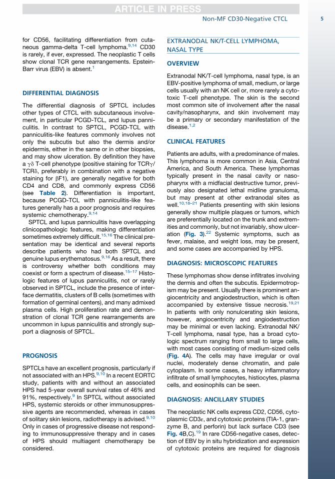
Patients are adults, with a predominance of males.This lymphoma is more common in Asia, CentralAmerica, and South America. These lymphomastypically present in the nasal cavity or naso-pharynx with a midfacial destructive tumor, previ-ously also designated lethal midline granuloma,but may present at other extranodal sites aswell.10,18–21 Patients presenting with skin lesionsgenerally show multiple plaques or tumors, whichare preferentially located on the trunk and extrem-ities and commonly, but not invariably, show ulcer-ation (Fig. 3).22 Systemic symptoms, such asfever, malaise, and weight loss, may be present,and some cases are accompanied by HPS.
DIAGNOSIS: MICROSCOPIC FEATURES
These lymphomas show dense infiltrates involvingthe dermis and often the subcutis. Epidermotrop-ismmay be present. Usually there is prominent an-giocentricity and angiodestruction, which is oftenaccompanied by extensive tissue necrosis.19,21
In patients with only nonulcerating skin lesions,however, angiocentricity and angiodestructionmay be minimal or even lacking. Extranodal NK/T-cell lymphoma, nasal type, has a broad cyto-logic spectrum ranging from small to large cells,with most cases consisting of medium-sized cells(Fig. 4A). The cells may have irregular or ovalnuclei, moderately dense chromatin, and palecytoplasm. In some cases, a heavy inflammatoryinfiltrate of small lymphocytes, histiocytes, plasmacells, and eosinophils can be seen.
DIAGNOSIS: ANCILLARY STUDIES
The neoplastic NK cells express CD2, CD56, cyto-plasmic CD3ε, and cytotoxic proteins (TIA-1, gran-zyme B, and perforin) but lack surface CD3 (seeFig. 4B,C).19 In rare CD56-negative cases, detec-tion of EBV by in situ hybridization and expressionof cytotoxic proteins are required for diagnosis
Fig. 3. Extranodal NK/T-cell lymphoma, nasal type.Presentationwith general-ized plaques and ulcerat-ing tumor on the rightwrist.
Willemze6
(see Fig. 4D). EBV latent membrane protein(LMP-1) is inconsistently expressed. Most caseshave a genuine NK-cell origin with no surfaceCD3 expression and no TCR expression, togetherwith TCR genes in germline configuration. Somecases, however, may show a true T-cell phenotypewith a monoclonal TCR gene rearrangement andsurface CD3 expression.23
DIFFERENTIAL DIAGNOSIS
Extranodal NK/T-cell lymphoma, nasal type,should bedifferentiated fromother types of aggres-sive cytotoxic CTCL (PCGD-TCL, CD81 AECTCL,and some cases of tumor-stage MF with a cyto-toxic phenotype) and from other EBV-associatedNK/T-cell LPDs, some of which preferentially affectchildren.2 One of these conditions, hydroa vaccini-forme–like lymphoma (HVL), is described later.Because extranodal NK/T-cell lymphoma, nasaltype, is strongly associated with EBV and mostcases have an NK-cell phenotype, demonstrationof EBV by EBER in situ hybridization or LMP-1staining, together with negative staining for T-cellmarkers and germline configuration of TCR genes,strongly supports a diagnosis of extranodal NK/T-cell lymphoma, nasal type (Table 3).
PROGNOSIS
Recent studies indicate that nasal cases have abetter prognosis than extranasal cases.24 Extra-nodal NK/T-cell lymphoma presenting in the skinis a highly aggressive tumor with a median sur-vival of less than 12 months.10,20–22 Although
patients presenting with only skin lesions mayhave a somewhat better prognosis than patientspresenting with both cutaneous and extracutane-ous disease, the overall prognosis remainspoor.22 CD301, CD561 cases reported to have abetter prognosis most probably have been exam-ples of C-ALCL with coexpression of CD56.25 Inpatients with stage I disease, radiotherapy is thefirst choice of treatment.26 Cases of moreadvanced disease show an aggressive clinicalbehavior and are often resistant to chemotherapy.Recently, an intensive chemotherapy regimen,including steroid (dexamethasone), methotrexate,ifoffamide, L-asparaginase and etoposide (theSMILE regimen), has shown promising effects.27
HYDROA VACCINIFORME–LIKE LYMPHOMA
OVERVIEW
HVL is a rare EBV-positive CTCL, clinically resem-bling hydroa vacciniforme (HV), which is a rarechronic photosensitivity disorder mainly affectingchildren and characterized by a necrotic papulo-vesicular eruption with scarring on sun-exposedareas.28 HVL has been described mainly in chil-dren and young adults from Latin American andAsian countries and has been included in theWHO 2008 classification as one of the EBV-positive LPDs of childhood.2
CLINICAL FEATURES
Patients present with ulceronecrotic skin lesionsassociated with blisters, facial edema, and
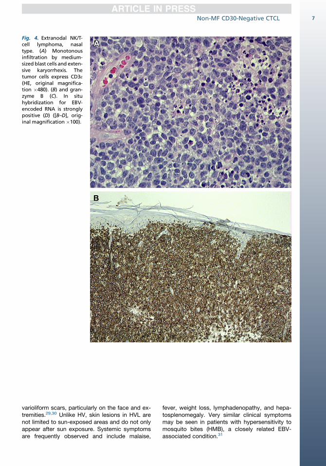
Fig. 4. Extranodal NK/T-cell lymphoma, nasaltype. (A) Monotonousinfiltration by medium-sized blast cells and exten-sive karyorrhexis. Thetumor cells express CD3ε(HE, original magnifica-tion �480). (B) and gran-zyme B (C). In situhybridization for EBV-encoded RNA is stronglypositive (D) ([B–D], orig-inal magnification �100).
Non-MF CD30-Negative CTCL 7
varioliform scars, particularly on the face and ex-tremities.29,30 Unlike HV, skin lesions in HVL arenot limited to sun-exposed areas and do not onlyappear after sun exposure. Systemic symptomsare frequently observed and include malaise,
fever, weight loss, lymphadenopathy, and hepa-tosplenomegaly. Very similar clinical symptomsmay be seen in patients with hypersensitivity tomosquito bites (HMB), a closely related EBV-associated condition.31
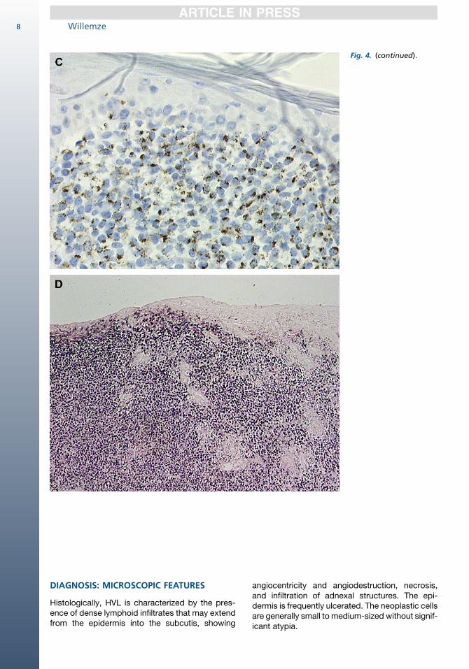
Fig. 4. (continued).
Willemze8
DIAGNOSIS: MICROSCOPIC FEATURES
Histologically, HVL is characterized by the pres-ence of dense lymphoid infiltrates that may extendfrom the epidermis into the subcutis, showing
angiocentricity and angiodestruction, necrosis,and infiltration of adnexal structures. The epi-dermis is frequently ulcerated. The neoplastic cellsare generally small to medium-sized without signif-icant atypia.
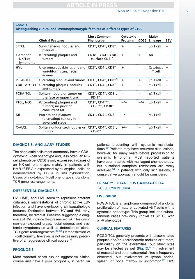
Table 3Distinguishing clinical and immunophenotypic features of different types of CTCL
Clinical FeaturesMost CommonPhenotype
CytotoxicProteins CD56
MajorLineage EBV
SPTCL Subcutaneous nodules andplaques
CD31, CD4�, CD81 1 � ab T cell �
ExtranodalNK/T-celllymphoma
(Ulcerating) plaques andtumors
CD3e1, CD4�, CD81
(surface CD3�)1 1 NK 1
HVL Ulceronecrotic skin lesions andvarioliform scars, facialedema
CD31, CD4�, CD81 1 � CytotoxicT cell
1
PCGD-TCL Ulcerating plaques and tumors CD31, CD4�, CD8�/1 1 1 gd T cell �CD81 AECTCL Ulcerating plaques, nodules
and tumorsCD31, CD4�, CD81 1 � ab T cell �
PCSM-TCL Solitary nodule or tumor onthe face or upper trunk
CD31, CD41, CD8�,PD-11
� � ab T cell �
PTCL, NOS (Ulcerating) plaques andtumors; no prior orconcurrent MF
CD31, CD41/�,CD8�/1, CD30�
�/1 �/1 ab T cell �
MF Patches and plaques;(ulcerating) tumors inadvanced stage
CD31, CD41, CD8� �/1 � ab T cell �
C-ALCL Solitary or localized nodules ortumors
CD31, CD41, CD8�,CD301
1/� � ab T cell �
Non-MF CD30-Negative CTCL 9
DIAGNOSIS: ANCILLARY STUDIES
The neoplastic cells most commonly have a CD81
cytotoxic T-cell phenotype and, less often, an NK-cell phenotype. CD56 is only expressed in cases ofan NK-cell phenotype, mainly in patients withHMB.32 EBV is expressed by all atypical cells, asdemonstrated by EBER in situ hybridization.Cases of a cytotoxic T-cell phenotype show clonalTCR gene rearrangements.
DIFFERENTIAL DIAGNOSIS
HV, HMB, and HVL seem to represent differentcutaneous manifestations of chronic active EBVinfection and have overlapping clinicopathologicfeatures. Distinction between HV and HVL may,therefore, be difficult. Features suggesting a diag-nosis of HVL include the presence of skin lesions innon–sun-exposed areas, facial edema, and sys-temic symptoms as well as detection of clonalTCR gene rearrangements.30,33 Demonstration ofT-cell clonality, however, is not necessarily predic-tive of an aggressive clinical course.33
PROGNOSIS
Most reported cases run an aggressive clinicalcourse and have a poor prognosis, in particular
patients presenting with systemic manifesta-tions.30 Patients may have recurrent skin lesions,however, for many years before progression tosystemic lymphoma. Most reported patientshave been treated with multiagent chemotherapy,but sustained complete remissions are rarelyachieved.34 In patients with only skin lesions, aconservative approach should be considered.
PRIMARY CUTANEOUS GAMMA-DELTA
T-CELL LYMPHOMA
OVERVIEW
PCGD-TCL is a lymphoma composed of a clonalproliferation of mature, activated gd T cells with acytotoxic phenotype. This group includes subcu-taneous cases previously known as SPTCL witha gd phenotype.
CLINICAL FEATURES
PCGD-TCL generally presents with disseminatedplaques and/or ulceronecrotic nodules or tumors,particularly on the extremities, but other sitesmay be affected as well (Fig. 5).9,14 Involvementof mucosal and other extranodal sites is frequentlyobserved, but involvement of lymph nodes,spleen, or bone marrow is uncommon.35 HPS
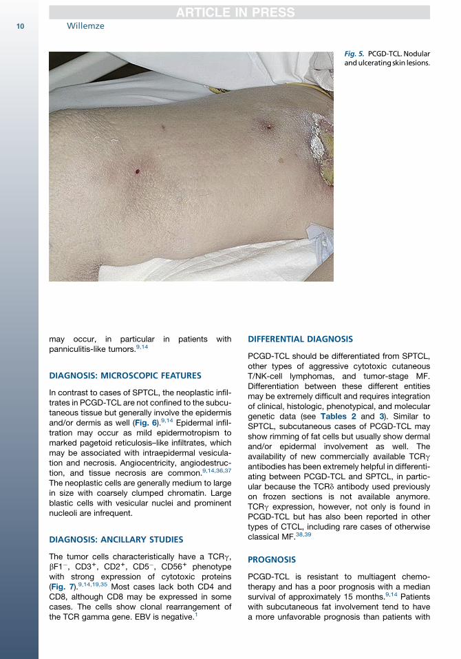
Fig. 5. PCGD-TCL.Nodularandulcerating skin lesions.
Willemze10
may occur, in particular in patients withpanniculitis-like tumors.9,14
DIAGNOSIS: MICROSCOPIC FEATURES
In contrast to cases of SPTCL, the neoplastic infil-trates in PCGD-TCL are not confined to the subcu-taneous tissue but generally involve the epidermisand/or dermis as well (Fig. 6).9,14 Epidermal infil-tration may occur as mild epidermotropism tomarked pagetoid reticulosis–like infiltrates, whichmay be associated with intraepidermal vesicula-tion and necrosis. Angiocentricity, angiodestruc-tion, and tissue necrosis are common.9,14,36,37
The neoplastic cells are generally medium to largein size with coarsely clumped chromatin. Largeblastic cells with vesicular nuclei and prominentnucleoli are infrequent.
DIAGNOSIS: ANCILLARY STUDIES
The tumor cells characteristically have a TCRg,bF1�, CD31, CD21, CD5�, CD561 phenotypewith strong expression of cytotoxic proteins(Fig. 7).9,14,19,35 Most cases lack both CD4 andCD8, although CD8 may be expressed in somecases. The cells show clonal rearrangement ofthe TCR gamma gene. EBV is negative.1
DIFFERENTIAL DIAGNOSIS
PCGD-TCL should be differentiated from SPTCL,other types of aggressive cytotoxic cutaneousT/NK-cell lymphomas, and tumor-stage MF.Differentiation between these different entitiesmay be extremely difficult and requires integrationof clinical, histologic, phenotypical, and moleculargenetic data (see Tables 2 and 3). Similar toSPTCL, subcutaneous cases of PCGD-TCL mayshow rimming of fat cells but usually show dermaland/or epidermal involvement as well. Theavailability of new commercially available TCRgantibodies has been extremely helpful in differenti-ating between PCGD-TCL and SPTCL, in partic-ular because the TCRd antibody used previouslyon frozen sections is not available anymore.TCRg expression, however, not only is found inPCGD-TCL but has also been reported in othertypes of CTCL, including rare cases of otherwiseclassical MF.38,39
PROGNOSIS
PCGD-TCL is resistant to multiagent chemo-therapy and has a poor prognosis with a mediansurvival of approximately 15 months.9,14 Patientswith subcutaneous fat involvement tend to havea more unfavorable prognosis than patients with
Fig. 6. PCGD-TCL. High-power viewof subcutane-ous infiltrate showingrimming of fat cells byneoplastic T cells and ex-tensive karyorrhexis (HE,original magnification�480).
Non-MF CD30-Negative CTCL 11
epidermal or dermal disease only.14 Rare cases ofPCGD-TCL with panniculitis-like features, how-ever, following a more indolent clinical coursehave been reported.40,41 Patients should betreated with systemic chemotherapy, but the re-sults are often disappointing. 14
PRIMARY CUTANEOUS AGGRESSIVE
EPIDERMOTROPIC CD8-POSITIVE CYTOTOXIC
T-CELL LYMPHOMA (PROVISIONAL ENTITY)
OVERVIEW
CD81 AECTCLs are characterized by a prolifera-tion of epidermotropic CD8-positive cytotoxicT cells and an aggressive clinical behavior. In theWHO 2008 classification, CD81 AECTCL is listedas a provisional entity.2
CLINICAL FEATURES
Clinically, these lymphomas show localized ordisseminated eruptive papules, nodules, andtumors showing central ulceration and necrosisor by superficial, hyperkeratotic patches and pla-ques (Fig. 8).13,42 CD81 AECTCL may disseminateto visceral sites (lung, testis, central nervous sys-tem, and oral mucosa), but lymph nodes are oftenspared.42,43
DIAGNOSIS: MICROSCOPIC FEATURES
The histologic appearance is variable, rangingfrom a lichenoid pattern with marked, pagetoid ep-idermotropism and subepidermal edema in earlypatchlike lesions to diffuse dermal infiltrates innodular and tumorous lesions (Fig. 9). Epidermalnecrosis and ulceration, as well as invasion anddestruction of adnexal structures, are commonlyfound.42,43 Angiocentricity and angioinvasionmay be present. Tumor cells are small–mediumor medium–large with pleomorphic or blasticnuclei.1
DIAGNOSIS: ANCILLARY STUDIES
The tumor cell have a bF11, CD31, CD81, gran-zyme B1, perforin1, TIA-11, CD45RA1/�,CD45RO�, CD2�/1, CD4�, CD5�, CD71/� pheno-type (Fig. 10).10,13,42,43 CD30 is rarely expressed.The neoplastic T cells show clonal TCR gene rear-rangements. Specific recurrent genetic abnormal-ities have not been described. EBV is negative.1
DIFFERENTIAL DIAGNOSIS
CD81 AECTCL should be differentiated from othertypes of CTCL expressing a CD8-positive cyto-toxic T-cell phenotype (see Table 3).41 Differentia-tion from SPTCL, early-stage MF, LyP, andpagetoid reticulosis is generally not difficult, inparticular when the clinical features are taken
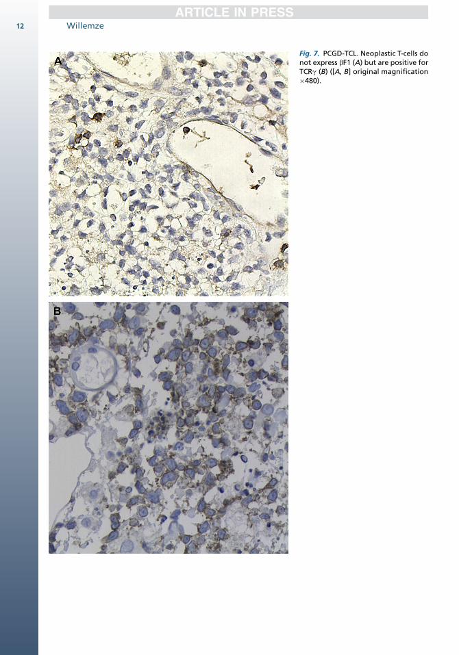
Fig. 7. PCGD-TCL. Neoplastic T-cells donot express bF1 (A) but are positive forTCRg (B) ([A, B] original magnification�480).
Willemze12
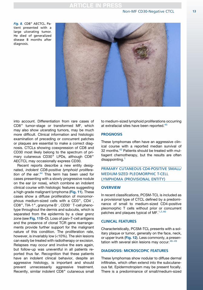
Fig. 8. CD81 AECTCL. Pa-tient presented with alarge ulcerating tumor.He died of generalizeddisease 8 months afterdiagnosis.
Non-MF CD30-Negative CTCL 13
into account. Differentiation from rare cases ofCD81 tumor-stage or transformed MF, whichmay also show ulcerating tumors, may be muchmore difficult. Clinical information and histologicexamination of preceding or concurrent patchesor plaques are essential to make a correct diag-nosis. CTCLs showing coexpression of CD8 andCD30 most likely belong to the spectrum of pri-mary cutaneous CD301 LPDs, although CD81
AECTCL may occasionally express CD30.Recent reports describe a new entity desig-
nated, indolent CD8-positive lymphoid prolifera-tion of the ear.44 This term has been used forcases presenting with a slowly progressive noduleon the ear (or nose), which combine an indolentclinical course with histologic features suggestinga high-grade malignant lymphoma (Fig. 11). Thesecases show a diffuse proliferation of monomor-phous medium-sized cells with a CD31, CD4�,CD81, TIA-11, granzyme B�, CD30� T-cell pheno-type throughout the dermis and subcutis, which isseparated from the epidermis by a clear grenzzone (see Fig. 11B–D). Loss of pan–T-cell antigensand the presence of clonal TCR gene rearrange-ments provide further support for the malignantnature of this condition. The proliferation rate,however, is invariably low (<10%). The skin lesionscan easily be treated with radiotherapy or excision.Relapses may occur and involve the ears again,but follow-up was uneventful in all patients re-ported thus far. Recognition that these patientshave an indolent clinical behavior, despite anaggressive histology, is important and shouldprevent unnecessarily aggressive treatment.Recently, similar indolent CD81 cutaneous small
to medium-sized lymphoid proliferations occurringat extrafacial sites have been reported.45
PROGNOSIS
These lymphomas often have an aggressive clin-ical course with a reported median survival of32 months.42 Patients should be treated with mul-tiagent chemotherapy, but the results are oftendisappointing.1
PRIMARY CUTANEOUS CD4-POSITIVE SMALL/
MEDIUM-SIZED PLEOMORPHIC T-CELL
LYMPHOMA (PROVISIONAL ENTITY)
OVERVIEW
In recent classifications, PCSM-TCL is included asa provisional type of CTCL defined by a predomi-nance of small to medium-sized CD4-positivepleomorphic T cells without prior or concurrentpatches and plaques typical of MF.1,2,46
CLINICAL FEATURES
Characteristically, PCSM-TCL presents with a sol-itary plaque or tumor, generally on the face, neck,or upper trunk (Fig. 12). Less commonly, a presen-tation with several skin lesions may occur.46–49
DIAGNOSIS: MICROSCOPIC FEATURES
These lymphomas show nodular to diffuse dermalinfiltrates, which often extend into the subcutane-ous fat. Epidermotropism may be present focally.There is a predominance of small/medium-sized

Fig. 9. CD81 AECTCL. (A) Diffuse dermalinfiltrate with marked epidermotropism(HE, original magnification �100). (B)Strong expression of CD8 (originalmagnification �100).
14
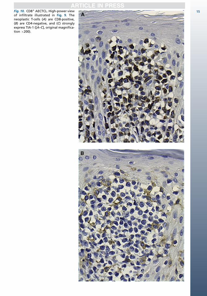
Fig. 10. CD81 AECTCL. High-power viewof infiltrate illustrated in Fig. 9. Theneoplastic T-cells (A) are CD8-positive,(B) are CD4-negative, and (C) stronglyexpress TIA-1 ([A–C], original magnifica-tion �200).
15
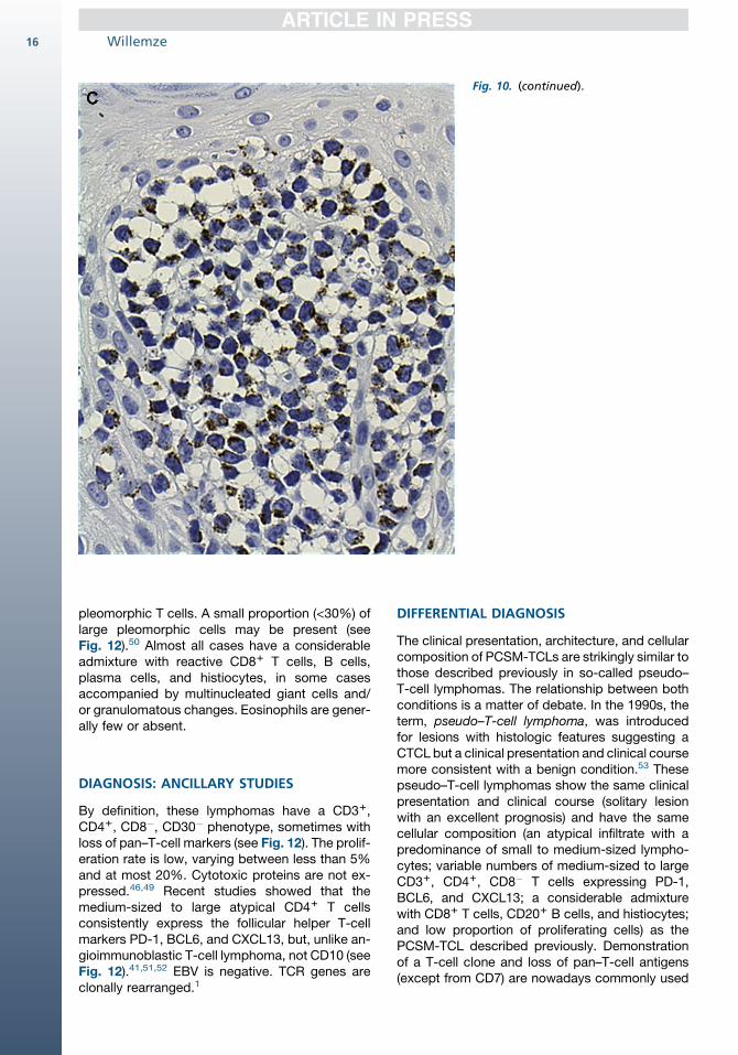
Fig. 10. (continued).
Willemze16
pleomorphic T cells. A small proportion (<30%) oflarge pleomorphic cells may be present (seeFig. 12).50 Almost all cases have a considerableadmixture with reactive CD81 T cells, B cells,plasma cells, and histiocytes, in some casesaccompanied by multinucleated giant cells and/or granulomatous changes. Eosinophils are gener-ally few or absent.
DIAGNOSIS: ANCILLARY STUDIES
By definition, these lymphomas have a CD31,CD41, CD8�, CD30� phenotype, sometimes withloss of pan–T-cell markers (see Fig. 12). The prolif-eration rate is low, varying between less than 5%and at most 20%. Cytotoxic proteins are not ex-pressed.46,49 Recent studies showed that themedium-sized to large atypical CD41 T cellsconsistently express the follicular helper T-cellmarkers PD-1, BCL6, and CXCL13, but, unlike an-gioimmunoblastic T-cell lymphoma, not CD10 (seeFig. 12).41,51,52 EBV is negative. TCR genes areclonally rearranged.1
DIFFERENTIAL DIAGNOSIS
The clinical presentation, architecture, and cellularcomposition of PCSM-TCLs are strikingly similar tothose described previously in so-called pseudo–T-cell lymphomas. The relationship between bothconditions is a matter of debate. In the 1990s, theterm, pseudo–T-cell lymphoma, was introducedfor lesions with histologic features suggesting aCTCL but a clinical presentation and clinical coursemore consistent with a benign condition.53 Thesepseudo–T-cell lymphomas show the same clinicalpresentation and clinical course (solitary lesionwith an excellent prognosis) and have the samecellular composition (an atypical infiltrate with apredominance of small to medium-sized lympho-cytes; variable numbers of medium-sized to largeCD31, CD41, CD8� T cells expressing PD-1,BCL6, and CXCL13; a considerable admixturewith CD81 T cells, CD201 B cells, and histiocytes;and low proportion of proliferating cells) as thePCSM-TCL described previously. Demonstrationof a T-cell clone and loss of pan–T-cell antigens(except from CD7) are nowadays commonly used
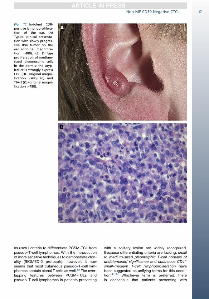
Fig. 11. Indolent CD8-positive lymphoprolifera-tion of the ear. (A)Typical clinical presenta-tion with slowly progres-sive skin tumor on theear (original magnifica-tion �480). (B) Diffuseproliferation of medium-sized pleomorphic cellsin the dermis; the atyp-ical cells strongly expressCD8 (HE, original magni-fication �480) (C) andTIA-1 (D) (original magni-fication �480).
Non-MF CD30-Negative CTCL 17
as useful criteria to differentiate PCSM-TCL frompseudo–T-cell lymphomas. With the introductionof more sensitive techniques to demonstrate clon-ality (BIOMED-2 protocols), however, it nowseems that most cutaneous pseudo–T-cell lym-phomas contain clonal T cells as well.52 The over-lapping features between PCSM-TCLs andpseudo–T-cell lymphomas in patients presenting
with a solitary lesion are widely recognized.Because differentiating criteria are lacking, smallto medium-sized pleomorphic T-cell nodules ofundetermined significance and cutaneous CD41
small–medium T-cell lymphoproliferation havebeen suggested as unifying terms for this condi-tion.41,54 Whichever term is preferred, thereis consensus that patients presenting with
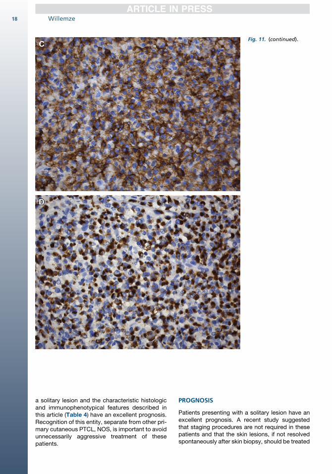
Fig. 11. (continued).
Willemze18
a solitary lesion and the characteristic histologicand immunophenotypical features described inthis article (Table 4) have an excellent prognosis.Recognition of this entity, separate from other pri-mary cutaneous PTCL, NOS, is important to avoidunnecessarily aggressive treatment of thesepatients.
PROGNOSIS
Patients presenting with a solitary lesion have anexcellent prognosis. A recent study suggestedthat staging procedures are not required in thesepatients and that the skin lesions, if not resolvedspontaneously after skin biopsy, should be treated
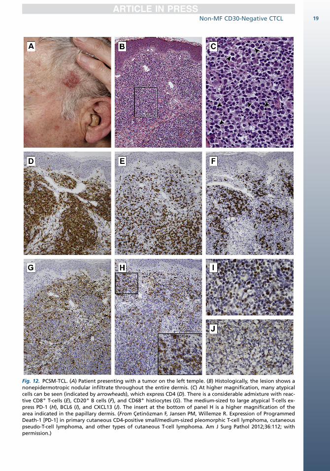
Fig. 12. PCSM-TCL. (A) Patient presenting with a tumor on the left temple. (B) Histologically, the lesion shows anonepidermotropic nodular infiltrate throughout the entire dermis. (C) At higher magnification, many atypicalcells can be seen (indicated by arrowheads), which express CD4 (D). There is a considerable admixture with reac-tive CD81 T-cells (E), CD201 B cells (F), and CD681 histiocytes (G). The medium-sized to large atypical T-cells ex-press PD-1 (H), BCL6 (I), and CXCL13 (J). The insert at the bottom of panel H is a higher magnification of thearea indicated in the papillary dermis. (From Cetinozman F, Jansen PM, Willemze R. Expression of ProgrammedDeath-1 [PD-1] in primary cutaneous CD4-positive small/medium-sized pleomorphic T-cell lymphoma, cutaneouspseudo-T-cell lymphoma, and other types of cutaneous T-cell lymphoma. Am J Surg Pathol 2012;36:112; withpermission.)
Non-MF CD30-Negative CTCL 19
Table 4Differential diagnosis of CD8D CTCL
Frequency of CD8Expression (%) Helpful Distinguishing Features
CD81 AECTCL 100 Ulcerating plaques, nodules, and tumors; no prioror concurrent eczematous patches/plaques
Early patch/plaque-stage MF 15 Prior or concurrent eczematous patches/plaquesa
Tumor-stage/transformed MF 5 (Ulcerating) tumors; prior or concurrenteczematous patches/plaquesa
Pagetoid reticulosis 50 Solitary, slowly expanding plaque; usually on theextremities
C-ALCL <5 Solitary or localized (ulcerating) tumors; tendencyfor spontaneous remissiona
LyP, type D 100 Recurrent, self-healing papular, nodular or smallulceronecrotic skin lesionsa
SPTCL >90 Subcutaneous nodules and plaques; no ulceration;no epidermal involvement
Indolent CD81 lymphoidproliferation of the ear
100 Slowly progressive nodule on ear (or nose);no ulceration; no epidermal involvement;low proliferation rate
a No difference in clinical presentation and prognosis between CD81 and more common CD41 cases.Adapted from Quintanilla-Martinez L, Jansen PM, Kinney MC, et al. Non-mycosis fungoides cutaneous T-cell lym-
phomas: report of the 2011 Society for Hematopathology/European Association for Haematopathology workshop. AmJ Clin Pathol 2013;139:507; with permission.
Willemze20
primarily with intralesional steroids or surgicalexcision and only by exception with radio-therapy.52 PCSM-TCLs that present with general-ized skin lesions and/or do not meet the criteriaof the cases described previously are rare andshould be fully staged. A recent study suggestedthat PCSM-TCLs with rapidly growing bulky tu-mors, a low percentage of admixed CD81 T cells,and/or a high proliferative fraction are at risk todevelop progressive disease; however, this shouldbe confirmed.48 The optimal treatment in suchcases has yet to be defined.
PRIMARY CUTANEOUS PERIPHERAL T-CELL
LYMPHOMA, NOT OTHERWISE SPECIFIED
OVERVIEW
The term, PTCL, NOS, is used for CTCLs that donot fit into any of the better-defined subtypes ofCTCLs, including the 3 rare subtypes of PTCL,NOS, which have been recognized in recent clas-sifications (see Table 1). PTCL, NOS can involvethe skin primarily or secondarily, but PTCL, NOSpresenting with only skin lesions (PTCL, NOS) isuncommon.
CLINICAL FEATURES
Patients are commonly adults who present withsolitary or localized, but more frequently general-ized, often ulcerating nodules or tumors(Fig. 13A).46,55
DIAGNOSIS: MICROSCOPIC FEATURES
Histologically, these lymphomas show nodularor diffuse infiltrates with variable numbers ofmedium-sized to large pleomorphic orimmunoblast-like T cells (see Fig. 13B). Epidermo-tropism is generally mild or absent. Largeneoplastic cells comprise more than 30% of theinfiltrate.1
DIAGNOSIS: ANCILLARY STUDIES
Most cases show an aberrant CD41 T-cell pheno-type with variable loss of pan–T-cell antigens.CD30 staining is negative or restricted to few scat-tered tumor cells. Rare casesmay show coexpres-sion of CD56. Expression of cytotoxic proteins isuncommon in CD41 cases but frequentlyobserved in cases with a CD4�, CD8� T-cellphenotype.46
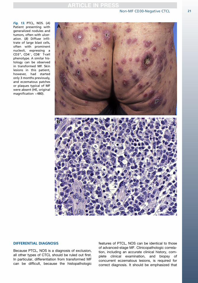
Fig. 13. PTCL, NOS. (A)Patient presenting withgeneralized nodules andtumors, often with ulcer-ation. (B) Diffuse infil-trate of large blast cells,often with prominentnucleoli, expressing aCD31, CD4�, CD8� T-cellphenotype. A similar his-tology can be observedin transformed MF. Skinlesions in this patient,however, had startedonly 3 months previously,and eczematous patchesor plaques typical of MFwere absent (HE, originalmagnification �480).
Non-MF CD30-Negative CTCL 21
DIFFERENTIAL DIAGNOSIS
Because PTCL, NOS is a diagnosis of exclusion,all other types of CTCL should be ruled out first.In particular, differentiation from transformed MFcan be difficult, because the histopathologic
features of PTCL, NOS can be identical to thoseof advanced-stage MF. Clinicopathologic correla-tion, including an accurate clinical history, com-plete clinical examination, and biopsy ofconcurrent eczematous lesions, is required forcorrect diagnosis. It should be emphasized that
Willemze22
patients with MF developing skin lesions orinvolved lymph nodes with the histologic featuresof a PTCL, NOS should not be considered tohave progressed to a PTCL, NOS and should notbe reclassified as such. They simply have tumor-stage MF or nodal involvement by MF. Differentia-tion is important, because most patients withskin-limited MF should still be treated with skin-directed therapies (eg, radiotherapy), whereaspatients with a PTCL, NOS should be treatedwith multiagent chemotherapy.
PROGNOSIS
The prognosis is generally poor (5-year survivalrates less than 20%) and independent of thepresence or absence of extracutaneous diseaseat the time of diagnosis, the extent of skinlesions at presentation, cell size, or expressionof CD41 or CD81 phenotype.46,47,50,55 Patientsare commonly treated with multiagent chemo-therapy as with treatment of aggressive T-celllymphomas.
REFERENCES
1. Willemze R, Jaffe ES, Burg G, et al. WHO-EORTC
classification for cutaneous lymphomas. Blood
2005;105:3768–85.
2. Swerdlow A, Campo E, Harris NL, et al. World
Health Organization classification of tumours of he-
matopoietic and lymphoid tissue. Lyon (France):
IARC Press; 2008.
3. Tan SH, Sim CS, Ong BH. Cutaneous lymphomas
other than mycosis fungoides in Singapore: a clin-
icopathological analysis using recent classification
systems. Br J Dermatol 2003;149:542–53.
4. Park JH, Shin HT, Lee DY, et al. World Health
Organization-European Organization for Research
and Treatment of Cancer classification of cuta-
neous lymphoma in Korea: a retrospective study
at a single tertiary institution. J Am Acad Dermatol
2012;67:1200–9.
5. Gonzalez CL, Medeiros LJ, Braziel RM, et al. T-cell
lymphoma involving subcutaneous tissue. A clini-
copathologic entity commonly associated with he-
mophagocytic syndrome. Am J Surg Pathol 1991;
15:17–27.
6. Jaffe ES, Harris NL, Stein H, et al. World Health Or-
ganization classification of tumours: pathology and
genetics of tumours of hematopoietic and lymphoid
tissues. Lyon (France): IARC Press; 2001.
7. Willemze R, Kerl H, Sterry W, et al. EORTC classifi-
cation for primary cutaneous lymphomas: a pro-
posal from the Cutaneous Lymphoma Study
Group of the European Organization for Research
and Treatment of Cancer. Blood 1997;90:354–71.
8. Salhany KE, Macon WR, Choi JK, et al. Subcutane-
ous panniculitis-like T-cell lymphoma: clinicopatho-
logic, immunophenotypic, and genotypic analysis
of alpha/beta and gamma/delta subtypes. Am J
Surg Pathol 1998;22:881–93.
9. Willemze R, Jansen PM, Cerroni L, et al. Subcu-
taneous panniculitis-like T-cell lymphoma: defini-
tion, classification, and prognostic factors: an
EORTC Cutaneous Lymphoma Group Study of 83
cases. Blood 2008;111:838–45.
10. Massone C, Chott A, Metze D, et al. Subcutaneous,
blastic natural killer (NK), NK/T-cell, and other cyto-
toxic lymphomas of the skin: a morphologic, immu-
nophenotypic, and molecular study of 50 patients.
Am J Surg Pathol 2004;28:719–35.
11. Hoque SR, Child FJ, Whittaker SJ, et al. Subcutane-
ous panniculitis-like T-cell lymphoma: a clinicopath-
ological, immunophenotypic and molecular
analysis of six patients. Br J Dermatol 2003;148:
516–25.
12. Marzano AV, Berti E, Paulli M, et al. Cytophagic
histiocytic panniculitis and subcutaneous
panniculitis-like T-cell lymphoma: report of 7 cases.
Arch Dermatol 2000;136:889–96.
13. Santucci M, Pimpinelli N, Massi D, et al. Cytotoxic/
natural killer cell cutaneous lymphomas. Report of
EORTC Cutaneous Lymphoma Task Force Work-
shop. Cancer 2003;97:610–27.
14. Toro JR, Liewehr DJ, Pabby N, et al. Gamma-delta
T-cell phenotype is associated with significantly
decreased survival in cutaneous T-cell lymphoma.
Blood 2003;101:3407–12.
15. Magro CM, Crowson AN, Kovatich AJ, et al. Lupus
profundus, indeterminate lymphocytic lobular pan-
niculitis and subcutaneous T-cell lymphoma: a
spectrum of subcuticular T-cell lymphoid
dyscrasia. J Cutan Pathol 2001;28:235–47.
16. Pincus LB, LeBoit PE, McCalmont TH, et al. Subcu-
taneous panniculitis-like T-cell lymphoma with over-
lapping clinicopathologic features of lupus
erythematosus: coexistence of 2 entities? Am J
Dermatopathol 2009;31:520–6.
17. Magro CM, Crowson AN, Byrd JC, et al. Atypical
lymphocytic lobular panniculitis. J Cutan Pathol
2004;31:300–6.
18. Miyamoto T, Yoshino T, Takehisa T, et al. Cutaneous
presentation of nasal/nasal type T/NK cell lym-
phoma: clinicopathological findings of four cases.
Br J Dermatol 1998;139:481–7.
19. Jaffe ES, Krenacs L, Raffeld M. Classification of
cytotoxic T-cell and natural killer cell lymphomas.
Semin Hematol 2003;40:175–84.
20. Cheung MM, Chan JK, Lau WH, et al. Primary non-
Hodgkin’s lymphoma of the nose and nasopharynx:
clinical features, tumor immunophenotype, and
treatment outcome in 113 patients. J Clin Oncol
1998;16:70–7.
Non-MF CD30-Negative CTCL 23
21. Chan JK, Sin VC, Wong KF, et al. Nonnasal lym-
phoma expressing the natural killer cell marker
CD56: a clinicopathologic study of 49 cases of an
uncommon aggressive neoplasm. Blood 1997;89:
4501–13.
22. Bekkenk MW, Jansen PM, Meijer CJ, et al. CD561
hematological neoplasms presenting in the skin: a
retrospective analysis of 23 new cases and 130
cases from the literature. Ann Oncol 2004;15:
1097–108.
23. Pongpruttipan T, Sukpanichnant S, Assanasen T,
et al. Extranodal NK/T-cell lymphoma, nasal type,
includes cases of natural killer cell and alphabeta,
gammadelta, and alphabeta/gammadelta T-cell
origin: a comprehensive clinicopathologic and
phenotypic study. Am J Surg Pathol 2012;36:
481–99.
24. Au WY, Weisenburger DD, Intragumtornchai T,
et al. Clinical differences between nasal and ex-
tranasal natural killer/T-cell lymphoma: a study
of 136 cases from the International Peripheral
T-Cell Lymphoma Project. Blood 2009;113:
3931–7.
25. Mraz-Gernhard S, Natkunam Y, Hoppe RT, et al.
Natural killer/natural killer-like T-cell lymphoma,
CD561, presenting in the skin: an increasingly
recognized entity with an aggressive course.
J Clin Oncol 2001;19:2179–88.
26. Li YX, Yao B, Jin J, et al. Radiotherapy as primary
treatment for stage IE and IIE nasal natural killer/
T-cell lymphoma. J Clin Oncol 2006;24:181–9.
27. Yamaguchi M, Kwong YL, Kim WS, et al. Phase II
study of SMILE chemotherapy for newly diagnosed
stage IV, relapsed, or refractory extranodal natural
killer (NK)/T-cell lymphoma, nasal type: the NK-
Cell Tumor Study Group study. J Clin Oncol 2011;
29:4410–6.
28. Iwatsuki K, Xu Z, Takata M, et al. The association of
latent Epstein-Barr virus infection with hydroa vac-
ciniforme. Br J Dermatol 1999;140:715–21.
29. Barrionuevo C, Anderson VM, Zevallos-
Giampietri E, et al. Hydroa-like cutaneous T-cell
lymphoma: a clinicopathologic and molecular ge-
netic study of 16 pediatric cases from Peru. Appl
Immunohistochem Mol Morphol 2002;10:7–14.
30. Sangueza M, Plaza JA. Hydroa vacciniforme-like
cutaneous T-cell lymphoma: clinicopathologic and
immunohistochemical study of 12 cases. J Am
Acad Dermatol 2013;69:112–9.
31. Kawa K, Okamura T, Yagi K, et al. Mosquito allergy
and Epstein-Barr virus-associated T/natural killer-
cell lymphoproliferative disease. Blood 2001;98:
3173–4.
32. Hirai Y, Yamamoto T, Kimura H, et al. Hydroa vacci-
niforme is associated with increased numbers of
Epstein-Barr virus-infected gammadeltaT cells.
J Invest Dermatol 2012;132:1401–8.
33. Cohen JI, Kimura H, Nakamura S, et al. Epstein-
Barr virus-associated lymphoproliferative disease
in non-immunocompromised hosts: a status report
and summary of an international meeting, 8-9
September 2008. Ann Oncol 2009;20:1472–82.
34. Kimura H, Ito Y, Kawabe S, et al. EBV-associated
T/NK-cell lymphoproliferative diseases in nonimmu-
nocompromised hosts: prospective analysis of 108
cases. Blood 2012;119:673–86.
35. De Wolf-Peeters C, Achten R. Gammadelta T-cell
lymphomas: a homogeneous entity? Histopa-
thology 2000;36:294–305.
36. Arnulf B, Copie-Bergman C, Delfau-Larue MH,
et al. Nonhepatosplenic gammadelta T-cell lym-
phoma: a subset of cytotoxic lymphomas with
mucosal or skin localization. Blood 1998;91:
1723–31.
37. Berti E, Cerri A, Cavicchini S, et al. Primary cuta-
neous gamma/delta T-cell lymphoma presenting
as disseminated pagetoid reticulosis. J Invest Der-
matol 1991;96:718–23.
38. Rodriguez-Pinilla SM, Ortiz-Romero PL,
Monsalvez V, et al. TCR-gamma expression in pri-
mary cutaneous T-cell lymphomas. Am J Surg
Pathol 2013;37:375–84.
39. Barzilai A, Goldberg I, Shibi R, et al. Mycosis fun-
goides expressing gamma/delta T-cell receptors.
J Am Acad Dermatol 1996;34:301–2.
40. Magro CM, Wang X. Indolent primary cutaneous
gamma/delta T-cell lymphoma localized to the sub-
cutaneous panniculus and its association with
atypical lymphocytic lobular panniculitis. Am J
Clin Pathol 2012;138:50–6.
41. Quintanilla-Martinez L, Jansen PM, Kinney MC,
et al. Non-mycosis fungoides cutaneous T-cell
lymphomas: report of the 2011 Society for Hemato-
pathology/European Association for Haematopa-
thology workshop. Am J Clin Pathol 2013;139:
491–514.
42. Berti E, Tomasini D, Vermeer MH, et al. Primary
cutaneous CD8-positive epidermotropic cytotoxic
T cell lymphomas. A distinct clinicopathological en-
tity with an aggressive clinical behavior. Am J
Pathol 1999;155:483–92.
43. Agnarsson BA, Vonderheid EC, Kadin ME. Cuta-
neous T cell lymphoma with suppressor/cytotoxic
(CD8) phenotype: identification of rapidly progres-
sive and chronic subtypes. J Am Acad Dermatol
1990;22:569–77.
44. Petrella T, Maubec E, Cornillet-Lefebvre P, et al.
Indolent CD8-positive lymphoid proliferation of the
ear: a distinct primary cutaneous T-cell lymphoma?
Am J Surg Pathol 2007;31:1887–92.
45. Kempf W, Kazakov DV, Cozzio A, et al.
Primary cutaneous CD8(1) small- to medium-
sized lymphoproliferative disorder in extrafacial
sites: clinicopathologic features and concept on
Willemze24
their classification. Am J Dermatopathol 2013;35:
159–66.
46. Bekkenk MW, Vermeer MH, Jansen PM, et al. Pe-
ripheral T-cell lymphomas unspecified presenting
in the skin: analysis of prognostic factors in a group
of 82 patients. Blood 2003;102:2213–9.
47. Fink-Puches R, Zenahlik P, Back B, et al. Primary
cutaneous lymphomas: applicability of current
classification schemes (European Organization for
Research and Treatment of Cancer, World Health
Organization) based on clinicopathologic features
observed in a large group of patients. Blood
2002;99:800–5.
48. Garcia-Herrera A, Colomo L, Camos M, et al. Pri-
mary cutaneous small/medium CD41 T-Cell lym-
phomas: a heterogeneous group of tumors with
different clinicopathologic features and outcome.
J Clin Oncol 2008;26(20):3364–71.
49. Grogg KL, Jung S, Erickson LA, et al. Primary
cutaneous CD4-positive small/medium-sized pleo-
morphic T-cell lymphoma: a clonal T-cell lympho-
proliferative disorder with indolent behavior. Mod
Pathol 2008;21:708–15.
50. Beljaards RC, Meijer CJ, Van der Putte SC, et al.
Primary cutaneous T-cell lymphoma: clinicopatho-
logical features and prognostic parameters of
35 cases other than mycosis fungoides and
CD30-positive large cell lymphoma. J Pathol
1994;172:53–60.
51. Rodriguez Pinilla SM, Roncador G, Rodriguez-
Peralto JL, et al. Primary cutaneous CD41 small/
medium-sized pleomorphic T-cell lymphoma ex-
presses follicular T-cell markers. Am J Surg Pathol
2009;33:81–90.
52. Cetinozman F, Jansen PM, Willemze R. Expression
of programmed death-1 in primary cutaneous CD4-
positive small/medium-sized pleomorphic T-cell
lymphoma, cutaneous pseudo-T-cell lymphoma,
and other types of cutaneous T-cell lymphoma.
Am J Surg Pathol 2012;36:109–16.
53. Rijlaarsdam JU, Scheffer E, Meijer CJ, et al. Cuta-
neous pseudo-T-cell lymphomas. A clinicopatho-
logic study of 20 patients. Cancer 1992;69:717–24.
54. Beltraminelli H, Leinweber B, Kerl H, et al. Pri-
mary cutaneous CD41 small-/medium-sized pleo-
morphic T-cell lymphoma: a cutaneous nodular
proliferation of pleomorphic T lymphocytes of un-
determined significance? A study of 136 cases.
Am J Dermatopathol 2009;31:317–22.
55. Grange F, Hedelin G, Joly P, et al. Prognostic fac-
tors in primary cutaneous lymphomas other than
mycosis fungoides and the Sezary syndrome. The
French Study Group on Cutaneous Lymphomas.
Blood 1999;93:3637–42.