Embed Size (px)
Citation preview
1
CCeellll IInnjjuurryy
By Prof.Dr. Maha Abuhashim
2013-2014
Objectives:- Cell injury, adaptation and cell death Types, examples, etiology, pathogenesis and pathological features of each.
Cellular Basis of Disease:
Cellular dysfunction Organ dysfunction Clinical expression
This concept dates to the 19th
century and Rudolph Virchow, the father of modern pathology
“…all forms of organ injury start with molecular or structural alterations in cells…”
Key Concepts
*Normal cells have a fairly narrow range of function or steady state: Homeostasis.
* Excess physiologic or pathologic stress may force the cell to a new steady state: Adaptation.
* Too much stress exceeds the cell’s adaptive capacity: Injury.

2
Adaptations
Definition:- It is modification of cell morphology and function to achieve a new steady but altered state, preserving the viability of cells, which include
1- Hypertrophy 2 –Atrophy 3- Hyperplasia 4- Metaplasia
1-Hypertrophy
- Hypertrophy is an increase in the size of individual cells, in response to a stimulus or injury. - A good example would be the hypertrophy of skeletal muscle myocytes in body builders.
2-Atrophy
- Shrinkage in the size of the cell (with or without accompanying shrinkage of the organ or tissue).
- Atrophied cells are smaller than normal but they are still viable
3-Hyperplasia
- Is an increase in the absolute number of cells, in response to a stimulus or
persistent cell injury.
Normal adult breast Lactating breast

3
4- Metaplasia
-A reversible change in which one mature/adult cell type (epithelial or mesenchymal) is replaced by another mature cell type of the same category.
Cell injury
- It is change in cell’s morphology and function in response to stress. - Cell injury occurs when the limits to an adaptive response (adaptation) have been exceeded or if the cells are not able to adapt.
The effects of injury depend on
A.Type,duration and severity of injury. B. Type of injured tissue, its adaptability and genetic makeup e.g. - brain tissue is very sensitive to hypoxia (2-5 min) - skeletal muscles can adapt hypoxia for (2-6 hours)

4
Causes of Cell Injury
1- Oxygen deprivation (hypoxia, ischemia)
2- Oxygen free radicals. 3- Physical agents (heat, cold, radiation, trauma). 4- Chemical agents e.g. drugs, toxins
5- Infectious organisms. 6- Immunologic reactions.
7- Genetic derangements. 8- Nutritional imbalances e.g. starvation, obesity
The role of mitochondria in cell injury
Mitochondria is concerned with cell respiration and the production of ATP which is responsible about important vital functions of the cell including :- 1- Cellular osmolarity (Na/K ) 2- membrane transport process. 3- Protein synthesis.
Reversible Injury- Pathogenesis
Mitochondrial oxidative phosphorylation is disrupted first Decreased ATP
1- Decreased Na/K pump gain of intracellular Na cell swelling
2- Altered metabolism depletion of glycogen (anaerobic respiration with glycogenolysis).
3- Lactic acid accumulation decreased pH and increased intracellular
osmotic pressure intracellular H2O…… cell swelling.

5
Reversible cell injury- Morphology
Light microscopic changes
- Cell swelling Cloudy swelling and hydropic change)
- Fatty change
Cloudy swelling and hydropic degeneration
- Occurs in organs rich in mitochondria e.g. renal tubules, cardiac muscles and hepatocytes.
Grossly- Organ is enlarged, soft, pale with tense capsule and rounded borders. - C/S is bulging.
M/E: Swollen cells with granular cytoplasm. - Nucleus is NORMAL
Hydropic degeneration renal tubules Hydropic degeneration liver
Hydropic degeneration:-
- A severe form of cloudy swelling. Cytoplasm accumulates vacuoles of water. In the liver, it may be caused by alcohol;, CCL4 toxicity, viral hepatitis.
6
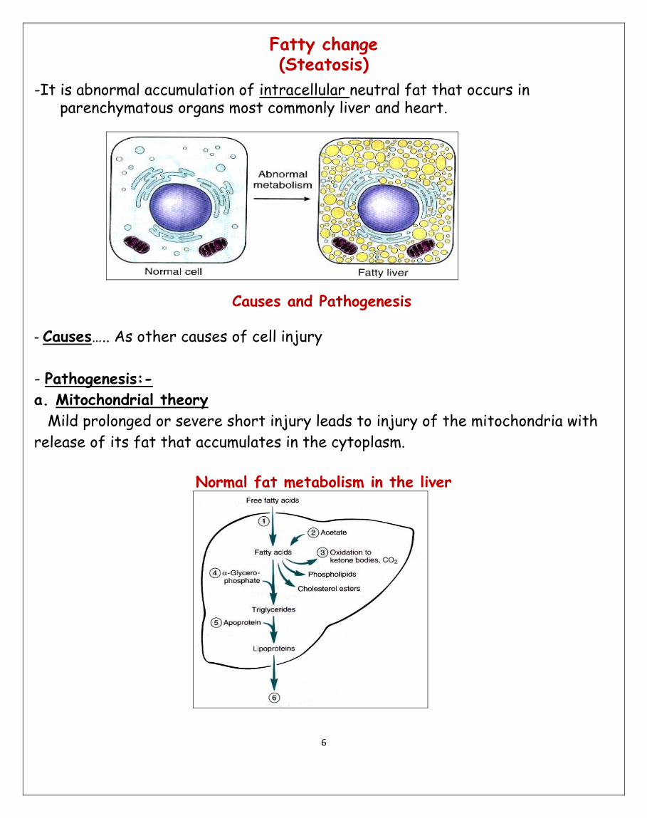
Fatty change (Steatosis)
-It is abnormal accumulation of intracellular neutral fat that occurs in parenchymatous organs most commonly liver and heart.
Causes and Pathogenesis
- Causes….. As other causes of cell injury
- Pathogenesis:-
a. Mitochondrial theory
Mild prolonged or severe short injury leads to injury of the mitochondria with
release of its fat that accumulates in the cytoplasm.
Normal fat metabolism in the liver

7
b. Causes of fatty change in the liver
1- Increased fatty acids entry to the liver
( Obesity, starvation and cortisone therapy). 2- Increased Fatty acid synthesis in the liver from acetate (alcoholism).
3- Decreased oxidation of fatty acids (hypoxia, anemia, respiratory failure).
4- Increased estrification of fatty acids to triglycerides (DM,alcoholism ). 5- Decreased formation of apoprotein (protein mal- nutrition, alcoholism and CCL4 toxicity)
Fatty liver
Grossly:- -Liver is enlarged, (3-6 kg) - Soft, with rounded borders. - Color is yellow. - C/S is bulging. - Greasy to touch.
M/E:- - liver cells accumulate fat which appears in H&E stained section as clear vacuoles. - These vacuoles are first small (microsteatosis). - Later on the vacuoles fuse to form one large vacuole which pushes the,nucleus to one side of the cell. The nucleus becomes flattened (signet ring cell *)

8
Patterns of fatty change in the liver
Focal…. Random single or small clusters e.g. in viral hepatitis C. Zonal…. Centrizonal (zone 3)….hypoxic injury – away from blood supply.
Peripheral zonal ( zone 1)…toxic injury – first area to meet blood. Massive or diffuse….Reye syndrome
Fat stains
- During routine staining of sections by Hx and E fat is dissolved during preparation by the organic solvents.
- For demonstration of fat, frozen sections are used and stained by - Sudan III and oil red O…….Orange red - Osmic acid…….Black
Fatty change of the heart
Two types:- 1- Localized… in moderate cases - Mostly due to anemia - Gives yellow streaks alternating with dark brown fibers (tigroid or tabby cat appearance).
Localized fatty change heart (tigroid skin) 2- Diffuse… in case of severe toxicity e.g. dyphtheria. - It leads to toxic myocarditis & acute heart failure.

9
Irreversible cell injury
Cell Death 1- Necrosis
x Definition
x Causes…the same as reversible injury but more severe
x Pathogenesis
x Pathological changes (gross and microscopic)
x Types
Necrosis
Definition:- Death of a group of cells within living organism.
Two factors characterize irreversibility of the cell damage
1- Irreversible mitochondrial damage.
2-Increased intracellular calcium (Ca).
Pathogenesis
1- Irreversible mitochondrial dysfunction markedly decreased ATP. 2-Increased level of intra-cellular Ca…..activation of many enzymes e.g.
a. Proteasesb. b. Phospholipases c. Endonucleases

10
Microscopicaly - Cell membrane disappears. - Cytoplasm is swollen, mitochondria is swollen, rupture, forms myelin figures and may be calcified. - Nuclear changes;- - Pyknosis: Nuclear shrinkage and increased basophilia
- Karyorrhexis: Fragmentation of the pyknoti nucleus
- Karyolysis: Fading of basophilia of chromatin
What is autolysis & heterolysis??
Types of Necrosis
1- Coagulative (most common) 2- Liquefactive
3- Caseous 4- Fat necrosis (traumatic and enzymatic)
5- Fibrinoid necrosis 6- Gangrenous necrosis
1- Coagulative Necrosis
- Protein denaturation predominates enzymatic digestion.
- Cell’s basic outline is preserved but details are lost.

11
- Cytoplasm becomes homogeneous, glassy eosinophilic in appearance due to loss
of cytoplasmic RNA (basophilic) and glycogen.
- Nucleus may show pyknosis, karyolysis or karyorrhexis.
Coagulative Necrosis of the kidney after infarction
Gross appearance of kidney
infarction (coagulative necrosis)
- Affected area is triangular in shape.
- It is pale, dry and opaque.
- It is surrounded by hyperemic area.
M/E appearance of kidney infarction
(coagulative necrosis)
- Affected area is triangular in shape.
- There is preservation of structural
outlines with loss of cellular details.
2- Liquefactive Necrosis
- Usually due to enzymatic dissolution of necrotic cells (usually due to release of proteolytic enzymes from neutrophils)
- Most often seen in CNS infarction and in abscesses.
An abscess Brain infarction with liqufaction

12
3- Caseous necrosis
Cause… - Mycobacterial or fungal infection (any organ).
Grossly….Necrosis appears cheese-like material.
. Microscopically…
- Large granuloma (histiocytes)
- Central region amorphous granular debris
- Loss of cell outlines - Loss of nuclei
Gross and microscopic picture of tuberculosis of the lung with evident caseation necrosis
4- Fat necrosis
Necrosis of fat cells ….release of triglycerides which are hydolysed by lipase into
fatty acids and glycerol.
- Fatty acids attracts Ca (from the blood) to form Ca. soaps.
- May be traumatic or enzymatic.
- a. Traumatic …trauma to the S.C fat in fatty areas (e.g. breast)…..liberation of
fat and initiation of a F.B reaction forming a hard mass.
b. Enzymatic Fat Necrosis
- Results from hydrolytic action of pancreatic lipases on fat
- Most often seen in and around the pancreas.

13
- Fatty acids released via hydrolysis react with calcium to form chalky white areas “saponification”
Fat necrosis with calcification
5- Fibrinoid Necrosis
- Usually seen in the walls of blood vessels (e.g., in vasculitides)
- Glassy, eosinophilic fibrin-like material is deposited within the vascular walls.
Renal glomerulus – influx of fibrin into the afferent arteriole, due, in this case, to
malignant hypertension.
Patterns of necrosis

14
Apoptosis
- A second type of cell death referred to as programmed cell death.
- It is an important mechanism for the removal of cells as occurs in cells with irreparable DNA damage (from viruses, free radicals, chemical….etc), protecting against neoplastic transformation.
- It may be physiological or pathological…
Physiologic apoptosis Pathologic apoptosis
1.Embryogenesis
2.Menstruation
3.Menopause
4.Intestinal homeostasis
5. Immune tolerance
1.Acute inflammation
2.Organ atrophy
3.Neoplasia
4.Graft rejection
5.Viral hepatitis
Mechanism of apoptosis:-
1. Cell shrinkage, loss of microvilli and cell junctions.
2. Regular fragmentation of DNA each 180 bp intervals.
3. Membrane bleeping.
4. Formation of apoptotic bodies.
5. Rapid phagocytosis of apoptotic bodies.
6. Absence of surrounding inflammation.(why??)
Apoptosis
1- Normal cells 2- Cell shrinkage, loss of junctions 3- Membrane blebing
4- Apoptotic bodies & phagocytosis

15
Differences between necrosis and apoptosis:-
Necrosis/Apoptosis

16
Mucinous and myxomatous degeneration
Normally mucin is a glycoprotein secreted by mucous secreting cells e.g.
Respiratory tract or GIT cells.
Exaggerated production of mucin may occur either in connective tissue or
epithelial cells.
N/E:Pale grey transparent slimy fluid.
M/E:Pale blue with Hx.
Mucoid adenocarcinoma
Sites: A) Epithelial (mucoid): As in catarrhal inflammation & mucoid adenocarcinoma. B) Connective tissue (myxomatous):
- In the subcutaneous tissue in myxoma – myxedema & connective tissue tumors. M/E (of myxomatous degeneration):
Few star shaped cells (with processes) separated by pale blue mucin with rounded hyperchromatic nuclei. The cytoplasm is pale.
Hyalinosis Hyalinosis means the deposition of a glassy, homogeneous, structurless
eosinophilic protein substance either inside cells or in connective tissue material.
Cellular haylinosis 1- Mallory hyaline ……liver of chronic alcoholics. 2- Corpora amylacia…. Prostate of old males. 3- Russell bodies….Plasma cells in rhinoscleroma

17
Mallory body Corpora amylacia Russel body
Connective tissue hyalinosis
1- Wall of blood vessels (arterioles and capillaries) in benign hypertension. 2- Old scars, thrombi, tumors.
Vascular hyalinosis Hyalinosis in leiomyoma
Amyloidosis Definition
Chemistry
Morphology
Stains
Clinical types
Organ amyloidosis
Definition Amyloidosis is a condition in which there’s a deposition of abnormal extra
cellular fibrillar protein (amyloid) in many tissues.

18
Amyloid..
- It is not a single substance but a group of substances that share a common physical structure and can be formed by different types of proteins. - It is derived from many different precursor peptides which represent fragments of larger proteins. - It is deposited as a meshwork of rigid, straight fibrils being composed of the precursor peptides lined up in an anti-parallel β-pleated sheet configuration. It is this physical arrangement of the constituent peptides that makes a protein an amyloid rather than any specific peptide sequence.
Physical nature of amyloid

19
Chemical nature of amyloid
It is formed of glycoprotein + fibrillary protein 1- Glycoprotein
- 5-10% of amyloid structure.
- Constant in all types of amyloid.
-Responsible for stability and staining properties of amyloid.
2- Fibrillary protein
- 90-95% of the amyloid molecule.
- 2 main types AL (Amyloid Light chain) and AA (Amyloid A associated)
- Both are antigenically different .
- Deposited in different clinical conditions
Types of fibrillary protein
Morphologic features of amyloid ♣ Amyloid is characteristically extracellular in distribution mostly near basement membranes.

20
- Gross amyloid stain… It appears Brown with iodin It turns blue after adding sulphoric acid to iodin.
3.+sulphoric acid 2. + iodine 1. Amyloid Histologic staining of amyloid ♣ In routine Hx and E stained sections it has an amorphous eosinophilic appearance.
Amyloid stained with Hx & E
♣ Amyloid is stained red by Congo Red giving an apple-green birefringence when
viewed under polarized light.
- It can also be demonstrated by metachromatic stains (gentian, methyl and
crystal violet), immunochemical methods and by electron microscope to
demonstrate its fibrillar ultra structure

21
Amyloid stained with CONGO RED Congo red under polarized light
Clinical patterns of amyloidosis
♣. The disease may be systemic or limited (localized) to a single organ.
♣.In systemic amyloidosis,the deposition of amyloid may occur as a primary disease (e.g. multiple myeloma) or in a secondary form in the course of other disease ( chronic inflammation) or it may be heredofamilial. - In each case, there is an accumulation of a precursor peptide which is processed into an amyloid protein.
Systemic amyloidosis
Primary amyloidosis
(Immunocyte dyscrasias with amyloidosis) Secondary amyloidosis
(Reactive systemic amyloidosis)

22
- In primary amyloidosis ,
Common sites of amyloid deposition are
Muscles including the cardiac muscle ,
alimentary tract, then solid organs.
- Common cause of death is…..heart
failure less commonly renal failure.
- In secondary amyloidosis however,
- Amyloid is deposited first in solid
organs e.g. kidney and liver.
- Common cause of death is renal failure
( why not hepatic failure ???)
Localized amyloidosis
Organ amyloidosis
Grossly, any organ affected by amyloid deposition will show the following features….. - Size……. Enlarged - Shape …. Preserved - Surface … Smooth - Consistency…. Firm & elastic - Color….. Pale grayish-brown - C/S …. Sharp edges to confirm amyloid deposition….. Gross stains of amyloid
23
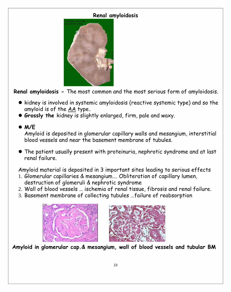
Renal amyloidosis
Renal amyloidosis - The most common and the most serious form of amyloidosis.
kidney is involved in systemic amyloidosis (reactive systemic type) and so the amyloid is of the AA type.
Grossly the kidney is slightly enlarged, firm, pale and waxy.
M/E Amyloid is deposited in glomerular capillary walls and mesangium, interstitial blood vessels and near the basement membrane of tubules.
The patient usually present with proteinuria, nephrotic syndrome and at last renal failure.
Amyloid material is deposited in 3 important sites leading to serious effects
1. Glomerular capillaries & mesangium…. Obliteration of capillary lumen, destruction of glomeruli & nephrotic syndrome
2. Wall of blood vessels … ischemia of renal tissue, fibrosis and renal failure. 3. Basement membrane of collecting tubules …failure of reabsorption
Amyloid in glomerular cap.& mesangium, wall of blood vessels and tubular BM

24
Liver amyloidosis
Hepatic amyloidosis Part of systemic amyloidosis (secondary more than primary)
…amyloid is deposited in the space of Disse causing pressure atrophy of hepatocytes in advanced cases.
Grossly the liver may be hugely enlarged firm, pale grey waxy
Clinically ,there is hepatomegaly but liver functions are rarely affected .
M/E
Amyloid strats to deposit in the space of Disse in the mid-zone of hepatic lobule then encroaches on the sinuses …. Atrophy of hepatocytes.
Wall of hepatic arterioles and venules.
Amyloid deposited in space of Disse &wall of blood vessels
Amyloid spleen
Splenic amyloidosis
The amyloid material is deposited either in white pulp (nodular pattern or sago spleen)
Or in the red pulp (map like) (diffuse) compressing the lymphoid follicles. Or it may be mixed both patterns

25
Nodular Diffuse
Grossly:-
The spleen shows moderate or marked splenomegaly .
the spleen is firm, C/S shows pale, grey waxy deposits
Cardiac amyloidosis
- May be part of systemic amyloidosis (usually with immunocyte dyscrasias)
with AL type amyloid, or in localized cardiac senile amyloidosis
- The heart is enlarged, with pale, grey-pink subendocardial deposits.
- M/E amyloid is deposited between cardiac muscle fibers causing their
atrophy.
- May interfere with the conducting system (arrythmia). The case ends with
cardiomyopathy and heart failure
Cardiac amyloidosis
Amyloidosis of GIT Tongue ….. Macroglossia.

26
Gingiva…. Thickened
Mucosa of stomach and intestine…amyloid deposition in mucosal blood vessels…atrophy of epithelial cells…… malabsorption and diarrhea.
Rectal biopsy is an easy way to assess systemic amyloidosis
Amyloidosis (Summary)
Pathologic Calcification
Definition It is deposition of calcium salts in sites other than bone and teeth.
Types
1. Dystrophic calcification
- Occurs in nonviable tissue with normal blood calcium level
2. Metastatic calcification
- Occurs in viable tissue
- Caused by hypercalcemia
3. Stone formation
1- Dystrophic Calcification
Normal blood calcium level + diseased or necrotic tissue. 1. Necrotic tissue - fat, coagulative, liquefactive, caseous necrosis
2. Atherosclerosis - Central necrotic core

27
3. Cells….Psammoma bodies
4. Damaged or aging heart valves
Atherosclerosis Calcified cardiac cusp Psammoma bodies
2- Metastatic Calcification
Increased blood calcium+ normal tissue
Causes of hypercalcemia
1. Increased PTH secretion
- parathyroid tumor - ectopic PTH secretion
2. Bone destruction
- osteolytic tumors - Paget’s disease - immobilization
3. Vitamin D disorders
- vitamin D toxicity - sarcoidosis
4. Excess calcium intake, as in milk alkali syndrome ( nephrocalcinosis and renal
stones caused by milk and antiacid self- therapy.
Sites for calcium deposition:-
1. Lungs - respiratory failure
2. Kidneys - nephrocalcinosis, kidney failure
3. Stomach
4. Arteries…. Sometimes affection of small blood vessels of the skin cause
ischemic ulcers with necrosis (calciphylaxix)
wall of a blood vessel Lung gastric submucosa

28
Gout
Gout… Increased serum uric acid leads to deposition of uric acid crystals in joints……inflammation with foreign body giant cell reaction (Tophi) mainly in the metatarsophalyngeal joint of the big toe.
Increased uric acid is deposited in the kidney leading to
1. Urate stones
2. Interstitial inflammation , fibrosis & chronic renal failure. 3. Damage to distal convoluting tubules… polyuria
Gouty arthritis
Pathological pigmentation Endogenous pigments
Lipofuscin
Melanin
Bilirubin Hemosidrin
Exogenous pigments
• Inhalation….anthracosis
• Ingestion…..lead poison
• Tatoo
Lipofuscin
It is an endogenous, insoluble pigment of complex lipid and protein structure. It is brownish-yellow granules on both sides of the nucleus. It is a wear and tear pigment formed by the action of free radicals on cell
membrane lipids.

29
It commonly accumulates in elderly patients, in whom the pigment is found most often within hepatocytes and at both poles of nuclei of myocardial cells.
The combination of lipofuscin accumulation and atrophy of organs is called (brown atrophy)
Hemoglobin derived pigments
Hemosidrin is an iron containing pigment consisting of aggregates of ferritin. It appears in tissues as golden brown aggregates that can be identified by
staining with Prussian blue dye. It is present normally in small amounts as physiologic iron store inside tissue
macrophages in the liver, bone marrow and spleen.
Hemosidrin
Hemosiderin is a hemoglobin derived pigment What are the three brown intracellular pigments? How to differentiate
Hemosidrin Prussian blue
Hemosidrosis:-
It is accumulation of hemosidrin, primarily within tissue macrophages without associated tissue or organ damage. It may local or systemic.

30
Local hemosidrosis:- -Is local deposition of hemosidrin due to local tissue hemorrhage. Systemic hemosidrosis:- - Is generalized hemosidrin deposition without tissue damage. - Causes include hemorrhage, multiple blood transfusion, excessive dietary iron intake often accompanied by alcohol consumption.
Hemochromatosis:- It is more extensive accumulation of hemosidrin often within parenchymal cells with associated tissue damage, scarring and organ dysfunction. It may be hereditary (primary) or (secondary).
(Primary) hemochromatosis:-
- Caused often due to gene mutation resulting in excessive iron absorption from the duodenum .
Hemosidrin deposits in..
- Liver….micronodular cirrhosis.
- Pancreas…. Diabetes mellitus bronzed diabetes
- Skin…..pigmentation
- Heart…..heart failure
(Secondary) hemochromatosis:- - Usually occurs after multiple blood transfusions to patients with hereditary hemolytic anemia.
Hematin… A parasitic pigment. Results from digestion RBCs in the parasit’s
intestine. It contains iron but it is PB negative . WHY?


![Cell Injury[1]](https://img.pdfslide.net/doc/110x75/563dba79550346aa9aa5f218/cell-injury1-56a51a5ef0c98.jpg)