Embed Size (px)
Citation preview
Research Article Open Access
Ramena Y and Ramena G, J Morphol Anat 2018, 2:1
Mini-Review Open Access
Journal of Morphology and AnatomyJour
nal o
f Morphology and Anatom
y
Volume 2 • Issue 1 • 1000111J Morphol Anat, an open access journal
Cell-Cell Junctions and Epithelial DifferentiationRamena Yathish1 and Ramena Grace2*1Great Salt Lake Brine Shrimp Cooperative Inc., Ogden, Utah, USA2Department of Aquaculture and Fisheries, University of Arkansas at Pine Bluff, Arkansas, USA
AbstractEpithelial tissues make up most organs in the body and perform various functions like protection, excretion,
secretion etc. The epithelial tissues consist of different cell type like squamous, cuboidal, glandular and columnar depending on the organ and function. These cells can be of single or multiple layers. The epithelial cells are tightly packed together and connected by junctional complexes. They are tight junctions, adherens junctions, gap junctions, desmosomes and hemi desmosomes. These junctional complexes are made up of trans-membrane proteins that form homo or hetero complexes. The proteins expressed at the cell-cell junctions act as binding docks for various adaptor molecules at their c-terminus and there regulate cell signaling. Some junctional molecules connect to cell cytoskeleton and basement membrane and maintain epithelial integrity, cell polarity and epithelial differentiation. Any dysregulation of cell-cell junctional molecules due to pathogenic conditions results in loss of epithelial integrity and differentiation leading the cells to undergo EMT.
*Corresponding author: Grace Ramena, Assistant Professor, Department of Aquaculture and Fisheries, University of Arkansas at Pine Bluff, Arkansas, USA, Tel: (870) 575-8137; E-mail: [email protected]
Received January 06th, 2018; Accepted February 08, 2018; Published February 19, 2018
Citation: Ramena Y, Ramena G (2018) Cell-Cell Junctions and Epithelial Differentiation. J Morphol Anat 2: 111.
Copyright: © 2018 Ramena Y, et al. This is an open-access article distributed under the terms of the Creative Commons Attribution License, which permits unrestricted use, distribution, and reproduction in any medium, provided the original author and source are credited.
Keywords: Epithelial to mesenchymal transition; Epithelial differentiation; Epithelial integrity; Cell-cell junctions; Junctional complexes
Epithelial CellsFour types of tissues are there, epithelial, connective, muscle and
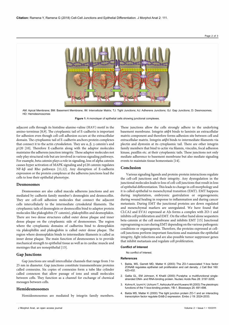
nervous which make up the human body. Among them epithelial tissues are avascular. Epithelial cells perform various functions like secretion, excretion, absorption, and protection. The nutrient or waste exchange takes place through diffusion with neighboring connective tissue. Cells in the epithelial tissue are closely packed together with very little intercellular matrix. It consists of nonliving material like salts and fibers that are tissue specific. Epithelial cells are wide spread throughout the body and perform many functions like protection, secretion, absorption etc. Epithelial cells are of different types like squamous, cuboidal or glandular, columnar and may be multiple or single layered depending on the organ. Glandular epithelium is specialized to produce and secrete substances and it forms two types of glands. They are endocrine and exocrine glands. The endocrine glands like thyroid gland secrete hormones like thyroxin directly into the blood stream that is spread through-out the body. On the other hand, exocrine glands secrete substances into ducts or tubes that are secreted to the epithelial surface. Exocrine glands are of three types based on their structure. They are merocrine, apocrine and holocrine glands. Mammary glands belong to apocrine type where the milk from the lobules is secreted into a duct that is released from the apical portion to the outside. In a mature mammary gland the alveolar and luminal epithelial cells are surrounded by myoepithelium. The epithelial cells are connected together at the lateral sides and basement membrane by junctional complexes. They are tight junctions, adherens junctions, desmosomes, gap junctions and hemi-desmosomes (Figure 1).
Tight JunctionsTight junctions (TJs) are present at the apical region above the
adherens junctions around the epithelial cell’s circumference. TJs play several important roles: they hold the cells together, act as a barrier, help maintain cell polarity, prevent lateral diffusion of proteins, ions, molecules. TJs also play an important role in maintaining the blood brain barrier. TJs are known to form strongest connections between adjacent cells which are not broken even until a dead cell is replaced by a new one. They perform these functions either by homotypic or
heterotypic interactions, by binding to peripheral proteins like ZO-1, ZO-2, ZO-3, ZONAB, PAR3, PAR6 etc. at their cytoplasmic tail that link them to the actin cytoskeleton. ZONAB is a Y-box transcription factor that binds to CDK4 and both these proteins are sequestered at the cell membrane via ZO-1 [1]. Y-box transcription factors play a role in cell proliferation [2,3]. Disruption of these junctions allows ZONAB to translocate to the nucleus, promote expression of cyclin D1, proliferating nuclear antigen and thus cell proliferation. It is known to regulate erbB-2 expression and cell proliferation based on cell density [4,5]. There are four main trans-membrane proteins found at TJs. They are occludins, claudins and junctional adhesion molecules (JAMs) and nectins. Occludins and claudins have four trans-membrane segments, two intracellular and 2 extracellular domains, while JAMs have single transmembrane segment. Occludins are involved in signaling event, claudins mediate ca+2 independent cell-cell adhesions, and JAMs are thought to mediate Para-cellular barrier.
Adherens JunctionsAdherens junctions are mediated by E-cadherin. By using
electron microscopy identified that E-cadherin is localized at the adherens junctions of epithelial cells in the intestine [6]. It is a calcium dependent cell-cell adhesion molecule [7]. All the epithelial cells express E-cadherin on their lateral sides at the adherens junctions. McNeill et al 1990 showed that E-cadherin helps maintain apico-basal polarity in addition to mediating cell-cell adhesion. E-cadherin is a type I transmembrane glycoprotein that is about 728 amino acids long [7]. It has 5 domains EC1 to EC5 that binds to calcium in the extracellular domain, a single transmembrane domain and cytoplasmic domain. E-cadherin interacts with other E-cadherin molecules on
Citation: Ramena Y, Ramena G (2018) Cell-Cell Junctions and Epithelial Differentiation. J Morphol Anat 2: 111.
Page 2 of 3
Volume 2 • Issue 1 • 1000111J Morphol Anat, an open access journal
adjacent cells through its histidine-alanine-valine (HAV) motif in the amino-terminus [8,9]. The cytoplasmic tail of E-cadherin is important for adhesion even though cell-cell adhesion occurs at the extracellular domain. The cytoplasmic tail of E-cadherin anchors protein complexes that connect it to the actin cytoskeleton. They are α, β, γ catenin’s and p120 [10]. Therefore E-cadherin along with the adaptor molecules maintains the adherens junction integrity. These adaptor molecules not only play structural role but are involved in various signaling pathways. For example, beta catenin plays a role in signaling, loss of alpha catenin causes hyper activation of MAPK signaling and p120 catenin regulates NF-kβ and Rho pathways [11,12]. Any disruption of E-cadherin expression or the protein complexes at the adherens junctions lead the cells to lose their epithelial phenotype.
DesmosomesDesmosomes are also called macula adherens junctions and are
mediated by cadherin family member’s desmoglein and desmocollin. They are cell-cell adhesion molecules that connect the adjacent cells intercellularly to the intermediate cytoskeletal filaments. The cytoplasmic tails of desmoglein or desmocollin bind to adaptor protein molecules like plakoglobin (Ƴ-catenin), plakophillin and desmoplakin. There are two dense structures called outer dense plaque and inner dense plaque on the cytoplasmic side of desmosomes. The region where the cytoplasmic domains of cadherins bind to demoplakin via plakophillin and plakoglobin is called outer dense plaque. The region where desmoplakin binds to intermediate filaments is called as inner dense plaque. The main function of desmosomes is to provide mechanical strength to epithelial tissue as well as in cardiac muscle and meninges that are nonepithelial [13].
Gap Junctions Gap junctions are small intercellular channels that range from 3 to
25 nm in diameter. Gap junctions constitute transmembrane proteins called connexins. Six copies of connexins form a tube like cylinder called connexon that allow passage of ions and small molecules between cells. They function as a channel for exchange of chemical messages between cells.
HemidesmosomesHemidesmosomes are mediated by integrin family members.
These junctions allow the cells strongly adhere to the underlying basement membrane. Integrin α6β4 binds to laminin an extracellular matrix component and therefore forms adhesion site between cell and extracellular matrix. Integrin α6β4 binds to intermediate filaments via plectin and dystonin at its cytoplasmic tail. There are other integrin family members that bind to actin via filamin, vinculin, focal adhesion kinase, paxillin etc. at their cytoplasmic tails. These junctions not only mediate adherence to basement membrane but also mediate signaling events to maintain tissue homeostasis [14].
ConclusionVarious signaling ligands and protein-protein interactions regulate
the cell-cell junctions and their integrity. Any dysregulation in the junctional molecules leads to loss of cell-cell junctions that result in loss of epithelial differentiation. This leads to change in cell morphology and it is called epithelial to mesenchymal transition (EMT). EMT happens during implantation, embryonic gastrulation or organogenesis; during wound healing in response to inflammation and during cancer metastasis. During EMT the junctional proteins are down regulated and mesenchymal markers are unregulated. We have found that CLCA2 and EVA1 expressed at AJs forms a complex with ZO-1 and inhibits cell proliferation and EMT. On the other hand alone sequesters beta-catenin at the cell membrane and inhibits EMT [15] Junctional reprograming occurs during EMT depending on the various pathogenic conditions or organogenesis. Therefore, the proteins expressed at cell-cell junctions perform important functions and maintain the epithelial integrity, fight infections and are also possible tumor suppressor genes that inhibit metastasis and regulate cell proliferation.
Conflict of Interest
No conflict of Interest.
References
1. Balda, MS, Garrett MD, Matter K (2003) The ZO-1-associated Y-box factor ZONAB regulates epithelial cell proliferation and cell density. J Cell Biol 160: 423-432.
2. Gallia GL, EM Johnson, K Khalili (2000) Puralpha: a multifunctional single-stranded DNA- and RNA-binding protein. Nucleic Acids Res 28: 3197-3205.
3. Kohno K, Izumi H, Uchiumi T, Ashizuka M and Kuwano M (2003) The pleiotropic functions of the Y-box-binding protein, YB-1. Bioessays 25: 691-698.
4. Balda MS, Matter K (2000) The tight junction protein ZO-1 and an interacting transcription factor regulate ErbB-2 expression. Embo J 19: 2024-2033.
AM: Apical Membrane; BM: Basement Membrane; IM: Intercellular Matrix; TJ: Tight Junctions; AJ: Adherens Junctions; GJ: Gap Junctions; D: Desmosomes; HD: Hemidesmosomes
Figure 1: A monolayer of epithelial cells showing junctional complexes.
Citation: Ramena Y, Ramena G (2018) Cell-Cell Junctions and Epithelial Differentiation. J Morphol Anat 2: 111.
Page 3 of 3
Volume 2 • Issue 1 • 1000111J Morphol Anat, an open access journal
5. Balda MS and Matter K (2003) Epithelial cell adhesion and the regulation of gene expression. Trends Cell Biol 13: 310-318.
6. Boller K, Vestweber D, Kemler R (1985) Cell-adhesion molecule uvomorulin is localized in the intermediate junctions of adult intestinal epithelial cells. J Cell Biol 100: 327-332.
7. Nagafuchi A, Shirayoshi Y, Okazaki K, Yasuda K, Takeichi M (1987) Transformation of cell adhesion properties by exogenously introduced E-cadherin cDNA. Nature 329: 341-343.
8. Blaschuk OW, Sullivan R, David S, Pouliot Y (1990) Identification of a cadherin cell adhesion recognition sequence. Dev Biol 139: 227-229.
9. Halbleib JM, Nelson WJ (2006) Cadherins in development: cell adhesion, sorting, and tissue morphogenesis. Genes Dev 20: 3199-3214.
10. Onder TT, Gupta PB, Mani SA, Yang J, Lander ES, et al. (2008) Loss of
E-cadherin promotes metastasis via multiple downstream transcriptional pathways. Cancer Res 68: 3645-3654.
11. Vasioukhin V, Bauer C, Degenstein L, Wise B, Fuchs E (2001) Hyperproliferation and defects in epithelial polarity on conditional ablation of a-catenin in skin. Cell 104: 605–617.
12. Wildenberg GA, Dohn MR, Carnahan RH, Davis MA, Lobdell NA, et al. (2006) p120-catenin and p190RhoGAP regulate cell-cell adhesion by coordinating antagonism between Rac and Rho. Cell 127: 1027-1039.
13. Holthofer B, Windoffer R, Troyanovsky S, Leube RE (2007) Structure and function of desmosomes. Int Rev Cytol. 264: 65-163.
14. Borradori L, Sonnenberg A (1999) Structure and Function of Hemidesmosomes: More Than Simple Adhesion Complexes. J Invest Dermatol 112: 411-418.
15. Ramena G, Yin Y, Yu Y, Walia V, Elble RC (2016) CLCA2 Interactor EVA1 Is Required for Mammary Epithelial Cell Differentiation. PLoS ONE 11: e0147489.


![Rom J Morphol Embryol 2011, 52(1):69–74 R J M E … · Rom J Morphol Embryol 2011, 52(1) ... blished by the World Health Organization (WHO) Classification [1], ... rehydrated in](https://img.pdfslide.net/doc/110x75/5b6443407f8b9a687e8d1c3f/rom-j-morphol-embryol-2011-5216974-r-j-m-e-rom-j-morphol-embryol-2011.jpg)