Embed Size (px)
Citation preview
‘Cell cycle’ and ‘cell death’- related genes are differentially expressed during long – term in vitro real-time cultivation of porcine oviductal epithelial cells
AbstractAlterations in cells depend on their genetic material, its activation and translation of the products. The genes responsible for the cell cycle processes and apoptosis of porcine oviductal cells have been presented in our study. The processes occurring in the reproductive system of females are extremely complex and require in-depth knowledge. Thanks to in vitro studies on the fallopian tube epithelium cells, we can get closer to understanding the biochemical and morphological changes occurring in mammalian organisms. Our research was conducted on fallopian tubes obtained from commercially bred pigs and its aim was to assess the expression profile of genes responsible for the most important processes of cellular life. Cell cul-tures were carried out for 30 days, with the obtained cells subjected to molecular analysis. We have shown significant regulation of “cell death” and “cell cycle” genes, some of which are related to the reproductive system. The alterations in transcriptomic profile and mutual relations between the genes were analyzed and related to the literature findings. The knowledge gained could help in identifying new potential mar-kers of the in vitro occurrence of processes described by the ontology groups of interest.
Running title: pig, oocytes, microarray assays, in vitro maturation (IVM)
Keywords: Regulation of metabolic enzymes receptor signaling pathway
Magdalena Kulus1, Jakub Kulus1, Małgorzata Popis2, Blanka Borowiec2,3, Katarzyna Stefańska3, Piotr Celichowski3, Mariusz J. Nawrocki2, Klaus P. Brüssow1, Bartosz Kempisty2,3,4, Michal Jeseta4, Paweł Antosik1, Dorota Bukowska1
1Veterinary Centre, Nicolaus Copernicus University in Torun, Torun, Poland2Department of Anatomy, Poznan University of Medical Sciences, Poznan, Poland3Department of Histology and Embryology, Poznan University of Medical Sciences, Poznan, Poland4Department of Obstetrics and Gynecology, University Hospital and Masaryk University, Brno, Czech Republic* Correspondence: [email protected] list of author information is available at the end of article
Kulus et al. Medical Journal of Cell Biology 2019DOI: 10.2478/acb-2019-0012Received: 6.08.2019Accepted: 20.09.2019
Kulus et al. Medical Journal of Cell Biology (2019)91
IntroductionDue to their utmost importance, the undisturbed
processes of cell life are subject to continuous ge-netic control, including segregation of material, correctness of subsequent events and cell division.The cell cycle is therefore an extremely complex process, with its mechanisms focused on biochem-ical and morphological changes of replicating cells [1]. In contrast, the permanent retention of the vital functions of cells causes their irreversible death. It can take place due to accidental damage or a physi-ological mechanism called apoptosis [2].
The processes associated with the femalerepro-ductive system are very complicateddue to a vast-ness of biochemical changes. Therefore, they have not yet been fully understood. Proper functioning of the reproductive systems requires many hor-monal changes in the body, especially the effective feedback of the hypothalamus, pituitary gland and gonads [3]. The transport of the oocyte through the fallopian tube and the interaction between the em-bryo and maternal tissues are of great importance in the context of effective fertilization. Morpholog-ical and biochemical changes in the oviductal cells are mainlycaused by processes occurring after ovu-lation, related to the influence of both oocyte and embryo on oviductal cells [4]. General changes oc-curring at this stage are relatively well described, but the changes on the molecular ground are not fully known. Therefore, in our study, we described the changes in expressionof genes associated with “cell cycle” and “cell death”gene ontology processes.
Recent studies indicate the possibility of short- and long-termin vitro culture of oviductal cells, in-cluding epithelial cells. This gives an opportunity to observe many morphological and biochemical changes during in vitro culture (IVC) processes of oviductal epithelial cells (OECs). These changes are related to the modification of genes and protein ex-pression [5], including the mechanisms of oocyte in-fluence on fallopian tube epithelial cells in relation to both cell cycle and cell death. The occurrence of the above changes in vivo enables effective fertiliza-tion and later development of the embryo through pathways that are not fully discovered. Therefore, the analysis of these in vitro changes can contribute a great deal to the understanding of the interactions between the mother and embryo.
Current studies conducted by our team (not pub-lished data) indicate the possibility of effective in vitro culture of epithelial cells of the fallopian tube and even changes in their proliferation. Changes in cell morphology, cell growth and differentiation may be related to cell cycle and cell death process-es. Therefore, the aim of our work was to find new cellular markers associatedwith cell cycle and cell death processes in in vitro OEC cultures.
Material and methodsAnimals
The animals in our study- crossbred gilts (n=45) at the age of around 9 months, came from a com-mercial breeding herd. The selected individuals ex-pressed two regular estrus cycles. All the animals were checked daily for estrus behavior and were slaughtered after reaching the anestrus phase of the estrus cycle. The uteri were then transported to the laboratory within 30 min at 38°C.
Oviductal epithelial cell (OEC) selection and culture
Oviducts were washed twice in Dulbecco’s phos-phate buffered saline (PBS) (137 mM NaCl, 27 mM KCl, 10 mM Na2HPO4, 2 mM KH2PO4, pH 7.4). Epi-thelial cells were surgically removed using sterile blades. Then, the epithelium was incubated with collagenase I (Sigma Aldrich, Madison, USA), 1mg/mL inDulbecco’s modified Eagle’s medium (DMEM; Sigma Aldrich, Madison, USA) for 1 h at 37oC. The cell suspension obtained from this digestion was fil-tered through 40 µm pore size strainer to remove blood and single cells. The residue was collected by rinsing the strainer with DMEM. The cell samples were then centrifuged (200 x g, 10 min.). Next, they were washed in PBS and centrifuged again. Later, they were incubated with 0.5% Trypsin/EDTA (Sig-ma Aldrich, Madison, USA) at 37oC for 10 min. The reaction was stopped with fetal calf serum (FCS; Sigma Aldrich, Madison, USA). After incubation, cells where filtered and centrifuged for the last time. The final cell pellet was suspended in DMEM supplemented with 10% FCS, 100U/mL penicillin, 100 µg/mL streptomycin and 1µg/mL amphotericin B. The cells were cultured at 37oC in a humidified atmosphere of 5% CO2. Once the OEC cultures at-tained 70–80% confluency, they were passaged by washing with PBS, digestion with 0.025% Trypsin/EDTA, neutralization by a 0.0125% trypsin inhibi-tor (Cascade Biologics, Portland, USA), centrifuga-tion, and resuspension at a seeding density of 2x10⁴ cells/cm2. The culture medium was changed every three days. The culture lasted 30 days.
RNA extraction from porcine oviductal epithelial cells (OECs)
Oviductal epithelial cells from specific time pe-riods were pooled into three independent samples for each experimental group. Total RNA was extract-ed from samples using TRI Reagent (Sigma, St Louis, MO, USA) and RNeasy MinElute cleanup Kit (Qiagen, Hilden, Germany). The amount of total mRNA was determined from the optical density at 260 nm, and the RNA purity was estimated using the 260/280 nm absorption ratio (higher than 1.8) (NanoDrop spectrophotometer, Thermo Scientific, ALAB, Po-
Kulus et al. Medical Journal of Cell Biology (2019)92
land). The RNA integrity and quality were checked on a Bioanalyzer 2100 (Agilent Technologies, Inc., Santa Clara, CA, USA). The resulting RNA integrity numbers (RINs) were between 8.5 and 10, with an average of 9.2 (Agilent Technologies, Inc., Santa Clara, CA, USA). The RNA in each sample was diluted to a concen-tration of 100 ng/μl with an OD260/OD280 ratio of 1.8/2.0. From each RNA sample, 500 ng of RNA was taken for microarray expression assays.
Microarray expression analysis and statisticsTotal RNA (100 ng) from each pooled sample was
subjected to two rounds of sense cDNA amplifica-tion (Ambion® WT Expression Kit). The obtained cDNA was used for biotin labeling and fragmenta-tion by Affymetrix GeneChip® WT Terminal Label-ing and Hybridization (Affymetrix). Biotin-labeled fragments of cDNA (5.5 μg) were hybridized to the Affymetrix® Porcine Gene 1.1 ST Array Strip (48°C/20 h). Microarrays were then washed and stained according to the technical protocol using the Affymetrix GeneAtlas Fluidics Station. The array strips were scanned with the use of Imaging Station of the GeneAtlas System. Preliminary analysis of the scanned chips was performed using Affymetrix Ge-neAtlasTM Operating Software. The quality of gene expression data was confirmed according to the quality control criteria provided by the software. The obtained CEL files were imported into down-stream data analysis software.
All of the presented analyses and graphs were performed using Bioconductor and R programming languages. Each CEL file was merged with a descrip-tion file. To correct background, normalize, and summarize the results, we used the Robust Multi-array Averaging (RMA) algorithm. To determine the statistical significance of the analyzed genes, mod-erated t-statistics from the empirical Bayes method were performed. The obtained p-value was correct-ed for multiple comparisons using Benjamini and Hochberg’s false discovery rate. The selection of significantly altered genes was based on a p-value beneath 0.05 and expression higher than two-fold.
Differentially expressed genes were subjected to selection by examination of their involvement in the gene ontologies of interest. The differentially ex-pressed gene lists (separate for up- and down-reg-ulated genes) were uploaded to the DAVID soft-ware (Database for Annotation, Visualization and Integrated Discovery) [6], where genes belonging to “cell cycle” and “cell death” GO BP terms were extracted. Expression data of these genes was also subjected to a hierarchical clusterization proce-dure, with results presented as a heat map.
Subsequently, we analyzed the relation between the genes belonging to chosen GO terms with theGO-plot package [7]. The GoPlot package calculated the z-score: the number of up- regulated genes minus the number of down- regulated genes divided by the
square root of the count. This information allowed estimating the change course of each gene-ontology term.
Interactions between differentially expressed genes/proteins belonging to the studied gene on-tology group were investigated using the STRING10 software (Search Tool for the Retrieval of Interact-ing Genes) [8]. The list of gene names was used as a query for an interaction prediction. The search criteria were based on co-occurrences of genes/proteins in scientific texts (text mining), co-expres-sion, and experimentally observed interactions. The results of such analyses generated a gene/protein interaction network where the intensity of the edg-es reflected the strength of the interaction score.
Finally, the functional interactions between genes belonging to the chosen GO BP terms were investi-gated using the REACTOME FIViz application to the Cytoscape 3.6.0 software. The ReactomeFIViz app is designed to find pathways and network patterns related to cancer and other types of diseases. This app accesses the pathways stored in the Reactome database, allowing to perform pathway enrichment analysis for a set of genes, visualize hit pathways us-ing manually laid-out pathway diagrams directly in Cytoscape and investigate functional relationships among the genes in hit pathways. The app can also access the Reactome Functional Interaction (FI) network, a highly reliable, manually curated path-way-based protein functional interaction network covering over 60% of human proteins.
Ethical approvalThe research related to animal use has been com-
plied with all the relevant national regulations and instructional policies for the care and use of ani-mals. Bioethical Committee approval no. 83/2012/DNT.
ResultsWhole transcriptome profiling using Affymetrix
microarrays allowedus to analyze the gene expres-sion changes between 7, 15 and 30 days of porcine oviductal epithelial cells culture. Using the Affyme-trix® Porcine Gene 1.1 ST Array Strip, we examined the expression of 12257 transcripts. Genes with fold change higher than abs (2) and with a correct-ed p-value lower than 0.05 were considered as dif-ferentially expressed. This set of genes consists of 2533 different transcripts.
DAVID (Database for Annotation, Visualiza-tion and Integrated Discovery) software was used for extraction of gene ontology biological process terms (GO BP) that contains differently expressed transcripts. Up and down regulated gene sets were subjected to the DAVID search separately,with only the sets of adj. p value lower than 0.05 selected. The DAVID software analysis showed that the different-ly expressed genes belonged to 657 Gene ontology
Kulus et al. Medical Journal of Cell Biology (2019)93
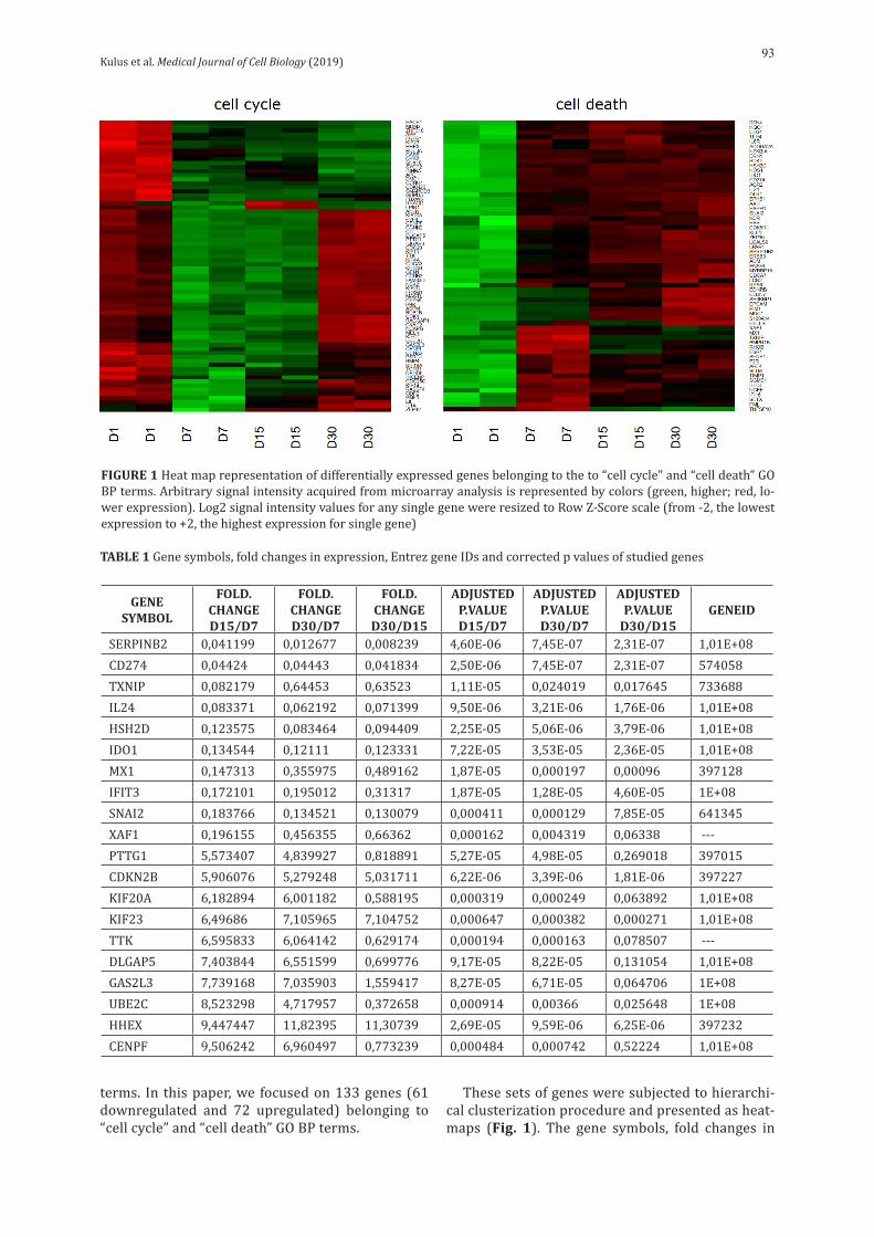
terms. In this paper, we focused on 133 genes (61 downregulated and 72 upregulated) belonging to “cell cycle” and “cell death” GO BP terms.
These sets of genes were subjected to hierarchi-cal clusterization procedure and presented as heat-maps (Fig. 1). The gene symbols, fold changes in
GENE SYMBOL
FOLD. CHANGE D15/D7
FOLD. CHANGE D30/D7
FOLD. CHANGE D30/D15
ADJUSTED P.VALUE D15/D7
ADJUSTED P.VALUE D30/D7
ADJUSTED P.VALUE
D30/D15GENEID
SERPINB2 0,041199 0,012677 0,008239 4,60E-06 7,45E-07 2,31E-07 1,01E+08CD274 0,04424 0,04443 0,041834 2,50E-06 7,45E-07 2,31E-07 574058TXNIP 0,082179 0,64453 0,63523 1,11E-05 0,024019 0,017645 733688IL24 0,083371 0,062192 0,071399 9,50E-06 3,21E-06 1,76E-06 1,01E+08HSH2D 0,123575 0,083464 0,094409 2,25E-05 5,06E-06 3,79E-06 1,01E+08IDO1 0,134544 0,12111 0,123331 7,22E-05 3,53E-05 2,36E-05 1,01E+08MX1 0,147313 0,355975 0,489162 1,87E-05 0,000197 0,00096 397128IFIT3 0,172101 0,195012 0,31317 1,87E-05 1,28E-05 4,60E-05 1E+08SNAI2 0,183766 0,134521 0,130079 0,000411 0,000129 7,85E-05 641345XAF1 0,196155 0,456355 0,66362 0,000162 0,004319 0,06338 ---PTTG1 5,573407 4,839927 0,818891 5,27E-05 4,98E-05 0,269018 397015CDKN2B 5,906076 5,279248 5,031711 6,22E-06 3,39E-06 1,81E-06 397227KIF20A 6,182894 6,001182 0,588195 0,000319 0,000249 0,063892 1,01E+08KIF23 6,49686 7,105965 7,104752 0,000647 0,000382 0,000271 1,01E+08TTK 6,595833 6,064142 0,629174 0,000194 0,000163 0,078507 ---DLGAP5 7,403844 6,551599 0,699776 9,17E-05 8,22E-05 0,131054 1,01E+08GAS2L3 7,739168 7,035903 1,559417 8,27E-05 6,71E-05 0,064706 1E+08UBE2C 8,523298 4,717957 0,372658 0,000914 0,00366 0,025648 1E+08HHEX 9,447447 11,82395 11,30739 2,69E-05 9,59E-06 6,25E-06 397232CENPF 9,506242 6,960497 0,773239 0,000484 0,000742 0,52224 1,01E+08
TABLE 1 Gene symbols, fold changes in expression, Entrez gene IDs and corrected p values of studied genes
FIGURE 1 Heat map representation of differentially expressed genes belonging to the to “cell cycle” and “cell death” GO BP terms. Arbitrary signal intensity acquired from microarray analysis is represented by colors (green, higher; red, lo-wer expression). Log2 signal intensity values for any single gene were resized to Row Z-Score scale (from -2, the lowest expression to +2, the highest expression for single gene)
Kulus et al. Medical Journal of Cell Biology (2019)94
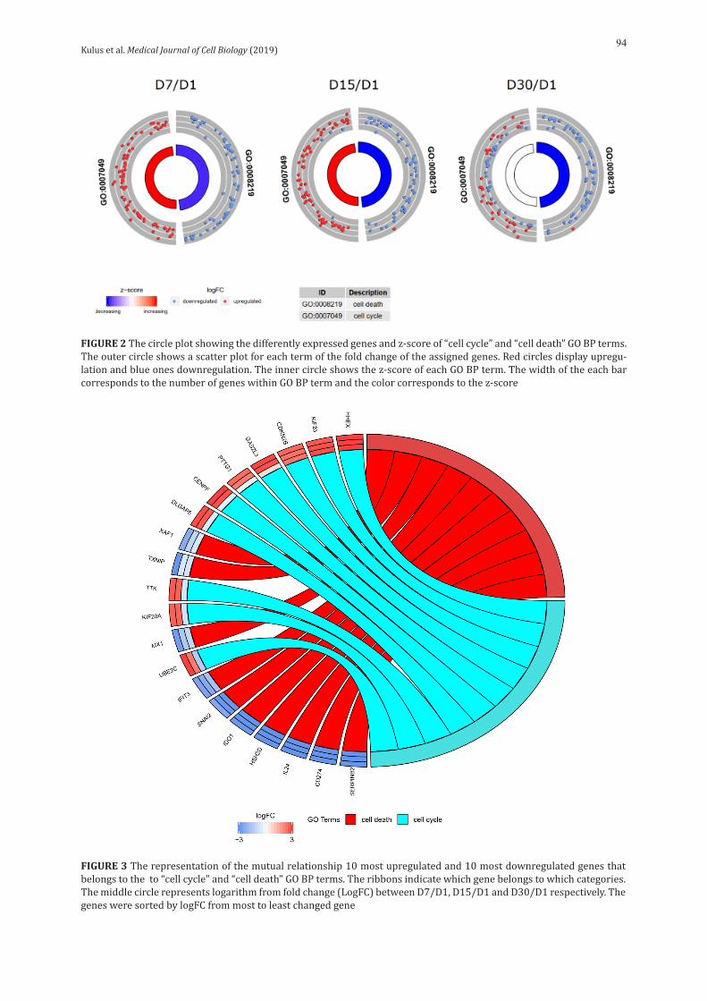
FIGURE 2 The circle plot showing the differently expressed genes and z-score of “cell cycle” and “cell death” GO BP terms. The outer circle shows a scatter plot for each term of the fold change of the assigned genes. Red circles display upregu-lation and blue ones downregulation. The inner circle shows the z-score of each GO BP term. The width of the each bar corresponds to the number of genes within GO BP term and the color corresponds to the z-score
FIGURE 3 The representation of the mutual relationship 10 most upregulated and 10 most downregulated genes that belongs to the to “cell cycle” and “cell death” GO BP terms. The ribbons indicate which gene belongs to which categories. The middle circle represents logarithm from fold change (LogFC) between D7/D1, D15/D1 and D30/D1 respectively. The genes were sorted by logFC from most to least changed gene
Kulus et al. Medical Journal of Cell Biology (2019)95
expression, Entrez gene IDs and corrected p-values of these genes were shown in table1.
The enrichment of each GO BP term was calculated as a z-score and shown on the circle diagram (Fig. 2).
From the differently expressed genes belong-ing to the studied GO BP terms, we chose 10 most downregulated and 10 most upregulated genes for further analysis. In Gene Ontology database, genes that formed one particular GO group can also be-long to other different GO term categories. For this reason, we explored the gene intersections between selected GO BP terms. The relation between those GO BP terms was presented as a circle plot (Fig. 3)
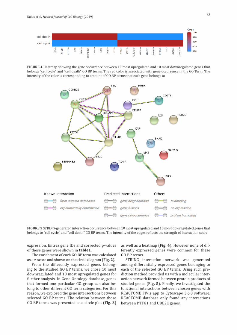
as well as a heatmap (Fig. 4). However none of dif-ferently expressed genes were common for these GO BP terms.
STRING interaction network was generated among differentially expressed genes belonging to each of the selected GO BP terms. Using such pre-diction method provided us with a molecular inter-action network formed between protein products of studied genes (Fig. 5). Finally, we investigated the functional interactions between chosen genes with REACTOME FIViz app to Cytoscape 3.6.0 software. REACTOME database only found any interactions between PTTG1 and UBE2C genes.
FIGURE 5 STRING-generated interaction occurrence between 10 most upregulated and 10 most dowregulated genes that belongs to “cell cycle” and “cell death” GO BP terms. The intensity of the edges reflects the strength of interaction score
FIGURE 4 Heatmap showing the gene occurrence between 10 most upregulated and 10 most downregulated genes that belongs “cell cycle” and “cell death” GO BP terms. The red color is associated with gene occurrence in the GO Term. The intensity of the color is corresponding to amount of GO BP terms that each gene belongs to
Kulus et al. Medical Journal of Cell Biology (2019)96
DiscussionThe complicated cellular processes that make
up the cell cycle are focused on the progress of bio-chemical and morphological changes during sub-sequent replications. Genetic control at the level of material segregation and correctness of subsequent events, including cell division, is essential for the life of the cell and the whole organism [1]. In contrast, the cell death process is defined as the permanent cessation of all cell functions, e.g. during the loss of cell membrane integrity or the complete fragmenta-tion of a cell including its nuclei. The cell death can be either accidental or programmed [2].
Thanks to our research on in vitroculturedpor-cine oviductal epithelial cells (OECs), it was possible to recognize the genes belonging to“cell cycle” and “cell death” ontological groups, as they showed high values of expression changes.We analyzed these genes, comparing their trends, mutual relations and level of expression. It is possible that these stud-ies could serve as potential data for establishing new cellular markers ofin vitro cultures. So far, the molecular mechanisms controlling changes in the mucous membrane of the fallopian tube have been little known. Hence, our research may bring a new value in a more detailed description.
During the in vitro culture (IVC) of OECs, we an-alyzed their transcriptomic profile at different time intervals: 1, 7, 15 and 30 days of cultivation. For both ontological groups, we compiled heat maps, which illustrate the distribution of expression in particular days of cultivation. As can be seen in figure 1, genes from the “cell death” group showed significant up – regulation on the first day of cultivation and then went into the down – regulation mode. This may be due to acute cell death on the first day, caused by mechanical damage during culture preparation, with lack of relation to cell death on the subsequent days. However, the ontological group associated with the “cell cycle”was characterized by the opposite expression in relation to “cell death” (mainly down – regulation)on the first day,which changed into up – regulation in subsequent culture days. Only at the end of cultivation, more precisely at day 30, down-regulation was observed again. Of the 20 genes we have selected, 10 belonged to the “cell cycle” group and 10 to “cell death”. Each gene belongs to only one ontological group (Fig. 4), with expression patterns corresponding to that the group (Fig. 3).
Within the “cell death” group, we distinguished genes typically associated with apoptotic processes (4 genes), immune response (2 genes), antiviral ef-fect (2 genes) and metabolic mechanisms (2 genes).
The first gene, associated with apoptotic process-es, is XAF1. XIAP associated factor 1 is a pro – apop-totic protein. Its activation sensitizes cancer cells to apoptotic stimuli [9]. It is an essential factor in inhibiting the growth of cancer cells and research is currently underway on its medical use [10]. In our
studies this gene, as a member of the “cell death” group, showed down – regulation. Also belonging to the “cell death” GO, the IL24 (interleukin 24) gene, known for inducing apoptotic processes, was also downregulated in our study. It has recently been shown that IL24 is one of the key regulators of atre-sia in the ovarian follicles of pigs [11].
The next two genes are related to apoptotic processes but also contribute to cell metabolism. HSH2D gene exhibits functions as an adapter pro-tein in tyrosine kinase signaling, cytokine signaling, as well as cytoskeleton reorganization, mainly in he-matopoietic cells [12]. It is worth emphasizing that it can be a modulator of apoptotic response through its ability to influence mitochondrial stability [13], which coincides with our results as it belongs to the “cell death” group. SNAI2 (snail family transcrip-tional repressor 2), as a transcription factor, occurs quite widely in various tissues, especially in subcu-taneous adipose tissue in pigs. Downregulation of this gene has also been reported in studies on por-cine skin – derived progenitor (pSKP) cultures, and the level of its mRNA has changed significantly in response to mitogen or growth factor stimulation by pSKP cells [14]. SNAI2 exhibits regulatory action over cell movement, promotes epithelial – mesen-chymal transformations and participates in anti – apoptotic processes [15]. The results suggest that it may be one of the main regulators of the pSKPs stem cell niche in vitro [14].
IDO1 and CD274 genes exhibit immunomodulato-ry properties. The IDO1 gene (2,3-dioxygenase 1 in-dolamine) is characterized by immunomodulatory actionthrough participation in tryptophan metabo-lism. It has been shown that increased IDO activity after LPS stimulation of porcine cells may be the ba-sis for immune function of IDO [16]. Another gene belonging to this group is CD274, which also has immunomodulating properties. As a gene, it codes programmed death ligand 1 (PD-L1), with its ex-pression depending on the concentration of oxygen [17]. Expression of this gene was demonstrated in the study of macrophages cultivated in co–culture with mesenchymal stem cells, where the phenotype of the former was changed from inflammatory to anti – inflammatory [18].
Another gene from the cell death group, exhibit-ing antiviral properties, is IFIT3 (interferon induced protein with tetratricopeptide repeats 3), the expres-sion of which is shown in different tissues of the swine [19]. Studies have been carried out using por-cine alveolar macrophages that have been infected with swine influenza virus. Significant up – regula-tion of IFIT3 was demonstrated in these cells, which correlated with the inhibition of virus proliferation [20]. Like the IFIT3 gene, the MX1 gene (MX dynamin like GTPase 1) is associated with antiviral activity. It is widespread in tissues and its action consists of inhibiting virus replication and blocking the endo-
Kulus et al. Medical Journal of Cell Biology (2019)97
cytic transport of viral particles [21]. Studies on cell lines show significant inhibition of classical swine fever virus through porcine MX1, suggesting its po-tential use in classical swine influenza therapy [22].
The genes associated with molecular mecha-nisms of cells in our study are SERPINB2 and TXNIP. SERPINB2, belonging to the cell death group, also known as the plasminogen activator inhibitor type 2 (PAI-2), as it inhibits serine protease plasminogen activators [23]. Its main cytoplasmic localization indicates intracellular function. It was shown that it exhibits cytoprotective propertiesin neurons. It is one of the most regulated proteins after cellular stress [24]. The TXNIP gene encodes thioredoxin - interacting proteins, which are important for glu-cose metabolism [25]. The relationship between the expression of this gene and hormonal activity of estrogens was demonstrated in Ożegowska’s work [26], porcine oocytes stimulated with this hormone exhibited down-regulation of TXNIP. This gene may be a good candidate for describing in vitro process-es of epithelial cells of the fallopian tube.
The genes that make up the “cell cycle” group in our research showed stronger or weaker links with mitosis, meiosis or intracellular organization processes.
The first, PTTG1 (pituitary tumor – transforming 1) plays an important role in mitosis, cellular trans-formation, DNA repair and transcription regulation [27]. In studies on porcine oocytes and early stag-es of porcine embryos, it has been shown that this gene may have an important connection with the process of maternal-to-embryonic transition (MET) [28]. During this process, the germ genome is ac-tivated,beginning the transcription. This gene can play an important role in the early development of mammalian embryogenesis.
The CDKN2B gene (cyclin dependent kinase inhib-itor 2B) is a strong cell cycle inhibitorprotein, acting jointly with CDK4 and CDK6. It may cause the cell cycle retention effect of TGF-beta [29,30].
The next gene from this GO iskinesin family mem-ber 20A (KIF20A). This gene is a mammalian mitotic kinesin – like motor protein of the kinesin superfam-ily proteins (KIFs). It plays a role in the dynamics of the Golgi Apparatus and in the regulation of cell cycle [31]. Recently, it has also been proven to take an ac-tive part in the maturation of pig oocytes and embry-os [32]. Similarly, KIF23 (kinesin family member 23) is involved in organelle processes, their transport, as well as the movement of microtubules and chromo-somes during the divisions [33]. It exhibits expres-sion in ovary and others tissues in pigs [19].
Another gene belonging to this group is TTK (TTK protein kinase), involved in aligning chromosomes during mitosis and duplicationof centrosomes. It is also involved in cell proliferation. Its expression in ovary, spleen and other organs in pigs has been shown [19,34]. The DLGAP5 gene (DLG associated
protein 5) encodes a building material of kineto-chore, as well as stabilizes the spindle apparatus and microtubules [34]. It belongs to the “cell cycle” ontological group and was upregulated during in vitro culture of porcine oviduct epithelial cells. In pigs, its expression is identified in several tissues, including the ovaries [19]. Another gene in this group, GAS2L3 (growth arrest specific 2 like 3), is also associated with chromosome segregation and alignment during cell division, particularly biding of microtubules [35,36]. In our study, we found that its expression increased during in vitro culture.
Thenext gene, UBE2C (ubiquitin conjugating en-zyme E2 C), which in our in vitro cultures of OECs showed upregulation. This gene functions as a factor involved in mitotic processes butits significant role in the course of meiosis of maturing pig oocytes has also been found [37]. The maintenance of hemato-poietic stem cell (HSC) populations throughout life is important for the hematopoietic processes. Ge-netic regulation of their self – renewal is subject to the HHEX (hematopoietically expressed homeobox) transcription factor [38], the expression of which wasdescribed in the endothelium of the dorsal aor-ta (DA) during development [39]. Linked to the cell cycle, the CENPF gene (centromere protein F) has shown up – regulationin our studies. This gene is re-sponsible for the formation of centromere proteins during attachment of microtubes to chromosomes. Their expression was demonstrated during all stag-es of porcine oocytes meiosis [40].
The network of STRING generated interactions (Fig. 5) presents mutual relations between genes. In our study, genes participating in the immune re-sponse (IDO1 and CD274) showed relations, includ-ing co-expression. In addition, similar relationships were possible in the case of IFIT3 and MX1genes, which have antiviral properties. It is also worth paying attention to the interrelationship of 7 genes closely related to cell division processes.
Thus, our research shows groups of genes that participate in opposing cellular processes. Some of them were closely related to the reproductive system, while others were responsible for normal physiological changes of various tissues. Signifi-cant changes in the expression of the selected genes may therefore indicate their potential as mark-ers of physiologychanges in the in vitro culturesof OECs. However, in order to preciselyunderstand all of these mechanisms that control cells in cultures and in living organisms, further research will be necessary.
AcknowledgementsPublication of this article was made possible by grant number 2014/13/D/NZ9/04798 “SONATA” from Polish National Centre of Science.This publication and its results are an outcome of a cooperation between Poznan University of Medical Sciences (Poznań, Poland) and Polish Ministry of Science and Higher Education, with Insti-

Kulus et al. Medical Journal of Cell Biology (2019)98
tute of Advanced Sciences Sp. z o.o. (Poznań, Poland), as a part of the “Professional PhD” programme.
Corresponding authorBartosz Kempisty PhD, Department of Histology and Embryology, Department of Anatomy, Poznań University of Medical Sciences, 6 S�więcickiego St., 60-781 Poznań, Poland Tel./Fax: +48 61 8546418 / +48 61 8546440, e-mail: [email protected].
Conflict of interest statementThe authors declare they have no conflict of interest.
References1. Harashima H, Dissmeyer N, Schnittger A. Cell cycle control across the
eukaryotic kingdom. Trends Cell Biol. 2013;23:345–56; DOI:10.1016/j.tcb.2013.03.002.
2. Overholtzer M, Mailleux AA, Mouneimne G, Normand G, Schnitt SJ, King RW, Cibas ES, Brugge JS. A nonapoptotic cell death process, entosis, that occurs by cell-in-cell invasion. Cell. 2007;131:966–79; DOI:10.1016/j.cell.2007.10.040.
3. Orisaka M, Tajima K, Tsang BK, Kotsuji F. Oocyte-granulosa-theca cell interactions during preantral follicular development. J Ovarian Res. 2009;2:9; DOI:10.1186/1757-2215-2-9.
4. Kim J-M, Park J-E, Yoo I, Han J, Kim N, Lim W-J, Cho E-S, Choi B, Choi S, Kim T-H, Te Pas MFW, Ka H, Lee K-T. Integrated transcriptomes thro-ughout swine oestrous cycle reveal dynamic changes in reproductive tissues interacting networks. Sci Rep. 2018;8:5436; DOI:10.1038/s41598-018-23655-1.
5. Kranc W, Budna J, Chachuła A, Borys S, Bryja A, Rybska M, Ciesiółka S, Sumelka E, Jeseta M, Brüssow KP, Bukowska D, Antosik P, Bruska M, No-wicki M, Zabel M, Kempisty B. “Cell Migration” Is the Ontology Group Differentially Expressed in Porcine Oocytes Before and After In Vitro Maturation: A Microarray Approach. DNA Cell Biol. 2017;36:273–82; DOI:10.1089/dna.2016.3425.
6. Huang DW, Sherman BT, Tan Q, Kir J, Liu D, Bryant D, Guo Y, Stephens R, Baseler MW, Lane HC, Lempicki RA. DAVID Bioinformatics Resources: expanded annotation database and novel algorithms to better extract biology from large gene lists. Nucleic Acids Res. 2007;35:W169-75; DOI:10.1093/nar/gkm415.
7. Walter W, Sánchez-Cabo F, Ricote M. GOplot: An R package for visual-ly combining expression data with functional analysis. Bioinformatics. 2015;31:2912–4; DOI:10.1093/bioinformatics/btv300.
8. von Mering C, Jensen LJ, Snel B, Hooper SD, Krupp M, Foglierini M, Jouf-fre N, Huynen MA, Bork P. STRING: known and predicted protein-pro-tein associations, integrated and transferred across organisms. Nucleic Acids Res. 2004;33:D433–7; DOI:10.1093/nar/gki005.
9. Schluckebier L, Aran V, De Moraes J, Paiva H, Sternberg C, Ferreira CG. XAF1 expression levels in a non-small cell lung cancer cohort and its potential association with carcinogenesis. Oncol Rep. 2017;38:402–10; DOI:10.3892/or.2017.5680.
10. Chen D, Zhang F, Sang Y, Zhu R, Zhang H, Chen Y. [XAF1 inhibits cell proli-feration and induces apoptosis in human lung adenocarcinoma cell line A549 in vitro]. Zhongguo Fei Ai Za Zhi. 2014;17:829–33; DOI:10.3779/j.issn.1009-3419.2014.12.01.
11. Terenina E, Fabre S, Bonnet A, Monniaux D, Robert-Granié C, SanCri-stobal M, Sarry J, Vignoles F, Gondret F, Monget P, Tosser-Klopp G. Dif-ferentially expressed genes and gene networks involved in pig ovarian follicular atresia. Physiol Genomics. 2017;49:67–80; DOI:10.1152/physiolgenomics.00069.2016.
12. Oda T, Muramatsu M, Isogai T, Masuho Y, Asano S, Yamashita T. HSH2: A Novel SH2 Domain-Containing Adapter Protein Involved in Tyrosine Kinase Signaling in Hematopoietic Cells. Biochem Biophys Res Commun. 2001;288:1078–86; DOI:10.1006/bbrc.2001.5890.
13. Herrin BR, Groeger AL, Justement LB. The adaptor protein HSH2 at-tenuates apoptosis in response to ligation of the B cell antigen re-ceptor complex on the B lymphoma cell line, WEHI-231. J Biol Chem. 2005;280:3507–15; DOI:10.1074/jbc.M407690200.
14. Zhao M, Isom SC, Lin H, Hao Y, Zhang Y, Zhao J, Whyte JJ, Dobbs KB, Prather RS. Tracing the stemness of porcine skin-derived progenitors (pSKP) back to specific marker gene expression. Cloning Stem Cells. 2009;11:111–22; DOI:10.1089/clo.2008.0071.
15. Taylor KM, LaBonne C. Modulating the activity of neural crest regula-tory factors. Curr Opin Genet Dev. 2007;17:326–31; DOI:10.1016/J.GDE.2007.05.012.
16. Wirthgen E, Tuchscherer M, Otten W, Domanska G, Wollenhaupt K, Tuchscherer A, Kanitz E. Activation of indoleamine 2,3-dioxy-genase by LPS in a porcine model. Innate Immun. 2014;20:30–9; DOI:10.1177/1753425913481252.
17. Holets LM, Hunt JS, Petroff MG. Trophoblast CD274 (B7-H1) Is Differen-tially Expressed Across Gestation: Influence of Oxygen Concentration1. Biol Reprod. 2006;74:352–8; DOI:10.1095/biolreprod.105.046581.
18. Abumaree MH, Al Jumah MA, Kalionis B, Jawdat D, Al Khaldi A, Aboma-ray FM, Fatani AS, Chamley LW, Knawy BA. Human Placental Mesenchy-mal Stem Cells (pMSCs) Play a Role as Immune Suppressive Cells by Shifting Macrophage Differentiation from Inflammatory M1 to Anti-in-flammatory M2 Macrophages. Stem Cell Rev Reports. 2013;9:620–41; DOI:10.1007/s12015-013-9455-2.
19. Li M, Chen L, Tian S, Lin Y, Tang Q, Zhou X, Li D, Yeung CKL, Che T, Jin L, Fu Y, Ma J, Wang X, Jiang A, Lan J, Pan Q, Liu Y, Luo Z, Guo Z, Liu H, Zhu L, Shuai S, Tang G, Zhao J, Jiang Y, Bai L, Zhang S, Mai M, Li C, Wang D, Gu Y, Wang G, Lu H, Li Y, Zhu H, Li Z, Li M, Gladyshev VN, Jiang Z, Zhao S, Wang J, Li R, Li X. Comprehensive variation discovery and recovery of missing sequence in the pig genome using multiple de novo assemblies. Genome Res. 2017;27:865–74; DOI:10.1101/gr.207456.116.
20. Li Y, Wen Z, Zhou H, Wu S, Jia G, Qian W, Jin M. Porcine interferon-in-duced protein with tetratricopeptide repeats 3, poIFIT3, inhibits swine influenza virus replication and potentiates IFN-β production. Dev Comp Immunol. 2015;50:49–57; DOI:10.1016/J.DCI.2014.10.008.
21. Palm M, Garigliany M-M, Cornet F, Desmecht D. Interferon-induced Sus scrofa Mx1 blocks endocytic traffic of incoming influenza A virus partic-les. Vet Res. 2010;41:29; DOI:10.1051/vetres/2010001.
22. He D, Zhang X, Liu K, Pang R, Zhao J, Zhou B, Chen P. In vitro inhibition of the replication of classical swine fever virus by porcine Mx1 protein. Antiviral Res. 2014;104:128–35; DOI:10.1016/j.antiviral.2014.01.020.
23. Bae SY, Park HJ, Hong J-Y, Lee H-J, Lee SK. Down-regulation of SerpinB2 is associated with gefitinib resistance in non-small cell lung cancer and en-hances invadopodia-like structure protrusions. Sci Rep. 2016;6:32258; DOI:10.1038/srep32258.
24. Lee JA, Yerbury JJ, Farrawell N, Shearer RF, Constantinescu P, Hatters DM, Schroder WA, Suhrbier A, Wilson MR, Saunders DN, Ranson M. SerpinB2 (PAI-2) Modulates Proteostasis via Binding Misfolded Pro-teins and Promotion of Cytoprotective Inclusion Formation. PLoS One. 2015;10:e0130136; DOI:10.1371/journal.pone.0130136.
25. Patwari P, Higgins LJ, Chutkow WA, Yoshioka J, Lee RT. The interaction of thioredoxin with Txnip. Evidence for formation of a mixed disulfide by disulfide exchange. J Biol Chem. 2006;281:21884–91; DOI:10.1074/jbc.M600427200.
26. Ożegowska K, Dyszkiewicz-Konwińska M, Celichowski P, Nawrocki MJ, Bryja A, Jankowski M, Kranc W, Brązert M, Knap S, Jeseta M, Skowroń-ski MT, Bukowska D, Antosik P, Brüssow KP, Bręborowicz A, Bruska M, Nowicki M, Pawelczyk L, Zabel M, Kempisty B. Expression pattern of new genes regulating female sex differentiation and in vitro matura-tional status of oocytes in pigs. Theriogenology. 2018;121:122–33; DO-I:10.1016/j.theriogenology.2018.08.019.
27. Bradshaw C, Kakar SS. Pituitary tumor transforming gene: an important gene in normal cellular functions and tumorigenesis. Histol Histopathol. 2007;22:219–26; DOI:10.14670/HH-22.219.
28. Xie B, Qin Z, Liu S, Nong S, Ma Q, Chen B, Liu M, Pan T, Liao DJ. Clo-ning of Porcine Pituitary Tumor Transforming Gene 1 and Its Expres-sion in Porcine Oocytes and Embryos. PLoS One. 2016;11:e0153189; DOI:10.1371/journal.pone.0153189.
29. Kojima Y, Downing K, Kundu R, Miller C, Dewey F, Lancero H, Raaz U, Perisic L, Hedin U, Schadt E, Maegdefessel L, Quertermous T, Leeper NJ. Cyclin-dependent kinase inhibitor 2B regulates efferocytosis and athe-rosclerosis. J Clin Invest. 2014;124:1083–97; DOI:10.1172/JCI70391.
30. Hu Z, He C. CDKN2B gene rs1063192 polymorphism decreases the risk of glaucoma. Oncotarget. 2017;8:21167–76; DOI:10.18632/oncotarget.15504.
31. Echard A, Jollivet F, Martinez O, Lacapère JJ, Rousselet A, Janoueix-Lero-sey I, Goud B. Interaction of a Golgi-associated kinesin-like protein with Rab6. Science. 1998;279:580–5.
32. Zhang Y, Liu J, Peng X, Zhu C-C, Han J, Luo J, Rui R. KIF20A Regulates Porcine Oocyte Maturation and Early Embryo Development. PLoS One. 2014;9:e102898; DOI:10.1371/journal.pone.0102898.
33. Miki H, Setou M, Kaneshiro K, Hirokawa N. All kinesin superfamily pro-tein, KIF, genes in mouse and human. Proc Natl Acad Sci. 2001;98:7004–11; DOI:10.1073/pnas.111145398.
34. Uenishi H, Eguchi T, Suzuki K, Sawazaki T, Toki D, Shinkai H, Okumura N, Hamasima N, Awata T. PEDE (Pig EST Data Explorer): construction of a database for ESTs derived from porcine full-length cDNA libraries. Nucleic Acids Res. 2004;32:D484; DOI:10.1093/NAR/GKH037.
35. Sharaby Y, Lahmi R, Amar O, Elbaz I, Lerer-Goldshtein T, Weiss AM, Appelbaum L, Tzur A. Gas2l3 is essential for brain morphogene-sis and development. Dev Biol. 2014;394:305–13; DOI:10.1016/j.ydbio.2014.08.006.
36. Wolter P, Schmitt K, Fackler M, Kremling H, Probst L, Hauser S, Gruss OJ, Gaubatz S. GAS2L3, a target gene of the DREAM complex, is required for

Kulus et al. Medical Journal of Cell Biology (2019)99
proper cytokinesis and genomic stability. J Cell Sci. 2012;125:2393–406; DOI:10.1242/jcs.097253.
37. Fujioka YA, Onuma A, Fujii W, Sugiura K, Naito K. Contributions of UBE2C and UBE2S to meiotic progression of porcine oocytes. J Reprod Dev. 2018;64:253–9; DOI:10.1262/jrd.2018-006.
38. Jackson JT, Shields BJ, Shi W, Di Rago L, Metcalf D, Nicola NA, McCormack MP. Hhex Regulates Hematopoietic Stem Cell Self-Renewal and Stress Hematopoiesis via Repression of Cdkn2a. Stem Cells. 2017;35:1948–57; DOI:10.1002/stem.2648.
39. Migueles RP, Shaw L, Rodrigues NP, May G, Henseleit K, Anderson KGV, Goker H, Jones CM, de Bruijn MFTR, Brickman JM, Enver T. Transcriptio-nal regulation of Hhex in hematopoiesis and hematopoietic stem cell on-togeny. Dev Biol. 2017;424:236–45; DOI:10.1016/j.ydbio.2016.12.021.
40. Ma W, Hou Y, Sun Q-Y, Sun X-F, Wang W-H. Localization of centromere pro-teins and their association with chromosomes and microtubules during meiotic maturation in pig oocytes. Reproduction. 2003;126:731–8.


![Cell death and Cell renewal.ppt [호환 모드]](https://img.pdfslide.net/doc/110x75/61a60371458c3f2fd3656b12/cell-death-and-cell-.jpg)