Embed Size (px)
Citation preview
1
© 2015 Sysmex America, Inc. All rights reserved.
Digital Cell Image Analysis
Move Away From The Microscope
Arby Uy, MT(ASCP)Product Marketing Manager
Sysmex America, Inc.
© 2015 Sysmex America, Inc. All rights reserved.© 2015 Sysmex America, Inc. All rights reserved.
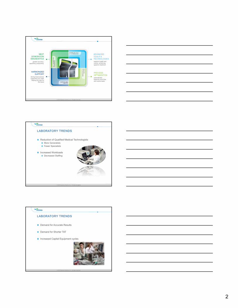
GOALS FOR TODAY
Obtain a working knowledge of digital cell image analysis.
Learn briefly about how automated digital systems work.
Obtain an understanding of some of the advantages of using automated cell image analysis in the clinical laboratory.
Demonstrate how automated cell image analysis can provide better and more rapid diagnoses through the use of examples
© 2015 Sysmex America, Inc. All rights reserved.© 2015 Sysmex America, Inc. All rights reserved.
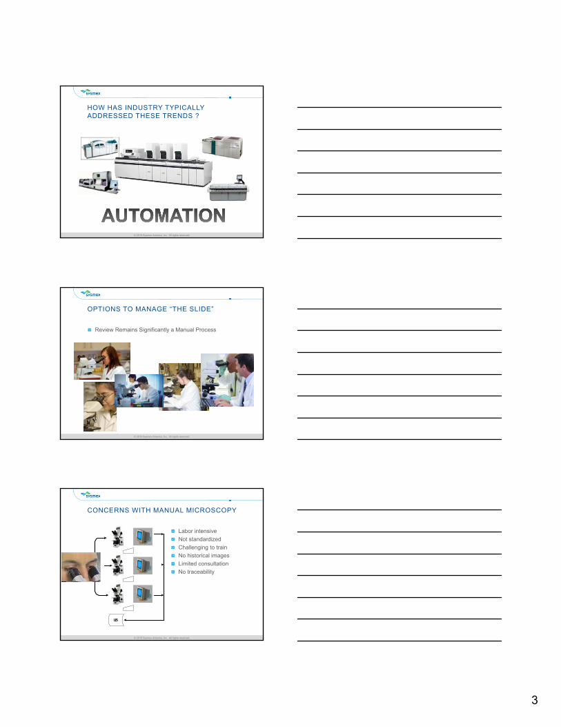
BEYOND TECHNOLOGY BEYOND EXPECTATIONS
Quality
Dependability
Accuracy
2
© 2015 Sysmex America, Inc. All rights reserved.© 2015 Sysmex America, Inc. All rights reserved.
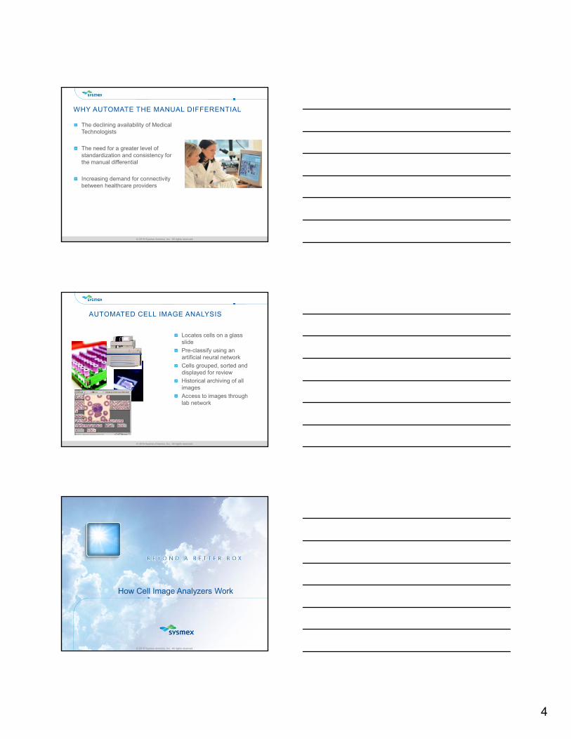
greater accuracy, specificity, productivity
NEXT GENERATION
DIAGNOSTICSsuperior insight and control, enhancing speed to treatment
ADVANCED TOOLS & TECHNOLOGIES
automatically balances work-flow and routine tasks
PROCESS OPTIMIZATION
driving improvement,supporting your staff,
helping you plan for the future
HARMONIZED SUPPORT
© 2015 Sysmex America, Inc. All rights reserved.© 2015 Sysmex America, Inc. All rights reserved.
LABORATORY TRENDS
Reduction of Qualified Medical TechnologistsMore Generalists
Fewer Specialists
Increased WorkloadsDecreased Staffing
© 2015 Sysmex America, Inc. All rights reserved.© 2015 Sysmex America, Inc. All rights reserved.
LABORATORY TRENDS
Demand for Accurate Results
Demand for Shorter TAT
Increased Capital Equipment cycles
3
© 2015 Sysmex America, Inc. All rights reserved.© 2015 Sysmex America, Inc. All rights reserved.
HOW HAS INDUSTRY TYPICALLY ADDRESSED THESE TRENDS ?
© 2015 Sysmex America, Inc. All rights reserved.© 2015 Sysmex America, Inc. All rights reserved.
OPTIONS TO MANAGE “THE SLIDE”
Review Remains Significantly a Manual Process
© 2015 Sysmex America, Inc. All rights reserved.© 2015 Sysmex America, Inc. All rights reserved.
CONCERNS WITH MANUAL MICROSCOPY
LIS
Labor intensive
Not standardized
Challenging to train
No historical images
Limited consultation
No traceability
4
© 2015 Sysmex America, Inc. All rights reserved.© 2015 Sysmex America, Inc. All rights reserved.
WHY AUTOMATE THE MANUAL DIFFERENTIAL
The declining availability of Medical Technologists
The need for a greater level of standardization and consistency for the manual differential
Increasing demand for connectivity between healthcare providers
© 2015 Sysmex America, Inc. All rights reserved.© 2015 Sysmex America, Inc. All rights reserved.
AUTOMATED CELL IMAGE ANALYSIS
Locates cells on a glass slide
Pre-classify using an artificial neural network
Cells grouped, sorted and displayed for review
Historical archiving of all images
Access to images through lab network
© 2015 Sysmex America, Inc. All rights reserved.
How Cell Image Analyzers Work
5
© 2015 Sysmex America, Inc. All rights reserved.© 2015 Sysmex America, Inc. All rights reserved.
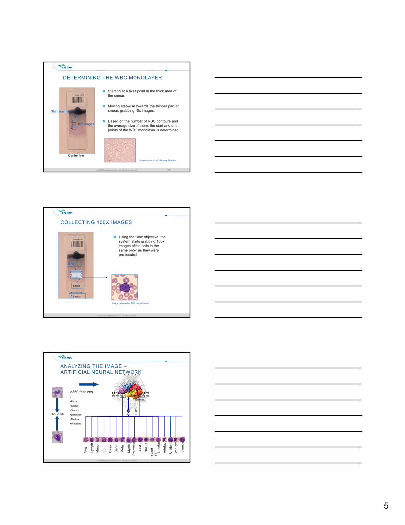
DETERMINING THE WBC MONOLAYER
Starting at a fixed point in the thick area of the smear.
Moving stepwise towards the thinner part of smear, grabbing 10x images.
Based on the number of RBC contours and the average size of them, the start and end points of the WBC monolayer is determined.
13
Start scanning
Center line
10x images
Image captured at 10x magnification
© 2015 Sysmex America, Inc. All rights reserved.© 2015 Sysmex America, Inc. All rights reserved.
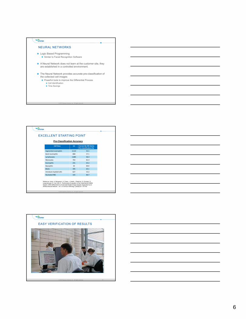
COLLECTING 100X IMAGES
Using the 100x objective, the system starts grabbing 100x images of the cells in the same order as they were pre-located
End
Start
12 mm
Image captured at 100x magnification
© 2015 Sysmex America, Inc. All rights reserved.© 2015 Sysmex America, Inc. All rights reserved.
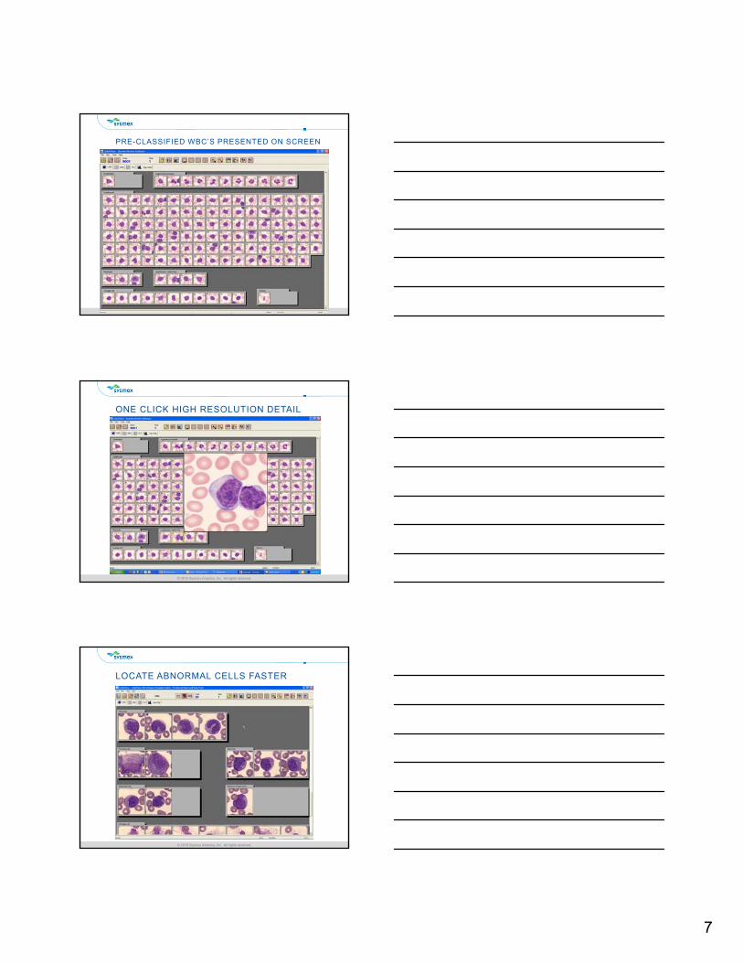
ANALYZING THE IMAGE –ARTIFICIAL NEURAL NETWORK
FEATURES
>350 features :•Form
•Colour
•Texture
•Detection
•Markov
•Wavelets
Se
g
Lym
ph
Mo
no
Eo
Ba
so
Ba
nd
Me
ta
Mye
lo
Pro
mye
lo
Bla
st
NR
BC
Gia
nt
PLT S
mu
dge
Art
efa
ct
Un
ide
nt.
Va
rLy
m
clu
mp
s
6
© 2015 Sysmex America, Inc. All rights reserved.© 2015 Sysmex America, Inc. All rights reserved.
NEURAL NETWORKS
Logic Based ProgrammingSimilar to Facial Recognition Software
A Neural Network does not learn at the customer site, they are established in a controlled environment.
The Neural Network provides accurate pre-classification of the collected cell images
Powerful tools to improve the Differential Process Cell Identification
Time Savings
© 2015 Sysmex America, Inc. All rights reserved.© 2015 Sysmex America, Inc. All rights reserved.
EXCELLENT STARTING POINT
Cell Class (n) Correct By DM‐Series Pre‐classification (%)
Segmented neutrophils 3,510 92.5
Band neutrophils 868 57.1
Lymphocytes 2,885 96.4
Monocytes 763 81.4
Eosinophils 231 63.2
Basosphils 50 80.0
Blasts 395 65.1
Immature myeloid cells 627 53.2
Nucleated RBC 165 86.7
Pre-Classification Accuracy
Reference: Kratz, A; Bengsston, H; Casey, J; Keefe, J; Beatrice, G; Grzybek, D; Lewandrowski, K; Van Cott, E : Performance Evaluation of the CellaVision® DM96 System: WBC Differentials by Automated Digital Image Analysis Supported by an Artificial Neural Network. Am J of Clinical Pathology; (2005)124: 770-781
© 2015 Sysmex America, Inc. All rights reserved.© 2015 Sysmex America, Inc. All rights reserved.
EASY VERIFICATION OF RESULTS
18
7
© 2015 Sysmex America, Inc. All rights reserved.© 2015 Sysmex America, Inc. All rights reserved.
PRE-CLASSIFIED WBC’S PRESENTED ON SCREEN
© 2015 Sysmex America, Inc. All rights reserved.© 2015 Sysmex America, Inc. All rights reserved.
ONE CLICK HIGH RESOLUTION DETAIL
© 2015 Sysmex America, Inc. All rights reserved.© 2015 Sysmex America, Inc. All rights reserved.
LOCATE ABNORMAL CELLS FASTER
8
© 2015 Sysmex America, Inc. All rights reserved.© 2015 Sysmex America, Inc. All rights reserved.
IMPROVED ACCURACY & PROFICIENCY
View cell classes side by side
Aid cell identification by seeing the “company they keep”
Compare patient cells to custom ref cell library
Using your Patient Population
Your Lab’s staining protocol
Access to historical imagesAbility to track patient changes over time
© 2015 Sysmex America, Inc. All rights reserved.© 2015 Sysmex America, Inc. All rights reserved.
STANDARDIZED RBC CHARACTERIZATION
© 2015 Sysmex America, Inc. All rights reserved.© 2015 Sysmex America, Inc. All rights reserved.
STANDARDIZE PLATELET ESTIMATES
9
© 2015 Sysmex America, Inc. All rights reserved.© 2015 Sysmex America, Inc. All rights reserved.
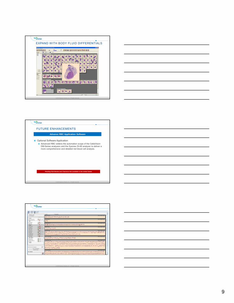
EXPAND WITH BODY FLUID DIFFERENTIALS
© 2015 Sysmex America, Inc. All rights reserved.© 2015 Sysmex America, Inc. All rights reserved.
FUTURE ENHANCEMENTS
Optional Software ApplicationAdvanced RBC widens the automation scope of the CellaVision DM-Series analyzers and the Sysmex DI-60 analyzer to deliver a more comprehensive and detailed red blood cell analysis.
Pending FDA Review and Clearance Not available in the United States
Advance RBC Application Software
© 2015 Sysmex America, Inc. All rights reserved.© 2015 Sysmex America, Inc. All rights reserved.
10
© 2015 Sysmex America, Inc. All rights reserved.
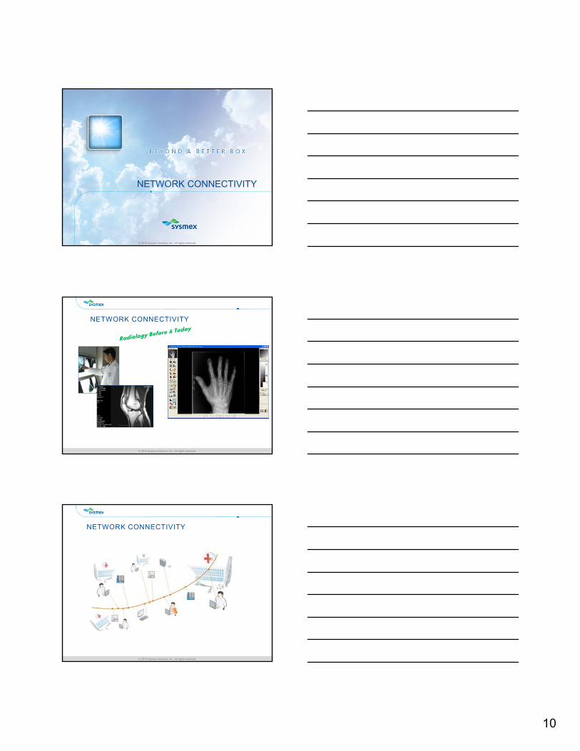
NETWORK CONNECTIVITY
© 2015 Sysmex America, Inc. All rights reserved.© 2015 Sysmex America, Inc. All rights reserved.
NETWORK CONNECTIVITY
© 2015 Sysmex America, Inc. All rights reserved.© 2015 Sysmex America, Inc. All rights reserved.
NETWORK CONNECTIVITY
11
© 2015 Sysmex America, Inc. All rights reserved.© 2015 Sysmex America, Inc. All rights reserved.
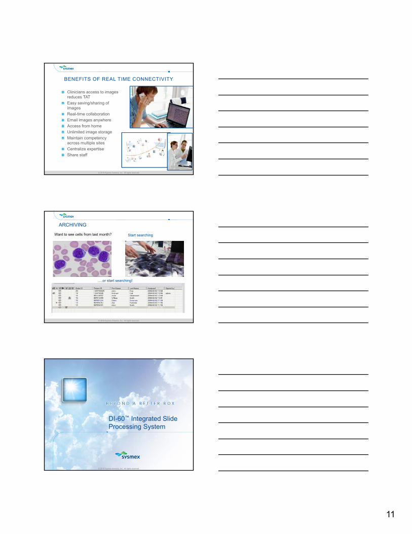
BENEFITS OF REAL TIME CONNECTIVITY
Clinicians access to images reduces TAT
Easy saving/sharing of images
Real-time collaboration
Email images anywhere
Access from home
Unlimited image storage
Maintain competency across multiple sites
Centralize expertise
Share staff
© 2015 Sysmex America, Inc. All rights reserved.© 2015 Sysmex America, Inc. All rights reserved.
ARCHIVING
Want to see cells from last month? Start searching
….or start searching!
© 2015 Sysmex America, Inc. All rights reserved.
DI-60™ Integrated Slide Processing System
12
© 2015 Sysmex America, Inc. All rights reserved.© 2015 Sysmex America, Inc. All rights reserved.
WHAT IS THE DI-60 ?
New Cell Image Analyzer Result of a collaborative R&D effort between Sysmex Corp and CellaVision
Full Integration for the hematology Work CellCBC
Slide Preparation/Staining
Digital Scan & Pre-classification
Network Compatible with Other DI-60’s or CellaVision DM-Series
© 2015 Sysmex America, Inc. All rights reserved.© 2015 Sysmex America, Inc. All rights reserved.
superior insight and control, enhancing speed to treatment
ADVANCED TOOLS & TECHNOLOGIES
automatically balances work-flow and routine tasks
PROCESS OPTIMIZATION
© 2015 Sysmex America, Inc. All rights reserved.© 2015 Sysmex America, Inc. All rights reserved.
SYSMEX DI-60™
13
© 2015 Sysmex America, Inc. All rights reserved.© 2015 Sysmex America, Inc. All rights reserved.
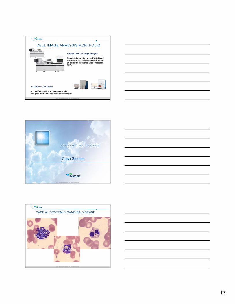
CELL IMAGE ANALYSIS PORTFOLIO
CellaVision® DM-Series:
A good fit for mid- and high volume labs. Analyzes both blood and body Fluid samples
Sysmex DI-60 Cell Image Analyzer:
Complete integration to the XN-3000 and XN-9000, or in configuration with an SP-10 called the Integrated Slide Processor (ISP).
© 2015 Sysmex America, Inc. All rights reserved.
Case Studies
© 2015 Sysmex America, Inc. All rights reserved.© 2015 Sysmex America, Inc. All rights reserved.
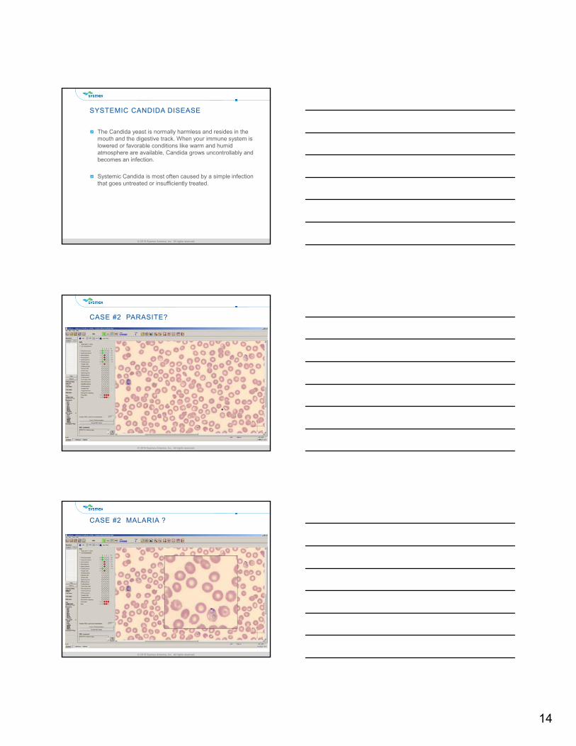
CASE #1 SYSTEMIC CANDIDA DISEASE
14
© 2015 Sysmex America, Inc. All rights reserved.© 2015 Sysmex America, Inc. All rights reserved.
SYSTEMIC CANDIDA DISEASE
The Candida yeast is normally harmless and resides in the mouth and the digestive track. When your immune system is lowered or favorable conditions like warm and humid atmosphere are available, Candida grows uncontrollably and becomes an infection.
Systemic Candida is most often caused by a simple infection that goes untreated or insufficiently treated.
© 2015 Sysmex America, Inc. All rights reserved.© 2015 Sysmex America, Inc. All rights reserved.
CASE #2 PARASITE?
© 2015 Sysmex America, Inc. All rights reserved.© 2015 Sysmex America, Inc. All rights reserved.
CASE #2 MALARIA ?
15
© 2015 Sysmex America, Inc. All rights reserved.© 2015 Sysmex America, Inc. All rights reserved.
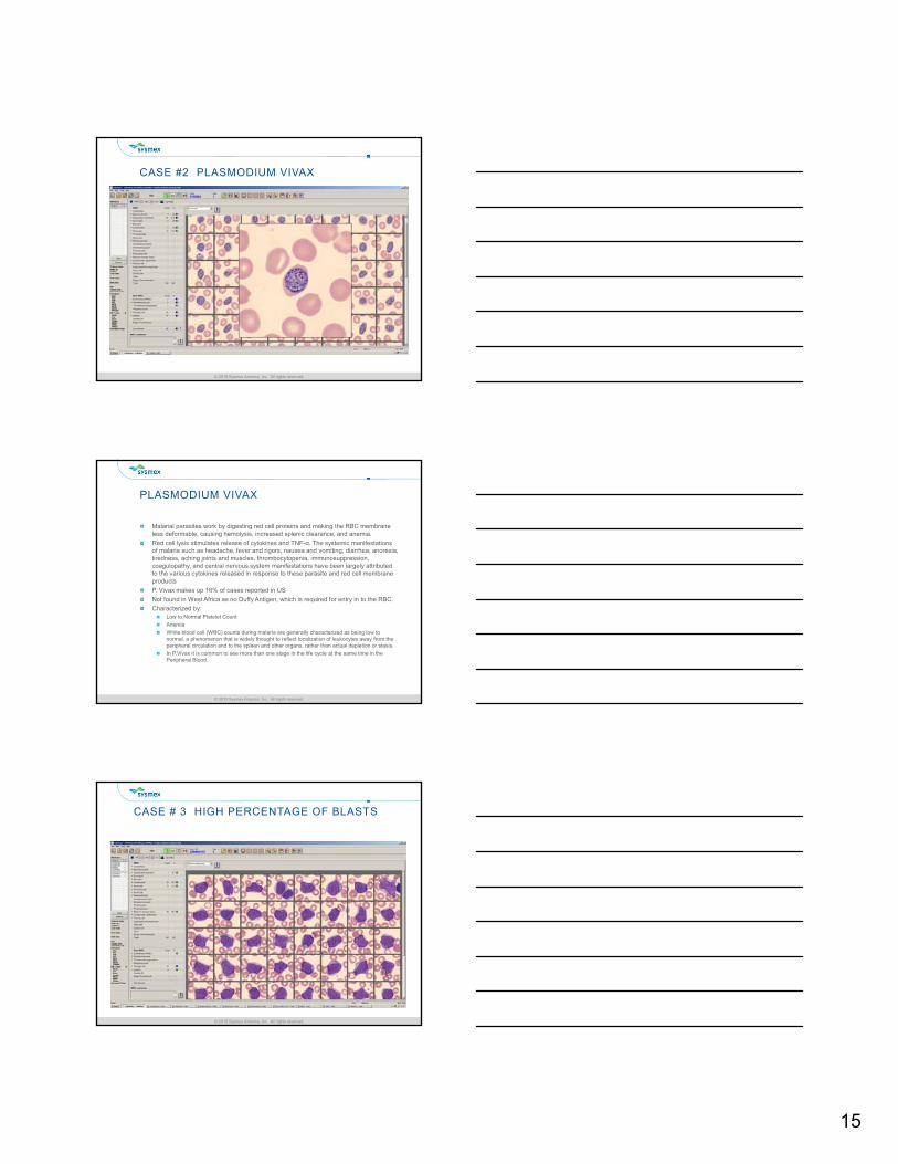
CASE #2 PLASMODIUM VIVAX
© 2015 Sysmex America, Inc. All rights reserved.© 2015 Sysmex America, Inc. All rights reserved.
PLASMODIUM VIVAX
Malarial parasites work by digesting red cell proteins and making the RBC membrane less deformable, causing hemolysis, increased splenic clearance, and anemia.
Red cell lysis stimulates release of cytokines and TNF-α. The systemic manifestations of malaria such as headache, fever and rigors, nausea and vomiting, diarrhea, anorexia, tiredness, aching joints and muscles, thrombocytopenia, immunosuppression, coagulopathy, and central nervous system manifestations have been largely attributed to the various cytokines released in response to these parasite and red cell membrane products
P. Vivax makes up 16% of cases reported in US
Not found in West Africa as no Duffy Antigen, which is required for entry in to the RBC.
Characterized by:Low to Normal Platelet Count
Anemia
White blood cell (WBC) counts during malaria are generally characterized as being low to normal, a phenomenon that is widely thought to reflect localization of leukocytes away from the peripheral circulation and to the spleen and other organs, rather than actual depletion or stasis.
In P.Vivax it is common to see more than one stage in the life cycle at the same time in the Peripheral Blood.
© 2015 Sysmex America, Inc. All rights reserved.© 2015 Sysmex America, Inc. All rights reserved.
CASE # 3 HIGH PERCENTAGE OF BLASTS
16
© 2015 Sysmex America, Inc. All rights reserved.© 2015 Sysmex America, Inc. All rights reserved.
CASE # 3 PRESENCE OF AUER RODS
© 2015 Sysmex America, Inc. All rights reserved.© 2015 Sysmex America, Inc. All rights reserved.
ACUTE MYELOID LEUKEMIA (AML)
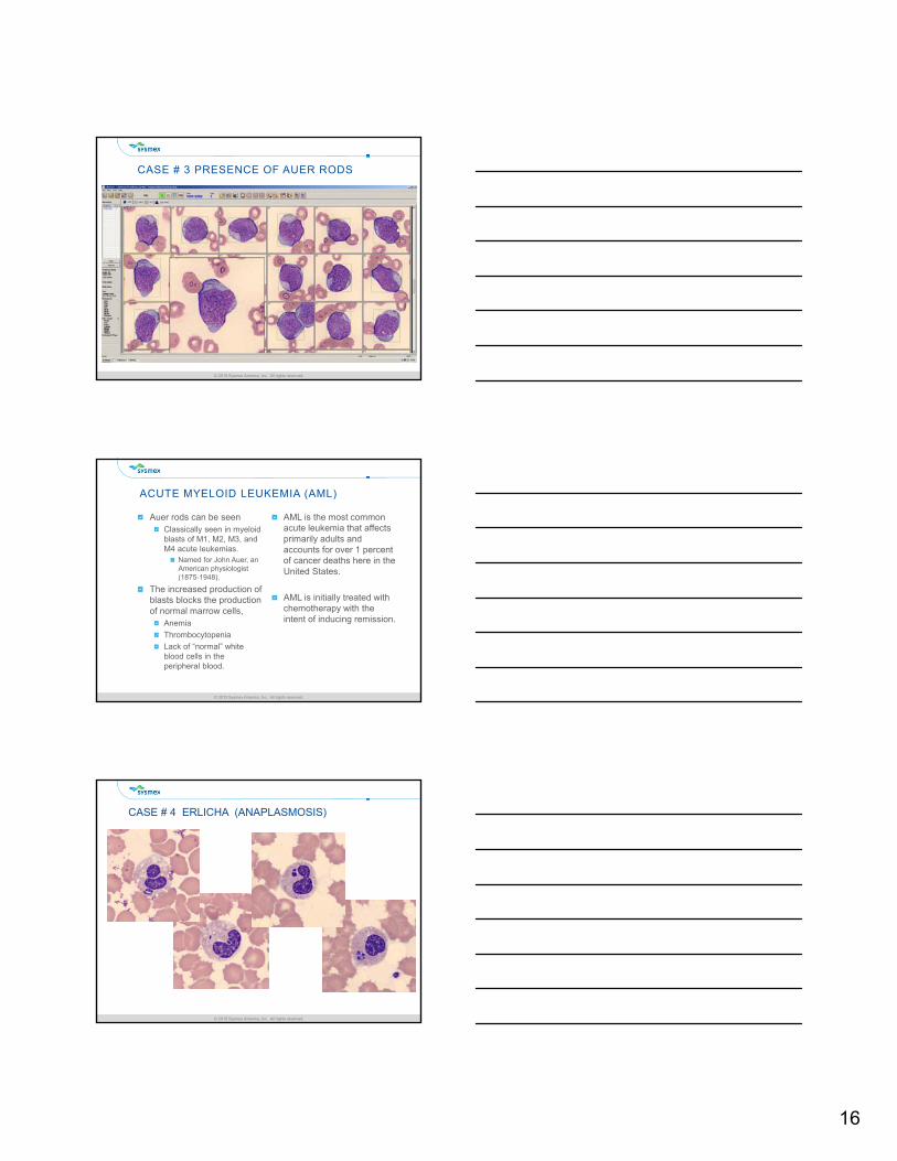
Auer rods can be seen Classically seen in myeloid blasts of M1, M2, M3, and M4 acute leukemias.
Named for John Auer, an American physiologist (1875-1948).
The increased production of blasts blocks the production of normal marrow cells,
Anemia
Thrombocytopenia
Lack of “normal” white blood cells in the peripheral blood.
AML is the most common acute leukemia that affects primarily adults and accounts for over 1 percent of cancer deaths here in the United States.
AML is initially treated with chemotherapy with the intent of inducing remission.
© 2015 Sysmex America, Inc. All rights reserved.© 2015 Sysmex America, Inc. All rights reserved.
CASE # 4 ERLICHA (ANAPLASMOSIS)
17
© 2015 Sysmex America, Inc. All rights reserved.© 2015 Sysmex America, Inc. All rights reserved.
ANAPLASMOSIS
Anaplasmosis is often characterized by sudden high fever, fatigue, muscle aches, headache. The disease can be mild or life-threatening. Severely ill patients can have low white blood cell count, low platelet count, anemia, elevated liver enzymes, kidney failure and respiratory insufficiency. Older people or people with immune suppression are more likely to require hospitalization. Deaths have occurred due to anaplasmosis.
There are two kinds of ehrlichiosis, both of which are caused by tick-borne rickettsial parasites called Ehrlichia that infect different kinds of white blood cells. In HME (human monocytic ehrlichiosis), they infect monocytes. In HGE (human granulocytic ehrlichiosis), they infect granulocytes. HGE was renamed anaplasmosis in 2003. It is likely that the lone star tick transmits HME and that the deer tick transmits HGE.
Ehrlichiosis (HME) was originally thought to be only an animal disease. It was described in humans in 1987 and is now found in 30 states, predominately in the southeast, south-central, and mid-Atlantic states, Europe and Africa.
© 2015 Sysmex America, Inc. All rights reserved.© 2015 Sysmex America, Inc. All rights reserved.
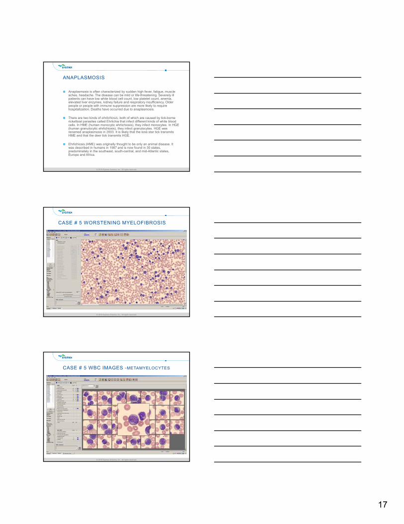
CASE # 5 WORSTENING MYELOFIBROSIS
© 2015 Sysmex America, Inc. All rights reserved.© 2015 Sysmex America, Inc. All rights reserved.
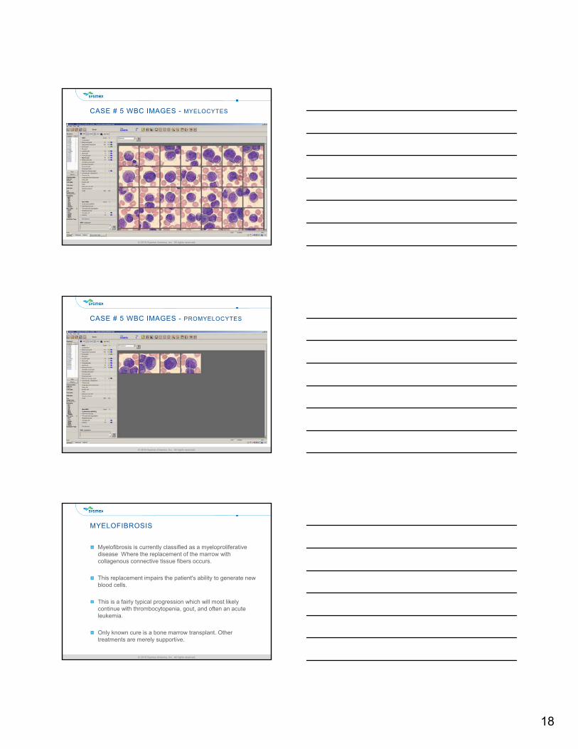
CASE # 5 WBC IMAGES -METAMYELOCYTES
18
© 2015 Sysmex America, Inc. All rights reserved.© 2015 Sysmex America, Inc. All rights reserved.
CASE # 5 WBC IMAGES - MYELOCYTES
© 2015 Sysmex America, Inc. All rights reserved.© 2015 Sysmex America, Inc. All rights reserved.
CASE # 5 WBC IMAGES - PROMYELOCYTES
© 2015 Sysmex America, Inc. All rights reserved.© 2015 Sysmex America, Inc. All rights reserved.
MYELOFIBROSIS
Myelofibrosis is currently classified as a myeloproliferative disease Where the replacement of the marrow with collagenous connective tissue fibers occurs.
This replacement impairs the patient's ability to generate new blood cells.
This is a fairly typical progression which will most likely continue with thrombocytopenia, gout, and often an acute leukemia.
Only known cure is a bone marrow transplant. Other treatments are merely supportive.
19
© 2015 Sysmex America, Inc. All rights reserved.© 2015 Sysmex America, Inc. All rights reserved.
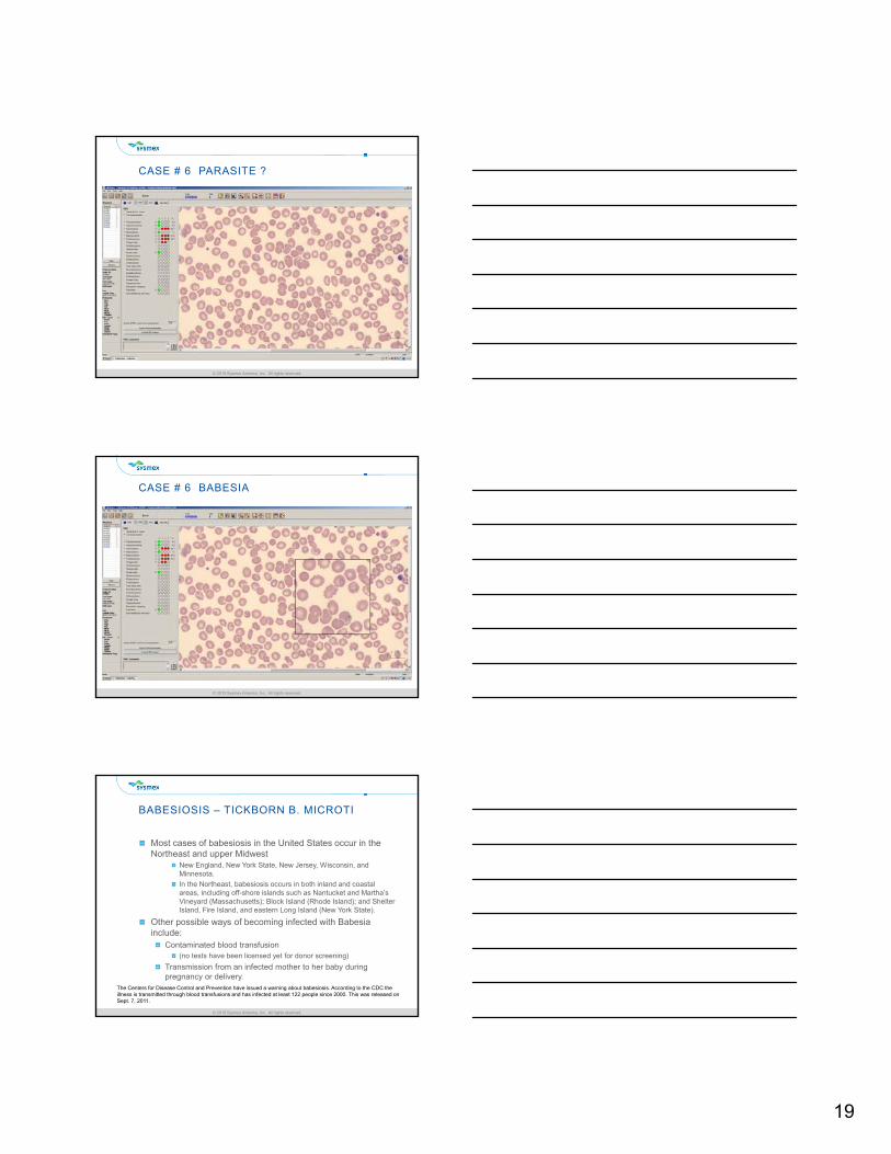
CASE # 6 PARASITE ?
© 2015 Sysmex America, Inc. All rights reserved.© 2015 Sysmex America, Inc. All rights reserved.
CASE # 6 BABESIA
© 2015 Sysmex America, Inc. All rights reserved.© 2015 Sysmex America, Inc. All rights reserved.
BABESIOSIS – TICKBORN B. MICROTI
Most cases of babesiosis in the United States occur in the Northeast and upper Midwest
New England, New York State, New Jersey, Wisconsin, and Minnesota.
In the Northeast, babesiosis occurs in both inland and coastal areas, including off-shore islands such as Nantucket and Martha’s Vineyard (Massachusetts); Block Island (Rhode Island); and Shelter Island, Fire Island, and eastern Long Island (New York State).
Other possible ways of becoming infected with Babesiainclude:
Contaminated blood transfusion (no tests have been licensed yet for donor screening)
Transmission from an infected mother to her baby during pregnancy or delivery.
The Centers for Disease Control and Prevention have issued a warning about babesiosis. According to the CDC the illness is transmitted through blood transfusions and has infected at least 122 people since 2000. This was released on Sept. 7, 2011.
20
© 2015 Sysmex America, Inc. All rights reserved.© 2015 Sysmex America, Inc. All rights reserved.
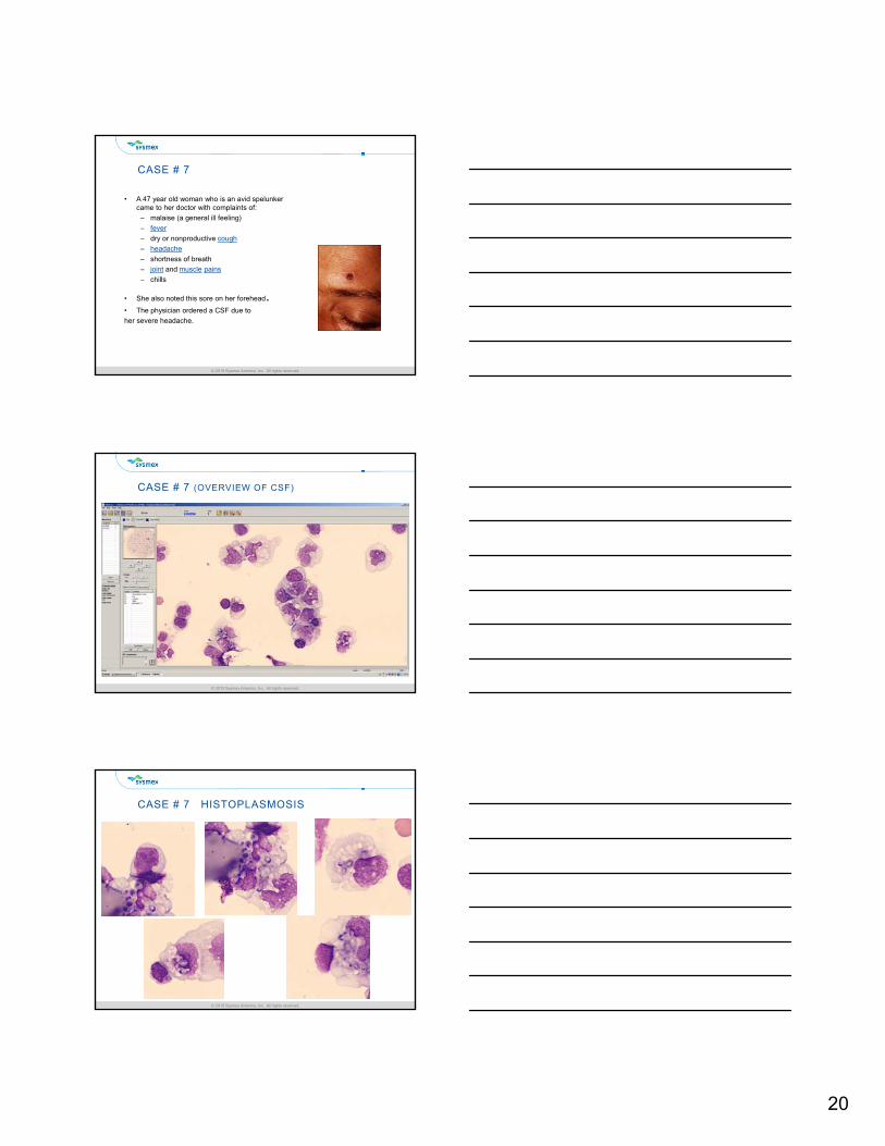
CASE # 7
• A 47 year old woman who is an avid spelunker came to her doctor with complaints of:
– malaise (a general ill feeling)
– fever
– dry or nonproductive cough
– headache
– shortness of breath
– joint and muscle pains
– chills
• She also noted this sore on her forehead.• The physician ordered a CSF due to
her severe headache.
© 2015 Sysmex America, Inc. All rights reserved.© 2015 Sysmex America, Inc. All rights reserved.
CASE # 7 (OVERVIEW OF CSF)
© 2015 Sysmex America, Inc. All rights reserved.© 2015 Sysmex America, Inc. All rights reserved.
CASE # 7 HISTOPLASMOSIS
21
© 2015 Sysmex America, Inc. All rights reserved.© 2015 Sysmex America, Inc. All rights reserved.
HISTOPLASMOSIS
HistoplasmosisCaused by the fungus Histoplasma capsulatum.
Symptoms of this infection vary greatly, but the disease primarily affects the lungs. Histoplasmosis is common among AIDS patients because of their suppressed immune system.
H. capsulatum grows in soil and material contaminated with bird or bat droppings (guano).
The fungus has been found in poultry house litter, caves, areas harboring bats, and in bird roosts (particularly those of starlings).
Histoplasmosis can be diagnosed by:Samples containing the fungus taken from sputum, blood, or infected organs.
Detection of antigens in blood or urine samples by ELISA or PCR.
A test for antibodies against Histoplasma in the blood.
© 2015 Sysmex America, Inc. All rights reserved.© 2015 Sysmex America, Inc. All rights reserved.
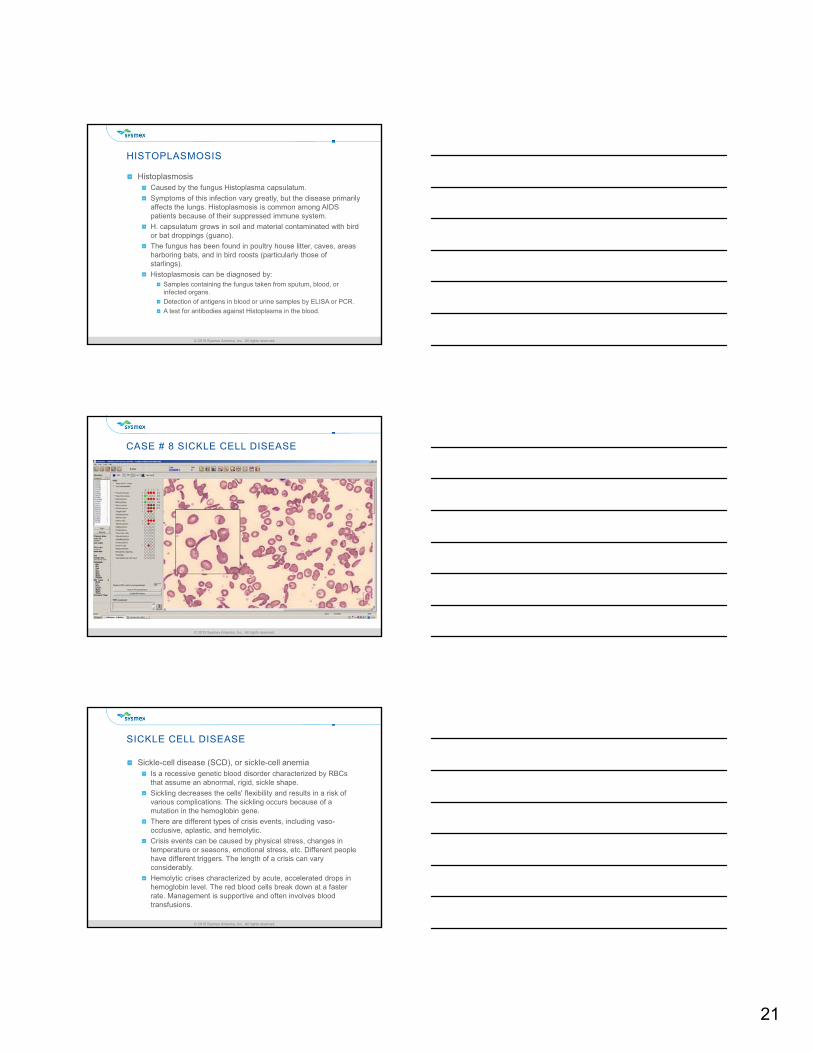
CASE # 8 SICKLE CELL DISEASE
© 2015 Sysmex America, Inc. All rights reserved.© 2015 Sysmex America, Inc. All rights reserved.
SICKLE CELL DISEASE
Sickle-cell disease (SCD), or sickle-cell anemiaIs a recessive genetic blood disorder characterized by RBCs that assume an abnormal, rigid, sickle shape.
Sickling decreases the cells' flexibility and results in a risk of various complications. The sickling occurs because of a mutation in the hemoglobin gene.
There are different types of crisis events, including vaso-occlusive, aplastic, and hemolytic.
Crisis events can be caused by physical stress, changes in temperature or seasons, emotional stress, etc. Different people have different triggers. The length of a crisis can vary considerably.
Hemolytic crises characterized by acute, accelerated drops in hemoglobin level. The red blood cells break down at a faster rate. Management is supportive and often involves blood transfusions.
22
© 2015 Sysmex America, Inc. All rights reserved.© 2015 Sysmex America, Inc. All rights reserved.
CASE STUDIES SUMMARY
Automated Cell Image Analysis in the Clinical Lab can be useful for many reasons including:
Clinical Capabilities
Operational Capabilities
Financial Capabilities
© 2015 Sysmex America, Inc. All rights reserved.© 2015 Sysmex America, Inc. All rights reserved.
VALUE TO LABORATORY
Improved WorkflowIncreased quality and consistency
Ability to review smears easily and cells classed
Identify issues with technicians and ability to educate
Improved ErgonomicsStaff Comfort
Cell Image adjustments
Competency and EducationStudents
Slide Review
Multiple Databases
Decreases tech time
© 2015 Sysmex America, Inc. All rights reserved.© 2015 Sysmex America, Inc. All rights reserved.
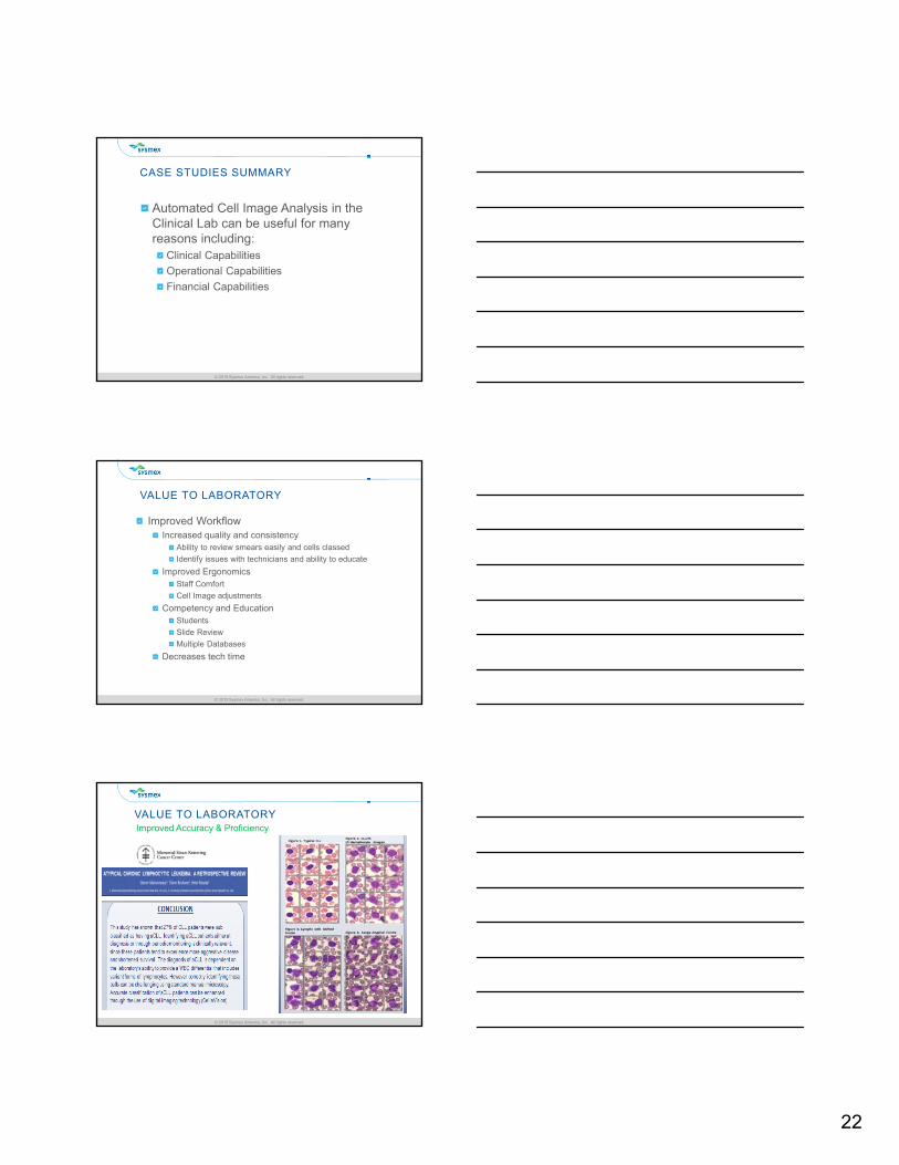
VALUE TO LABORATORYImproved Accuracy & Proficiency
23
© 2015 Sysmex America, Inc. All rights reserved.© 2015 Sysmex America, Inc. All rights reserved.
VALUE TO LABORATORY
Cells are automatically located
Pre-classification of cells removes the need to manually search the slide
Display of all WBC on one screen allows fast confirmation of cell counter Diff
Multiple slide merge on low counts eliminates buffy coat preps
Streamlines collaborations & consultations
Eliminates searching for slides
Enhanced Efficiency
© 2015 Sysmex America, Inc. All rights reserved.© 2015 Sysmex America, Inc. All rights reserved.
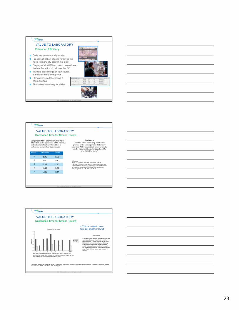
VALUE TO LABORATORYDecreased Time for Smear Review
Operator Manual Diff CIA DIFF
A 1:45 1:05
B 1:40 1:10
C 3:45 1:30
D 4:10 1:40
E 3:10 1:14
Conclusions:
“The time saved when using the DM96 is greatest for the less experienced laboratory
scientists. With increased instrument familiarity with the instrument there may be potential for
even more time saved”
Comparison of time taken to complete the 30 differentials on the CellaVision DM96 including reclassification of cells with time taken to perform the same differentials manually
Reference:Briggs C., Longhair I., Slavik M., Thwaite K., Mills R., ThavarajaV., Foster A., Romanin D., Machin S.J. (2009) Can automated blood film analysis replace the manual differential? An evaluation of the CellaVision DM96 automated image analysis system. Jnl. Lab. Hem. 31, 48–60
© 2015 Sysmex America, Inc. All rights reserved.© 2015 Sysmex America, Inc. All rights reserved.
VALUE TO LABORATORYDecreased Time for Smear Review
Conclusions:
Automated image analysis and classification has now reached a stage where it can meet the requirements of a modern routine hematological laboratory in terms of reliability and efficiency. (The systems) are reliable and accurate and enable automated analysis of blood smears in the hematology laboratory. The systems operate most effectively in screening routine blood samples….
Reference: Ceelie H, Dinkelaar RB, van GW. Examination of peripheral blood films using automated microscopy; evaluation of Diffmaster Octavia and Cellavision DM96. J Clin Pathol 2007 Jan;60(1):72-9.
Figure 4: Results for the manual differential count (n = 220) and the DM96 (n = 315). For each method, the mean time of analysis per sample was reduced by 63% with the automated system.
~ 63% reduction in mean time per smear reviewed
24
© 2015 Sysmex America, Inc. All rights reserved.© 2015 Sysmex America, Inc. All rights reserved.
VALUE TO PATHOLOGY
Enables Real Time CollaborationRemote Review Software
Pathologist complete reviewOn-site or Off-site
Real time consultations
Email cell images for reviewSpecific cells emailed
Decrease time to search for abnormal samples
Electronic record of abnormalities
© 2015 Sysmex America, Inc. All rights reserved.© 2015 Sysmex America, Inc. All rights reserved.
IMPACT TO THE INSTITUTION
Expanded Cell Image Analysis Program to Include Their Legacy Campus in Plano, TX
Connected Multiple Cell Imaging Systems Into a Single Production Database
Expanded Laboratory Remote Review Capabilities to the Medical Staff
Medical Service Utilizes the Images for Diagnosis and Patient Follow-up
© 2015 Sysmex America, Inc. All rights reserved.© 2015 Sysmex America, Inc. All rights reserved.
IMPACT TO THE INSTITUTION
“This New Infrastructure has allowed the Lab to provide improved services to physicians and patients. Prior to having the connectivity, the laboratory had to prepare and deliver the slide to the physicians. This new system alleviates the need to remember to make extra slides, and deliver them to the physician in a timely manner”…..
“Connectivity has made a huge difference. From a workload standpoint, we have been able to handle a significantly increased workload due to the expansion of the automated cell imaging capability”…..
25
© 2015 Sysmex America, Inc. All rights reserved.© 2015 Sysmex America, Inc. All rights reserved.
IMPACT TO THE INSTITUTION
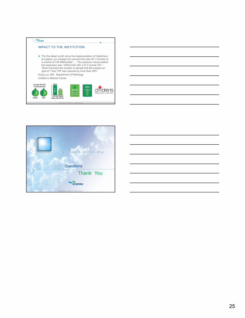
“For the latest month since the implementation of CellaVision at Legacy, our average turn around time was 34.7 minutes on a volume of 739 differentials”…. “Our previous volume before the expansion was ~200/month with a 37.2 minute TAT.”.. “More important the number of sample that fell outside our goal of 1 hour TAT was reduced by more than 50%”
Hung Luu, MD. Department of Pathology
Children’s Medical Center
© 2015 Sysmex America, Inc. All rights reserved.
Questions
Thank You