Embed Size (px)
Citation preview
J. Embryol exp. Morph. Vol. 72, pp. 197-208, 1982 1 9 7
Printed in Great Britain © Company of Biologists Limited 1982
Cell lineage of the Drosophila abdomen: theepidermis, oenocytes and ventral muscles
By PETER A. L A W R E N C E U N D PAUL JOHNSTON1
From the MRC Laboratory of Molecular Biology, Cambridge
SUMMARY
We use a new cell marker to study cell lineage of the epidermis, oenocytes and ventralmuscles in the Drosophila abdomen. We find that while the epidermal cells and the oenocytesshare common precursors until late in development, the muscles derive from a separatelineage. Marked clones in the muscles, but not in the epidermis and oenocytes, can occasion-ally extend between neighbouring segments, supporting a previous hypothesis that mesodermcells may not be determined as to segment.
INTRODUCTION
Every segment of the adult Drosophila abdomen consists of an epidermalpolyclone (Szabad, Schiipbach & Wieschaus, 1979; Lawrence, 1981a). Associ-ated with each segment there is a band of oenocytes, two strips of fat body anda specific pattern of superficial muscles (Robertson, 1936; Koch, 1945; Miller,1950). The adult epidermis develops from cells which, although integrated into thelarval epidermis (Pearson, 1972), remain small. In each hemisegment these cellsor 'histoblasts' are gathered into three nests and their development has beenwell described by both direct observation (Madhavan & Schneiderman, 1977;Madhavan & Madhavan, 1980) and by clonal analysis (Garcia-Bellido &Merriam, 1971; Guerra, Postlethwait & Schneiderman, 1973; Lawrence, Green& Johnston, 1978).
The lineage relationship between the histoblasts and the non-epidermaltissues is unclear. Robertson (1936) and Koch (1945) suggested that the epi-dermis, oenocytes and fat body arise from these histoblasts, but could notestablish a lineage relationship between them. Ferrus & Kankel (1981), usingmitotic recombination and a cell marker, presented some evidence that clonesextend between all three cell types. The lineage relationship between the pre-cursors of abdominal muscles and histoblasts is unknown (Perez, 1910; Crossley,1965, 1978). In this paper we use a new cell marker to study the cell lineage ofthe epidermis, oenocytes and muscles; we find that while the ventral epidermis
1 Authors' address: MRC Laboratory of Molecular Biology, Hills Road, CambridgeCB2 2QH, U.K.
198 P.A.LAWRENCE AND P.JOHNSTON
and oenocytes of each segment do derive from a common cell pool, the muscleshave a separate origin. We were unable to study the cell lineage of the fat body.
MATERIALS AND METHODS
To mark clones of cells we used the cuticular cell markers straw, (see Lindsley& Grell, 1968 for all genetic nomenclature, except when other references aregiven), pawn (Garcia-Bellido & Dapena, 1974) and yellow. The oenocytes andmuscles were marked with the cell-autonomous marker sdh8, an allele whichgives clones of cells with altered activity for succinate dehydrogenase (Lawrence,19816). Staining solutions were made up as described in Lawrence (19816).For muscles, whole abdomens were heated in Ringer solution at 52° for 10 min,washed in buffer in ice for 30 min, and dissected free of gut and fat body. Thesepieces were then stained for 5-6 h, washed in water, dehydrated in absolutealcohol, cleared in methyl salicylate and mounted in Euparal or Struhl'smountant (see Struhl, 1981). For the epidermal cells and oenocytes the optimumheating temperature was 47-48 °C: other steps were as for muscles. We failedto find suitable conditions for studying cell lineage of the fat body.
Embryos and larvae of the following genotypes were irradiated and 1-week-old females were studied because they are larger and easier to dissect than males.
genotype 1 en sdh8 bw/cn bw M(2)c33a
genotype 2 stw pwn sdh8 bw/cn bw M{2)c2Z&
genotype 3 stw pwn en10 sdh8 Zw/M(2)c33a (en10 is a lethal allele at theengrailed locus (Kornberg, 1981 a) made and kindly provided byChristiane Niisslein-Volhard and Eric Wieschaus - see theirpaper, 1980)
genotype 4 y/DfscS2, y; en sdh8 bw/M(2)S7 Dpsc82, y+
Irradiations were performed at 3-5±0-7 h after egg laying (h AEL), 48 ±4 hAELand60±12hAEL.
Recognizing muscle clones
In the thoracic muscles, Minute+ clones growing in a Minute* backgroundare not seen, while clones of the same genotype, which grow excessively in aMinute background, are (Lawrence, 1982). We believe this is because a fibrewhich contains even a few sdh+ nuclei is indistinguishable from entirely sdh+
fibres. The probability of a single fibre being entirely sdh and therefore recog-nized, will depend mainly on the growth rate of the sdh clone (which will in-crease the probability), and the degree of intermingling (which will reduce it).In the abdominal muscles of irradiated insects unstained patches were rarelyseen and frequently extended to only a few fibres. In order to recognize theclones and distinguish them from artefacts, such as general leaching of stain,two criteria were used: the individual muscle fibres must be entirely unstained
Cell lineage of the Drosophila abdomen 199while associated nerve fibres should be stained. Neighbouring muscle fibres,oenocytes and fat body must be stained. When these criteria were applied tothe dorsal muscles of the tergites the few unstained patches failed to meet them.Consequently, results on only the ventral abdomen are reported. Evidence thatventral clones scored by these criteria are genuine comes from the results; theirfrequency depends on X-ray dose (Table 2). Muscle clones were most easilydetected under polarized light when the birefringence of unstained fibres wasconspicuous.
Recognizing clones in the oenocytes
Oenocytes often stain capriciously: clones were only registered when theunstained cells were adjacent to and formed a sharp contrast with, stained cells.Again, neighbouring muscles and fat body must be stained. To ascertain whethercuticle clones extended to oenocytes, patches of mutant bristles were sometimesdetected under the dissecting microscope and those abdomens stained andmounted.
RESULTS
Clones in the epidermis and oenocytes
Clones in the epidermal cells were either marked directly with sdh and seenafter staining or indirectly with cuticle markers that affected the bristles andtrichomes {yellow, straw, pawn). Epidermal stw pwn sdh8 M(2)c+ clones wereas expected, the stw pwn trichomes in the cuticle being coextensive with theunstained epidermal cells beneath. Frequently, stw+ pwn+ bristles entered theepidermal territory, and stw pwn bristles were found at some distance from theirparent clone (Garcia-Bellido & Merriam, 1971).
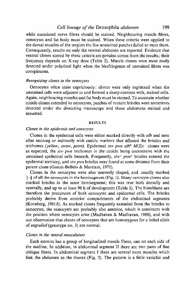
Clones in the oenocytes were also unevenly shaped, and usually marked£-•J of all the oenocytes in the hemisegment (Fig. 1). Many oenocyte clones alsomarked bristles in the same hemisegment; this was true both dorsally andventrally, and up to at least 96 h of development (Table 1). The histoblasts aretherefore the precursors of both oenocytes and epidermal cells. The bristlesprobably derive from anterior compartments of the abdominal segments(Kornberg, 19816). As marked clones frequently extended from the bristles tooenocytes, the oenocytes are probably also anterior, which is consistent withthe position where oenocytes arise (Madhavan & Madhavan, 1980), and withour observation that clones of oenocytes that are homozygous for a lethal alleleof engrailed (genotype no. 3) are normal.
Clones in the ventral musculature
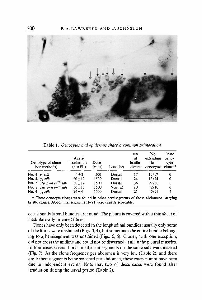
Each sternite has a group of longitudinal muscle fibres, one on each side ofthe midline. In addition, in abdominal segment II there are two pairs of fineoblique fibres. In abdominal segment I there are several extra muscles whichlink the abdomen to the thorax (Fig. 2). The pattern is a little variable and
200 P.A.LAWRENCE AND P.JOHNSTON
Table 1. Oenocytes and epidermis share a common primordium
Genotype of clone(see methods)
Age atirradiation
(h AEL)Dose(rads) Location
No.of
bristleclones
No.extending
tooenocytes
Pureoeno-cyte
clones*
No. 4. y,sdh 4±2 500 Dorsal 17 10/17 0No. 4. y,sdh 60 ±12 1500 Dorsal 24 13/24 0No. 3. stw pwn en10 sdh 60 ±12 1500 Dorsal 36 27/36 6No. 3. stwpwn en10 sdh 60±12 1500 Ventral 10 2/10 0No. 4. y, sdh 96±4 1500 Dorsal 21 5/21 4
* These oenocyte clones were found in other hemisegments of those abdomens carryingbristle clones. Abdominal segments II-VI were usually scoreable.
occasionally lateral bundles are found. The pleura is covered with a thin sheet ofmediolaterally oriented fibres.
Clones have only been detected in the longitudinal bundles; usually only someof the fibres were unstained (Figs. 3,4), but sometimes the entire bundle belong-ing to a, hemisegment was unstained (Figs. 5, 6). Clones, with one exception,did not cross the midline and could not be discerned at all in the pleural muscles.In four cases several fibres in adjacent segments on the same side were marked(Fig. 7). As the clone frequency per abdpmen is very low (Table 2), and thereare 10 hemisegments being screened per abdomen, these cases cannot have beendue to independent events. Note that two of these cases were found afterirradiation during the larval period (Table 2).
Cell lineage of the Drosophila abdomen 201
Fig. 2. Diagram of the dissected ventral abdomen of a female. Each segment(numbered I-VI) bears a pair of sensilla (s) and spiracles (sp), a patch of oenocytes(oe), and some longitudinal muscles (black bands). In the second segment, in additionto the longitudinal muscles, there are two pairs of fine muscles and a cuticularWheeler's organ (wo). Each spiracle bears a closing muscle and the pleura is coveredwith a fine sheet of mediolateral muscle fibres (pm).
202 P.A.LAWRENCE AND P.JOHNSTON
Table 2. Clones in the ventral abdomens {segments II-VI)
Genotype
No. 1. sdhNo. 1. sdhNo. 2. stw pwn sdhNo.3. stw pwn en10 sdhNo. 4. y, sdh
Age atirradia-
tion
3-5±0-748±448±460 ±1260 ±12
Dose(rads)
7501500150015001500
No. ofabdo-mens
174447751427t
280
No. ofmuscleclones
(1 segment)
15232238
No. ofmuscleclones
(2 segment)
22000
No. ofcuticleclones*
NSNS762130
* All bristle clones counted in the ventral abdomens screened (sternites II-VI) for geno-types no. 2, 3 and 4. NS = not scored.
t These 27 abdomens were selected because cuticle clones were detected (sometimes inerror) under the dissecting microscope.
Table 2 shows that, even allowing for the different X-ray doses, the frequencyof muscle clones rises between blastoderm (3-5 ±0-7 h AEL) and the late firststage larva (48 ± 4 h AEL): following irradiation at blastoderm the frequency isabout 1/1000 hemisegments while following irradiation in the first larval stage,with twice the X-ray dose, it rises to ca. 1/200 hemisegments. This might suggestan approximate doubling in the number of precursor cells between blastodermand the first stage larva but, as there are several important unknowns (the effectsof X-rays on cell number at the two different stages, the possibility that manyevents of somatic recombination go undetected because of myoblast fusion),this estimate should be taken with caution.
In genotypes 2, 3 and 4, 127 cuticle clones and 33 muscle clones were de-tected in 821 abdomens; in only one case (simply explained as a chance event)did a cuticle and a muscle clone coincide in the same hemisegment. This resultshows that the muscles and anterior epidermis do not share common precursorcells in the larva (the posterior compartments of the abdominal segments werenot studied since they cannot be easily marked, Kornberg, 19816).
The three muscle clones presumed to be homozygous for a lethal allele ofengrailed (genotype no. 3, Table 2) were normal in appearance. This is similar tothe result in the thorax which is discussed in Lawrence (1982).
DISCUSSION
We describe the cell lineage of three abdominal tissues of adult Drosophila:the epidermis, the oenocytes and the ventral muscles. Epidermal and oenocyteclones are confined to individual hemisegments while muscle clones can extendbetween two hemisegments. The epidermis and oenocytes derive from commonprecursor cells, but the muscles have a separate lineage.
Cell lineage of the Drosophila abdomen 203
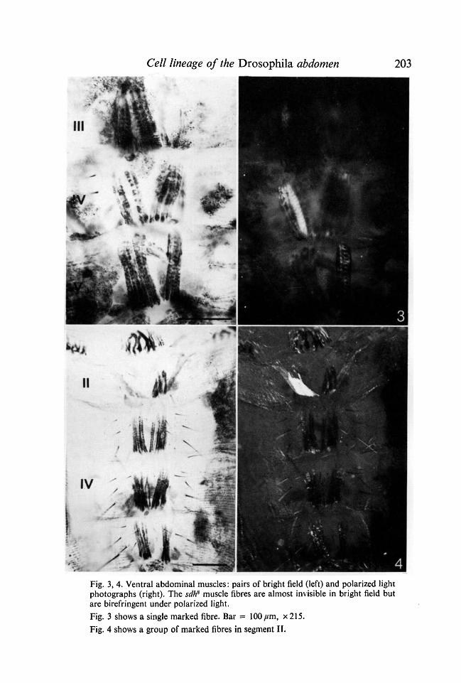
4Fig. 3, 4. Ventral abdominal muscles: pairs of bright field (left) and polarized lightphotographs (right). The sdh8 muscle fibres are almost invisible in bright field butare birefringent under polarized light.Fig. 3 shows a single marked fibre. Bar = 100 jum, x 215.Fig. 4 shows a group of marked fibres in segment II.
204 P.A.LAWRENCE AND P.JOHNSTON
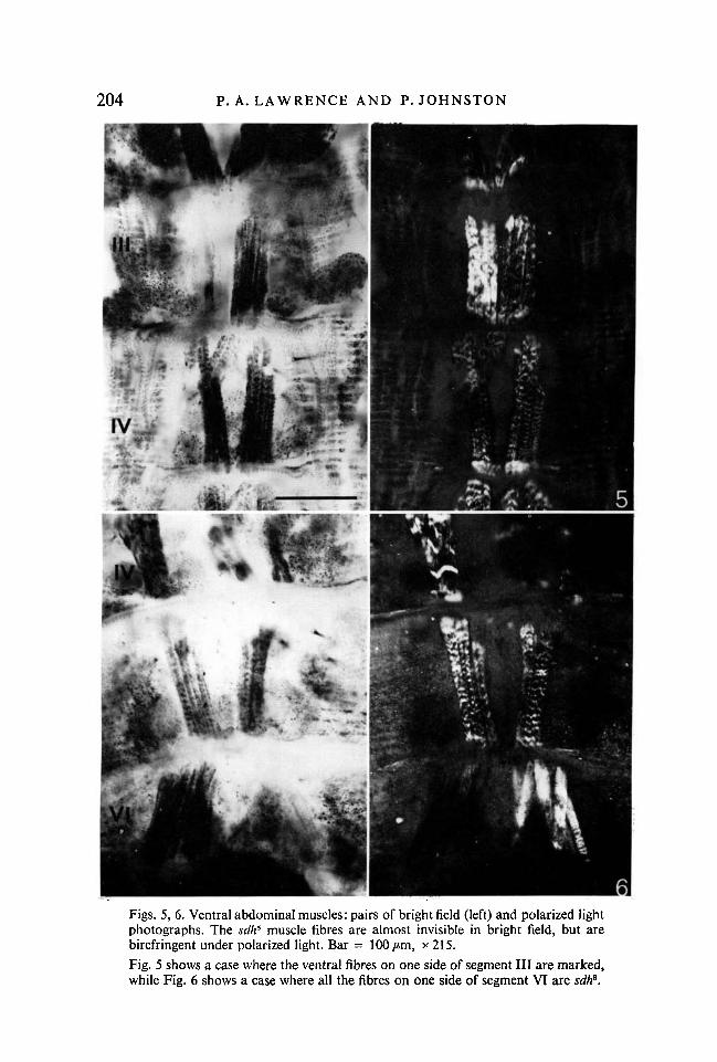
6Figs. 5, 6. Ventral abdominal muscles: pairs of bright field (left) and polarized lightphotographs. The sdh8 muscle fibres are almost invisible in bright field, but arebirefringent under polarized light. Bar = 100/tm, x 215.Fig. 5 shows a case where the ventral fibres on one side of segment III are marked,while Fig. 6 shows a case where all the fibres on one side of segment VI are sdh6.
Cell lineage of the Drosophila abdomen 205
o
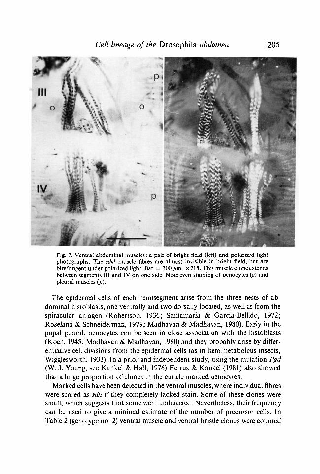
Fig. 7. Ventral abdominal muscles: a pair of bright field (left) and polarized lightphotographs. The sdh8 muscle fibres are almost invisible in bright field, but arebirefringent under polarized light. Bar = 100 pm, x 215. This muscle clone extendsbetween segments III and IV on one side. Note even staining of oenocytes (o) andpleural muscles (p).
The epidermal cells of each hemisegment arise from the three nests of ab-dominal histoblasts, one ventrally and two dorsally located, as well as from thespiracular anlagen (Robertson, 1936; Santamaria & Garcia-Bellido, 1972;Roseland & Schneiderman, 1979; Madhavan & Madhavan, 1980). Early in thepupal period, oenocytes can be seen in close association with the histoblasts(Koch, 1945; Madhavan & Madhavan, 1980) and they probably arise by differ-entiative cell divisions from the epidermal cells (as in hemimetabolous insects,Wigglesworth, 1933). In a prior and independent study, using the mutation Pgd(W. J. Young, see Kankel & Hall, 1976) Ferrus & Kankel (1981) also showedthat a large proportion of clones in the cuticle marked oenocytes.
Marked cells have been detected in the ventral muscles, where individual fibreswere scored as sdh if they completely lacked stain. Some of these clones weresmall, which suggests that some went undetected. Nevertheless, their frequencycan be used to give a minimal estimate of the number of precursor cells. InTable 2 (genotype no. 2) ventral muscle and ventral bristle clones were counted
206 P.A.LAWRENCE AND P.JOHNSTON
in one set of abdomens, and there were three to four times as many cuticleclones. Since there are 10-14 ventral abdominal histoblasts which produce thesternite and the pleural cuticle (Madhavan & Schneiderman, 1977; Lawrenceet al. 1978) the number of muscle precursors for each hemisegment of the ventralabdomen in the larva should be four or more. It is not known where these cellsare; there are some small cells near the histoblasts (Madhavan & Schneiderman,1977) which could be the muscle precursors and groups of small cells are alsoseen near the muscles of the body wall in the mature larva (Perez, 1910; ourobservations).
Following blastoderm irradiation, and allowing for different X-ray doses,the frequency of clones was about half that following larval irradiation. Thissuggests that the presumptive adult myoblasts divide about once in the embryo.In the thorax, muscle clone frequencies are remarkably similar to those in theabdomen: a frequency of 2 clones/1000 dorsal hemisegments was recorded inthe thorax following irradiation with 750 R at the blastoderm stage (Lawrence,1982) compared to 1/1000 ventral hemisegments in the abdomen (Table 2).Probably therefore, at the blastoderm stage, similar numbers of adult muscleprecursors are found in the segments of the thorax and abdomen. In the thorax(Lawrence, 1982), as in the abdomen, these precursors divide one to two timesbefore the end of the first instar. These findings support the hypothesis that atsegmentation every metameric unit is similar, diversification coming only laterin development (Lohs-Schardin, Cremer & Niisslein-Volhard, 1979; Lawrence& Morata, 1979).
Ventral muscle and epidermis of the anterior compartments of the abdominalsegments do not have common precursor cells, at least in the larva. This is theclear conclusion based on 160 ventral muscle and cuticle clones in about 9800half segments (genotypes nos. 2, 3, 4, Table 2) when there was only one casewhere a muscle and a cuticle clone was found in the same hemisegment. Sincethere is only about one division between the blastoderm stage and the larva,both in the epidermis (Szabad et al. 1979) and in the muscle, it is likely that themuscle and epidermal precursors are always separate (as is probably the case inthe thorax, Lawrence, 1982; Ferrus & Kankel, 1981). This is in accord with theallocation of abdominal muscle precursors to the mesoderm (Perez, 1910;Poulson, 1950).
Four muscle clones extended between two neighbouring segments, two ofthese being induced in the larval stage. While this could perhaps be an artefactproduced by the high X-ray dose and consequent cell death we think that it isnot. In descriptions of abdominal muscle development (Perez, 1910; Crossley,1965) the impression given is one of free, or almost free, myoblasts which canwander. If this is an accurate impression, then, even if separate primordia wereestablished by segmentation of the embryo, their descendant myoblasts couldmingle later on in the pupa. This interpretation is supported by an experiment:if myoblasts from the wing disc are released into the abdomen, they can con-
Cell lineage of the Drosophila abdomen 207tribute to the ventral and dorsal abdominal muscles (Lawrence & Brower, 1982).If free myoblasts can contribute to form the normal muscle pattern of any seg-ment, what determines the different patterns of muscles that are found in thedifferent segments? The answer appears to be the ectoderm, with which themyoblasts are associated (Bock, 1942; see Lawrence, 1982, for discussion).Unlike the myoblasts, the epidermal cells of each segment never mix with thoseof other segments (Lawrence, 1973, 1981a).
We have not been able to study the fat body, which, in the embryo at least,is classified as mesoderm (Johannsen & Butt, 1941; Poulson, 1950). However,Ferrus & Kankel (1981) find many epidermal clones which extend to the oeno-cytes and the fat body. There is room for some doubt because, as they point out,the autonomy of the marker they used is not completely established. If con-firmed, their results would show that at least part of the adult fat body isectodermal.
We thank Christiane Niisslein-Volhard and Eric Wieschaus for giving us engrailed10, andGary Struhl for criticism of the manuscript.
REFERENCES
BOCK, E. (1942). Wechselbeziehung zwischen den Keimblattern bei der Organbildung vonChrysopa perla (L.) I. Die Entwicklung des Ektoderms in mesodermdefekten Keimteilen.Wilhelm Roux" Arch. EntwMech. Org. 141, 159-247.
CROSSLEY, A. C. S. (1965). Transformations in the abdominal muscles of the blue blow-fly,Calliphora erythrocephala (Meig), during metamorphosis. / . Embryol. exp. Morph. 14,89-110.
CROSSLEY, A. C. S. (1978). The morphology and development of the Drosophila muscularsystem. In The Genetics and Biology of Drosophila, vol. 2 (ed., M. Ashburner & T. R. F.Wright), pp. 499-560. London: Academic Press.
FERRUS, A. & KANKEL, D. R. (1981). Cell lineage relationships in Drosophila melanogaster:the relationships of cuticular to internal tissues. Devi Biol. 85, 484-504.
GARCIA-BELLIDO, A. & DAPENA, J. (1974). Induction, detection and characterisation of celldifferentiation mutants in Drosophila. Molec. gen. Genet. 128, 117-130.
GARCIA-BELLIDO, A. & MERRIAM, J. R. (1971). Clonal parameters of tergite development inDrosophila. Devi Biol. 26, 264-276.
GUERRA, M., POSTLETHWAIT, J. H. & SCHNEIDERMAN, H. A. (1973). The development of theimaginal abdomen of Drosophila melanogaster. Devi Biol. 32, 361-372.
JOHANNSEN, O. A. & BUTT, F. H. (1941). Embryology of Insects and Myriapods. New York:McGraw-Hill.
KANKEL, D. R. & HALL, J. C. (1976). Fate mapping of nervous system and other internaltissues in genetic mosaics of Drosophila melanogaster. Devi Biol. 48, 1-24.
KOCH, J. (1945). Die Oenocyten von Drosophila melanogaster. Rev. Suisse Zool. 52, 415-420.KORNBERG, T. (1981 a), engrailed: a gene controlling compartment and segment formation in
Drosophila. Proc. natn. Acad. ScL, U.S.A. 78, 1095-1099.KORNBERG, T. (19816). Compartments in the abdomen of Drosophila and the role of the
engrailed locus. Devi Biol. 86, 363-381.LAWRENCE, P. A. (1973). Clonal analysis of segment development in Oncopeltus (Hemiptera).
J. Embryol. exp. Morph. 30, 681-699.LAWRENCE, P. A. (1981a). The cellular basis of segmentation in insects. Cell 26, 3-10.LAWRENCE, P. A. (19816). A general cell marker for clonal analysis of Drosophila develop-
ment. / . Embryol. exp. Morph. 64, 321-332.
208 P . A . L A W R E N C E AND P . JOHNSTON
LAWRENCE, P. A. (1982). Cell lineage of the thoracic muscles of Drosophila. Cell 29, 493-503.LAWRENCE, P. A. & BROWER, D. L. (1982). Myoblasts from Drosophila wing disks can contri-
bute to developing muscles throughout the fly. Nature, Lond. 295, 55-57.LAWRENCE, P. A., GREEN, S. M. & JOHNSTON, P. (1978). Compartmentalization and growth
of the Drosophila abdomen. / . Embryol. exp. Morph. 43, 233-245.LAWRENCE, P. A. & MORATA, G. (1979). Early development of the thoracic discs of Droso-
phila. Wilhelm Roux' Arch, devl Biol. 187, 375-379.LINDSLEY, D. L. & GRELL, E. H. (1968). Genetic variations of Drosophila melanogaster. Publs
Carnegie Instn.LOHS-SCHARDIN, M., CREMER, C. & NUSSLEIN-VOLHARD, C. (1979). A fate map for the larval
epidermis of Drosophila melanogaster. Devi Biol. 73, 239-255.MADHAVAN, M. M. & MADHAVAN, K. (1980). Morphogenesis of adult abdomen of Droso-
jjhila. J. Embryol. exp. Morph. 60, 1-31.MADHAVAN, M. M. & SCHNEIDERMAN, H. A. (1977). Histological analysis of the dynamics of
growth of imaginal discs and histoblast nests during the larval development of Drosophilamelanogaster. Wilhelm Roux1 Arch, devl Biol. 166, 1-44.
MILLER, A. (1950). The internal anatomy and histology of the imago of Drosophila melano-gaster. In Biology of Drosophila (ed. M. Demerec), pp. 420-534. New York: Wiley.
NUSSLEIN-VOLHARD, C. & WIESCHAUS, E. (1980). Mutations affecting segment number andpolarity in Drosophila. Nature 287, 795-801.
PEARSON, M. J. (1972). Imaginal disks and the abdominal histoblasts of Calliphora erythro-cephala. Nature 238, 349-351.
PEREZ, C. (1910). Recherches histologique sur la metamorphose des muscides. Archs Zool.exp. Gen. 5, 1-274.
POULSON, D. F. (1950). Histogenesis, organogenesis, and differentiation in the embryo ofDrosophila melanogaster Meigen. In The Biology of Drosophila (ed. M. Demerec), pp. 168-274. New York: Wiley.
ROBERTSON, C. W. (1936). The metamorphosis of Drosophila melanogaster, including anaccurately timed account of the principal morphological changes. / . Morph. 59, 351-399.
ROSELAND, C. R. & SCHNEIDERMAN, H. A. (1979). Regulation and metamorphosis of theabdominal histoblasts of Drosophila melanogaster. Wilhelm Roux" Arch, devl Biol. 186,235-265.
SANTAMARIA, P. & GARCIA-BELLIDO, A. (1972). Localization and growth pattern of the tergiteanlage of Drosophila. J. Embryol. exp. Morph. 28, 397-415.
STRUHL, G. (1981). Anterior and posterior compartments in the proboscis of Drosophila.Devl Biol. 84, 372-385.
SZABAD, J., SCHUPBACH, T. & WIESCHAUS, E. (1979). Cell lineage and development in thelarval epidermis of Drosophila melanogaster. Devl Biol. 73, 265-271.
WIGGLESWORTH, V. B. (1933). The physiology of the cuticle and of ecdysis in Rhodnius pro-lixus (Triatomidae, Hemiptera); with special reference to the function of the oenocytesand of the dermal glands. Q. J. microsc. Sci. 76, 269-318.
(Received 4 May 1982, revised 16 June 1982)


![Estafilococos Epidermis [Recuperado]](https://img.pdfslide.net/doc/110x75/563db8b9550346aa9a965480/estafilococos-epidermis-recuperado.jpg)