Embed Size (px)
Citation preview

1
Anatomy and Physiology for EngineersSlide 3-1
Cell Structure and Function
Cell theory:Cells are basic structural units of all plants andanimals.Cells are the smallest functioning units of life.Cells are produced only by the division ofprexisting cells.Each cell maintains homeostasis.
Our bodies maintain homeostasis through combinedaction of trillions of cells.
Anatomy and Physiology for EngineersSlide 3-2
Types of Cells (X 500 mag.)

2
Anatomy and Physiology for EngineersSlide 3-3
Cellular Anatomy
Anatomy and Physiology for EngineersSlide 3-4
Lipid bilayer that separates cyotplasm from extracellular fluid.Functions of cellular membrane:
Physical isolation; Regulation of exchange with environment;Sensitivity; Structural support.
Composed of: lipids proteinscarbohydrates.
Cellular Anatomy: Membrane

3
Anatomy and Physiology for EngineersSlide 3-5
Major components: lipids, proteins, carbohydrates.Lipids:
PhospholipidsPhosphate group (PO4
3-) linked to diglyceride (glycerol +2 fatty acid tails) and to a non-lipid head.2 layers: heads on outside, tails on inside.Often called the phospholipid bilayer.Allows lipid-soluble substances (alcohol, fattyacids,steroids) to diffuse through.Water soluble compounds must find other ways to movethrough the membrane (i.e., through selective channels).
Cell Membrane
Anatomy and Physiology for EngineersSlide 3-6
Major components: lipids, proteins, carbohydrates.Proteins:
Several types of proteins, found either embeddedwithin the layer or bound to inner or outersurfaces.Various functions:
Receptors,
Cell Membrane

4
Anatomy and Physiology for EngineersSlide 3-7
Protein Receptors: Insulin
Anatomy and Physiology for EngineersSlide 3-8
Major components: lipids, proteins, carbohydrates.Proteins:
Several types of proteins, found either embeddedwithin the layer or bound to inner or outersurfaces.Various functions:
Receptors, channels,
Cell Membrane

5
Anatomy and Physiology for EngineersSlide 3-9
Protein channels: Ion Channels
Anatomy and Physiology for EngineersSlide 3-10
Major components: lipids, proteins, carbohydrates.Proteins:
Several types of proteins, found either embeddedwithin the layer or bound to inner or outersurfaces.Various functions:
Receptors, channels, carriers,
Cell Membrane

6
Anatomy and Physiology for EngineersSlide 3-11
Protein carriers: Glucose Carrier
Anatomy and Physiology for EngineersSlide 3-12
Major components: lipids, proteins, carbohydrates.Proteins:
Several types of proteins, found either embeddedwithin the layer or bound to inner or outersurfaces.Various functions:
Receptors, channels, carriers, enzymes,
Cell Membrane

7
Anatomy and Physiology for EngineersSlide 3-13
Protein Enzymes: Digestive Enzymes lining GI Tract,
Anatomy and Physiology for EngineersSlide 3-14
Major components: lipids, proteins, carbohydrates.Proteins:
Several types of proteins, found either embeddedwithin the layer or bound to inner or outersurfaces.Various functions:
Receptors, channels, carriers, enzymes, anchors,
Cell Membrane

8
Anatomy and Physiology for EngineersSlide 3-15
Protein Anchors: Epithelial cells anchoredto basement membrane
Anatomy and Physiology for EngineersSlide 3-16
Major components: lipids, proteins, carbohydrates.Proteins:
Several types of proteins, found either embeddedwithin the layer or bound to inner or outersurfaces.Various functions:
Receptors, channels, carriers, enzymes, anchors,identifiers.
Cell Membrane

9
Anatomy and Physiology for EngineersSlide 3-17
Protein Identifiers:Recognition of AntigenBound to theMajorHistocompatibilityComplex (MHC)Protein byCytotoxic T cells
Anatomy and Physiology for EngineersSlide 3-18
Major components: lipids, proteins, carbohydrates.Proteins:
Several types of proteins, found either embedded withinthe layer or bound to inner or outer surfaces.Various functions:
Receptors, channels, carriers, enzymes, anchors, identifiers.Proteins “float” from location to location on the cellmembrane.Protein composition of membrane can change from timeto time as proteins are added or removed.
Cell Membrane

10
Anatomy and Physiology for EngineersSlide 3-19
Major components: lipids, proteins, carbohydrates.Carbohydrates:
Act as lubricants on outer surface of cellmembrane.Act as receptors for extracellular components.Prevent immune system from attacking cell.
Provides recognition signals to immune-systemproteins.
Cell Membrane
Anatomy and Physiology for EngineersSlide 3-20
Membrane TransportPermeability of cell membrane determines whichsubstances enter/leave cell.
Selectively permeable.Passage based on size, electron charge, shape, and lipidsolubility.
Passive –vs- active transport.Passive: no energy expenditure.Active: requires energy (usually in the form of ATP ‡ADP + Energy).
DiffusionNet passive movement of molecules along a concentrationgradient.Examples: CO2 diffusion out of a cell; O2 diffusion into acell.

11
Anatomy and Physiology for EngineersSlide 3-21
Diffusion across cell membranesAlthough water and dissolved solutes diffuse freelythrough extracellular fluid, cell membrane permitsonly selective diffusion.
Primary factors thatdetermine diffusionacross cell membrane:
Size & lipid solubility.Size‡ pass throughvarious membranechannels (< 0.8 nm).Solubility ‡ passthrough lipid bilayer.Water solublemolecules must gothrough channels.
Anatomy and Physiology for EngineersSlide 3-22
OsmosisSpecial type of diffusion involving only water molecules.Depends of the total concentration of solutes in watersolution.Total concentration of solutes on both sides of cellularmembrane always remains the same ‡ Why ?
Cell membrane is freely permeable to water but not to other largermolecules.Water molecules will diffuse in or out based on the trans-membraneH2O concentration gradient. Osmotic pressure: Amount of hydrostatic pressure required toprevent osmosis across a membrane.Greater the solute concentration ‡ greater the osmotic pressure.

12
Anatomy and Physiology for EngineersSlide 3-23
OsmosisOsmotic pressure
Hypertonic: Solution with largernumber of solutes.
Isotonic: Solution with equalconcentration ofsolutes.
Hypotonic: Solution with smallersolute concentration.
Extracellular solution can be:
Effect of extracellularconcentration on RBC
Normal saline: 0.9% NaCl in solution‡mimics normal osmoticconcentration of extracellular fluid.
Anatomy and Physiology for EngineersSlide 3-24
FiltrationPassage of H2O and other small moleculesthrough a membrane as a result of ahydrostatic pressure gradient.Larger solutes may not pass through due tosize of membrane pores.Occurs across capillary membrane; kidneys;etc.
Parterial=100 mm Hg Pvenous=30 mm Hg
Filtration of H2O, dissolved nutrients from capillaries into tissue

13
Anatomy and Physiology for EngineersSlide 3-25
Carrier-Mediated TransportProteins on cell membranes that transport carriers of specificions and/or organic substances across cellular membrane.Substances move by “piggy-backing” on protein.Transport process can be passive or active (ATP required).
Passive: Facilitated diffusion.Active: Active transport.
Facilitated DiffusionActive Transport
Anatomy and Physiology for EngineersSlide 3-26
Vesicular TransportMovement of material into or out ofcell through the formation of vesicles.
Vesicles: small membranous sacs thatenclose the compound to be moved.
Endocytosis: Transport into the cell.Pinocytosis (cell drinking):
Vesicle forms around extracellular fluidat cell membrane which then pinchesoff inside the cell.
Phagocytosis (cell eating)Vesicle forms around solid objectsUsually part of the immune response.
Exocytosis: Transport out of cell.
Vesicular transport requires energy.
WBC ‘eating’ old RBC

14
Anatomy and Physiology for EngineersSlide 3-27
Back to Cell StructureThe Cytoplasm
Between membrane and nucleus, divided into cytosol and organelles.Cytosol:
Intracellular fluid (55% of total cell volume).Contains dissolved nutrients, ions, soluble and insoluble proteins.Very different in composition from extracellular fluid.
High concentration of K+; low concentration of Na+.High concentration of dissolved proteins.Large reserves of amino acids and lipids.
Enzymatic regulation of intermediary metabolism.Enzymes for degradation, synthesis and transformation of simple sugars, aminoacids, fatty acids are all found in cytosol.
Ribosome protein synthesis.Free ribosomes found in cytoplasm synthesize proteins for use in cytosol.
Storage of fat and glycogen.Excess nutrients not used for ATP production are stored in cytosol.Ex: Adipose tissue ‡ specialized cells for storage of fat.
Anatomy and Physiology for EngineersSlide 3-28
Adipocyte Cytosol is composed mainly of a large lipid droplet.

15
Anatomy and Physiology for EngineersSlide 3-29
Organelles
Structures that form specific functions necessary fornormal cell activity.
Like intra-cellular “specialty shops.”Multiple activities can take place simultaneously withoutchemicals from one activity (ex: enzymatic destruction ofproteins in lysosomes) harming other parts of the cell.
Two types:Membranous (containing an outer membrane).
Nucleus, Mitochondria, Endoplasmic Reticulum, Golgi apparatus,Lysosomes.
Non-membranous (no covering membrane).Cytoskeleton, Micovilli, Centrioles, Cilia, Flagella, Ribosomes.
Anatomy and Physiology for EngineersSlide 3-30
Organelles – Cytoskeleton (Non-membranous)
Microfilaments:Thinnest strands (6 nm diam.)Made from the protein actin.Actin ‘pearls’ wound as a double strand.Act as mechanical stiffeners for variouscellular projections.Also important in cellular contraction.Attach cell membrane to cytoplasm.
Microtubules:Hollow tubes (22 nm diam) made from thesmall globular protein tubulin.Main function is to provide mechanicalrigidity, especially for asymmetrically shapedcells (ex: nerve cell).Form the spindle apparatus that distributesduplicated chromosomes to opposite sides ofthe dividing cell.
Complex protein network that acts as cellular “bone and muscle.”Composed of microfilaments and microtubules.

16
Anatomy and Physiology for EngineersSlide 3-31
Actin Molecules in Muscle Cell
Anatomy and Physiology for EngineersSlide 3-32
Small micro-filament supported finger-likeprojections of the cellular membrane.Presence of microvilli increases the area ofcontact between cell membrane and extra-cellular fluid.Found in cells lining the digestive tract andthe kidneys (facilitates absorption ofmaterials).
Organelles – Microvilli (non-membranous)

17
Anatomy and Physiology for EngineersSlide 3-33
Centrioles Short cylindrical structures important in cell division. Composed of microtubules.
CiliaLonger finger-like extensions of cell membrane.Internal structure of microtubules provides mechanical support.Coordinated movements of cilia produce movement of fluids, etc., acrosscell membrane.
Ex: movement of mucus and trapped particles away from respiratorytract and toward the throat.Damage to cilia (excessive smoking /other metabolic problem) willreduce “cleansing,” leading to chronic respiratory infections.
FlagellaResemble cilia but much longer.Responsible for moving the cell (ex: sperm cell).
Organelles – Centrioles, Cilia, Flagella (non-membranous)
Anatomy and Physiology for EngineersSlide 3-34
Small particles that synthesize proteins underdirection from DNA.Consists of ribosomal RNA and proteins.Number of ribosomes in each cell varies dependingon cell function.
Liver cells contain many more ribosomes to synthesizeliver proteins than fat cells that synthesize triglycerides.
Ribosomes act as the “workbenches” for proteinassembly.Two types:
Free ribosomes: scattered throughout the cytoplasmFixed ribosomes: part of the endoplasmic reticulum.
Organelles – Ribosomes (non-membranous)

18
Anatomy and Physiology for EngineersSlide 3-35
Elaborate fluid-filled membranous system distributedthroughout the cytosol.
Organelles – Endplasmic Reticulum (membranous)
ER has 3 major functions:Synthesis of proteins, carbohydratesand lipids.Storage of synthesized substances.Transport of material within cell.
Two types of ER:Smooth ER (SER)
Lacks ribosomes.Site of lipid and carbohydratesynthesis.
Rough ER (RER)Protein synthesis (lots of fixedribosomes).
Anatomy and Physiology for EngineersSlide 3-36
Steps in the muscular Contraction ProcessImportance of the Sarcoplasmic (Endoplasmic) Reticulum
in Storing Ca2+ Ions

19
Anatomy and Physiology for EngineersSlide 3-37
Sets of flattened, slightly curved membranous sacs stacked in layers.Number varies from 1 to 100’s depending on cell type.Most newly synthesized molecules arrive at Golgi apparatus throughtransport vesicles.Finished products released via 3 different types of vesicles: secretoryvesicles (exocytosis); membrane protein transport vesicles; and lysosomes.Functions:
Processing and direction of molecules into finished products.Packaging of special enzymes for use in the cytosol.Renewal or modification of cell membrane.
Organelles – Golgi Apparatus (membranous)
Anatomy and Physiology for EngineersSlide 3-38
Small vesicles (0.2 – 0.5 mm) filled with powerful digestive(hydrolytic) enzymes.Membrane and enzymes are derived from Golgi apparatus.Functions include:
Cleanup and recycling.Fuse with membranes of damaged organelles.Enzymes break down contents; nutrients enter cytoplasm; waste is ejectedvia exocytosis.
DefenseCells engulf bacteria and other debris (phagocytocis).Lysosome fuses with membrane of the internalized vesicle.Enzymes released into vesicle to destroy bacteria.
Apoptosis (programmed cell death)Deliberate destruction of certain cells (ex: normal part of embryonicdevelopment).
Organelles – Lysosomes (membranous)
20
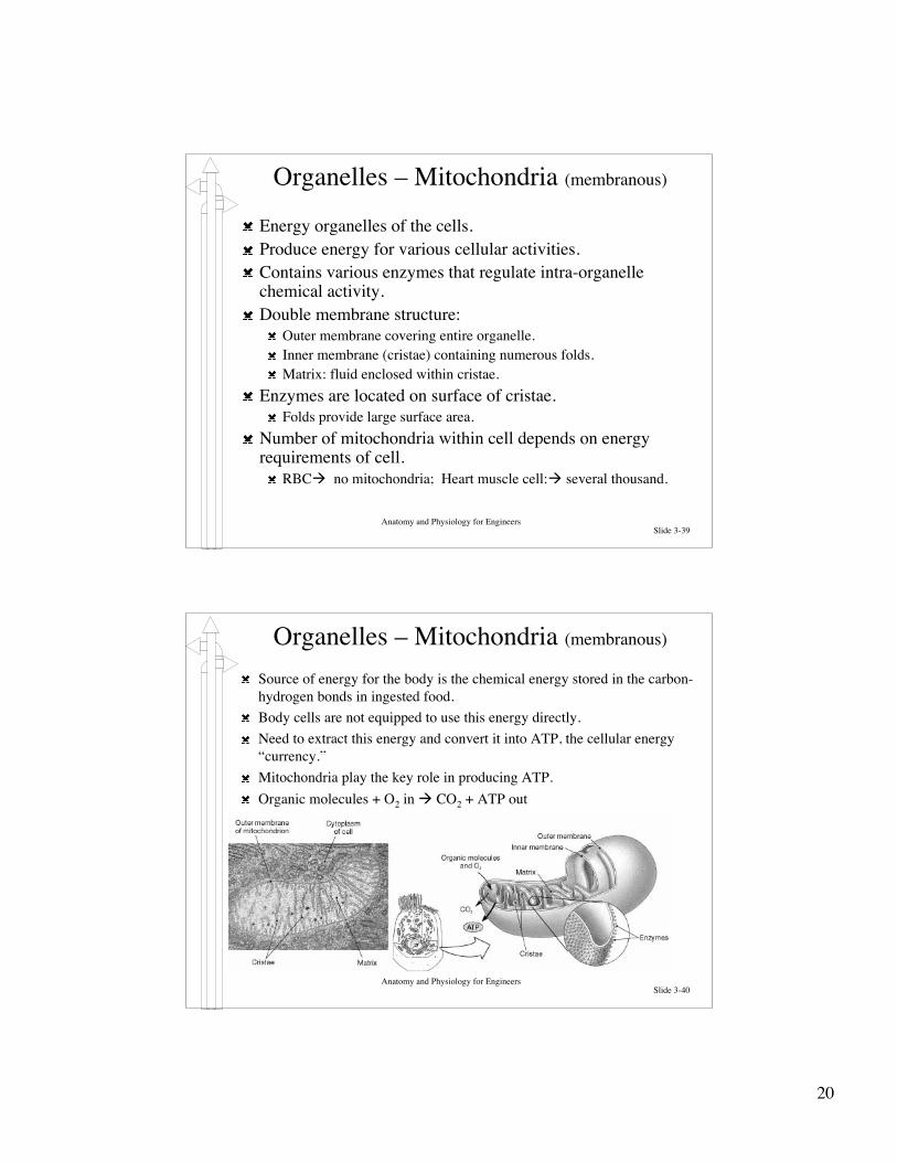
Anatomy and Physiology for EngineersSlide 3-39
Energy organelles of the cells.Produce energy for various cellular activities.Contains various enzymes that regulate intra-organellechemical activity.Double membrane structure:
Outer membrane covering entire organelle.Inner membrane (cristae) containing numerous folds.Matrix: fluid enclosed within cristae.
Enzymes are located on surface of cristae.Folds provide large surface area.
Number of mitochondria within cell depends on energyrequirements of cell.
RBC‡ no mitochondria; Heart muscle cell:‡ several thousand.
Organelles – Mitochondria (membranous)
Anatomy and Physiology for EngineersSlide 3-40
Source of energy for the body is the chemical energy stored in the carbon-hydrogen bonds in ingested food.Body cells are not equipped to use this energy directly.Need to extract this energy and convert it into ATP, the cellular energy“currency.”Mitochondria play the key role in producing ATP.Organic molecules + O2 in ‡ CO2 + ATP out
Organelles – Mitochondria (membranous)

21
Anatomy and Physiology for EngineersSlide 3-41
Control center of cellular operation.Most cells contain a single nucleus.
Exceptions (mature RBC – no nucleus, skeletal muscle cells – many nuclei).Structure: Nuclear envelope surrounding the nucleoplasm.
Nucleoplasm contains ions, enzymes, RNA & DNA nucleotides, proteins, smallamounts of RNA and DNA.
Chemical communication to cytoplasm takes place via nuclear pores: smallholes for passage of H2O, ions, small molecules (No proteins or DNA).Nucleoli: Organelles that synthesize components of ribosomes.
Organelles – Nucleus (membranous)
Anatomy and Physiology for EngineersSlide 3-42
Chromosomes (within nucleus)DNA + histones (proteins) ‡chromosomes.Each nucleus contains 23 pairschromosomes.One member of each pair fromfather; other from mother.Level of coiling depends onwhether cell is dividing or not.
Extremely tight coiling withindividing cells.Loosely coiled structures(chromatin) in non-dividingcells.
DNA contains information tocode > 100,000 differentproteins.
Organelles – Nucleus (membranous)

22
Anatomy and Physiology for EngineersSlide 3-43
Genes and the Genetic CodeBasic structure of nucleic acids was discussed earlier.
Single DNA molecule consists of a pair of strands held together byhydrogen bonding between complimentary nitrogenous bases (A-Adenosine; T-Thymine; C-Cytosine; G-Guanine).Information is stored in the sequence of the nitrogenous bases – this isthe genetic code.
Basic code segment is a triplet.Sequence of 3 bases can specify identity of a single amino acid.
Each gene consists of all triplets needed to produce a specificprotein.Since proteins consist of varying number of amino acids,number of triplets within a gene can vary, i.e., size of genecan vary.Triplets also have ‘file start’ and ‘file end’ information.
Anatomy and Physiology for EngineersSlide 3-44
Protein SynthesisEach molecule of DNA consists of 1000’s of genes‡ can code many 1000 proteins.Genes are normally tightly coiled and bound tohistones: cannot be activated.In order for gene to be activated, enzymes musttemporarily break down hydrogen bonds betweenbases and detach gene from the histones.Process of gene activation is not completelyunderstood.However, more is known about protein synthesis.
Transcription and translation.

23
Anatomy and Physiology for EngineersSlide 3-45
TranscriptionGenes are found in nucleus but protein synthesis occurs in cytoplasm(ribosomes); need a messenger (mRNA) to carry blueprint information tomanufacturing site.Process of creating mRNA is called transcription.Enzyme (RNA polymerase) binds to the initial segment of activated gene.RNA polymerase begins replicating nitrogenous basesWill code Uracil for Thiamine (U for A -- instead of T).Sequence of 3 bases on RNA ‡ codon: complementary to DNA triplet.At ‘file end’ signal, RNA polymerase detaches; mRNA strand moves throughnuclear pores.
Anatomy and Physiology for EngineersSlide 3-46
Synthesis of proteins using information encoded in mRNA.Every amino acid has at least 1 unique and specific codon.Initiated when a newly created mRNA strand binds with a ribosome.
Translation
Transfer RNA (tRNA) deliversamino acids to ribosomes forassembly of proteins (> 20 types oftRNA).1st tRNA with 1 amino acid arrivesand binds to 1st mRNA codon.2nd tRNA arrives with 2nd aminoacid‡ binds to 2nd codon.Ribosomal enzymes attach aminoacids 1 and 2 with a peptide bond,and shifts up 1 codon.Dipeptide is now attached to aminoacid # 3 arriving on 3rd tRNA.Continue adding amino acids untilstop codon is reached.Ribosome detaches.

24
Anatomy and Physiology for EngineersSlide 3-47
Cell Division and MitosisIntegral part of growth.
Single cell to 75 X 1012 cells for mature adult.Occurs via cell reproduction.Central to reproduction is accurate duplication of cell’s genetic materialinto 2 daughter cells ‡ Mitosis.Mitosis involves division of somatic cells (mature cells).Production of reproductive cells (sperm and ova) involve different kind ofcell division ‡ meiosis.Time between cell division ‡ interphase.
Majority of cell life is spent in interphase.Some cells (nerve, heart, skeletal) never leave interphase.Preparation for mitosis requires increased production of organelles andcytosol ‡ may take hours, days, weeks.
Digestive cells divide every few days; cells lining a wound only divide after aninjury.
Once preparations are complete, cell replicates DNA in the nucleus (6 – 8hours).
Anatomy and Physiology for EngineersSlide 3-48
DNA ReplicationPurpose of DNA replication is to copy genetic information in nucleus.
One set of chromosomes can go to each of the daughter cells.Process begins when complementary strands separate and unwind.DNA polymerase (enzyme) bind to exposed nitrogenous base pairs.Complementary nucleotides in nucleoplasm attach to exposed DNA bases.Two copies of the DNA molecule are thereby created; mitosis beginsshortly thereafter.

25
Anatomy and Physiology for EngineersSlide 3-49
Stages of MitosisOnce duplicated chromosomes are created, need to separate and enclose these into2 identical nuclei.Cytokinesis: separation of the cytoplasm to form 2 separate but identical cells.Mitosis involves:
Prophase:Chromosomes coil tightly.Chromatids: chromosome copies; attached at the centromere.As chromosomes appear, 2 centriole pairs move to opposite poles of the nucleus.Array of microtubules (spindle fibers) extend between centriole pairs.Prophase ends with the disappearance of the nuclear envelope.
MetaphaseSpindle fibers enter nuclear region.Chromatids attach.Chromatids move to a narrow plane at center of cell (metaphase plate).
AnaphaseCentromere of each chromatid pair splits.2 daughter chromosomes move to opposite sides of cell.
TelophaseTwo nuclear membranes form.Nuclei enlarge.Chromosomes uncoil.
Anatomy and Physiology for EngineersSlide 3-50
Stages of MitosisOnce duplicated chromosomes are created, need to separate and enclosethese into 2 identical nuclei.Cytokinesis: separation of the cytoplasm to form 2 separate but identicalcells.Mitosis involves:
Prophase:Chromosomes coil tightly.Chromatids: chromosome copies;attached at the centromere.As chromosomes appear, 2centriole pairs move to oppositepoles of the nucleus.Array of microtubules (spindlefibers) extend between centriolepairs.Prophase ends with thedisappearance of the nuclearenvelope.

26
Anatomy and Physiology for EngineersSlide 3-51
Stages of Mitosis
Mitosis involves:Prophase:Metaphase
Spindle fibers enternuclear region.Chromatids attach.Chromatids moveto a narrow planeat center of cell(metaphase plate).
Anatomy and Physiology for EngineersSlide 3-52
Stages of MitosisMitosis involves:
Prophase:MetaphaseAnaphase
Centromere ofeach chromatidpair splits.2 daughterchromosomesmove to oppositesides of cell.

27
Anatomy and Physiology for EngineersSlide 3-53
Stages of Mitosis
Mitosis involves:Prophase:MetaphaseAnaphaseTelophase
Two nuclearmembranes form.Nuclei enlarge.Chromosomes uncoil.
Anatomy and Physiology for EngineersSlide 3-54
Cytokinesis
Once daughter chromosomes separate, cellpinches off via constriction of the cytoplasmalong the metaphase plate.After nuclear membranes are formed, cellpinches off completely, forming 2 separatecells.Completion of cytokinesis forms end of theprocess of cell division.

28
Anatomy and Physiology for EngineersSlide 3-55
Mitosis