Embed Size (px)
Citation preview
Cell Unit I: Organelles and Cell Membranes - Chapters 7 and 8
Chapter 7: A Tour of the Cell• Themes: THE CELL, Structure and Function,
Evolution, Regulation, Heritable Information, • Objectives:
– Microscopes– Organelles– Prokaryotic vs. Eukaryotic– Membranes– Cytoskeleton– Cell Surface and Junctions
Root Words• Centro-• Chloro-• Cili-• Cyto –• -ell• Endo –• Eu –• Extra-• Flagell-• Glyco –• -tubul• -oid• - pod• Thylaco -
• Lamin – • Lyso –• Micro – • Nucle –• Phago –• Plasm –• Pro-• -karyo• -soma• -kytos• -desma• Pseudo – • Tono – • -plast
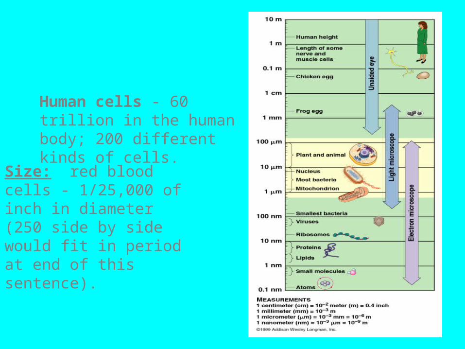
Note: MOST CELLS ARE BETWEEN 1 & 100 MICROMETERS.
Human cells - 60 trillion in the human body; 200 different kinds of cells.
Size: red blood cells - 1/25,000 of inch in diameter (250 side by side would fit in period at end of this sentence).
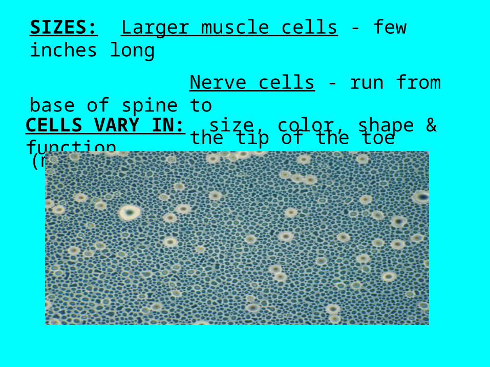
SIZES: Larger muscle cells - few inches long
Nerve cells - run from base of spine to
the tip of the toe (many feet long!!)CELLS VARY IN: size, color, shape & function.
Since all living things have cells, cells hold the secrets to many of life’s most intriguing questions:
(A) Aging (telomeres)(B) Cures to most if not all diseases. Example:
4,200 human diseases are caused by a defect in a single gene (which lead to mis-shapened proteins.)
(C) Stem cell research. (D) Evolutionary questions & problems (E) Behavior of cells.(F) Cancer
CYTOLOGY: The study of cells.
MICROSCOPY: The art of examining objects under the microscope.
Electron Microscopes (EM). 2 types:
(A) Transmission electron microscope (TEM)
(B) Scanning electron microscope (SEM)
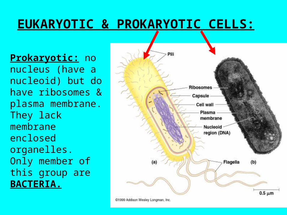
EUKARYOTIC & PROKARYOTIC CELLS:
Prokaryotic: no nucleus (have a nucleoid) but do have ribosomes & plasma membrane. They lack membrane enclosed organelles. Only member of this group are BACTERIA.
EUKAROYTIC CELLS: Have extensive and elaborately arranged internal membranes surrounding their organelles to allow for COMPARTMENTALIZATION.This allows different metabolic processes to
go on simultaneously inside the cell without one process interfering with another.
EX: The lysosome digests macromolecules that are worn out or no longer needed. The enzymes that do the digesting need a pH of around 5.
What problems would be caused if this organelle did not have a membrane????
NUCLEUS & PARTS: Nucleolus - Components of ribosome (precursors) are synthesized. Remember - Nuclear envelope is a double membrane as is all membranes. (See Fig. 7.6). CHROMATIN: Contains nuclear DNA. When a cell divides this becomes the chromosomes.
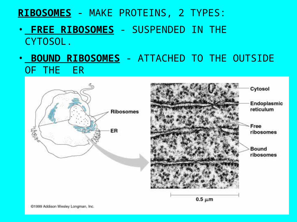
RIBOSOMES - MAKE PROTEINS, 2 TYPES:
• FREE RIBOSOMES - SUSPENDED IN THE CYTOSOL.
• BOUND RIBOSOMES - ATTACHED TO THE OUTSIDE OF THE ER
ENDOMEMBRANE SYSTEM: RELATED THROUGH DIRECT PHYSICAL CONTINUITY OR BY THE TRANSFER OF MEMBRANE SEGMENTS AS TINY VESICLES (MEMBRANE ENCLOSED SACS).
Include the nuclear envelope, ER, Golgi apparatus, lysosomes, various kinds of vacuoles, and the plasma membrane.
ENDOPLASMIC RETICULUM: Made of membranous tubules and sacs called cisternae.
SMOOTH ER: No ribosomes.
ROUGH ER: Ribosomes on the outside edge.
Index Cards• On each card, draw a picture and write a
description:– Peroxisome– Centrosome– Microfilaments and Microtubules– Plasmodesmata– Tonoplast– Plastid
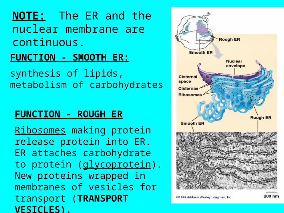
NOTE: The ER and the nuclear membrane are continuous.FUNCTION - SMOOTH ER:
synthesis of lipids, metabolism of carbohydrates
FUNCTION - ROUGH ER
Ribosomes making protein release protein into ER. ER attaches carbohydrate to protein (glycoprotein). New proteins wrapped in membranes of vesicles for transport (TRANSPORT VESICLES).
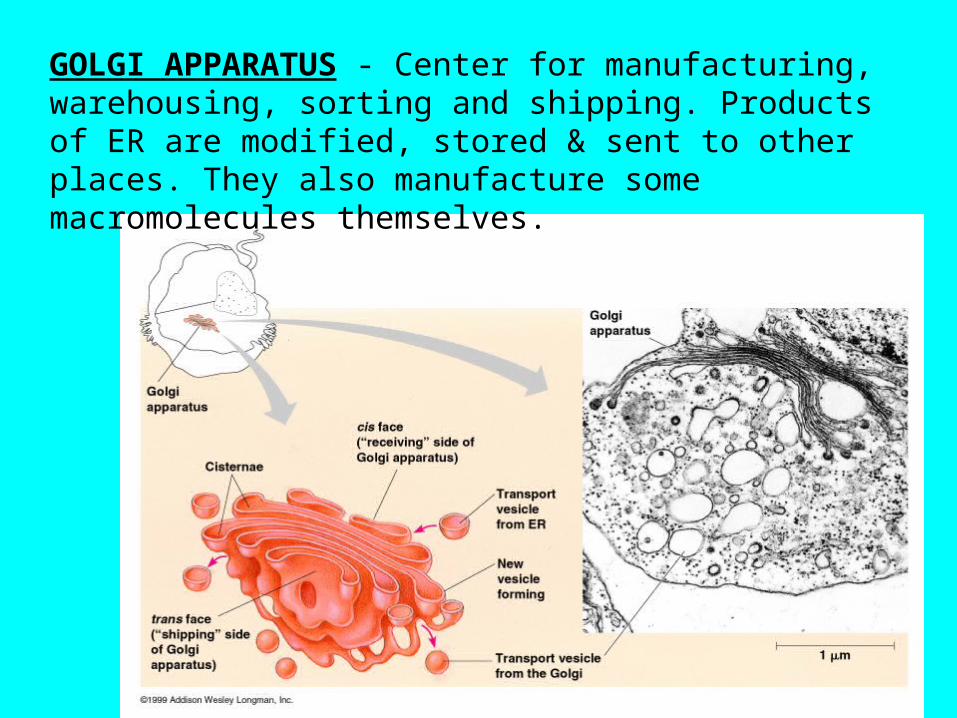
GOLGI APPARATUS - Center for manufacturing, warehousing, sorting and shipping. Products of ER are modified, stored & sent to other places. They also manufacture some macromolecules themselves.
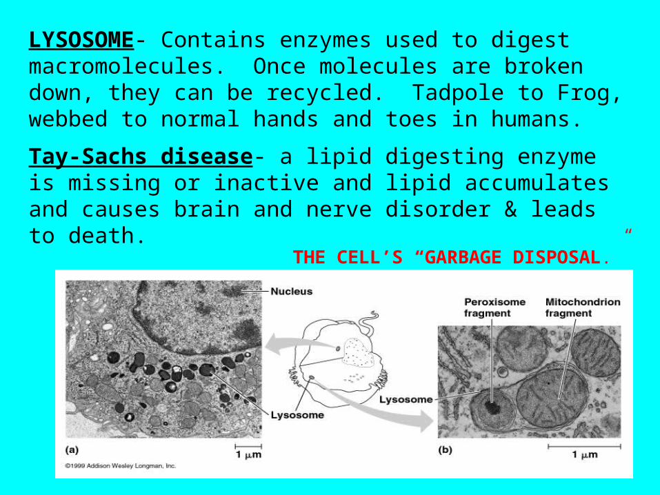
LYSOSOME- Contains enzymes used to digest macromolecules. Once molecules are broken down, they can be recycled. Tadpole to Frog, webbed to normal hands and toes in humans.
Tay-Sachs disease- a lipid digesting enzyme is missing or inactive and lipid accumulates and causes brain and nerve disorder & leads to death.
THE CELL’S “GARBAGE DISPOSAL.”
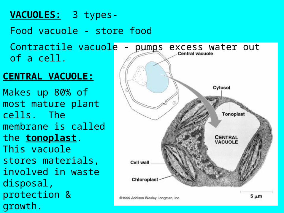
VACUOLES: 3 types-
Food vacuole - store food
Contractile vacuole - pumps excess water out of a cell.
CENTRAL VACUOLE:
Makes up 80% of most mature plant cells. The membrane is called the tonoplast. This vacuole stores materials, involved in waste disposal, protection & growth.
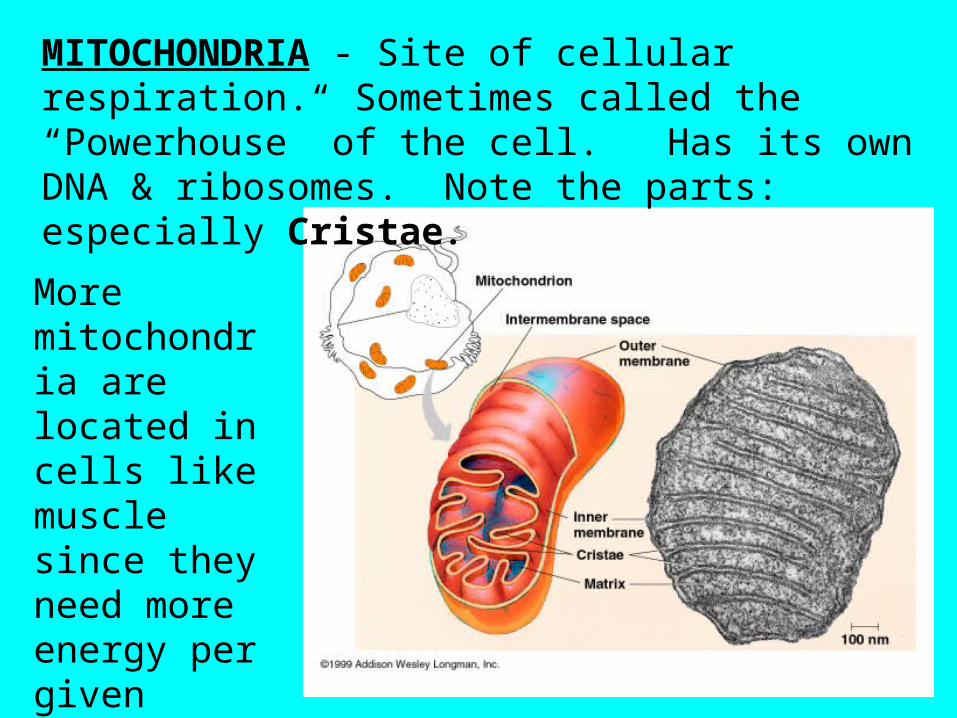
MITOCHONDRIA - Site of cellular respiration. Sometimes called the “Powerhouse” of the cell. Has its own DNA & ribosomes. Note the parts: especially Cristae.
More mitochondria are located in cells like muscle since they need more energy per given time.
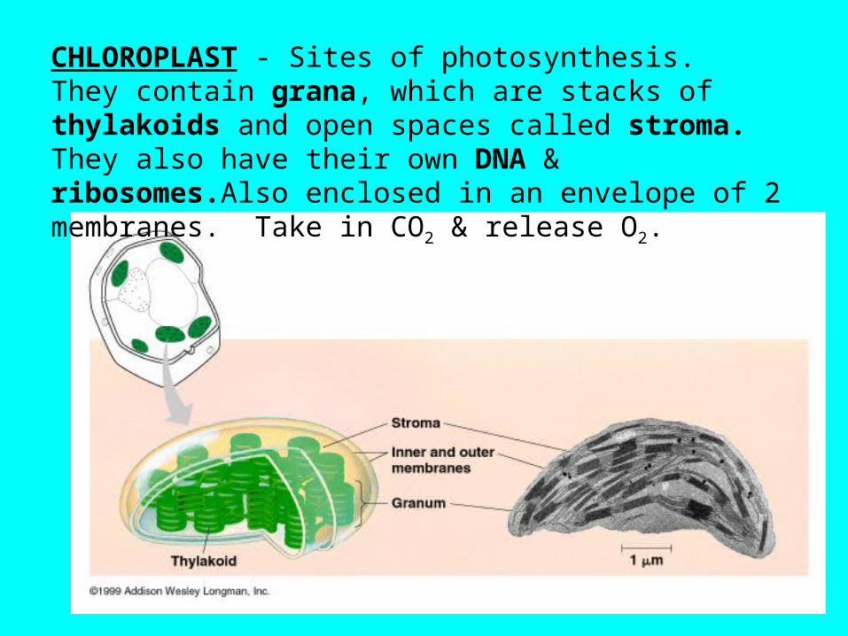
CHLOROPLAST - Sites of photosynthesis. They contain grana, which are stacks of thylakoids and open spaces called stroma. They also have their own DNA & ribosomes.Also enclosed in an envelope of 2 membranes. Take in CO2 & release O2.
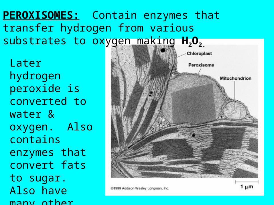
PEROXISOMES: Contain enzymes that transfer hydrogen from various substrates to oxygen making H2O2.
Later hydrogen peroxide is converted to water & oxygen. Also contains enzymes that convert fats to sugar. Also have many other functions.
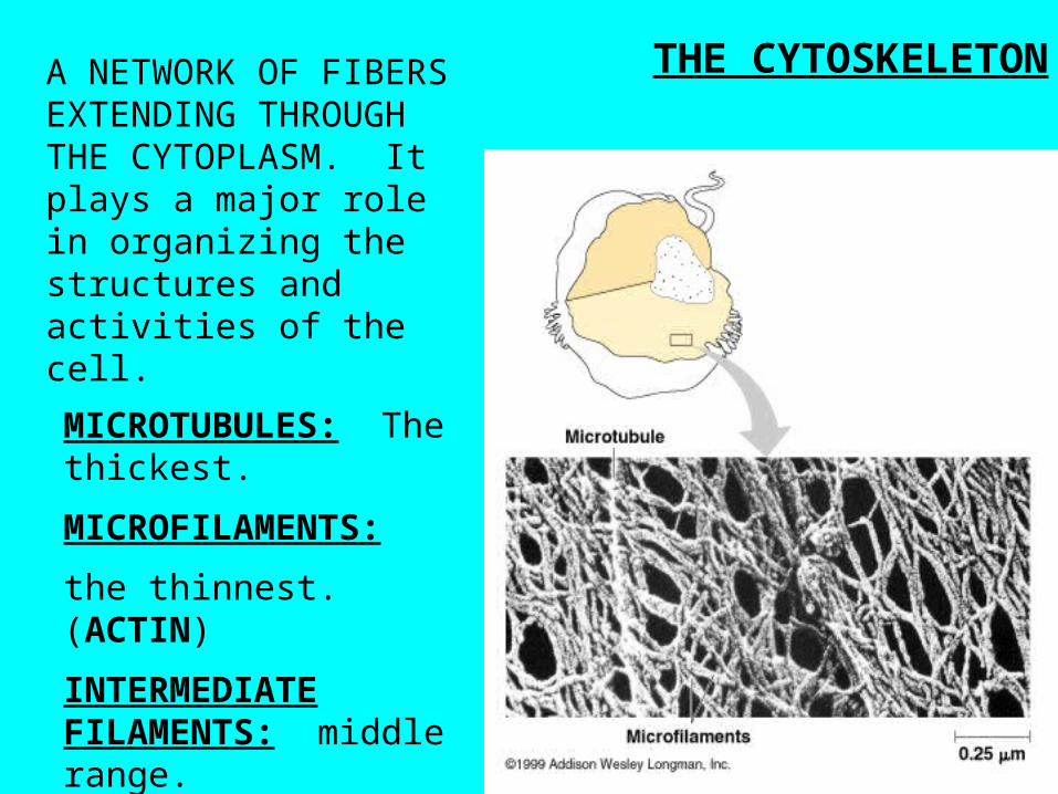
THE CYTOSKELETONA NETWORK OF FIBERS EXTENDING THROUGH THE CYTOPLASM. It plays a major role in organizing the structures and activities of the cell.
MICROTUBULES: The thickest.
MICROFILAMENTS:
the thinnest. (ACTIN)
INTERMEDIATE FILAMENTS: middle range.
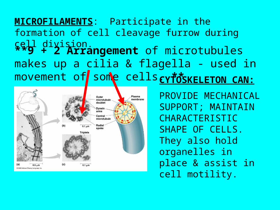
MICROFILAMENTS: Participate in the formation of cell cleavage furrow during cell division.
**9 + 2 Arrangement of microtubules makes up a cilia & flagella - used in movement of some cells. **CYTOSKELETON
CAN:
PROVIDE MECHANICAL SUPPORT; MAINTAIN CHARACTERISTIC SHAPE OF CELLS. They also hold organelles in place & assist in cell motility.
CELL SURFACES & JUNCTIONS:-----------------------------------------------------------
CELL WALLS: (Plants, Prokaryotes, Fungi & some Protists)
Young Plant: Primary cell wall.
Middle lamella; between primary walls of adjacent cells. This “glues” the cells together.
Secondary cell wall - between the plasma membrane & the primary wall. Innermost portion of a mature plant cell wall.
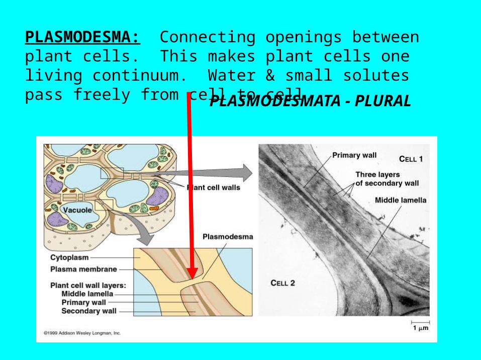
PLASMODESMA: Connecting openings between plant cells. This makes plant cells one living continuum. Water & small solutes pass freely from cell to cell. PLASMODESMATA - PLURAL
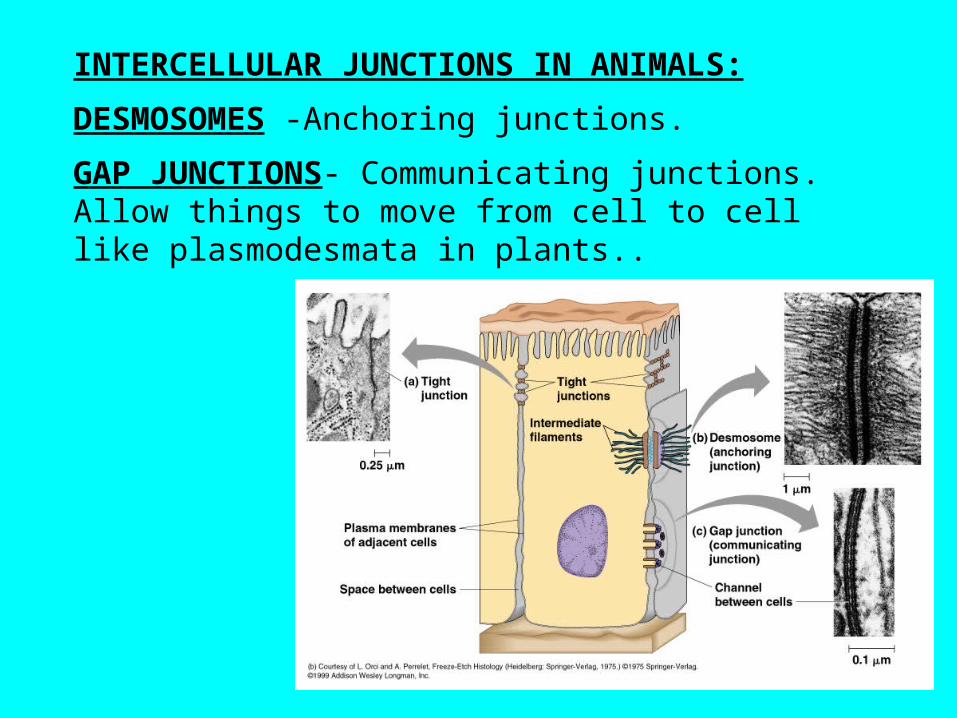
INTERCELLULAR JUNCTIONS IN ANIMALS:
DESMOSOMES -Anchoring junctions.
GAP JUNCTIONS- Communicating junctions. Allow things to move from cell to cell like plasmodesmata in plants..
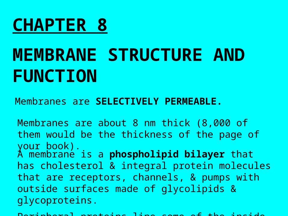
CHAPTER 8
MEMBRANE STRUCTURE AND FUNCTIONMembranes are SELECTIVELY PERMEABLE.
Membranes are about 8 nm thick (8,000 of them would be the thickness of the page of your book).
A membrane is a phospholipid bilayer that has cholesterol & integral protein molecules that are receptors, channels, & pumps with outside surfaces made of glycolipids & glycoproteins.
Peripheral proteins line some of the inside of the cell.
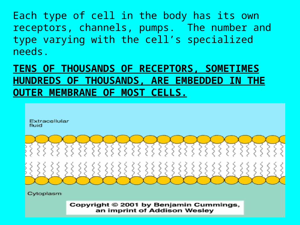
Each type of cell in the body has its own receptors, channels, pumps. The number and type varying with the cell’s specialized needs.
TENS OF THOUSANDS OF RECEPTORS, SOMETIMES HUNDREDS OF THOUSANDS, ARE EMBEDDED IN THE OUTER MEMBRANE OF MOST CELLS.
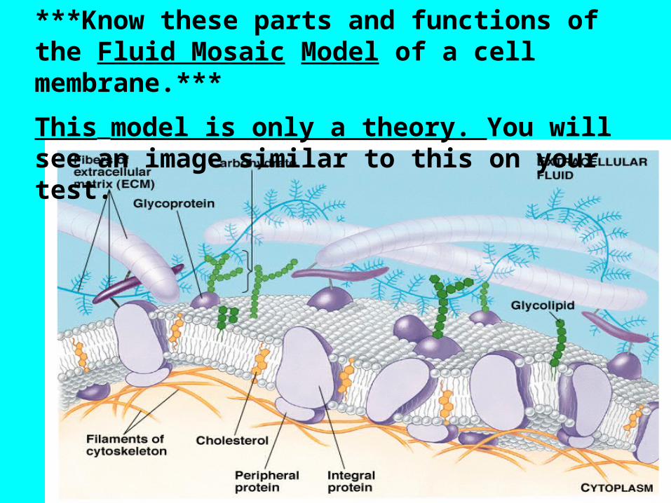
***Know these parts and functions of the Fluid Mosaic Model of a cell membrane.***
This model is only a theory. You will see an image similar to this on your test.
Important concepts to know about cell membrane structure:
• The membrane is called fluid because it moves
• The phospholipids move laterally along the plane of the membrane. (Membranes are asymmetric & fluid)• Glycoproteins & Glycolipids in animal cells are used to recognize similar and different cells. (Glyco- means a sugar has been added to the molecule).• Cholesterol in animal cells is used to maintain membrane fluidity & stabilize the membrane. Especially helps cells stay fluid when temperatures drop.• Integral proteins (embedded in the membrane) help cells recognize each other. These are usually glycoproteins. (Usually transmembrane proteins).
Oligosaccharides (short polysaccharides) attached to lipids are called glycolipids; attached to proteins, they are called glycoproteins. (These aid in cell to cell recognition).MEMBRANE POTENTIAL: All cells have voltage across their membrane. (The inside is (-) compared to the outside).
IONS - Pass through a membrane down an ELECTROCHEMICAL GRADIENT. (Imp. Concept)
Outside of membrane - proteins are attached to fibers of the EXTRACELLULAR MATRIX.(ECM).Small polar molecules that are uncharged like water & ethanol can pass easily through the lipid membrane as well as small molecules (O2 )

Fig. 8.9 Some Functions of membrane proteins:A single protein can sometimes perform combinations of these functions.
------------------------------------------------------------
Other concepts about membrane (Due to Lipid bilayer):• Hydrophobic
molecules (CO2 & O2) can dissolve membrane & cross it.
Relatively impermeable to ions (Like H+ & Na+)
Cell Signaling (Chapter 11)
Signal Transduction Pathway• Hormones -
3 Stages of Cell Signaling:
• Reception• Transduction• Response
Cell Signaling• Ligand Gated Ion Channels –
– Ligands are small molecules that bind to a specific larger one
– Channels are part of plasma membrane that allows the flow or blockage of ions (Ca2+)
– EX: Synapse between nerve cells
History of Cell Signaling
• How do yeasts and sex show the history of cell signaling? (5 lines) Page 197
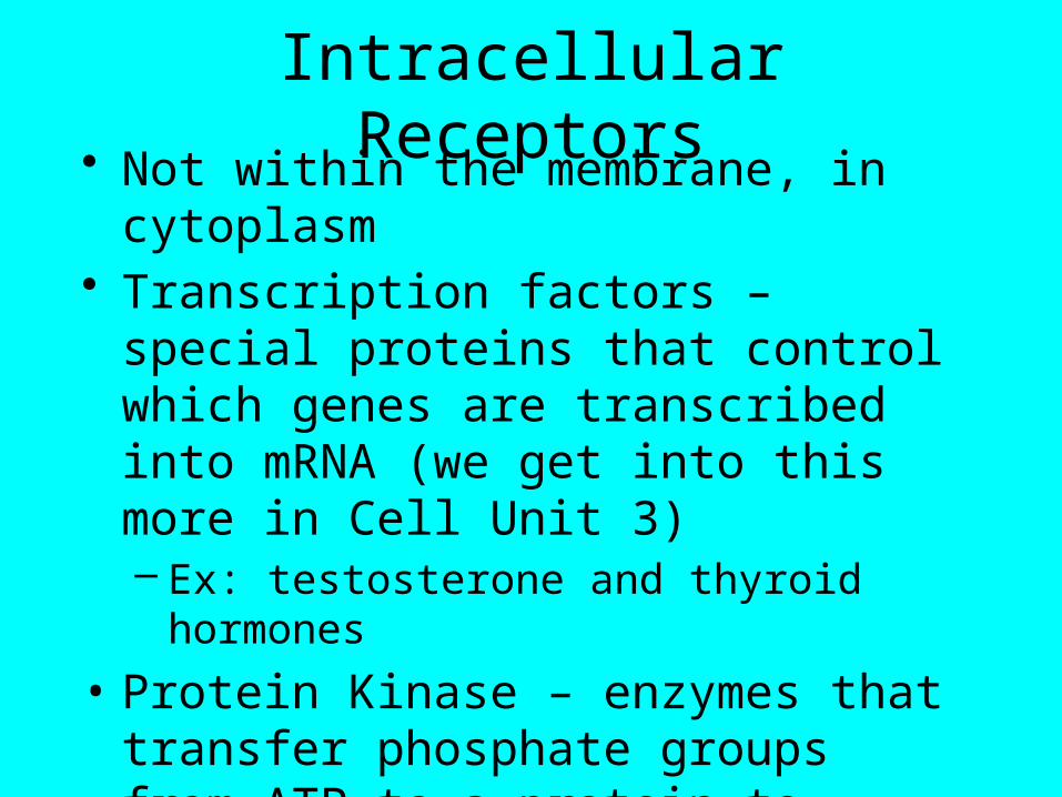
Intracellular Receptors• Not within the membrane, in cytoplasm• Transcription factors – special proteins that
control which genes are transcribed into mRNA (we get into this more in Cell Unit 3)– Ex: testosterone and thyroid hormones
• Protein Kinase – enzymes that transfer phosphate groups from ATP to a protein to activate it (phosporylation)
• Review Sutherland’s work with Cyclic AMP
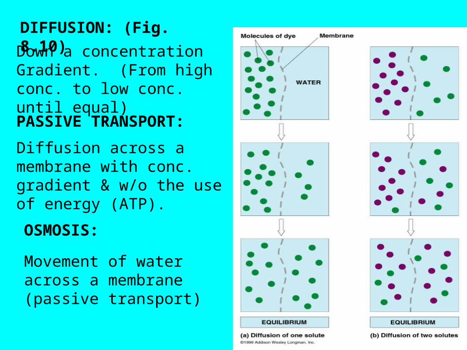
DIFFUSION: (Fig. 8.10)
Down a concentration Gradient. (From high conc. to low conc. until equal)
PASSIVE TRANSPORT:
Diffusion across a membrane with conc. gradient & w/o the use of energy (ATP).
OSMOSIS:
Movement of water across a membrane (passive transport)
AP LAB: #1
Osmosis and Diffusion
OSMOSIS & THE PASSIVE TRANSPORT OF WATER:HYPERTONIC (Solution). One that has higher concentration of solute than solvent compared to another solution.
HYPOTONIC (Solution). One that has a lower concentration of solute than solvent compared to another solution.
EXAMPLE: Tap water is hypertonic to distilled water but hypotonic to seawater. These terms are relative terms & only meaningful in a comparative sense.
ISOTONIC (Solution). 2 Solutions are equal in solute concentration.
Remember Lab. 1 !!
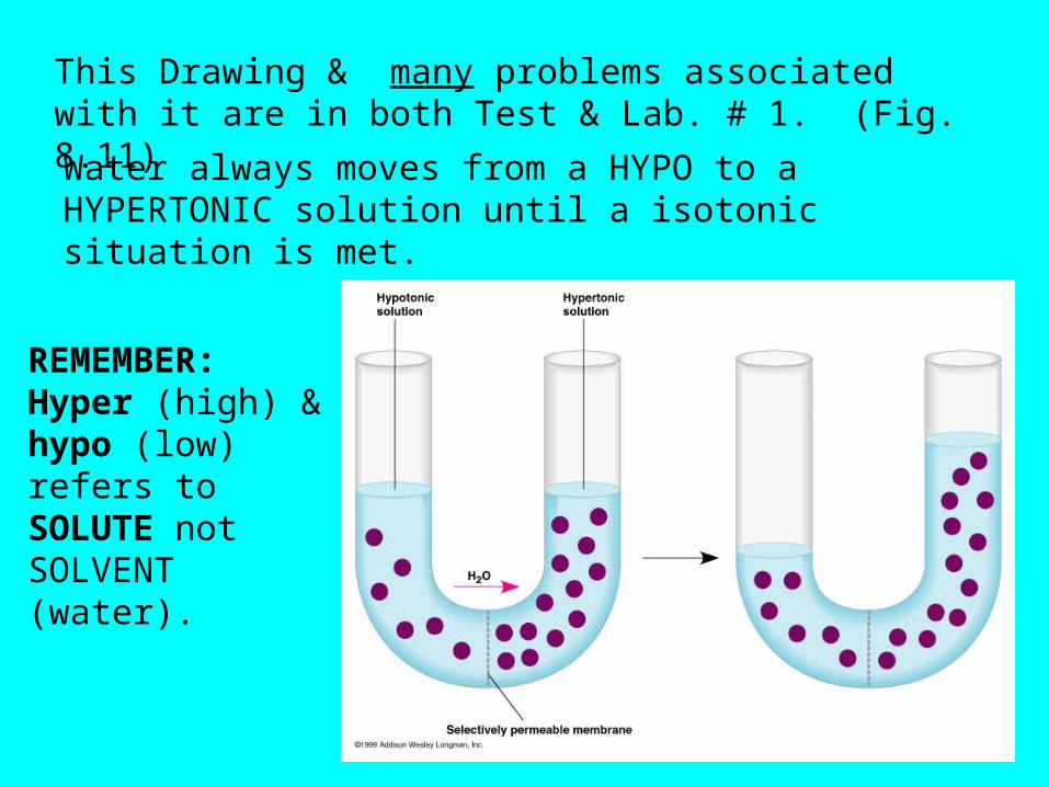
This Drawing & many problems associated with it are in both Test & Lab. # 1. (Fig. 8.11)
Water always moves from a HYPO to a HYPERTONIC solution until a isotonic situation is met.
REMEMBER: Hyper (high) & hypo (low) refers to SOLUTE not SOLVENT (water).
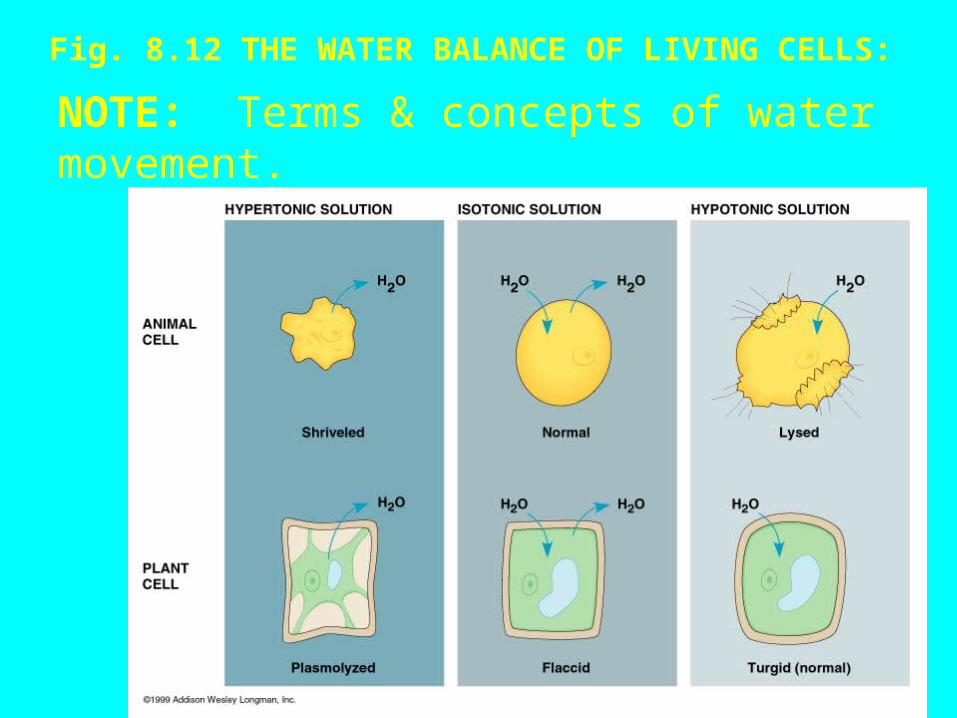
Fig. 8.12 THE WATER BALANCE OF LIVING CELLS:NOTE: Terms & concepts of water movement.
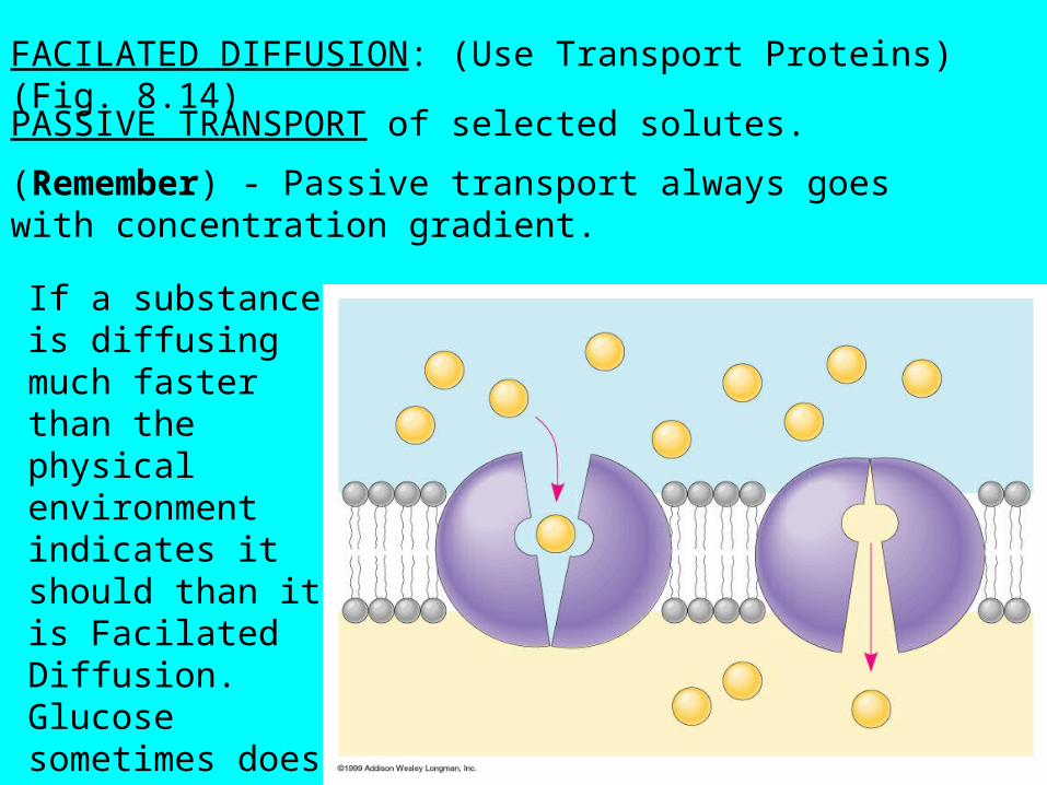
FACILATED DIFFUSION: (Use Transport Proteins) (Fig. 8.14)
PASSIVE TRANSPORT of selected solutes.
(Remember) - Passive transport always goes with concentration gradient.
If a substance is diffusing much faster than the physical environment indicates it should than it is Facilated Diffusion. Glucose sometimes does this.
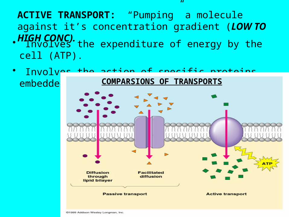
ACTIVE TRANSPORT: “Pumping” a molecule against it’s concentration gradient (LOW TO HIGH CONC).
• Involves the expenditure of energy by the cell (ATP).
• Involves the action of specific proteins embedded in the membrane. COMPARSIONS OF TRANSPORTS
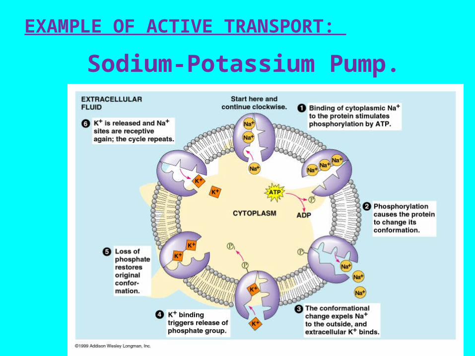
EXAMPLE OF ACTIVE TRANSPORT:
Sodium-Potassium Pump.
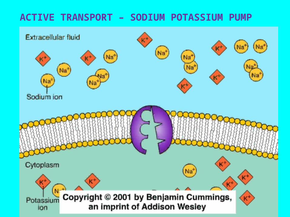
ACTIVE TRANSPORT – SODIUM POTASSIUM PUMP
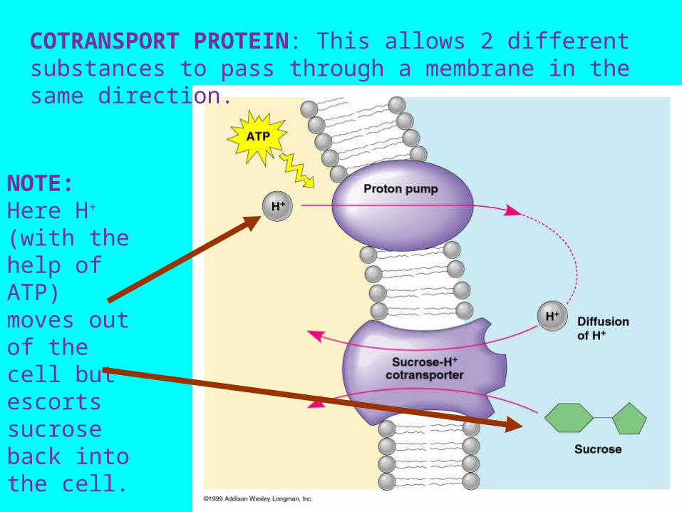
COTRANSPORT PROTEIN: This allows 2 different substances to pass through a membrane in the same direction.
NOTE: Here H+
(with the help of ATP) moves out of the cell but escorts sucrose back into the cell.
Test Hints: Use your CD & look at all the videos.
Make sure your study Fig. 8.18 and know the difference between:
Exocytosis & EndocytosisTypes of Endocytosis: Phagocytosis & Pinocytosis.