Embed Size (px)
Citation preview
���������������� ������ � ��������������������������
�������������
���� ����������
�

Md. Imtiyaz Aslam et. al. (2013) Int J Appl Sci Biotechnol, Vol 1(4): 208-213
DOI: 10.3126/ijasbt.v1i4.�9104 �
���������������� ������ � ��������������������������
Research Article
EFFECT OF POLY (N-ISOPROPYLACRYLAMIDE) “PNIPAM” ON HEPATIC
CELLS OF SWISS ALBINO MICE, MUS MUSCULUS
Md. Imtiyaz Aslam, Rohit Kumar Verma, Richa Roy and S. P. Roy* P.G. Department of Biotechnology, T. M. Bhagalpur University, Bhagalpur-812007
*Corresponding author e-mail: [email protected]
Abstract
The present paper deals with the effect of polymeric compound poly (N-isopropyl acrylamide) “PNIPAM” for their toxicity on
hepatic cells. The nanoparticle is a xenobiotic compound that accumulates in the liver for their metabolism. Non-metabolizing
xenobiotic compounds such as “PNIPAM” produces anomalies in the hepatic cells. Certain enzymes such as ALT and ALP can be
assayed for the hepatocytic toxicity. An attempt has been made to know the toxic effect of “PNIPAM” in a concentration of
0.8mg/ml on the hepatic cells of Swiss Albino mice, Mus musculus. The ALT and ALP analysis were performed through test kits for
their quantitative estimation. The histological result shows that several lesions were produced after the introduction of aqueous
solution of PNIPAM for an incubation period of 48 hours. The toxicity was confirmed Spectrophotometrically by the assessment of
enzyme ALT and ALP. The increased concentration of ALT (55.0 IU/L) and slight decrease in ALP (40.0 IU/L) concentration was
responsible for the metabolic alteration and production of hepatocytic anomalies in the mice.
Keywords: PNIPAM, Polymeric compound, Nanoparticle, ALT, ALP, Hepatocytes, Toxicity.
Introduction
“PNIPAM”, poly (N-isopropyl acrylamide) is a
polymeric nanoparticle. It is also called as “Smart
Polymers” (Galaev and Mattiasson, 1999, Aslam et al.,
2013). They are considered as an important class of
polymeric materials having several important
biomedical and industrial applications. These polymers
undergo sharp reversible phase transition in response
to small changes in relation to environmental stimuli
such as pH, temperature; ionic strength etc. Poly (N-
isopropyl acrylamide) “PNIPAM” is one of the typical
examples that have been widely studied for various
biotechnological applications. It has a lower critical
solution temperature (LCST) of precipitation around
32.580C in water and changes reversibly from
hydrophilic below this temperature to hydrophobic
above it. The reversible phase transition of “PNIPAM”
can be used for colloid suspension formation together
with an antigen or any other biomolecule as the
temperature is increased above the LCST, thus,
providing the possibility of their use as an adjuvant in
combination with a specific antigen.
The perusal of pertinent literature in this field reveals
that considerable information is available on the role of
ALT and ALP for knowing the liver and pancreas
dysfunction (Hillesheim et. al., 1995, Gebhardt, 1992
and Gebhardt and Gaunitz, 1997, Kikawa, 2006; Wang
et al., 2012).
However, practically no information is available on the
effect of “PNIPAM” on the hepatic cells, although
much work has been done on its application for
toxicity assays (Schild, 1992; Galaev and Mattiasson,
1999; Kumar et al., 2007 and Aslam et al., 2013). The
objective of the present study is to pin point the effect
of “PNIPAM” on the hepatic tissues of the Swiss
Albino mice, Mus musculus for assessing the extent of
hepatotoxicity.
ALT is known as Alanine Transaminase or alanine
aminotransferase (ALAT) or serum glutamic pyruvic
transaminase (sGPT). It is a homodimeric cytoplasmic
pyridoxal phosphate-dependent enzyme. It involved in
cellular nitrogen metabolism, amino acid metabolism,
and liver gluconeogenesis (Gebhardt, 1992). ALT
mediates conversion of major intermediate
metabolites, catalyzing reversible transamination
between alanine and �-ketoglutarate to form pyruvate
and glutamate (Lott et.al, 1986). ALT is widely
distributed in many tissues but is found in greatest
abundance in the liver, and to a much lesser extent in
the kidneys, heart, and brain (Gebhardt et.al, 1997).
The major role of ALT in the liver is the conversion of
alanine to glucose which is then exported to the body
to be utilized in a multitude of processes. Serum ALT
levels are generally low, but may spike during disease
states or in the event of tissue injury (Amacher,
1998). As such, ALT levels are routinely used as
��������� ����� ������� ������������������������� ����� � � � � � � � � �������� !"�#� �
� � �
Md. Imtiyaz Aslam et. al. (2013) Int J Appl Sci Biotechnol, Vol 1(4): 208-213
���������������� ������ � ��������������������������
indicators of medical issues, particularly liver diseases.
An increased level of ALT can be seen in patients with
diabetes, cirrhosis, fatty liver disease, and hepatitis.
Alanine Transaminase Assay provides ALT activity in
serum, plasma, tissue samples, and cell lysates (Wang
et al., 2012). Measurement of the ALT activity is
carried out by monitoring the rate of NADH oxidation
in a coupled reaction system employing lactate
dehydrogenase (LDH). The oxidation of NADH to
NAD+ is accompanied by a decrease in absorbance at
340 nm (Dawson, 1985). Under circumstances in
which the ALT activity is rate limiting, the rate
decrease is directly proportional to the ALT activity in
the sample.
Alkaline phosphatase (ALP) catalyzes the hydrolysis
of phosphate esters in alkaline buffer and produces an
organic radical and inorganic phosphate (Ambler et al.,
1970 and Willamson ,1972). Changes in alkaline
phosphatase level and activity are associated with
various disease states in the liver and bone. The
statistical validation through Student’s t-test for the
data related to ALP and ALT using softwares provided
online by different servers using the usual method of
calculation (Buda and Jarynowski, 2010). The
correlation between ALP and ALT can be made by
Pearson correlation using the statistical tool of
Wessa.net.
Materials and Methods
Model Organism
The model organism Swiss Albino mice, Mus
musculus balb/C strain is widely used to study toxic
effects on various organs such as liver, kidney,
pancreas, testis etc. The Swiss Albino mice, Mus
musculus balb/C strain is easily available, culturable
and ethically approved. 4 female Swiss albino mice,
Mus musculus Balb/C strain with average body weight
ranging from 25.0g to 30.0g were obtained from
Animal house of the University Department of
Zoology, T. M. Bhagalpur University, Bhagalpur,
India. Mice were fed with the help of ad libitum
(prepared mixed formulated feed by the laboratory
itself). Animals were housed in colony rooms with 12
hrs light/dark cycle at 30 ± 200C in the Post-Graduate
Department of Biotechnology, T.M. Bhagalpur
University, Bhagalpur. Approval of Institutional
Ethical Committee was sought prior to the
commencement of experiment.
Chemicals
“PNIPAM”, was purchased from Sigma Aldarich. The
ALT (Alanine Transaminase Activity) Assay Kit and
ALP (Alkaline Phosphatase) Assay Kit were purchased
from Cayman Chemicals and Abcam Chemicals
respectively.
Treatment Protocol
The four mice were grouped into test and control
containing two mice /group. The mice were fed upon
normal diet containing carbohydrate and protein
sources. After the rearing of fifteen days test group
were introduced with 0.8mg/ml of “PNIPAM”
nanopolymer. 24 hours starvation was performed
before the introduction of “PNIPAM” in test group
along with control. The same procedure was repeated
after every two days for thirteen days. “PNIPAM” was
introduced orally as aqueous solution having a
concentration of 0.8 mg/ml.
After thirteen days of dosing the test groups along with
control the animals were sacrificed to isolate liver.
Liver samples were taken and placed into formalin
solution. Small pieces of the liver tissues were
removed from the formalin solution and then placed
into the alcohol solution with an increasing percentage
of solubility (70%, 80%, 90%, and 100% w/w) for the
dehydration of samples, then after tissues were kept
into destaining xylene solution. Afterwards, the
samples were mounted into paraffin wax for
microtomy. The samples were cut into pieces of 3–5
nm. The tissue samples were stained with eosin for
microscopic examination. The slides were used for
microscopic analysis for the morphological anomalies
of hepatocytes. For enzymatic assessment the blood
samples were taken from supraorbital plexes. The
blood sample was mixed with EDTA to prevent
clotting and then centrifuged at 5000rpm for 10
minutes to isolate supernatant. Cayman’s Alanine
Transaminase Assay Kit was used for detecting ALT
activity in Supernatant (plasma). Measurement of the
ALT activity is carried out by monitoring the rate of
NADH oxidation in a coupled reaction system
employing lactate dehydrogenase (LDH). The
oxidation of NADH to NAD+is accompanied by a
decrease in absorbance at 340 nm.
Abcam’s Alkaline Phosphatase Assay Kit
(Colorimetric) was used to measure ALP activity in
plasma. Abcam’s Alkaline Phosphatase Assay Kit
(Colorimetric) is a highly sensitive, simple, direct and
HTS-ready colorimetric assay designed to measure
ALP activity in serum and biological samples. It
contains 10 substrate tablets providing convenience for
multiple usages. The kit uses p-nitrophenyl phosphate
(pNPP) as a phosphatase substrate which turns yellow
(�max= 405 nm) when dephosphorylated by ALP. The
Kit can detect 10-250 �U ALP in samples. � � �
Md. Imtiyaz Aslam et. al. (2013) Int J Appl Sci Biotechnol, Vol 1(4): 208-213
���������������� ������ � ��������������������������
The kit uses p-nitrophenyl phosphate (pNPP) as a
phosphatase substrate which turns yellow (�max= 405
nm) when dephosphorylated by ALP.
With the help of the alkaline phosphatase (ALP), ALT,
aspartate aminotransferase (AST) enzyme recognition
kits and the relative spectrometer functions, the
relative numbers for the enzymes were studied and
analyzed. Student’s’s t-test was used to estimate the
differences.
Assessment of hepatotoxicity
Hepatotoxicity was analyzed at both morphological
and physiological level. The alteration in tissue
organization depicted by various microbiological slides
at morphological level.
Statistical Analysis
The data obtained were analyzed for their statistical
validity and correlation using Online Statistical tools
provided by University of Delaware and Wessa.net for
the calculation of Student’s t- test and Pearson
correlation. These tools provide the validation
parameters and correlation values (-1, 0, +1).
Results
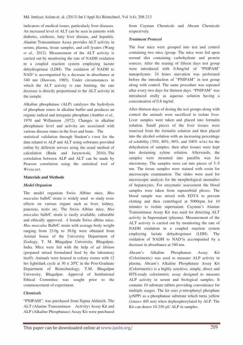
The effect of “PNIPAM” on hepatic cells of Swiss
albino mice, Mus musculus is depicted through
microphotographs showing control and in shots 1.0 -
4.0 respectively (Fig-1). The test slides show necrotic
regions in patches as compared with the control. Table-
1 shows variation in ALP and ALT concentration on
the different dosing days. It was observed that there
was a decrease in ALP concentration and increase in
ALT concentration was observed on different dosing
days. The data was tested through Student’s t-test
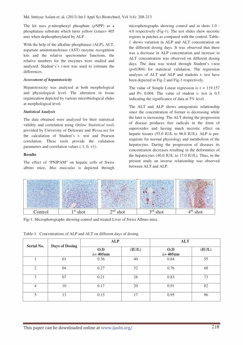
(p=0.004) for statistical validation. The regression
analyses of ALT and ALP and students t- test have
been depicted in Fig-2 and Fig-3 respectively.
The value of Simple Linear regression is r = 119.157
and P= 0.004. The value of student t- test is 0.5
indicating the significance of data at 5% level.
The ALT and ALP shows antagonistic relationship
since the concentration of former is decreasing while
the later is increasing. The ALT during the progression
of disease produces free radicals in the form of
superoxides and having much necrotic effect on
hepatic tissues (55.0 IU/L to 96.0 IU/L). ALP is pre-
requisite for normal physiology and metabolism of the
hepatocytes. During the progression of diseases its
concentration decreases resulting in the deformities of
the hepatocytes (40.0 IU/L to 17.0 IU/L). Thus, in the
present study an inverse relationship was observed
between ALT and ALP.
Control 1st shot 2nd shot 3rd shot 4th shot
Fig-1: Microphotographs showing control and treated Liver of Swiss Albino mice.
Table-1: Concentrations of ALP and ALT on different days of dosing.
Serial No.
Days of Dosing
ALP ALT
O.D
�= 405nm
(IU/L) O.D
�= 405nm
(IU/L)
1 01 0.36 40 0.64 55
2 04 0.27 32 0.76 68
3 07 0.21 28 0.83 73
4 10 0.17 20 0.91 82
5 13 0.15 17 0.95 96
� � �
���������������� ������ � ��������������������������
Fig-2: Regression graph of ALT and ALP
Fig-3: Student’s t-test graph
The results were analysed with the help of online
statistical tool provided by University of Delaware
unpaired t- test to know the statistical significance and
p - value. The confidence interval, degree of freedom,
intermediate value, the mean standard deviation and
SEM were calculated for ALP &ALT group
individually (Table-2).
P value and statistical significance
The two-tailed P value equals 0.0004
By conventional criteria; this difference is considered
to be extremely statistically significant.
Confidence interval
The mean of Group One minus Group Two equals
-47.40
95% confidence interval of this difference: From -
65.89 to -28.91
Intermediate values used in calculations
t = 5.9112
df = 8
Standard error of difference = 8.019
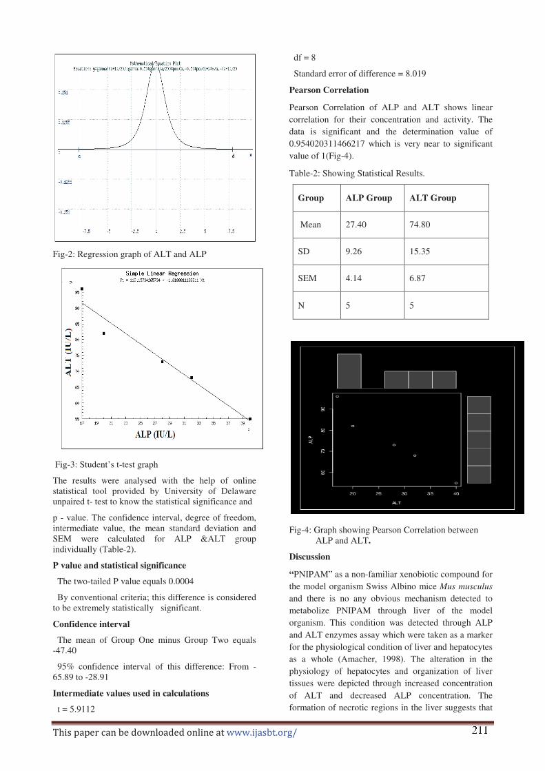
Pearson Correlation
Pearson Correlation of ALP and ALT shows linear
correlation for their concentration and activity. The
data is significant and the determination value of
0.954020311466217 which is very near to significant
value of 1(Fig-4).
Table-2: Showing Statistical Results.
Group ALP Group ALT Group
Mean 27.40 74.80
SD 9.26 15.35
SEM 4.14 6.87
N 5 5
Fig-4: Graph showing Pearson Correlation between
ALP and ALT.
Discussion
“PNIPAM” as a non-familiar xenobiotic compound for
the model organism Swiss Albino mice Mus musculus
and there is no any obvious mechanism detected to
metabolize PNIPAM through liver of the model
organism. This condition was detected through ALP
and ALT enzymes assay which were taken as a marker
for the physiological condition of liver and hepatocytes
as a whole (Amacher, 1998). The alteration in the
physiology of hepatocytes and organization of liver
tissues were depicted through increased concentration
of ALT and decreased ALP concentration. The
formation of necrotic regions in the liver suggests that � � �

Md. Imtiyaz Aslam et. al. (2013) Int J Appl Sci Biotechnol, Vol 1(4): 208-213
���������������� ������ � ��������������������������
PNIPAM has slightly toxic effect if not metabolized
through hepatocytes. As a result disorganization of
liver tissues has been occur. The aqueous solution of
PNIPAM at the concentration of 0.8mg/ml shows
deleterious effect suggesting that the same
concentration cannot be tolerable by the liver size of
the model organism.
The present result is in the conformity with the work of
Cooperstein and Canavan (2010) who observed
biological cell detachment from Poly N-isopropyl
acrylamide.
The statistical analysis through Student’s t-test gives
the p-value 0.004, confidence interval of 95% with a
standard error of difference 8.019 which shows the
statistical validity of the data obtained. The correlation
between ALP and ALT was made through Pearson
correlation which suggests a direct and linear
relationship between ALP and ALT activity,
antagonizing each other.
The further study in the field of xenobiotic
susceptibility in mammals must be practiced due to
endless and ever-growing list of newly synthesized
compounds. The effect of these xenobiotic compounds
must be studied at hematological, physiological and
therapeutic level to prevent harmful effects on vital
organs of the concerned organism. These xenobiotic
compounds have no any immune system yet evolved in
animals. They are toxic even in minute quantities.
Several information is available on the level of the
toxicity of these compounds. Rinaki et al., (2003) have
suggested dose/solubility ratio of biopharmaceutics.
Acosta et al., (1985) have given an in vitro approach to
the study of target organ toxicity of drug and
chemicals.
Acknowledgement
The authors are thankful to U.G.C, New Delhi for
Financial Support and Course Coordinator, P. G.
Department of Biotechnology, T. M. Bhagalpur
University, Bhagalpur (India) for providing the
laboratory facilities and Head, University Department
of Zoology, for the supply of test animal.
References
Acosta D, Sorensen EM, Anuforo DC, Mitchell DB, Ramos
K, Santone KS and Smith MA (1985) An in vitro
approach to the study of target organ toxicity of drugs
and chemicals. In Vitro Cell Dev Biol. 21: 495–504.
Amacher DE (1998) Serum transaminase elevations as
indicators of hepatic injury following the
administration of drugs. Regul. Toxicol. Pharmacol. 27
(2), 119–130.
Ambler J, Arnold DF and Green AG (1970) A study of the
phosphatase activity in some commercial quality
control sera with p- nitrophenyl phosphate and phenyl
phosphate substrates. Clin Chim Acta. 27 (2): 350-3.
Aslam Imtiyaz, Verma Rohit Kumar and Roy SP (2013)
Synthesis of Polymeric Nanoparticle “PNIPAM” Poly
(N-isopropylacrylamide) and their Toxicity Assay on
Swiss Albino Mice, Mus musculus. Indian Journal of
Fundamental and Applied Life Sciences.Vol. 3 (1);
116-119
Buda A and Jarynowski A (2010) Life-time of correlations
and its applications vol.1, Wydawnictwo Niezalezne:
5–21,
Cooperstein MA and Canavan HE (2010) Biological cell
detachment from poly (N-isopropyl acrylamide) and its
applications. Langmuir 26, 7695–7707.
Dawson R Ben (1985) Data for Biochemical Research (3rd
ed.). Oxford: Clarendon Press. P. 122
Galaev IY and Mattiasson B (1999) ‘Smart’ polymers and
what they could do in biotechnology and medicine.
Trends Biotechnol. 17, 335–340.
Gebhardt R (1992). Metabolic zonation of the liver:
Regulation and implications for liver function.
Pharmacol Ther 53: 275–354.
Gebhardt R and Gaunitz F (1997). Cell-cell interactions in
the regulation of the expression of hepatic enzymes.
Cell Biol Toxicol 13: 263–273.
Hazewinkel Michiel ed (2001), "Student’s
test", Encyclopedia of Mathematics, Springer.
Hillesheim W Jaeschke and H Neumann HG (1995)
Cytotoxicity of aromatic amines in rat liver and
oxidative stress. Chem Biol Interact 98: 85–95.
Kikkawa R, Fujikawa M, Yamamoto T, Hamada Y, Yamada
H and Horii I (2006) In vitro hepatotoxicity study of
rats in comparison with in vitro hepatotoxicity
screening system, J. Toxicol. Sci., 31, 23-34.
Kumar A, Srivastava A, Galaev IY and Mattiasson B (2007)
Smart polymers: physical forms and bioengineering
applications. Prog. Polym. Sci. 32, 1205–1237.
Lott JA and Wolf PL (1986) Alanine and aspartate
aminotransferase (ALT and AST). Clinical
enzymology: a case-oriented approach. Chicago, Year
Book Medical Publishers, 111-138.
Rinaki E, Valsami G and Macheras P (2003) Quantitative
biopharmaceutics classification system: The central
role of dose/solubility ratio. Pharm Res; 20:1917–
1925.
Schild HG (1992) Poly (N-isopropylacrylamide):
experiment, theory and application. Prog. Polym. Sci.
17, 163–249. � � �
Md. Imtiyaz Aslam et. al. (2013) Int J Appl Sci Biotechnol, Vol 1(4): 208-213
���������������� ������ � ��������������������������
Wang CS, Chang Ting-Tsung, Yao Wei-Jen, Wang Shan-
Tair and Chou Pesus (2012) "Impact of increasing
alanine aminotransferase levels within normal range on
incident diabetes". J Formos Med Assoc 111 (4): 201–8
Williamson T (1972) A comparision between the
phosphastrate and phenyl phosphate methods of
alkaline phosphatase assay. Med Lab Technol. 29 (2):
182-7.
� � �