Embed Size (px)
Citation preview
Nanomedicine: Nanotechnology, Biology, and Medicine11 (2015) 815–824
nanomedjournal.com
1
Cellular uptake and biocompatibility of bismuth ferrite harmonicadvanced nanoparticles
Davide Staedlera,1, Solène Passemard, MSc, PhDa,1, Thibaud Magouroux, MSc, PhDb,Andrii Rogov, MScb, Ciaran Manus Maguire, MScc, Bashir M. Mohamed, PhDc,Sebastian Schwung, MScd, Daniel Rytz, MSc, PhDd, Thomas Jüstel, MS, PhDe,
Stéphanie Hwu, MScb, Yannick Mugnier, MSc, PhDf, Ronan Le Dantec, MSc, PhDf,Yuri Volkov, MD, PhDc,g, Sandrine Gerber-Lemaire, MSc, PhDa,
Adriele Prina-Mello, MSc, PhDc,g, Luigi Bonacina, MSc, PhDb,⁎, Jean-Pierre Wolf, MSc, PhDb
aInstitute of Chemical Sciences and Engineering, EPFL, Batochime, 1015, Lausanne, SwitzerlandbGAP-Biophotonics, Université de Genève, 22 Chemin de Pinchat, 1211 Genève 4, Switzerland
cNanomedicine Laboratory and Molecular Imaging Group, School of Medicine, Trinity Centre for Health Sciences, Trinity College, D8, Dublin, IrelanddFEE Gmbh, Struthstrasse 2, 55743 Idar-Oberstein, Germany
eFachbereich Chemieingenieurwesen, Fachhochschule Münster, Stegerwaldstrasse 39, 48565 Steinfurt, GermanyfUniv. Savoie, SYMME, F-74000, Annecy, France
gAMBER Centre and CRANN Institute, Trinity College, D2, Dublin, Ireland
Received 17 August 2014; accepted 22 December 2014
Abstract
Bismuth Ferrite (BFO) nanoparticles (BFO-NP) display interesting optical (nonlinear response) and magnetic properties which makethem amenable for bio-oriented diagnostic applications as intra- and extra membrane contrast agents. Due to the relatively recent availabilityof this material in well dispersed nanometric form, its biocompatibility was not known to date. In this study, we present a thoroughassessment of the effects of in vitro exposure of human adenocarcinoma (A549), lung squamous carcinoma (NCI-H520), and acutemonocytic leukemia (THP-1) cell lines to uncoated and poly(ethylene glycol)-coated BFO-NP in the form of cytotoxicity, haemolyticresponse and biocompatibility. Our results support the attractiveness of the functional-BFO towards biomedical applications focused onadvanced diagnostic imaging.© 2015 Elsevier Inc. All rights reserved.
Key words: Nanophotonic; Non-linear imaging; Bismuth ferrite; PEGylation; Biocompatibility
Funded by: Partially funded by European Commission funded projectNAMDIATREAM project (FP7 LSP ref 246479) and CAN project(European Regional Development Fund through the Ireland WalesProgramme 2007-13 INTERREG 4A). The study was performed in thecontext of the European COST Action MP1302 Nanospectroscopy.
Conflicts of interest: the authors declare no conflict of interest.⁎Corresponding author.E-mail address: [email protected] (L. Bonacina).
These authors contributed equally to this work.
http://dx.doi.org/10.1016/j.nano.2014.12.0181549-9634/© 2015 Elsevier Inc. All rights reserved.
Background
Most of nanophotonics approaches (quantum dots, plasmonicnanoparticles (NP), up-conversion NP) for health applicationspresent static optical properties (absorption bands, surfaceplasmon resonances) often in the UV-visible spectral regionand do not fully allow for exploiting the tuning capabilities ofnew laser sources and their latest extensions in the infrared. Tocircumvent these limitations, a few research groups in the lastyears have introduced a new nanotechnology approach based oninorganic nanocrystals with non-centrosymmetric structures.Such nanomaterials present a very efficient nonlinear response,and can be easily imaged by their second harmonic generation
816 D. Staedler et al / Nanomedicine: Nanotechnology, Biology, and Medicine 11 (2015) 815–824
(SHG) in multi-photon imaging platforms.1-6 Such harmonic NP(HNP) do not suffer from conventional optical limitations suchas photobleaching and blinking allowing long-term monitoringof developing tissues.5,7 Several HNP have been recentlysynthesized and tested for biological applications.4-7 Particularcare should be paid to assess the ability of these NP to reachintracellular targets without causing major interferences to thecell metabolism. In this context, this subcellular targetingbecomes increasingly important as key parameters for theunderstating of complex events in living cells.8,9 In fact, thepossibility to freely change detection wavelength can beexploited for subtle co-localization studies with organelle-spe-cific dyes, as the signal from NP can be freely shifted to avoidoverlap with fluorophores emission bands. However, one aspectthat is not fully understood and10 remains uncertain is hownanomaterials interact with cellular interfaces such as cytoskel-etal membranes since it is known that small alterations in theirphysicochemical properties can drastically influence thecells-NP interactions, especially the uptake mechanisms.9,11
Therefore, lead-NP-candidate identification process based onhigh throughput screening as decision-making process is aprerequisite for the validation of new SHG NP for bio-imagingapplications. Here we present a study based on BiFeO3 (bismuthferrite, abbreviated as BFO) NP (BFO-NP), which were recentlysuccessfully introduced as photodynamic tools and imagingprobes.12 Nonetheless, such is the technological novelty of thisnew group of materials that there is still a knowledge gap thatrequires the scientific community attention towards the investi-gation of the interaction at the cellular and subcellular levels. Theopportunity of closing this gap is presented by providing the firstthorough investigation on the effects of BFO-NP in cellularmetabolism and uptake mechanisms. Toxicity and biocompati-bility were assessed by automated high content screening,recording cytotoxicity, lysosomal mass and cell permeability, inline with previously published works.13,14 Cellular uptake wasinvestigated by co-localizing the NP with specific fluorophoresfor cell membranes and endosomes. Moreover, in this paper wepresent for the first time to our knowledge the most efficientprotocol for the coating of these HNP with poly(ethylene glycol)(PEG) derivatives to promote colloidal stability and biocompat-ibility in biological media, and to allow post-functionalizationwith bioactive molecules.15,16 In this context, the biocompati-bility, cellular uptake and intracellular localization of free andPEG coated BFO-NP were compared.
Methods
Preparation of a polydisperse suspension of BFO
The starting BFO suspension (2 mL, 62.5 wt % in ZrO2balls), provided by the company FEE (Germany) under acollaboration agreement, was diluted in EtOH (2 L) andultra-sonicated for 12 h. After 10 days sedimentation, theupper portion of the polydisperse suspension (50 mL) wastaken and mixed with oleic acid (4 mL). The volatiles wereremoved under vacuum and the residue was weighed andsuspended in EtOH to obtain a stock solution at 3.6 mg/mL.
Coating of BFO-NP
BFO-NP (suspended in EtOH, 3.6 mg/mL, 583 μL) wered i lu t ed in E tOH: to luene :25% aqueous ammonia(0.50:0.50:0.32 mL) and ultra-sonicated for 30 min. PEGoligomers 1 and 214 (1:1 ratio, 100 mg) were added and thesuspension ultra-sonicated at 40 °C for 16 hr. The suspensionwas reduced to a small volume and distributed in plastic tubes fordilution with a mixture of dichloromethane (DCM):EtOH:water(1:1:1, 1 mL). After centrifugation (10 min, 13 000 rpm), theaqueous layer, containing the excess of unreacted polymers, wasremoved and a mixture of EtOH:water (1:1, 0.5 mL) was addedto each plastic tube. The procedure was repeated 5 times toobtain a pure suspension of coated BFO-NP in DCM. Afterevaporation of DCM and addition of EtOH, the BFO-NPconcentration was calculated by measuring the turbidity of thesolution by spectrometry at 600 nm (Synergy HT) and bycomparing the values with a standard-curve prepared using thestock solution at 3.6 mg/mL.
Characterization of uncoated and coated BFO-NP
Advanced physico-chemical characterization of uncoated andcoated BFO-NP was recently performed and reported. In thepresent work, BFO-NP were characterized by dynamic lightscattering (DLS) using a Zetasizer NanoZ (Malvern) fordetermination of mean hydrodynamic volume and zeta potential.Suspension of uncoated or coated BFO-NP (20 μL) was dilutedin 1 mL of distilled water. Acetic acid (100 μL) was added andthe resulting suspensions were ultra-sonicated for 30 min andanalysed by DLS.
Nanoparticles characterization in biological media
The physico-chemical characterization of the NP was carriedout by nanoparticle tracking analysis (NTA, Nanosight NS500).BFO-NP at 25 μg/mL were vortexed for 5 s to disperse theparticles and then diluted at 1 μg/mL in different solutions(0.22 μm filtered): diethylpyrocarbonate (DEPC) water,Dulbecco's Modified Eagle Medium (DMEM), Roswell ParkMemorial Institute (RPMI) and Ham's F-12 K (Kaighn's)Medium (F12K) culture media, and their supplemented formwith 10% fetal bovine serum (FBS). Three culture media werechosen since commonly utilised for the in vitro culture cellmodels selected. The different dispersions were then analyzedvia NTA for the physico-chemical characterization measurementof hydrodynamic radius and polydispersity index (PDI) at roomtemperature of BFO-NP. All measurements were carried outthree times at physiologically relevant pH (pH = 7.4) and meansand standard deviations (SD) were calculated. Quality assuranceover the measurements carried out was guaranteed by theadoption of Quality Nano (QNano, FP7 project) standardoperating procedures (SOPs), which have been developed aspart of large inter laboratory comparative study focused onnanoparticle physico-chemical characterization.17
Cell model and culturing conditions
Human lung-derived A549 and NCI-H520 cancer cell linesand human monocytic THP-1 cell line are available from ATCC
817D. Staedler et al / Nanomedicine: Nanotechnology, Biology, and Medicine 11 (2015) 815–824
(American Tissue Culture Collection, Manassas, VA, USA).A549 were grown in DMEM medium containing 4.5 g/Lglucose, 10% FBS and penicillin/streptomycin (PS) in a 37 °Cincubator with 5% CO2 at 95% humidity. NCI-H520 and THP-1were grown in complete Roswell Park Memorial Institute(RPMI) 1640 medium supplemented with 10% FCS and PS ina 37 °C incubator with 5% CO2 at 95% humidity. Fordifferentiation into macrophages, THP-1 cells were plated at adensity of 20'000 cells/cm2 in RPMI 1640 supplemented with10% FBS and 100 ng/mL phorbol 12-myristate 13-acetate(PMA, Sigma-Aldrich) for 72 h. Differentiated THP-1 cellsadhered to the bottom of the wells.
Fluorescent staining for cellular imaging
The cells (20’000 cells/cm2) were grown for 48 h or 72 h foractivated PMA THP-1 cells, in a 24 well plate containing onerod-shaped microscope slide (BD Falcon). After this time cellswere exposed to BFO-NP at 25 μg/mL or to vehicle (ethanol) atindicated time. For endosomes imaging the cell layers wereexposed for 2 h to 15 μg/mL of the fluorescent probeFM1-43FX (Invitrogen). After incubation, the cells were fixedin 4% formaldehyde in PBS for 30 min, subsequently washedonce in 0.1% Triton X-100 in PBS and twice in PBS, thenmaintained in formaldehyde. For membrane staining, cells werefixed and then exposed to 0.1 μg/mL Nile Red (Invitrogen) inPBS for 5 min, then rinsed twice in PBS and maintained informaldehyde. Cell nuclei were stained with 4',6'-diamidino-2-phenylindole (DAPI).
Multiphoton laser scanning microscopy
Samples were observed using a Nikon multiphoton invertedmicroscope (A1R-MP) coupled with a Mai-Tai tunable Ti:sap-phire oscillator from Spectra-Physics (100 fs, 80 MHz,700-1000 nm). A Plan APO 40× WI N.A. 1.25 objective wasused to focus the excitation laser and to epi-collect the SHGsignal and dye markers fluorescence. Nanoparticles andfluorescent dyes (FM1-43FX and Nile Red) were excited at790 nm and observed through tailored pairs of dichroic mirrorsand interferometric filters (Semrock, FF01- 395/11-25 for SH,FF01-607/70-25 for fluorescence). Statistics were calculated byaveraging measured values from samples between 40 and 170cells per condition for cell labelling and between 8 and 100 cellsper condition for co-localization measurements. Cell labellingwas computed by dividing the number of cells labelled by at leastone nanoparticle to the total number of cells in the microscopefield of view and expressed as % of total cells. Co-localizationwith endosomes was estimated by counting the number ofnanoparticles co-localizing with the fluorescence signal from cellmembrane dye FM1-43FX-and dividing this value by the totalnumber of nanoparticles present inside each cell. The result ofthis procedure is expressed as % of all particles internalized ineach cell.
In vitro dose and exposure endpoint determination
Cytotoxicity on three-cell line models was investigated in vitroafter 24 h and 72 h incubation with BFO-NP. Followingstandardization of the BFO preparation protocols, all were injected
into 96 well plates to a final volume of 200 μL/well.A549,NCI-H520 and activated PMA THP-1 cells were initially plated ata cell density of 10’000 cell/cm2 and incubated with 1.0, 2.5, 5.0,7.5 or 10 μg/mL of uncoated and coated BFO-NP for 24 h and72 h in a 37 °C incubator with 5% CO2 at 95% humidity.Experiments were repeated three times, using triplicate wells eachtime for each formulation tested. Positive and negative controlswere also included into each experiment in order to quantify theextent of toxicity response induced by each particle. Three positivecontrols were Valinomycin (VAL, Fisher Scientific) (finalconcentration 120 μM) to measure changes in mitochondrialtransmembrane potential, Tacrine (TAC, Sigma-Aldrich) (finalconcentration 100 μM) to measure changes in lysosomal mass/pHand Quantum Dots (CdSe) (final concentration at 1 μM) tomeasure nanoparticle-induced uptake.14 After 24 h and 72 hincubation, cells were washed in phosphate-buffered salinesolution (PBS) at pH 7.4 and fixed in 3% paraformaldehyde(PFA). A multiparametric cytotoxicity assay was performed usingthe Cellomics® HCS reagent HitKitTM as per manufacturer’sinstructions (Thermo Fisher Scientific Inc.). For each experiment,each plate well was scanned and acquired in a stereologyconfiguration of 6 randomly selected fields. In total, each endpointdata plot in the heatmaps represents the analysis of an average of270,000 cells = 3 runs × 3 triplicates × 6 fields × 5000 cells (onaverage from t = 0 h). Images were acquired at 10x magnificationusing three detection channels with different excitation filters.These included a DAPI filter (channel 1), which detected bluefluorescence of the Cellomics® Hoechst 33342 probe indicatingnuclear intensity at a wavelength of 461 nm; FITC filter (channel2), which detected green fluorescence of the Cellomics® cellpermeability probe indicating cell permeability at a wavelength of509 nm and a TRITC filter (channel 3), which detected thelysosomal mass and pH changes of the Cellomics® LysoTrackerprobe with red fluorescence at a wavelength of 599 nm.
Statistical analysis
Response of each cell type to the coated and uncoatedBFO-NP was analysed by 2-way ANOVA with Bonferronipost-test analysis. A P-value b0.05 was considered to bestatistically significant. In this work we are comparing 4 cellparameters associated with the cytotoxicity response of 3 celllines exposed to uncoated or coated BFO, at 5 doses, with 3controls; therefore, the statistical value associated with our workcarried out by High Content Screening and data mining is ofsignificance. To visualize the data, KNIME (http://KNIME.org,2.0.3) data exploration platform and the screening module HiTS(http://code.google.com/p/hits, 0.3.0) were used. Knime is amodular open-source data manipulation and visualizationprogramme, as previously reported.14,18-20 All measured param-eters were normalized using the per cent of the positive controls.Z score was used for scoring the normalized values. These scoreswere summarized using the mean function as follows Z score(x-mean)/SD, as from previous work.14,18-20 Heatmaps graphicalillustration in a colorimetric gradient table format was adopted asthe most suitable schematic representation to report on anystatistical significance and variation from normalized controlsbased on their Z score value. Heatmap tables illustrate the range
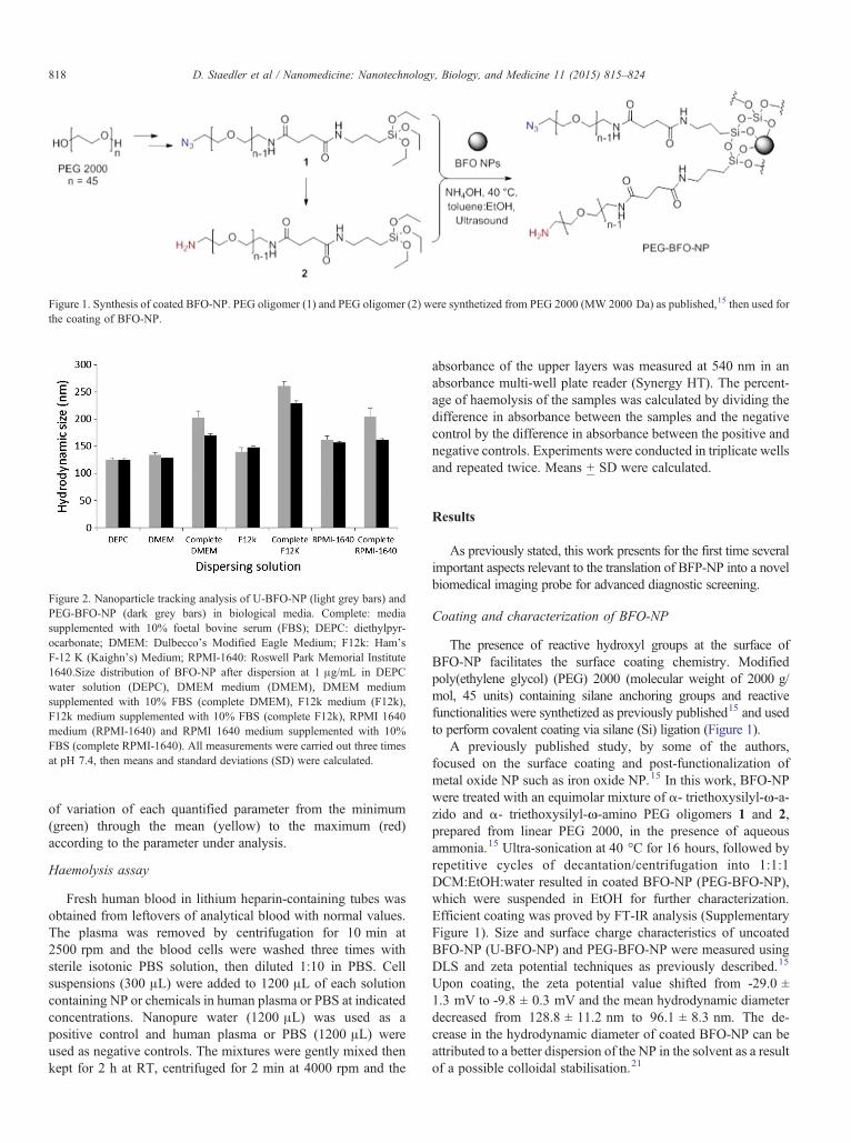
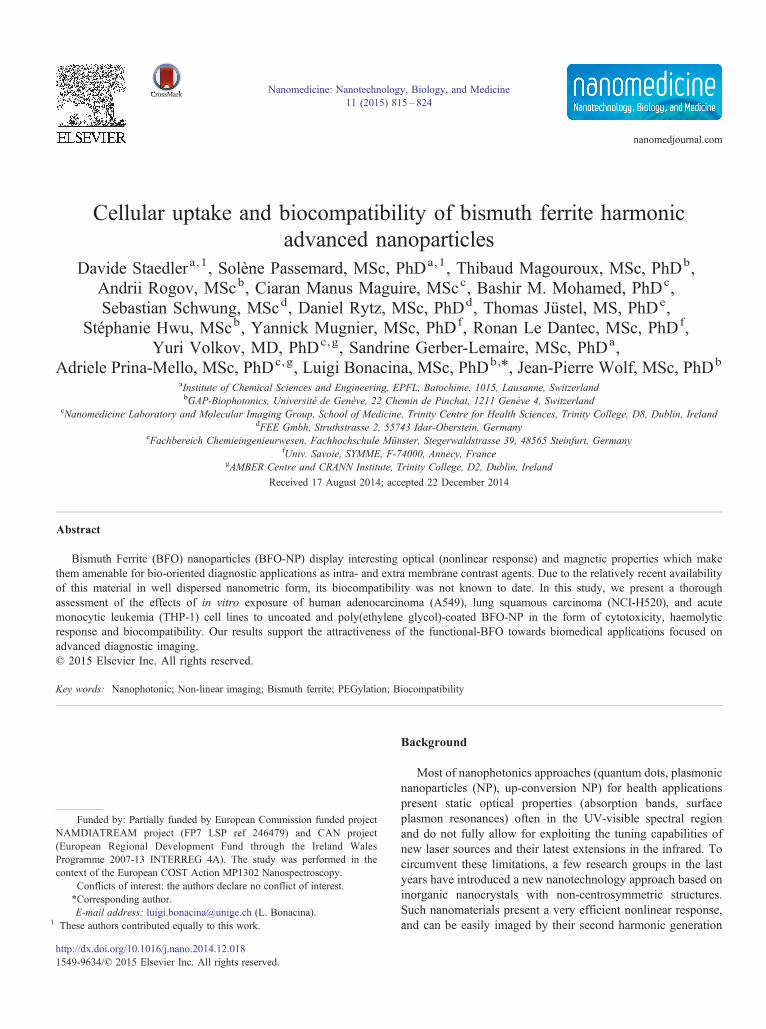
Figure 1. Synthesis of coated BFO-NP. PEG oligomer (1) and PEG oligomer (2) were synthetized from PEG 2000 (MW 2000 Da) as published,15 then used forthe coating of BFO-NP.
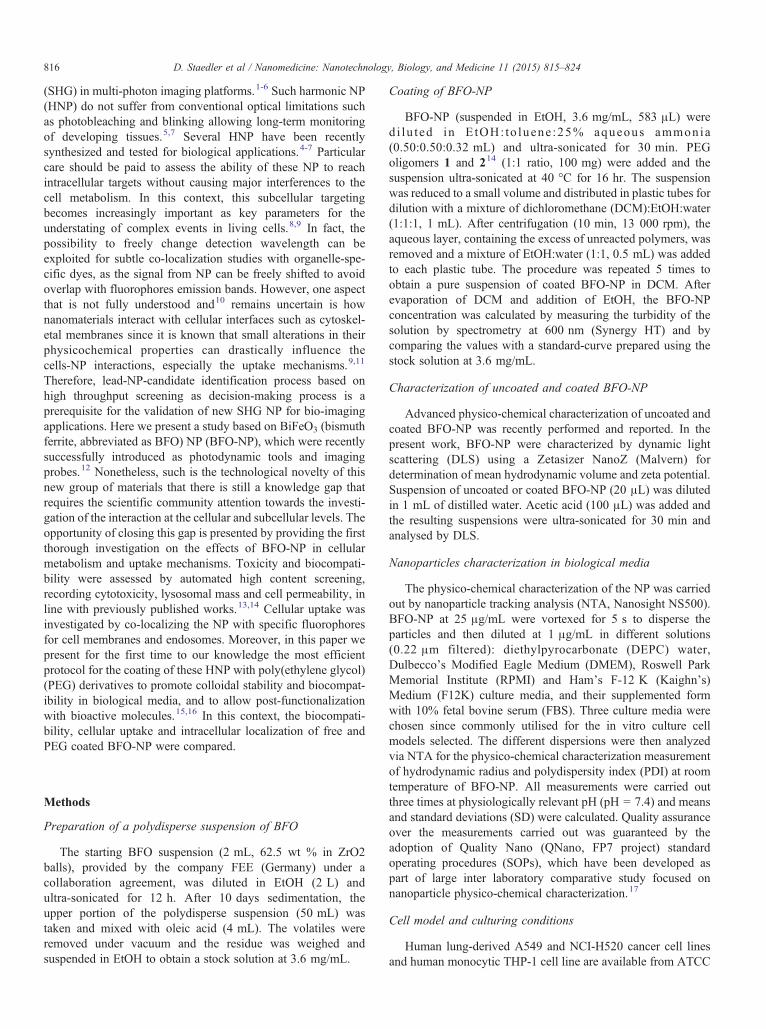
Figure 2. Nanoparticle tracking analysis of U-BFO-NP (light grey bars) andPEG-BFO-NP (dark grey bars) in biological media. Complete: mediasupplemented with 10% foetal bovine serum (FBS); DEPC: diethylpyr-ocarbonate; DMEM: Dulbecco's Modified Eagle Medium; F12k: Ham'sF-12 K (Kaighn's) Medium; RPMI-1640: Roswell Park Memorial Institute1640.Size distribution of BFO-NP after dispersion at 1 μg/mL in DEPCwater solution (DEPC), DMEM medium (DMEM), DMEM mediumsupplemented with 10% FBS (complete DMEM), F12k medium (F12k),F12k medium supplemented with 10% FBS (complete F12k), RPMI 1640medium (RPMI-1640) and RPMI 1640 medium supplemented with 10%FBS (complete RPMI-1640). All measurements were carried out three timesat pH 7.4, then means and standard deviations (SD) were calculated.
818 D. Staedler et al / Nanomedicine: Nanotechnology, Biology, and Medicine 11 (2015) 815–824
of variation of each quantified parameter from the minimum(green) through the mean (yellow) to the maximum (red)according to the parameter under analysis.
Haemolysis assay
Fresh human blood in lithium heparin-containing tubes wasobtained from leftovers of analytical blood with normal values.The plasma was removed by centrifugation for 10 min at2500 rpm and the blood cells were washed three times withsterile isotonic PBS solution, then diluted 1:10 in PBS. Cellsuspensions (300 μL) were added to 1200 μL of each solutioncontaining NP or chemicals in human plasma or PBS at indicatedconcentrations. Nanopure water (1200 μL) was used as apositive control and human plasma or PBS (1200 μL) wereused as negative controls. The mixtures were gently mixed thenkept for 2 h at RT, centrifuged for 2 min at 4000 rpm and the
absorbance of the upper layers was measured at 540 nm in anabsorbance multi-well plate reader (Synergy HT). The percent-age of haemolysis of the samples was calculated by dividing thedifference in absorbance between the samples and the negativecontrol by the difference in absorbance between the positive andnegative controls. Experiments were conducted in triplicate wellsand repeated twice. Means + SD were calculated.
Results
As previously stated, this work presents for the first time severalimportant aspects relevant to the translation of BFP-NP into a novelbiomedical imaging probe for advanced diagnostic screening.
Coating and characterization of BFO-NP
The presence of reactive hydroxyl groups at the surface ofBFO-NP facilitates the surface coating chemistry. Modifiedpoly(ethylene glycol) (PEG) 2000 (molecular weight of 2000 g/mol, 45 units) containing silane anchoring groups and reactivefunctionalities were synthetized as previously published15 and usedto perform covalent coating via silane (Si) ligation (Figure 1).
A previously published study, by some of the authors,focused on the surface coating and post-functionalization ofmetal oxide NP such as iron oxide NP.15 In this work, BFO-NPwere treated with an equimolar mixture of α- triethoxysilyl-ω-a-zido and α- triethoxysilyl-ω-amino PEG oligomers 1 and 2,prepared from linear PEG 2000, in the presence of aqueousammonia.15 Ultra-sonication at 40 °C for 16 hours, followed byrepetitive cycles of decantation/centrifugation into 1:1:1DCM:EtOH:water resulted in coated BFO-NP (PEG-BFO-NP),which were suspended in EtOH for further characterization.Efficient coating was proved by FT-IR analysis (SupplementaryFigure 1). Size and surface charge characteristics of uncoatedBFO-NP (U-BFO-NP) and PEG-BFO-NP were measured usingDLS and zeta potential techniques as previously described.15
Upon coating, the zeta potential value shifted from -29.0 ±1.3 mV to -9.8 ± 0.3 mV and the mean hydrodynamic diameterdecreased from 128.8 ± 11.2 nm to 96.1 ± 8.3 nm. The de-crease in the hydrodynamic diameter of coated BFO-NP can beattributed to a better dispersion of the NP in the solvent as a resultof a possible colloidal stabilisation.21
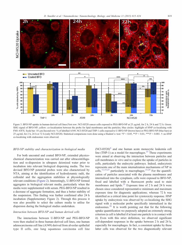
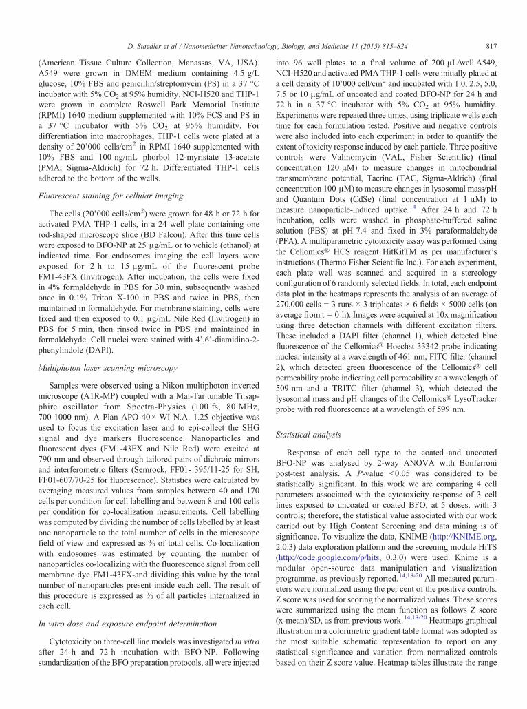
Figure 3. BFO-NP uptake in human-derived cell lines.First row: NCI-H520 cancer cells exposed to PEG-BFO-NP at 25 μg/mL for 2 h, 24 h and 72 h. Green:SHG signal of BFO-NP; yellow: co-localization between the probe for lipid membranes and the particles; blue circles: highlight of HNP co-localizing withFM1-43FX. Scale bar: 10 μm.Second row: % of labelled A549, NCI-H520 and THP-1 cells exposed to U-BFO-NP (brown bars) or PEG-BFO-NP (blue bars) at25 μg/mL for 2 h, 24 h or 72 h (only NCI-H520). Statistical comparisons were done using a Student's t test: *P b 0.05, **P b 0.01, ***P b 0.001. †: no HNPco-localizing with endosomes were observed.
819D. Staedler et al / Nanomedicine: Nanotechnology, Biology, and Medicine 11 (2015) 815–824
BFO-NP stability and characterization in biological media
For both uncoated and coated BFO-NP, extended physico-chemical characterization was carried out after ultracentrifuga-tion and re-dispersion in ultrapure deionized water prior toincubation into relevant biological dispersing media. The twodevised BFO-NP potential probes were also characterized byNTA, aiming at the identification of hydrodynamic radii, thecolloidal and the aggregation stabilities at physiologicallyrelevant conditions (Figure 2). Interestingly, U-BFO-NP formedaggregates in biological relevant media, particularly when themedia were supplemented with serum. PEG-BFO-NP resulted ina decrease of aggregate formation, and thus a better stability ofthe suspension. This finding was further confirmed after 24 hincubation (Supplementary Figure 2). Through this process itwas also possible to select the culture media to utilise fordispersion during the biological studies undertaken.
Interaction between BFO-NP and human derived cells
The interactions between U-BFO-NP and PEG-BFO-NPwere then studied in three human-derived cell lines, one humanadenocarcinoma cell line (A549) derived from alveolar epithelialtype II cells, one lung squamous carcinoma cell line
(NCI-H520)6 and one human acute monocytic leukemia cellline (THP-1) as a model for macrophages.22 These experimentswere aimed at observing the interaction between particles andcell membranes in vitro and to explore the uptake of particles incells, particularly the endocytic pathways. Indeed, endocytosisrepresents one of the main internalization mechanisms of NP incells,11,15,23 particularly in macrophages.23,24 For the quantifi-cation of particles associated with the plasma membranes andinternalized into the cytoplasm, cells were exposed to BFO-NP,fixed and labelled with a fluorescent probe used to stainmembranes and lipids.25 Exposure time of 2 h and 24 h werechosen since considered representative minimum and maximumexposure time for diagnostic applications; whereas 72 h wasidentified as a critical time point for cytotoxicity response. HNPuptake by endocytosis was observed by co-localizing the SHGsignal with a molecular probe specifically internalized in theendosomes.26 It is worth pointing out that for the followinguptake quantification we purposely adopted an extremely strongcriterion (a cell is labelled if at least one particle is in contact withit). Even with this strict definition, we observed significantcell-dependent responses in the uptake between 2 h and 24 h,especially for macrophages. In fact, a consistent uptake by theselatter cells was observed for the two diagnostically relevant
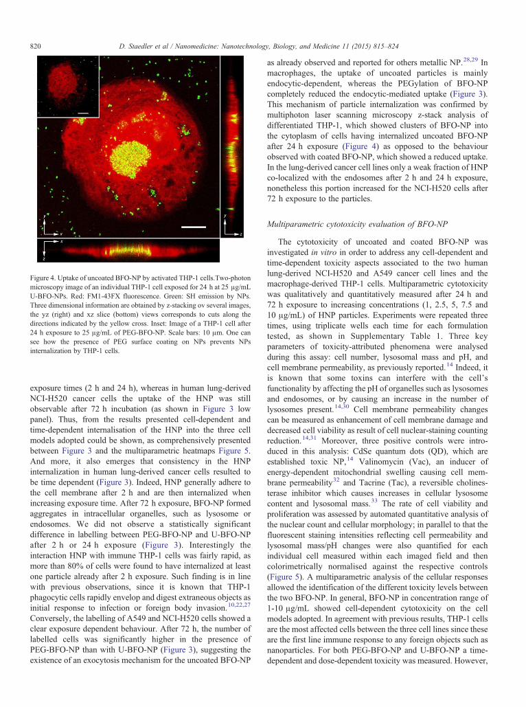
Figure 4. Uptake of uncoated BFO-NP by activated THP-1 cells.Two-photonmicroscopy image of an individual THP-1 cell exposed for 24 h at 25 μg/mLU-BFO-NPs. Red: FM1-43FX fluorescence. Green: SH emission by NPs.Three dimensional information are obtained by z-stacking ov several images,the yz (right) and xz slice (bottom) views corresponds to cuts along thedirections indicated by the yellow cross. Inset: Image of a THP-1 cell after24 h exposure to 25 μg/mL of PEG-BFO-NP. Scale bars: 10 μm. One cansee how the presence of PEG surface coating on NPs prevents NPsinternalization by THP-1 cells.
820 D. Staedler et al / Nanomedicine: Nanotechnology, Biology, and Medicine 11 (2015) 815–824
exposure times (2 h and 24 h), whereas in human lung-derivedNCI-H520 cancer cells the uptake of the HNP was stillobservable after 72 h incubation (as shown in Figure 3 lowpanel). Thus, from the results presented cell-dependent andtime-dependent internalisation of the HNP into the three cellmodels adopted could be shown, as comprehensively presentedbetween Figure 3 and the multiparametric heatmaps Figure 5.And more, it also emerges that consistency in the HNPinternalization in human lung-derived cancer cells resulted tobe time dependent (Figure 3). Indeed, HNP generally adhere tothe cell membrane after 2 h and are then internalized whenincreasing exposure time. After 72 h exposure, BFO-NP formedaggregates in intracellular organelles, such as lysosome orendosomes. We did not observe a statistically significantdifference in labelling between PEG-BFO-NP and U-BFO-NPafter 2 h or 24 h exposure (Figure 3). Interestingly theinteraction HNP with immune THP-1 cells was fairly rapid, asmore than 80% of cells were found to have internalized at leastone particle already after 2 h exposure. Such finding is in linewith previous observations, since it is known that THP-1phagocytic cells rapidly envelop and digest extraneous objects asinitial response to infection or foreign body invasion.10,22,27
Conversely, the labelling of A549 and NCI-H520 cells showed aclear exposure dependent behaviour. After 72 h, the number oflabelled cells was significantly higher in the presence ofPEG-BFO-NP than with U-BFO-NP (Figure 3), suggesting theexistence of an exocytosis mechanism for the uncoated BFO-NP
as already observed and reported for others metallic NP.28,29 Inmacrophages, the uptake of uncoated particles is mainlyendocytic-dependent, whereas the PEGylation of BFO-NPcompletely reduced the endocytic-mediated uptake (Figure 3).This mechanism of particle internalization was confirmed bymultiphoton laser scanning microscopy z-stack analysis ofdifferentiated THP-1, which showed clusters of BFO-NP intothe cytoplasm of cells having internalized uncoated BFO-NPafter 24 h exposure (Figure 4) as opposed to the behaviourobserved with coated BFO-NP, which showed a reduced uptake.In the lung-derived cancer cell lines only a weak fraction of HNPco-localized with the endosomes after 2 h and 24 h exposure,nonetheless this portion increased for the NCI-H520 cells after72 h exposure to the particles.
Multiparametric cytotoxicity evaluation of BFO-NP
The cytotoxicity of uncoated and coated BFO-NP wasinvestigated in vitro in order to address any cell-dependent andtime-dependent toxicity aspects associated to the two humanlung-derived NCI-H520 and A549 cancer cell lines and themacrophage-derived THP-1 cells. Multiparametric cytotoxicitywas qualitatively and quantitatively measured after 24 h and72 h exposure to increasing concentrations (1, 2.5, 5, 7.5 and10 μg/mL) of HNP particles. Experiments were repeated threetimes, using triplicate wells each time for each formulationtested, as shown in Supplementary Table 1. Three keyparameters of toxicity-attributed phenomena were analysedduring this assay: cell number, lysosomal mass and pH, andcell membrane permeability, as previously reported.14 Indeed, itis known that some toxins can interfere with the cell’sfunctionality by affecting the pH of organelles such as lysosomesand endosomes, or by causing an increase in the number oflysosomes present.14,30 Cell membrane permeability changescan be measured as enhancement of cell membrane damage anddecreased cell viability as result of cell nuclear-staining countingreduction.14,31 Moreover, three positive controls were intro-duced in this analysis: CdSe quantum dots (QD), which areestablished toxic NP,14 Valinomycin (Vac), an inducer ofenergy-dependent mitochondrial swelling causing cell mem-brane permeability32 and Tacrine (Tac), a reversible cholines-terase inhibitor which causes increases in cellular lysosomecontent and lysosomal mass.33 The rate of cell viability andproliferation was assessed by automated quantitative analysis ofthe nuclear count and cellular morphology; in parallel to that thefluorescent staining intensities reflecting cell permeability andlysosomal mass/pH changes were also quantified for eachindividual cell measured within each imaged field and thencolorimetrically normalised against the respective controls(Figure 5). A multiparametric analysis of the cellular responsesallowed the identification of the different toxicity levels betweenthe two BFO-NP. In general, BFO-NP in concentration range of1-10 μg/mL showed cell-dependent cytotoxicity on the cellmodels adopted. In agreement with previous results, THP-1 cellsare the most affected cells between the three cell lines since theseare the first line immune response to any foreign objects such asnanoparticles. For both PEG-BFO-NP and U-BFO-NP a time-dependent and dose-dependent toxicity was measured. However,
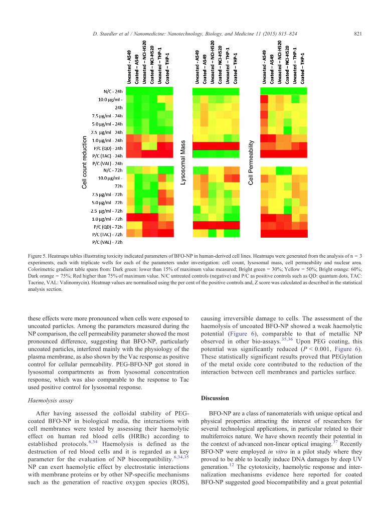
Figure 5. Heatmaps tables illustrating toxicity indicated parameters of BFO-NP in human-derived cell lines. Heatmaps were generated from the analysis of n = 3experiments, each with triplicate wells for each of the parameters under investigation: cell count, lysosomal mass, cell permeability and nuclear area.Colorimetric gradient table spans from: Dark green: lower than 15% of maximum value measured; Bright green = 30%; Yellow = 50%; Bright orange: 60%;Dark orange = 75%; Red higher than 75% of maximum value. N/C untreated controls (negative) and P/C as positive controls such as QD: quantum dots, TAC:Tacrine, VAL: Valinomycin). Heatmap values are normalised using the per cent of the positive controls and, Z score was calculated as described in the statisticalanalysis section.
821D. Staedler et al / Nanomedicine: Nanotechnology, Biology, and Medicine 11 (2015) 815–824
these effects were more pronounced when cells were exposed touncoated particles. Among the parameters measured during theNP comparison, the cell permeability parameter showed the mostpronounced difference, suggesting that BFO-NP, particularlyuncoated particles, interfered mainly with the physiology of theplasma membrane, as also shown by the Vac response as positivecontrol for cellular permeability. PEG-BFO-NP got stored inlysosomal compartments as from lysosomal concentrationresponse, which was also comparable to the response to Tacused positive control for lysosomal response.
Haemolysis assay
After having assessed the colloidal stability of PEG-coated BFO-NP in biological media, the interactions withcell membranes were tested by assessing their haemolyticeffect on human red blood cells (HRBc) according toestablished protocols.6,34 Haemolysis is defined as thedestruction of red blood cells and it is regarded as a keyparameter for the evaluation of NP biocompatibility.6,34,35
NP can exert haemolytic effect by electrostatic interactionswith membrane proteins or by other NP-specific mechanismssuch as the generation of reactive oxygen species (ROS),
causing irreversible damage to cells. The assessment of thehaemolysis of uncoated BFO-NP showed a weak haemolyticpotential (Figure 6), comparable to that of metallic NPobserved in other bio-assays.35,36 Upon PEG coating, thispotential was significantly reduced (P b 0.001, Figure 6).These statistically significant results proved that PEGylationof the metal oxide core contributed to the reduction of theinteraction between cell membranes and particles surface.
Discussion
BFO-NP are a class of nanomaterials with unique optical andphysical properties attracting the interest of researchers forseveral technological applications, in particular related to theirmultiferroics nature. We have shown recently their potential inthe context of advanced non-linear optical imaging.37 RecentlyBFO-NP were employed in vitro in a pilot study where theyproved to be able to locally induce DNA damages by deep UVgeneration.12 The cytotoxicity, haemolytic response and inter-nalization mechanisms evidence here reported for coatedBFO-NP suggested good biocompatibility and a great potential
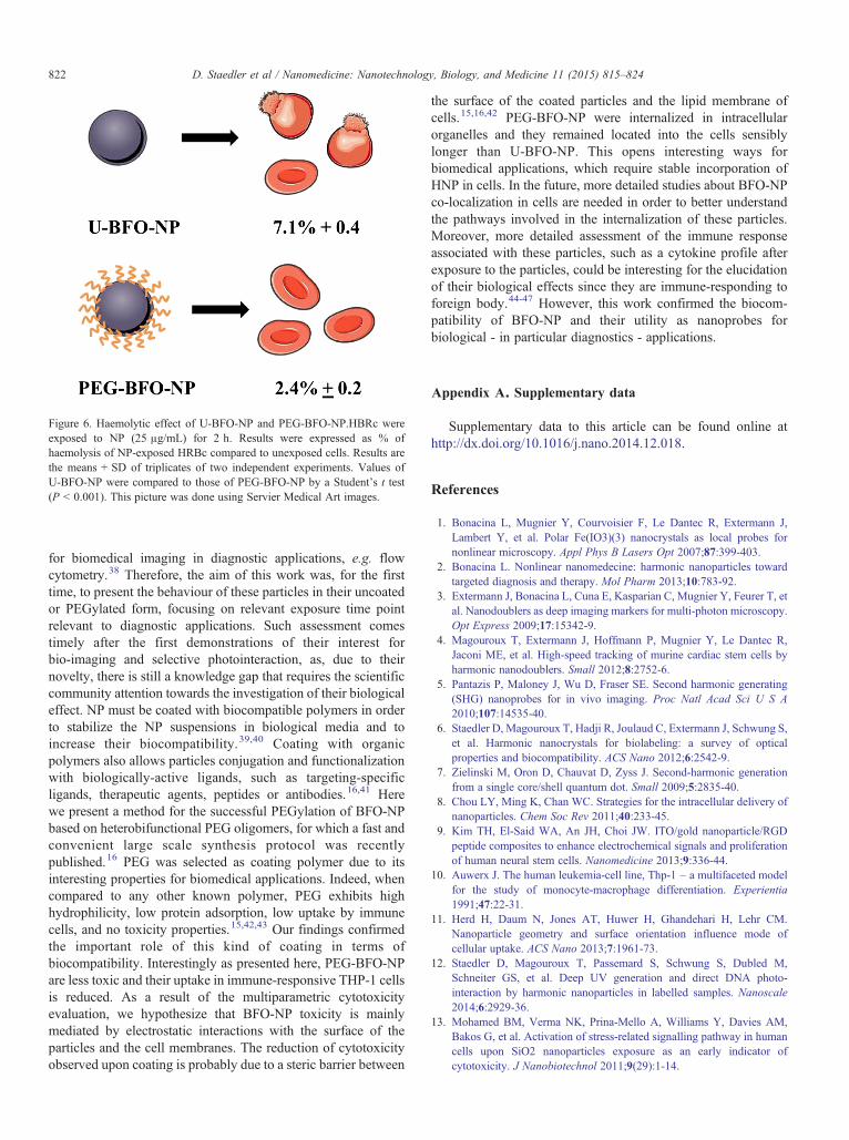
Figure 6. Haemolytic effect of U-BFO-NP and PEG-BFO-NP.HBRc wereexposed to NP (25 μg/mL) for 2 h. Results were expressed as % ofhaemolysis of NP-exposed HRBc compared to unexposed cells. Results arethe means + SD of triplicates of two independent experiments. Values ofU-BFO-NP were compared to those of PEG-BFO-NP by a Student's t test(P b 0.001). This picture was done using Servier Medical Art images.
822 D. Staedler et al / Nanomedicine: Nanotechnology, Biology, and Medicine 11 (2015) 815–824
for biomedical imaging in diagnostic applications, e.g. flowcytometry.38 Therefore, the aim of this work was, for the firsttime, to present the behaviour of these particles in their uncoatedor PEGylated form, focusing on relevant exposure time pointrelevant to diagnostic applications. Such assessment comestimely after the first demonstrations of their interest forbio-imaging and selective photointeraction, as, due to theirnovelty, there is still a knowledge gap that requires the scientificcommunity attention towards the investigation of their biologicaleffect. NP must be coated with biocompatible polymers in orderto stabilize the NP suspensions in biological media and toincrease their biocompatibility.39,40 Coating with organicpolymers also allows particles conjugation and functionalizationwith biologically-active ligands, such as targeting-specificligands, therapeutic agents, peptides or antibodies.16,41 Herewe present a method for the successful PEGylation of BFO-NPbased on heterobifunctional PEG oligomers, for which a fast andconvenient large scale synthesis protocol was recentlypublished.16 PEG was selected as coating polymer due to itsinteresting properties for biomedical applications. Indeed, whencompared to any other known polymer, PEG exhibits highhydrophilicity, low protein adsorption, low uptake by immunecells, and no toxicity properties.15,42,43 Our findings confirmedthe important role of this kind of coating in terms ofbiocompatibility. Interestingly as presented here, PEG-BFO-NPare less toxic and their uptake in immune-responsive THP-1 cellsis reduced. As a result of the multiparametric cytotoxicityevaluation, we hypothesize that BFO-NP toxicity is mainlymediated by electrostatic interactions with the surface of theparticles and the cell membranes. The reduction of cytotoxicityobserved upon coating is probably due to a steric barrier between
the surface of the coated particles and the lipid membrane ofcells.15,16,42 PEG-BFO-NP were internalized in intracellularorganelles and they remained located into the cells sensiblylonger than U-BFO-NP. This opens interesting ways forbiomedical applications, which require stable incorporation ofHNP in cells. In the future, more detailed studies about BFO-NPco-localization in cells are needed in order to better understandthe pathways involved in the internalization of these particles.Moreover, more detailed assessment of the immune responseassociated with these particles, such as a cytokine profile afterexposure to the particles, could be interesting for the elucidationof their biological effects since they are immune-responding toforeign body.44-47 However, this work confirmed the biocom-patibility of BFO-NP and their utility as nanoprobes forbiological - in particular diagnostics - applications.
Appendix A. Supplementary data
Supplementary data to this article can be found online athttp://dx.doi.org/10.1016/j.nano.2014.12.018.
References
1. Bonacina L, Mugnier Y, Courvoisier F, Le Dantec R, Extermann J,Lambert Y, et al. Polar Fe(IO3)(3) nanocrystals as local probes fornonlinear microscopy. Appl Phys B Lasers Opt 2007;87:399-403.
2. Bonacina L. Nonlinear nanomedecine: harmonic nanoparticles towardtargeted diagnosis and therapy. Mol Pharm 2013;10:783-92.
3. Extermann J, Bonacina L, Cuna E, Kasparian C, Mugnier Y, Feurer T, etal. Nanodoublers as deep imaging markers for multi-photon microscopy.Opt Express 2009;17:15342-9.
4. Magouroux T, Extermann J, Hoffmann P, Mugnier Y, Le Dantec R,Jaconi ME, et al. High-speed tracking of murine cardiac stem cells byharmonic nanodoublers. Small 2012;8:2752-6.
5. Pantazis P, Maloney J, Wu D, Fraser SE. Second harmonic generating(SHG) nanoprobes for in vivo imaging. Proc Natl Acad Sci U S A2010;107:14535-40.
6. Staedler D, Magouroux T, Hadji R, Joulaud C, Extermann J, Schwung S,et al. Harmonic nanocrystals for biolabeling: a survey of opticalproperties and biocompatibility. ACS Nano 2012;6:2542-9.
7. Zielinski M, Oron D, Chauvat D, Zyss J. Second-harmonic generationfrom a single core/shell quantum dot. Small 2009;5:2835-40.
8. Chou LY, Ming K, Chan WC. Strategies for the intracellular delivery ofnanoparticles. Chem Soc Rev 2011;40:233-45.
9. Kim TH, El-Said WA, An JH, Choi JW. ITO/gold nanoparticle/RGDpeptide composites to enhance electrochemical signals and proliferationof human neural stem cells. Nanomedicine 2013;9:336-44.
10. Auwerx J. The human leukemia-cell line, Thp-1 – a multifaceted modelfor the study of monocyte-macrophage differentiation. Experientia1991;47:22-31.
11. Herd H, Daum N, Jones AT, Huwer H, Ghandehari H, Lehr CM.Nanoparticle geometry and surface orientation influence mode ofcellular uptake. ACS Nano 2013;7:1961-73.
12. Staedler D, Magouroux T, Passemard S, Schwung S, Dubled M,Schneiter GS, et al. Deep UV generation and direct DNA photo-interaction by harmonic nanoparticles in labelled samples. Nanoscale2014;6:2929-36.
13. Mohamed BM, Verma NK, Prina-Mello A, Williams Y, Davies AM,Bakos G, et al. Activation of stress-related signalling pathway in humancells upon SiO2 nanoparticles exposure as an early indicator ofcytotoxicity. J Nanobiotechnol 2011;9(29):1-14.
823D. Staedler et al / Nanomedicine: Nanotechnology, Biology, and Medicine 11 (2015) 815–824
14. Prina-Mello A, Crosbie-Staunton K, Salas G, Morales MD, Volkov Y.Multiparametric toxicity evaluation of SPIONs by high contentscreening technique: identification of biocompatible multifunctionalnanoparticles for nanomedicine. IEEE Trans Magn 2013;49:377-82.
15. HakS,HelgesenE,HektoenHH,Huuse EM, JarzynaPA,MulderWJ, et al.The effect of nanoparticle polyethylene glycol surface density on ligand-directed tumor targeting studied in vivo by dual modality imaging. ACSNano 2012;6:5648-58.
16. Passemard S, Staedler D, Ucnova L, Schneiter GS, Kong P, Bonacina L,et al. Convenient synthesis of heterobifunctional poly(ethylene glycol)suitable for the functionalization of iron oxide nanoparticles forbiomedical applications. Bioorg Med Chem Lett 2013;23:5006-10.
17. Hole P, SillenceK, Hannell C,Maguire CM,RoessleinM, SuarezG, et al.Interlaboratory comparison of size measurements on nanoparticles usingnanoparticle tracking analysis (NTA). J Nanopart Res 2013;15:2101.
18. Mohamed BM, Verma NK, Davies AM, McGowan A, Crosbie-StauntonK, Prina-Mello A, et al. Citrullination of proteins: a common post-translational modification pathway induced by different nanoparticles invitro and in vivo. Nanomedicine (Lond) 2012;7:1181-95.
19. Movia D, Prina-Mello A, Volkov Y, Giordani S. Determination ofspiropyran cytotoxicity by high content screening and analysis for safeapplication in bionanosensing. Chem Res Toxicol 2010;23:1459-66.
20. Movia D, Prina-Mello A, Bazou D, Volkov Y, Giordani S. Screening thecytotoxicity of single-walled carbon nanotubes using novel 3D tissue-mimetic models. ACS Nano 2011;5:9278-90.
21. Gupta AK, Wells S. Surface-modified superparamagnetic nanoparticlesfor drug delivery: preparation, characterization, and cytotoxicity studies.IEEE Trans Nanobioscience 2004;3:66-73.
22. Kohro T, Tanaka T, Murakami T, Wada Y, Aburatani H, Hamakubo T,et al. A comparison of differences in the gene expression profiles ofphorbol 12-myristate 13-acetate differentiated THP-1 cells and humanmonocyte-derived macrophage. J Atheroscler Thromb 2004;11:88-97.
23. Shukla R, Bansal V, Chaudhary M, Basu A, Bhonde RR, Sastry M.Biocompatibility of gold nanoparticles and their endocytotic fate inside thecellular compartment: amicroscopic overview.Langmuir 2005;21:10644-54.
24. Lankoff A, Sandberg WJ, Wegierek-Ciuk A, Lisowska H, Refsnes M,Sartowska B, et al. The effect of agglomeration state of silver andtitanium dioxide nanoparticles on cellular response of HepG2, A549 andTHP-1 cells. Toxicol Lett 2012;208:197-213.
25. Lee Y, Yang I, Lee JE, Hwang S, Lee JW, Um SS, et al. Enhancedphotocurrent generation by Forster resonance energy transfer betweenphospholipid-assembled conjugated oligoelectrolytes and Nile red. JPhys Chem C 2013;117:3298-307.
26. Hansen GH, Rasmussen K, Niels-Christiansen LL, Danielsen EM.Endocytic trafficking from the small intestinal brush border probed withFM dye. Am J Physiol Gastrointest Liver Physiol 2009;297:G708-15.
27. Tsuchiya S, Yamabe M, Yamaguchi Y, Kobayashi Y, Konno T, Tada K.Establishment and characterization of a human acute monocyticleukemia cell line (THP-1). Int J Cancer 1980;26:171-6.
28. Chithrani BD, Chan WC. Elucidating the mechanism of cellular uptakeand removal of protein-coated gold nanoparticles of different sizes andshapes. Nano Lett 2007;7:1542-50.
29. Jin H, Heller DA, Sharma R, Strano MS. Size-dependent cellular uptakeand expulsion of single-walled carbon nanotubes: single particle trackingand a generic uptake model for nanoparticles. ACS Nano 2009;3:149-58.
30. Marquis BJ, Love SA, Braun KL, Haynes CL. Analytical methods toassess nanoparticle toxicity. Analyst 2009;134:425-39.
31. Jeng HA, Swanson J. Toxicity of metal oxide nanoparticles inmammalian cells. J Environ Sci Health A Tox Hazard Subst EnvironEng 2006;41:2699-711.
32. Lofrumento DD, La Piana G, Abbrescia DI, Palmitessa V, La Pesa V,Marzulli D, et al. Valinomycin induced energy-dependent mitochondrialswelling, cytochrome c release, cytosolic NADH/cytochrome c oxida-tion and apoptosis. Apoptosis 2011;16:1004-13.
33. Monteith DK, Theiss JC, Haskins JR, de la Iglesia FA. Functional andsubcellular organelle changes in isolated rat and human hepatocytesinduced by tetrahydroaminoacridine. Arch Toxicol 1998;72:147-56.
34. He Q, Zhang J, Shi J, Zhu Z, Zhang L, Bu W, et al. The effect ofPEGylation of mesoporous silica nanoparticles on nonspecific binding ofserum proteins and cellular responses. Biomaterials 2010;31:1085-92.
35. Choi J, Reipa V, Hitchins VM, Goering PL, Malinauskas RA.Physicochemical characterization and in vitro hemolysis evaluation ofsilver nanoparticles. Toxicol Sci 2011;123:133-43.
36. Zhao YN, Sun XX, Zhang GN, Trewyn BG, Slowing II, Lin VSY.Interaction of mesoporous silica nanoparticles with human red blood cellmembranes: size and surface effects. ACS Nano 2011;5:1366-75.
37. Schwung S, RogovA, ClarkeG, JoulaudC,Magouroux T, Staedler D, et al.Nonlinear optical and magnetic properties of BiFeO3 harmonic nanopar-ticles. J Appl Phys 2014;116:114306, http://dx.doi.org/10.1063/1.4895836.
38. Geissbuehler M, Bonacina L, Shcheslavskiy V, Bocchio NL, Geiss-buehler S, Leutenegger M, et al. Nonlinear correlation spectroscopy(NLCS). Nano Lett 2012;12:1668-72.
39. Lin MM, Kim HH, Kim H, Dobson J, Kim do K. Surface activation andtargeting strategies of superparamagnetic iron oxide nanoparticles incancer-oriented diagnosis and therapy. Nanomedicine (Lond)2010;5:109-33.
40. Shah NB, Vercellotti GM, White JG, Fegan A, Wagner CR, Bischof JC.Blood-nanoparticle interactions and in vivo biodistribution: impact ofsurface PEG and ligand properties. Mol Pharm 2012;9:2146-55.
41. Gil PR, Parak WJ. Composite nanoparticles take aim at cancer. ACSNano 2008;2:2200-5.
42. Perry JL, Reuter KG, Kai MP, Herlihy KP, Jones SW, Luft JC, et al.PEGylated PRINT nanoparticles: the impact of PEG density on proteinbinding, macrophage association, biodistribution, and pharmacokinetics.Nano Lett 2012;12:5304-10.
43. Verma A, Stellacci F. Effect of surface properties on nanoparticle-cellinteractions. Small 2010;6:12-21.
44. Hanley C, Thurber A, Hanna C, Punnoose A, Zhang J, Wingett DG.The influences of cell type and ZnO nanoparticle size on immunecell cytotoxicity and cytokine induction. Nanoscale Res Lett2009;4:1409-20.
45. Lesniak A, Fenaroli F, Monopoli MR, Aberg C, Dawson KA, Salvati A.Effects of the presence or absence of a protein corona on silicananoparticle uptake and impact on cells. ACS Nano 2012;6:5845-57.
46. Sayes CM, Reed KL, Warheit DB. Assessing toxicity of fine andnanoparticles: comparing in vitro measurements to in vivo pulmonarytoxicity profiles. Toxicol Sci 2007;97:163-80.
47. Siglienti I, Bendszus M, Kleinschnitz C, Stoll G. Cytokine profile ofiron-laden macrophages: implications for cellular magnetic resonanceimaging. J Neuroimmunol 2006;173:166-73.