Embed Size (px)
Citation preview
Central Nervous System Central Nervous System
Complications of HIVComplications of HIV
Tracey A. Cho, M.D.
AWACC
October 1, 2010
HIV Neurology
Tracey A. Cho, M.D.Tracey A. Cho, M.D.
Neurology-Infectious Diseases Section
Department of Neurology
Massachusetts General Hospital
Boston, MA 02114
USA
Nothing to disclose.
Professor Yunus Moosa
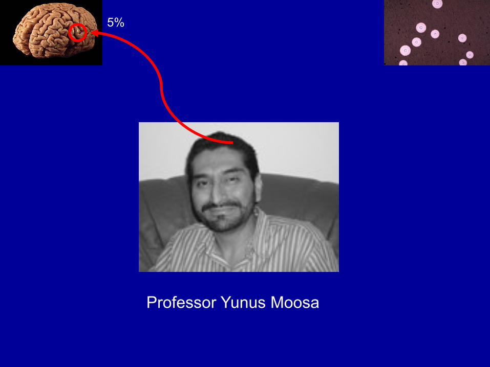
5%
Professor Yunus Moosa
Roadmap
• Spectrum of neurological syndromes in
HIV
• General diagnostic patterns of CNS
syndromes
• Cases with syndromic differential
diagnosis and treatment
• Role of ART in CNS
• Questions and discussion
HIV Neurology:
Overview• Neurological complications are common
• All stages of infection/immune status
• All parts of the nervous system affected
• Diverse syndromes
• Multiple pathologies often coexist
• Cumulative neurological burden – the
limited ability of recovery in CNS is unique
and raises the stakes for early treatment
Case 1
27M with advanced HIV presents with
vomiting, HA, neck pain, and mental status
change worsening over weeks.
Case 2
38M with HIV presents with 1 day of N/V
followed by generalized tonic clonic
seizure.
Case 3
52M with HIV presents with 5 weeks of right
arm clumsiness, unsteady gait.)
Case 4
64M with HIV presents with gradually
worsening mental function over 1-2 years.
HIV Neurology:
Diagnostic Principles• Epidemiology
• Host factors
• Clinical symptoms and signs
• Serological and other non-CNS studies
• CSF studies
• Imaging where available
• Response to treatment
Epidemiology
• Limited data in resource-limited settings
• Little confirmatory evidence of definitive
diagnosis
• Malaria, tuberculosis, and
neurocysticercosis increased in sub-
Saharan Africa
• HIV prevalence impacts CNS infectious
epidemiology
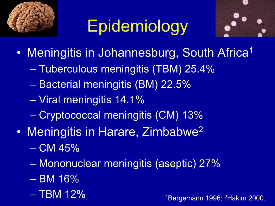
Epidemiology
• Meningitis in Johannesburg, South Africa1
– Tuberculous meningitis (TBM) 25.4%
– Bacterial meningitis (BM) 22.5%
– Viral meningitis 14.1%
– Cryptococcal meningitis (CM) 13%
• Meningitis in Harare, Zimbabwe2
– CM 45%
– Mononuclear meningitis (aseptic) 27%
– BM 16%
– TBM 12% 1Bergemann 1996; 2Hakim 2000.
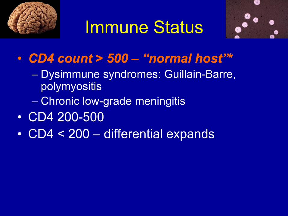
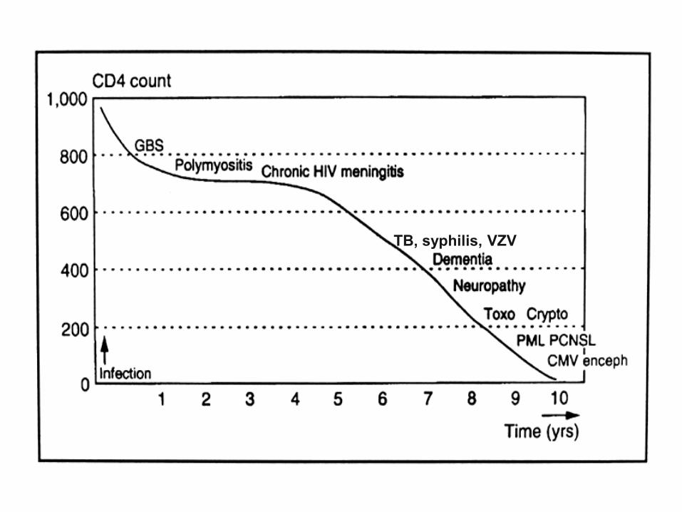
Immune Status
•• CD4 count > 500 CD4 count > 500 –– “normal host”*“normal host”*
– Dysimmune syndromes: Guillain-Barre, polymyositis
– Chronic low-grade meningitis
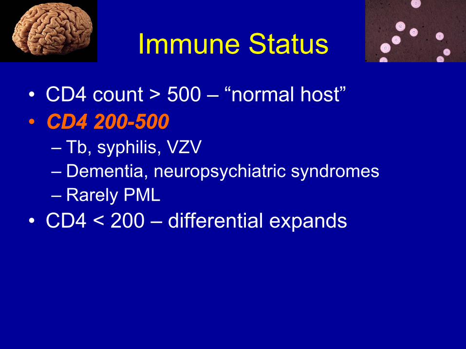
Immune Status
•• CD4 200CD4 200--500500
– Tb, syphilis, VZV
– Dementia, neuropsychiatric syndromes
– Rarely PML
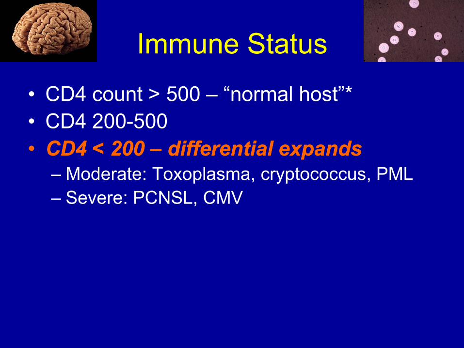
Immune Status
•• CD4 < 200 CD4 < 200 –– differential expandsdifferential expands
– Moderate: Toxoplasma, cryptococcus, PML
– Severe: PCNSL, CMV
TB, syphilis, VZV
Viral Status/
Prophylaxis• High viral load, even with preserved CD4
count, carries increased risk for neurological complications
• ART effectiveness and timing changes differential, including IRIS
• Prophylaxis with TMP-SMX lowers risk of toxoplasmosis
• Prophylaxis with fluconazole lowers risk cryptococcal meningitis (but not mortality)1
1Parkes-Ratanshi 2009
Clinical Localization
Localization within nervous system
• Meninges
• Diffuse brain lesions
• Focal brain lesions
• Spinal cord
• Nerve root and peripheral nerve
• Muscle
Clinical Localization
Localization within nervous system
•• MeningesMeninges
•• Diffuse Diffuse brain lesionsbrain lesions
•• Focal brain lesionsFocal brain lesions
Clinical Localization
Localization within nervous system
•• MeningesMeninges
– Headache, nucchal rigidity, photophobia, confusion (may overlap with encephalitis)
Clinical Localization
Localization within nervous system
•• Diffuse brain lesionsDiffuse brain lesions
– Encephalopathy, dementia, neuro-psychiatric
Clinical Localization
Localization within nervous system
•• Focal Focal brain lesionsbrain lesions
– Hemiparesis, ataxia, aphasia, visual field deficit, seizure
Clinical Localization
Localization within nervous system
•• In practice, there is often overlap of In practice, there is often overlap of syndromes (syndromes (meningoencephalitismeningoencephalitis))
–– Seizures, altered mental status, CSF Seizures, altered mental status, CSF abnormalities may be seen in each syndromeabnormalities may be seen in each syndrome
Etiology
• Meninges: acuteacute
– Aseptic meningitis
– Pyogenic
– HIV seroconversion
– HSV-2, VZV, neurosyphilis
– HIV rebound
– HIV IRIS
Etiology
• Meninges: subacutesubacute
– Cryptococcal meningitis
– Tuberculous meningitis
– Other fungal (histoplasma, coccidiodes)
– Neurosyphilis
– Neoplastic (lymphomatous)
– HIV (usu asymptomatic)
Etiology
• Diffuse brain lesions: acuteacute
– HIV encephalitis
– CMV encephalitis
– VZV encephalitis
– Post-infectious encephalitis (ADEM)
– Toxic (efavirenz, illicit drugs, EtOH)
– Neurosyphilis
– Cerebral malaria (HIV or non-HIV)
Etiology
• Diffuse brain lesions: subacutesubacute
– HIV-associated dementia
– HIV rebound meningoencephalitis
– Neuro-IRIS
– Neurosyphilis
Etiology
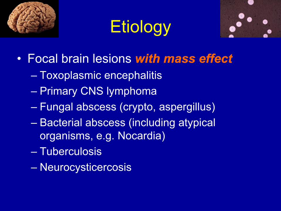
• Focal brain lesions with mass effectwith mass effect
– Toxoplasmic encephalitis
– Primary CNS lymphoma
– Fungal abscess (crypto, aspergillus)
– Bacterial abscess (including atypical
organisms, e.g. Nocardia)
– Tuberculosis
– Neurocysticercosis
Etiology
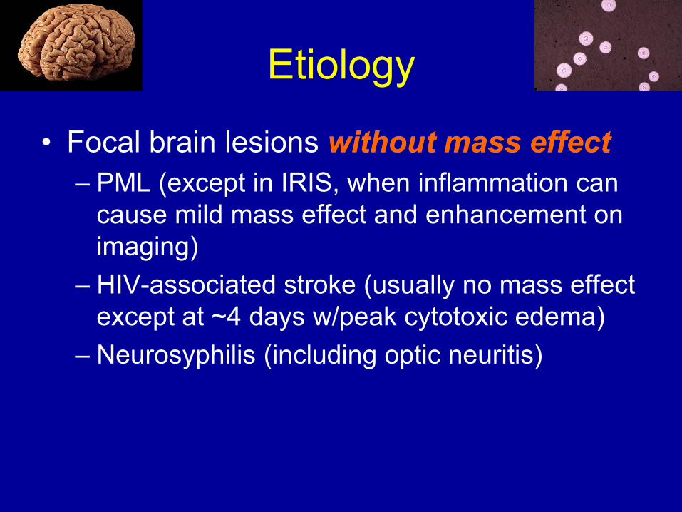
• Focal brain lesions without mass effectwithout mass effect
– PML (except in IRIS, when inflammation can
cause mild mass effect and enhancement on
imaging)
– HIV-associated stroke (usually no mass effect
except at ~4 days w/peak cytotoxic edema)
– Neurosyphilis (including optic neuritis)
Diagnostic Studies:
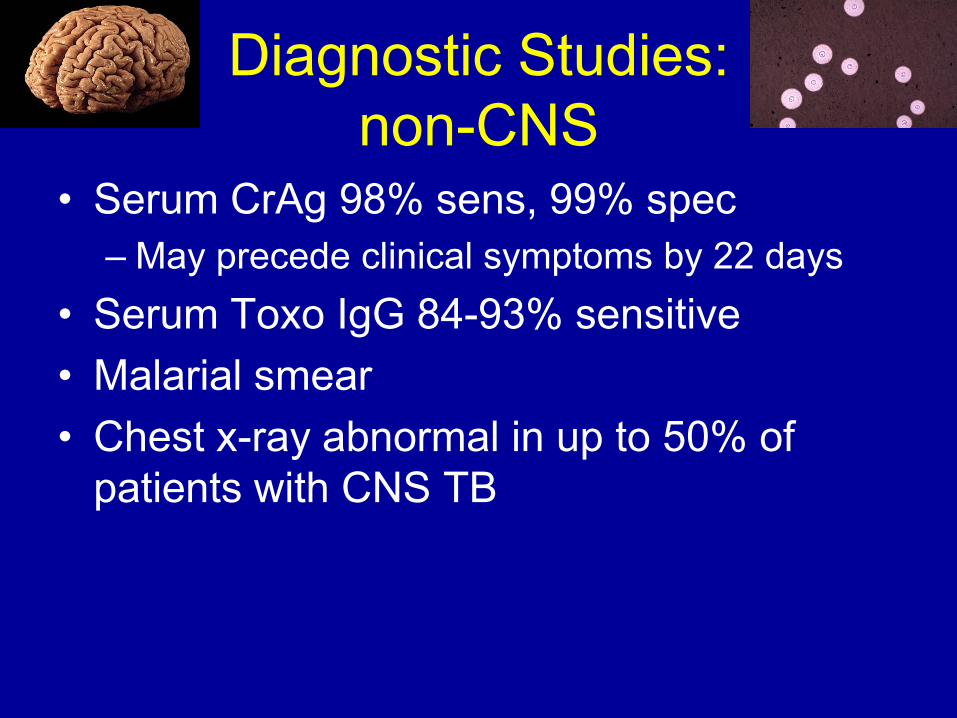
non-CNS• Serum CrAg 98% sens, 99% spec
– May precede clinical symptoms by 22 days
• Serum Toxo IgG 84-93% sensitive
• Malarial smear
• Chest x-ray abnormal in up to 50% of
patients with CNS TB
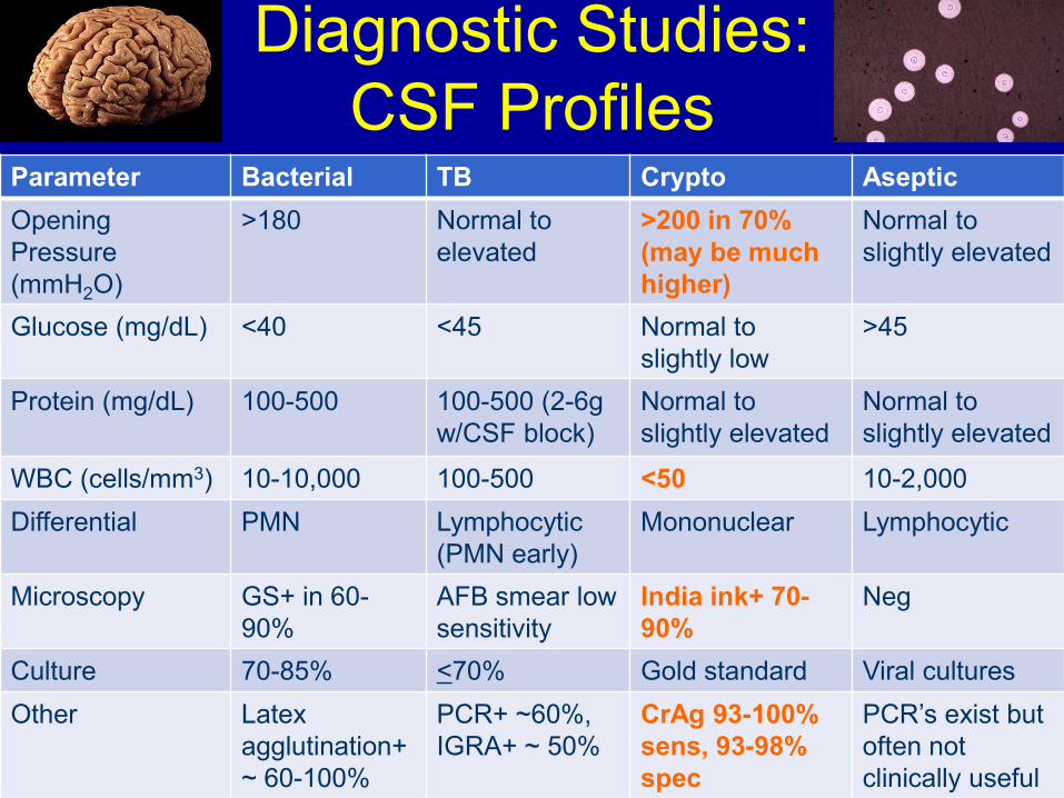
Diagnostic Studies:
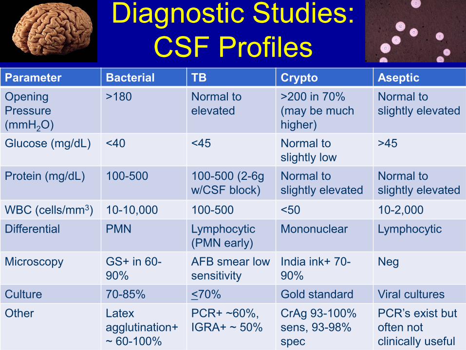
CSF ProfilesParameter Bacterial TB Crypto Aseptic
Opening
Pressure
(mmH2O)
>180 Normal to
elevated
>200 in 70%
(may be much
higher)
Normal to
slightly elevated
Glucose (mg/dL) <40 <45 Normal to
slightly low
>45
Protein (mg/dL) 100-500 100-500 (2-6g
w/CSF block)
Normal to
slightly elevated
Normal to
slightly elevated
WBC (cells/mm3) 10-10,000 100-500 <50 10-2,000
Differential PMN Lymphocytic
(PMN early)
Mononuclear Lymphocytic
Microscopy GS+ in 60-
90%
AFB smear low
sensitivity
India ink+ 70-
90%
Neg
Culture 70-85% <70% Gold standard Viral cultures
Other Latex
agglutination+
~ 60-100%
PCR+ ~60%,
IGRA+ ~ 50%
CrAg 93-100%
sens, 93-98%
spec
PCR’s exist but
often not
clinically useful
Diagnostic Studies:
CSF ProfilesParameter Bacterial TB Crypto Aseptic
Opening
Pressure
(mmH2O)
>180 Normal to
elevated
>200 in 70%
(may be much
higher)
Normal to
slightly elevated
Glucose (mg/dL) <40 <45 Normal to
slightly low
>45
Protein (mg/dL) 100-500 100-500 (2-6g
w/CSF block)
Normal to
slightly elevated
Normal to
slightly elevated
WBC (cells/mm3) 10-10,000 100-500 <50 10-2,000
Differential PMN Lymphocytic
(PMN early)
Mononuclear Lymphocytic
Microscopy GS+ in 60-
90%
AFB smear low
sensitivity
India ink+ 70-
90%
Neg
Culture 70-85% <70% Gold standard Viral cultures
Other Latex
agglutination+
~ 60-100%
PCR+ ~60%,
IGRA+ ~ 50%
CrAg 93-100%
sens, 93-98%
spec
PCR’s exist but
often not
clinically useful
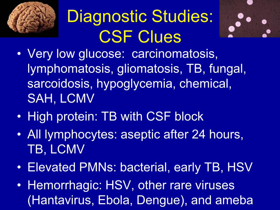
Diagnostic Studies:
CSF Clues• Very low glucose: carcinomatosis,
lymphomatosis, gliomatosis, TB, fungal,
sarcoidosis, hypoglycemia, chemical,
SAH, LCMV
• High protein: TB with CSF block
• All lymphocytes: aseptic after 24 hours,
TB, LCMV
• Elevated PMNs: bacterial, early TB, HSV
• Hemorrhagic: HSV, other rare viruses
(Hantavirus, Ebola, Dengue), and ameba

Diagnostic Studies:
CSF Clues•• Very low glucoseVery low glucose: carcinomatosis,
lymphomatosis, gliomatosis, TB, fungal,
sarcoidosis, hypoglycemia, chemical,
SAH, LCMV

Diagnostic Studies:
CSF Clues
•• High proteinHigh protein: TB with CSF block

Diagnostic Studies:
CSF Clues
•• All lymphocytesAll lymphocytes: aseptic after 24 hours,
TB, LCMV
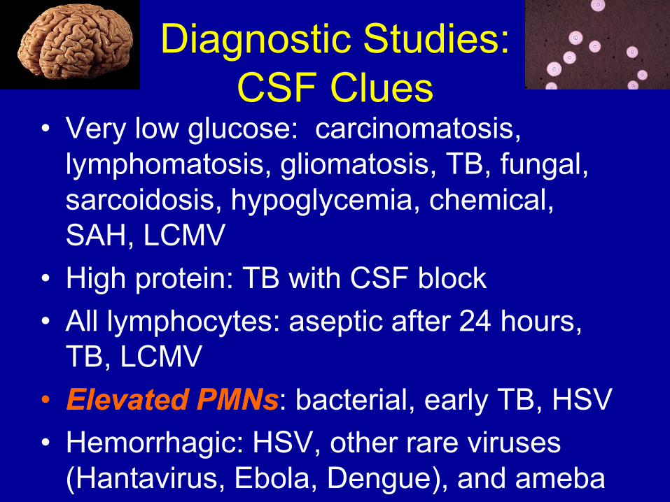
Diagnostic Studies:
CSF Clues
•• Elevated Elevated PMNsPMNs: bacterial, early TB, HSV
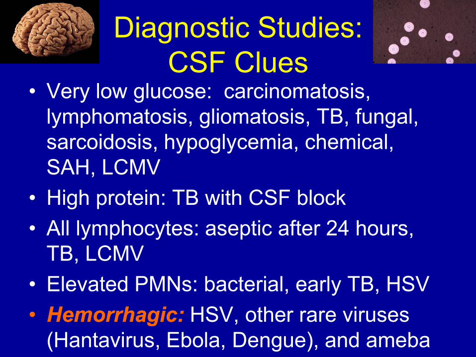
Diagnostic Studies:
CSF Clues
•• Hemorrhagic:Hemorrhagic: HSV, other rare viruses
(Hantavirus, Ebola, Dengue), and ameba
Diagnostic Studies:
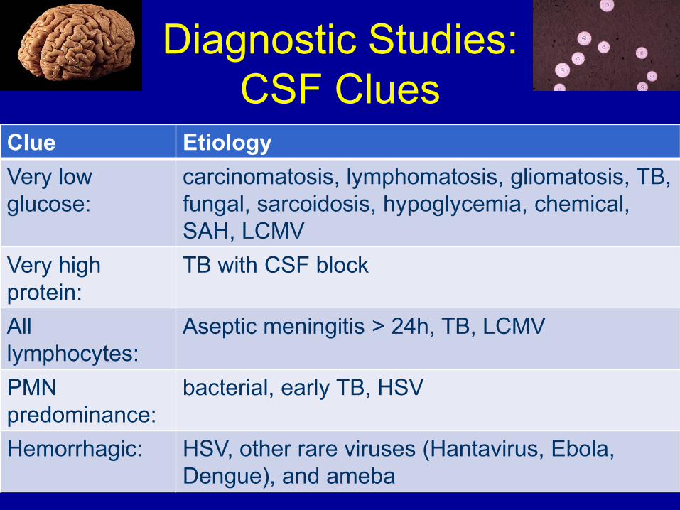
CSF CluesClue Etiology
Very low
glucose:
carcinomatosis, lymphomatosis, gliomatosis, TB,
fungal, sarcoidosis, hypoglycemia, chemical,
SAH, LCMV
Very high
protein:
TB with CSF block
All
lymphocytes:
Aseptic meningitis > 24h, TB, LCMV
PMN
predominance:
bacterial, early TB, HSV
Hemorrhagic: HSV, other rare viruses (Hantavirus, Ebola,
Dengue), and ameba
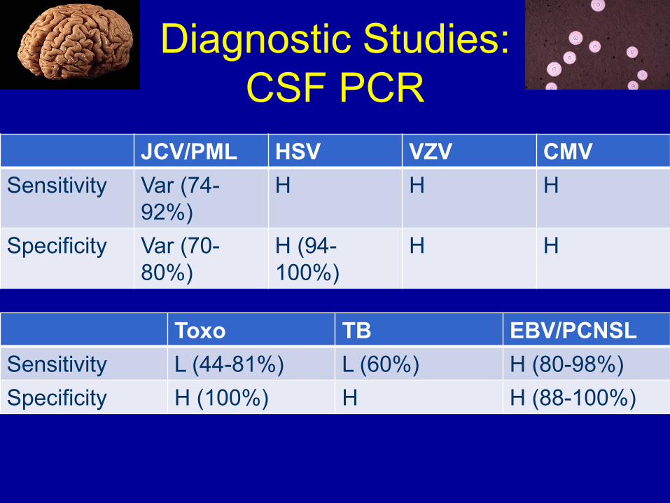
Diagnostic Studies:
CSF PCR
JCV/PML HSV VZV CMV
Sensitivity Var (74-
92%)
H H H
Specificity Var (70-
80%)
H (94-
100%)
H H
Toxo TB EBV/PCNSL
Sensitivity L (44-81%) L (60%) H (80-98%)
Specificity H (100%) H H (88-100%)
Resource-Limited
CNS Infection• Mortality much higher than in resource-rich
countries
• Definitive diagnosis elusive
• Presentation delayed
• LP often delayed
• Treatment delayed
Resource-Limited
CNS Infection• Reasons for LP delay
– No equipment available
– Limited laboratory hours
– Patient response to empiric therapy prior to
LP
– Patient mortality prior to LP
– Patient refusal
– Concern for herniation
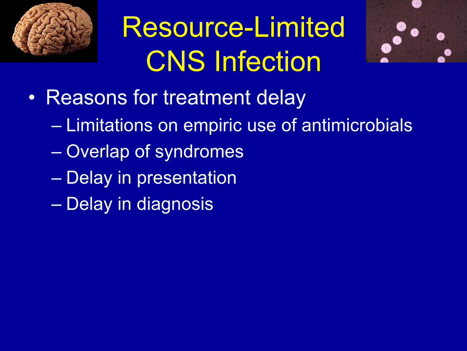
Resource-Limited
CNS Infection• Reasons for treatment delay
– Limitations on empiric use of antimicrobials
– Overlap of syndromes
– Delay in presentation
– Delay in diagnosis

Resource-Limited CNS
Infection: Proposed Algorithm• Immediate CXR and malarial smear
• HIV +/-
• CRAG +/-
• Empiric Rx based on CSF profile
• Concurrent adjunct studies: toxo IgG (focal
signs), VDRL, lymph node biopsy
(lymphadenopathy), CSF AFB &
mycobacterial culture, head CT, transfer
• Reassess based on responseTrachtenberg 2007
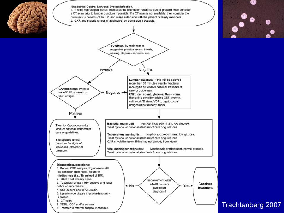
Trachtenberg 2007
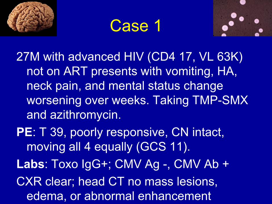
Case 1
27M with advanced HIV (CD4 17, VL 63K)
not on ART presents with vomiting, HA,
neck pain, and mental status change
worsening over weeks. Taking TMP-SMX
and azithromycin.
PE: T 39, poorly responsive, CN intact,
moving all 4 equally (GCS 11).
Labs: Toxo IgG+; CMV Ag -, CMV Ab +
CXR clear; head CT no mass lesions,
edema, or abnormal enhancement
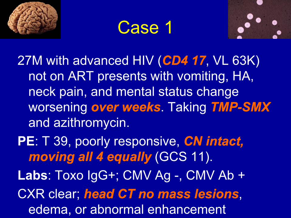
Case 1
27M with advanced HIV (CD4 17CD4 17, VL 63K)
not on ART presents with vomiting, HA,
neck pain, and mental status change
worsening over weeksover weeks. Taking TMPTMP--SMXSMX
and azithromycin.
PE: T 39, poorly responsive, CN intact, CN intact,
moving all 4 equallymoving all 4 equally (GCS 11).
Labs: Toxo IgG+; CMV Ag -, CMV Ab +
CXR clear; head CT no mass lesionshead CT no mass lesions,
edema, or abnormal enhancement
Case 1
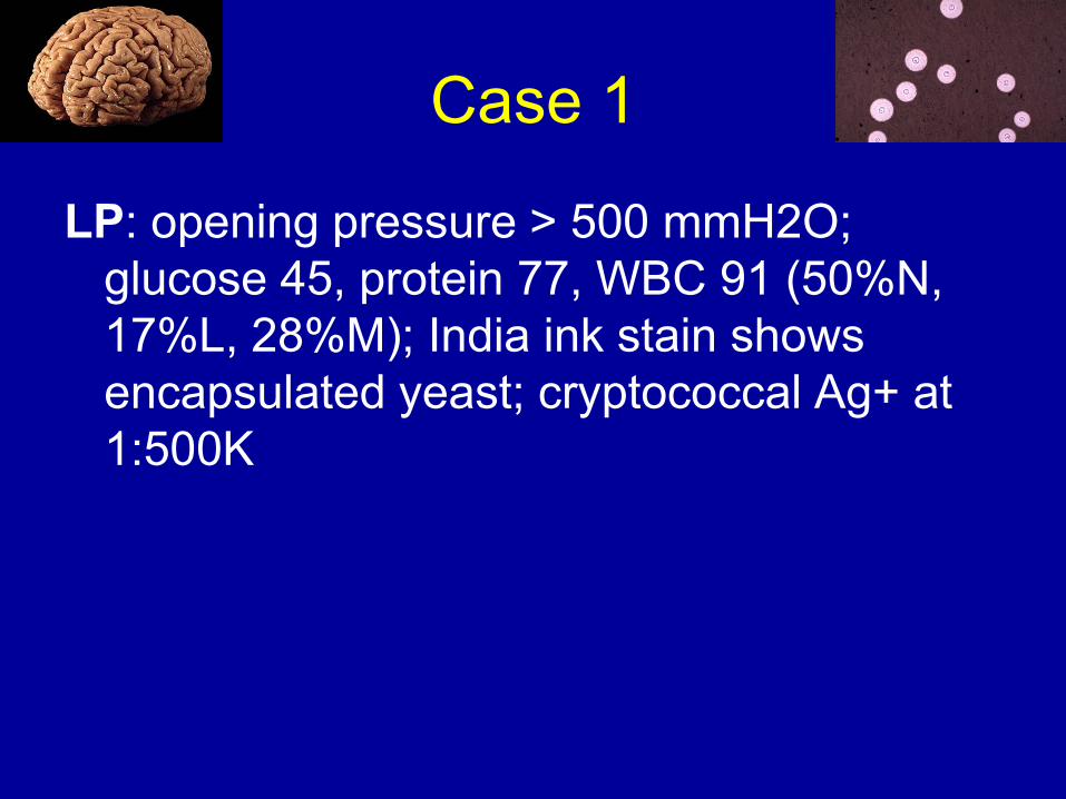
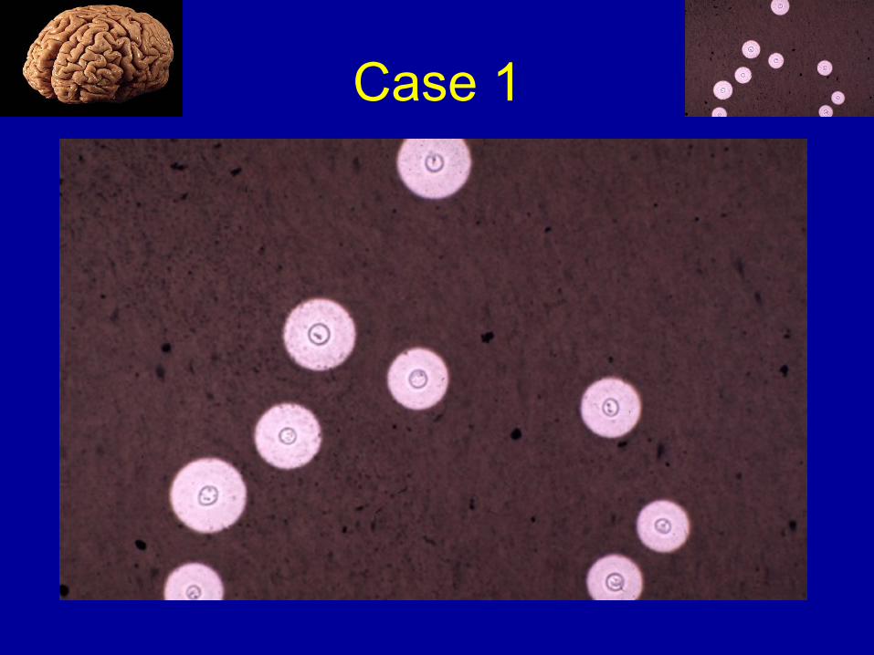
LP: opening pressure > 500 mmH2O;
glucose 45, protein 77, WBC 91 (50%N,
17%L, 28%M); India ink stain shows
encapsulated yeast; cryptococcal Ag+ at
1:500K
Case 1
Meningitis
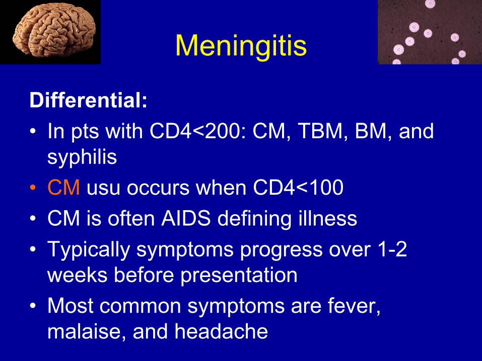
Differential:
• In pts with CD4<200: CM, TBM, BM, and
syphilis
• CM usu occurs when CD4<100
• CM is often AIDS defining illness
• Typically symptoms progress over 1-2
weeks before presentation
• Most common symptoms are fever,
malaise, and headache
Meningitis
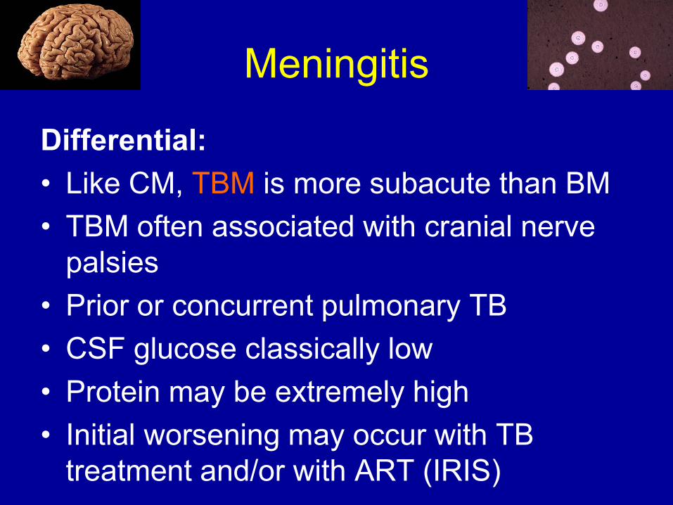
Differential:
• Like CM, TBM is more subacute than BM
• TBM often associated with cranial nerve
palsies
• Prior or concurrent pulmonary TB
• CSF glucose classically low
• Protein may be extremely high
• Initial worsening may occur with TB
treatment and/or with ART (IRIS)
Meningitis
Differential:
• BM more often acute, higher WBC count,
PMN predominance
• Associated stroke should suggest
meningovascular syphilis (or TBM)
• HIV “aseptic” meningitis occurs at
seroconversion
• HIV viral breakthrough (rebound) occurs
with ART failure or non-adherence
Meningitis
Differential considerations:
• HIV pts are susceptible to the same
causes of meningitis as the normal
population, especially where bacterial
meningitis is endemic.
• In advanced HIV, symptoms typical for
meningitis may be mild due to lack of
appropriate inflammatory response; CSF
may be bland or minimally inflamed.
Meningitis
Diagnosis:
• Blood cultures; serum CrAg, RPR/TPPA
• CXR to look for pulmonary Tb
• LP for opening pressure (typically high in
CM); glucose, protein, WBC with
differential; bacterial and fungal cultures,
India ink, CrAg, AFB and mycobacterial
culture
Meningitis
Diagnosis:
• CT is helpful as many pts with advanced
HIV have multiple infections
• Presence of mass lesions would change
differential
• Mass lesions causing downward pressure
increase risk for herniation with LP
• In practice, CT is limited in resource-
limited settings
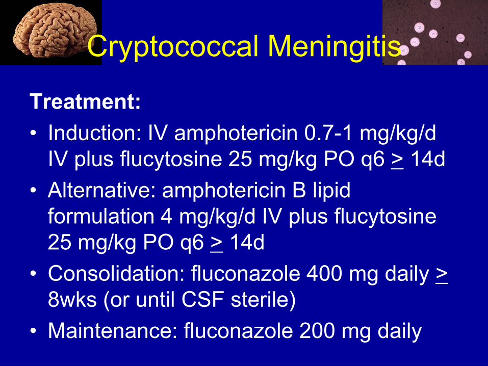
Cryptococcal Meningitis
Treatment:
• Induction: IV amphotericin 0.7-1 mg/kg/d
IV plus flucytosine 25 mg/kg PO q6 > 14d
• Alternative: amphotericin B lipid
formulation 4 mg/kg/d IV plus flucytosine
25 mg/kg PO q6 > 14d
• Consolidation: fluconazole 400 mg daily >
8wks (or until CSF sterile)
• Maintenance: fluconazole 200 mg daily
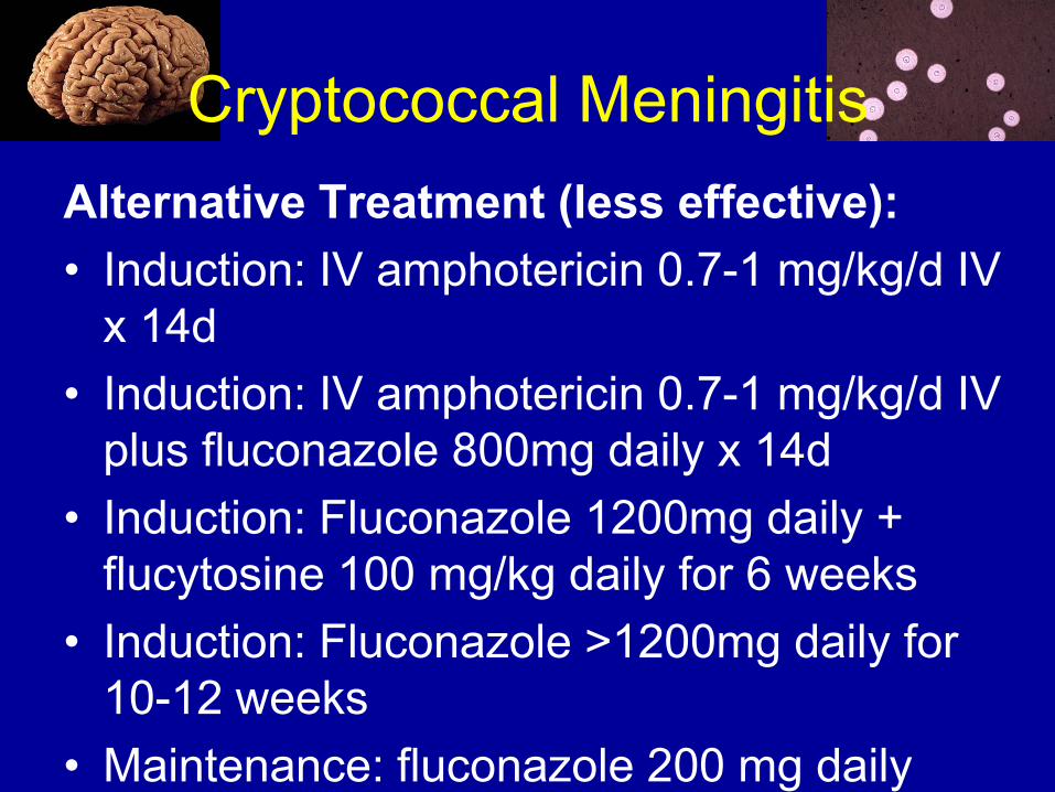
Cryptococcal Meningitis
Alternative Treatment (less effective):
• Induction: IV amphotericin 0.7-1 mg/kg/d IV
x 14d
• Induction: IV amphotericin 0.7-1 mg/kg/d IV
plus fluconazole 800mg daily x 14d
• Induction: Fluconazole 1200mg daily +
flucytosine 100 mg/kg daily for 6 weeks
• Induction: Fluconazole >1200mg daily for
10-12 weeks
• Maintenance: fluconazole 200 mg daily
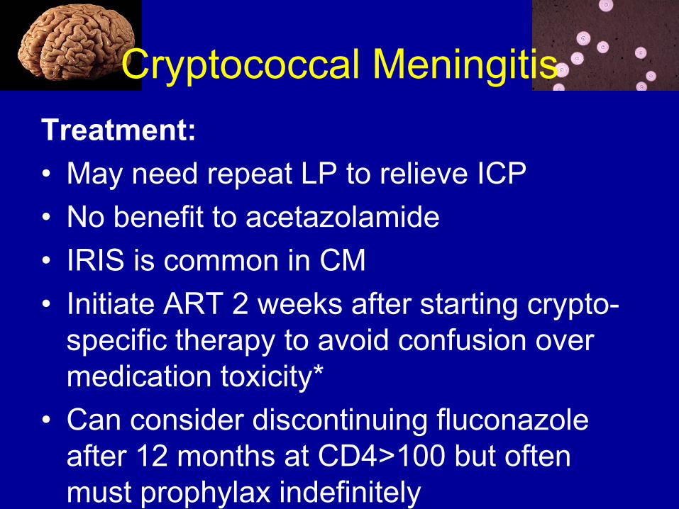
Cryptococcal Meningitis
Treatment:
• May need repeat LP to relieve ICP
• No benefit to acetazolamide
• IRIS is common in CM
• Initiate ART 2 weeks after starting crypto-
specific therapy to avoid confusion over
medication toxicity*
• Can consider discontinuing fluconazole
after 12 months at CD4>100 but often
must prophylax indefinitely
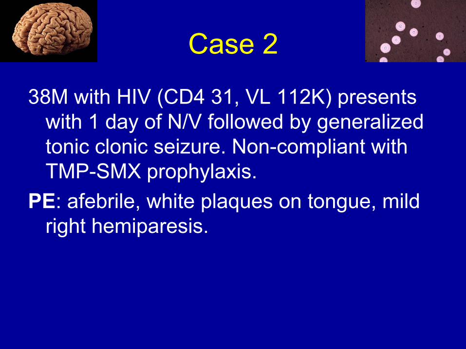
Case 2
38M with HIV (CD4 31, VL 112K) presents
with 1 day of N/V followed by generalized
tonic clonic seizure. Non-compliant with
TMP-SMX prophylaxis.
PE: afebrile, white plaques on tongue, mild
right hemiparesis.
Case 2
38M with HIV (CD4 31CD4 31, VL 112K) presents
with 1 day1 day of N/V followed by generalized
tonic clonic seizureseizure. NonNon--compliant with compliant with
TMPTMP--SMX SMX prophylaxis.
PE: afebrile, white plaques on tongue, mild mild
right hemiparesisright hemiparesis.
Focal Brain Lesion
Differential:
• Toxoplasma gondii
• Primary central nervous system lymphoma
(PCNSL)
• Progressive multifocal leuko-
encephalopathy (PML)
• Tuberculosis
• Cysticercosis
• Bacterial abscess

Focal Brain Lesion
Differential:
• All commonly present with fever,
headache, confusion
• Symptoms in toxo generally develop
rapidly over days, as opposed to PCNSL
(over a few weeks) and PML (over weeks
to months)
• Focal tuberculous lesions can present
similarly over days to weeks
• NCC often presents first with seizure

Focal Brain Lesion
Diagnosis:
• Most patients with toxo are serum IgG positive, so a negative serology makes toxo doubtful (7-16% false negative)
• If patient is adherent to TMP-SMX prophylaxis for PCP, toxo is less likely.
• Gold standard for diagnosis is brain biopsy
Focal Brain Lesion
Diagnosis:
• Therapeutic trial.
• Approximately 70-80% of pts have clinical and radiographic response.
• Vast majority have at least 50% improvement from baseline at 14 days of treatment.
• If no improvement within three weeks, other diagnoses should be pursued and treated.
Case 2
Diagnostic studies:
• Serum toxo IgG+, IgM-
• Chest X-ray clear
• CSF normal; EBV and JCV PCR negative
• Empiric toxo treatment lead to rapid clinical and radiographic improvement
• Continued on anti-epileptic for one year and then tapered off
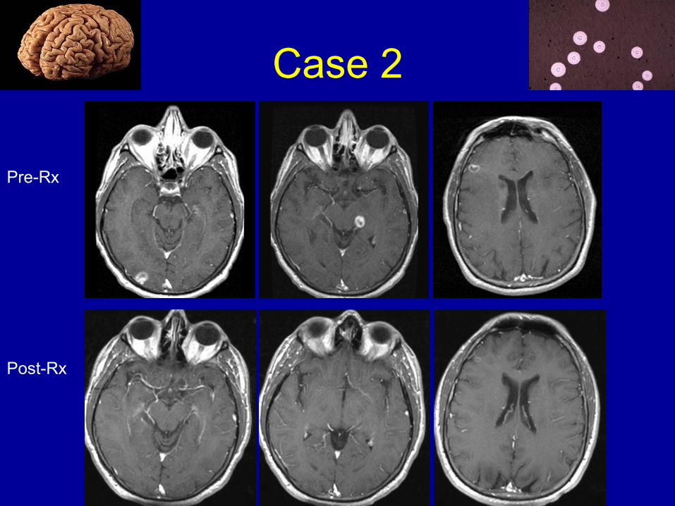
Case 2
Pre-Rx
Post-Rx
Toxoplasmosis
Treatment:
• Pyrimethamine 200 mg PO x1, then 50 mg
(for <60 kg body wt) or 75 mg (for >60 kg
body wt) PO qd
• Plus folinic acid 10 mg PO qd
• Plus sulfadiazine 1000 mg (for <60 kg
body wt) or 1500 mg (for >60 kg body wt)
PO q6h
• Treat for 6-8 weeks until good response
Toxoplasmosis
Treatment:
• Alternative to sulfadiazine: Clindamycin 600 mg IV or PO q6h
• Suppressive therapy: Pyrimethamine 50 mg PO qd + sulfadiazine 0.5-1 g PO q6h + folinic acid 10 mg PO daily
• May be discontinued when CD4>200 for at least 6 months
• Recovery is variable; may have lifelong epilepsy
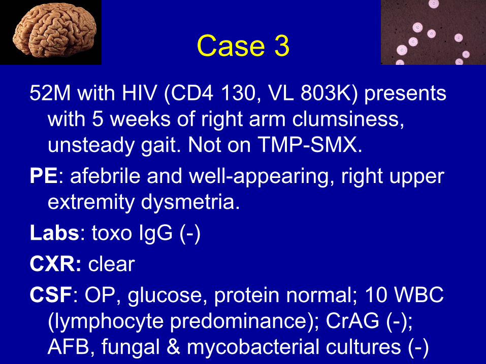
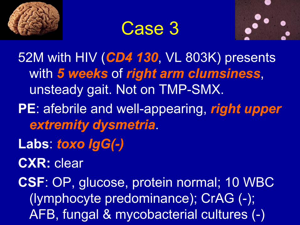
Case 3
52M with HIV (CD4 130, VL 803K) presents
with 5 weeks of right arm clumsiness,
unsteady gait. Not on TMP-SMX.
PE: afebrile and well-appearing, right upper
extremity dysmetria.
Labs: toxo IgG (-)
CXR: clear
CSF: OP, glucose, protein normal; 10 WBC
(lymphocyte predominance); CrAG (-);
AFB, fungal & mycobacterial cultures (-)
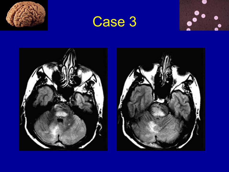
Case 3
52M with HIV (CD4 130CD4 130, VL 803K) presents
with 5 weeks5 weeks of right arm clumsinessright arm clumsiness,
unsteady gait. Not on TMP-SMX.
PE: afebrile and well-appearing, right upper right upper
extremity extremity dysmetriadysmetria.
Labs: toxotoxo IgGIgG((--))
CXR: clear
CSF: OP, glucose, protein normal; 10 WBC
(lymphocyte predominance); CrAG (-);
AFB, fungal & mycobacterial cultures (-)

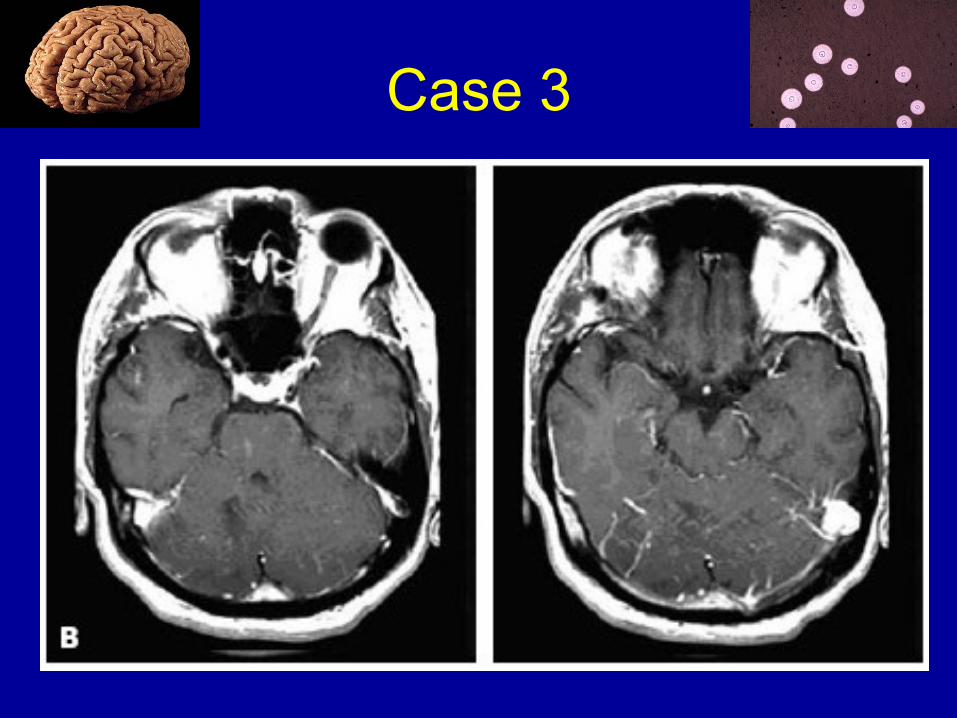
Case 3
Case 3
Focal Brain Lesion
Differential:
• Subacute (weeks to months) onset of focal
neurological symptoms
• Without contrast-enhanced imaging,
differential remains broad
• Toxo IgG (-), moderate CD4 count make
toxo and PCNSL less likely
• PML, PCNSL, tuberculous abscess or
tuberculoma, bacterial abscess all
possible
Focal Brain Lesion
Diagnosis:
• For PML, CSF JCV PCR 72-92% sensitive
and 92-100% specific in the pre-ART era
(less sensitive w/cART).
• Diagnosis in resource-rich countries
usually based on clinical and radiographic
patterns combined with CSF PCR studies
and (lack of) response to other treatments.
Focal Brain Lesion
Treatment:
• ART is only effective therapy for PML (or PCNSL)
• Survival in PML with ART has improved from 10 to 50% in resource-rich countries
• PML-IRIS may have enhancement, edema
• Steroids controversial but may be considered for severe cases
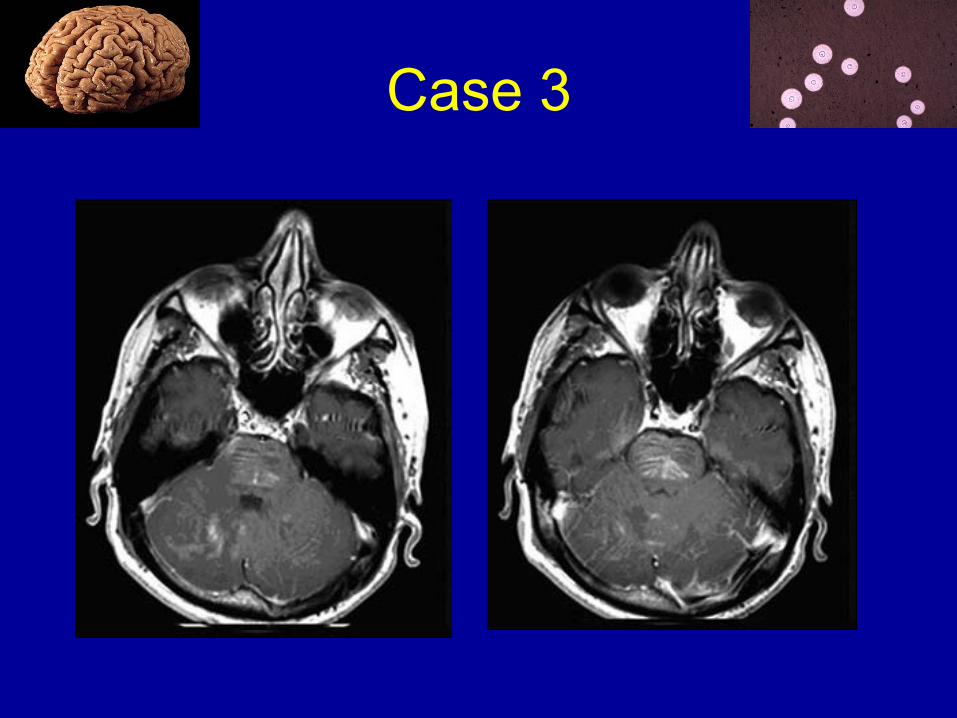
Case 3
The patient is started on cART and TMP-
SMX. Over 2-4 weeks he develops
worsening symptoms.
PE: new brainstem signs.
Labs: stable CD4 count, decrease in VL
from 803K to 4K.
Repeat MRI w/worsening, enhancement
Treated with prednisone 60mg tapered over
2 weeks, with stabilization but poor
recovery.
Case 3
Case 3
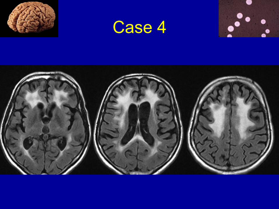
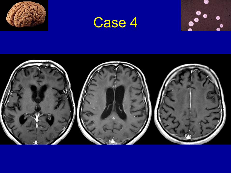
Case 4
64 RHM h/o HIV 3 years prior in setting of
PCP with initial CD4 in 30s and viral load
180K; wild-type HIV genotype. Started
emtricitabine/tenofovir/efavirenz 2 months
later. One month later he developed
behavioral changes and had GTC seizure.
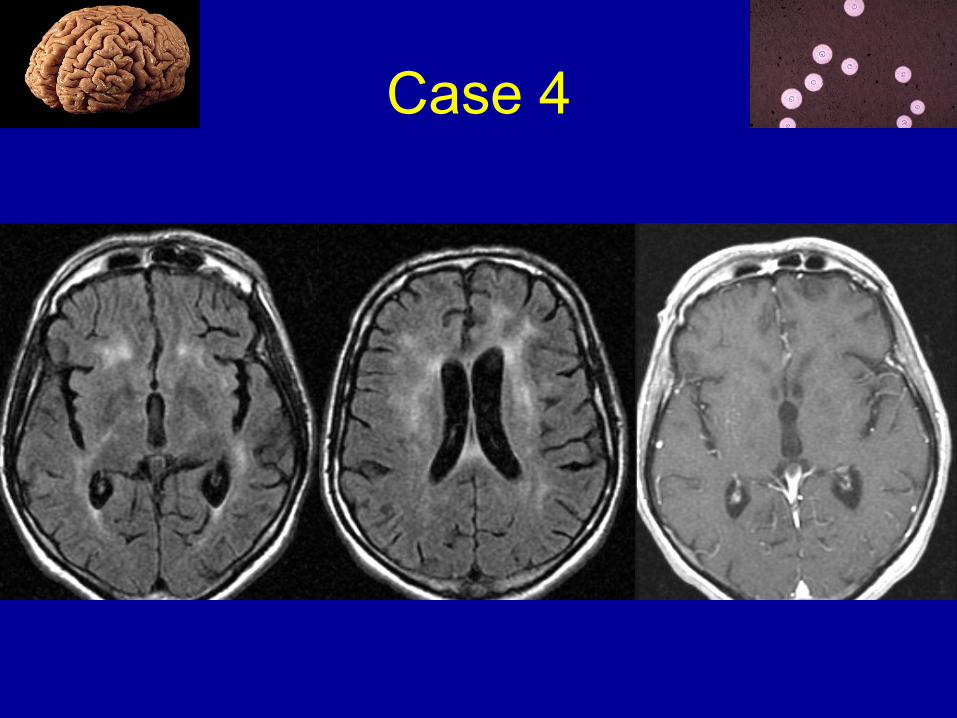
MRI showed bilateral moderate atrophy and
confluent subcortical T2 hyperintensities
without enhancement or mass effect:
Case 4
Case 4
Serum: CD4 had risen from 34 to 115; viral load had dropped from 180K to 9K.
CSF: glucose 72, protein 140, WBC 4 (65L, 25M); JCV, VDRL, mycobacterial culture neg; CSF HIV viral load > 750K.
Uncontrolled CNS viral replication vs. IRIS. ART changed to efavirenz + lamivudine/zidovudine for better CNS penetration, later changed to atazanavir/ritonavir, lamivudine/ zidovudine, and tenofovir.
Case 4
He continued to have behavioral changes
including apathy, requiring skilled nursing
facility care for about a year but then he
moved into a supported living facility with
his own apartment.
His serum viral load was undetectable and
was doing well for about a year.

Case 4
Two years after diagnosis he gradually
developed poor hygiene and had several
falls. He reported a "limp" in his left leg
and endorsed some "slowing down" and
"not taking care of myself." He also had
incontinence and weight loss and was
admitted to the hospital.
PE: frontal dysfunction (grasp, Meyerson,
verbal fluency), bradykinesia, rigidity,
hyper-reflexia, and marked gait instability.
Case 4
Case 4
Diffuse brain lesion
Differential: subacute to chronic mental
status changes in a patient with HIV
• HIV-associated dementia (HAD)
• Neurosyphilis
• HIV rebound meningoencephalitis
• Neuro-IRIS
• Cytomegalovirus (CMV) encephalitis
• PML (usu with focal deficits)
Diffuse brain lesion
CMV Clinical:
• CMV encephalitis occurs with profound
immunosuppression (usu CD4 < 50)
• Can manifest with progressive dementia
similar to HAD, but is a rare cause of
dementia
• Imaging may show inflammation around
the ventricles and meninges, but this is not
specific
Diffuse brain lesion
CMV Diagnosis:
• PCR for CMV DNA from the CSF is both
sensitive and specific.
• CMV also infects the retina (retinitis),
ventricles (ventriculitis), spinal cord
(myelitis), nerve roots (lumbosacral
polyradiculopathy), or peripheral nerves
(mononeuritis multiplex).
Diffuse brain lesion
HIV-associated dementia:
• Subacute to chronic subcortical dementia
• Early cognitive deficits in short-term
memory, mental slowing, reading and
comprehension problems, concentration
problems, and apathy.
Diffuse brain lesion
HIV-associated dementia:
• Early motor deficits are often subclinical
but can be detected on examination as
slowing of rapid movements.
• Later patients have a more diffuse
dementia, frontal release signs, and more
obvious extra-pyramidal symptoms.
Diffuse brain lesion
HIV-associated dementia:
• Diagnosis of exclusion.
• Minimal focal findings compared to PML
• CSF findings are nonspecific, but often
mild lymphocytic pleiocytosis; HIV RNA
viral load is often detectable and may even
be elevated above that in the blood; with
cART, however, CSF viral load correlates
poorly with severity of dementia
Case 4
HAD Diagnosis:
• Neuropsychological testing can help
distinguish HAD from other causes of
dementia, which is becoming more
important as patients are living longer and
are thus at risk for AD and vascular
dementia.
• In Uganda, older age and lower CD4 count
were associated with increased risk for
HAD Wong 2007
Case 4
Serum: viral load < 50, macrocytic anemia.
HIV genotype: TAM (41L) [6-fold
decreased potency of zidovudine].
CSF: glucose 79, protein 76, WBC 4 (90L);
JCV neg; HIV viral load 5,270; CSF HIV
genotype same as serum.
Case 4
ART regimen ultimately changed to lopinavir/ritonavir, emtricitabine/tenofovir, zidovudine; nevirapine later added for optimal “Neuro-HAART”. Donepezil added.
As of last visit, his mental status was improved (no apathy, normal verbal fluency, continued grasp), improved speed of movements, and significant improvement in gait. Viral load remained < 50 although CD4 still 160s. MRI was stable.
Neuro-ART
• Incidence of HIV-associated dementia (as
well as toxo and PCNSL) has decreased
with effective ART
• Even with good immune and viral
response, however, neurocognitive
impairment is common
• Emerging evidence that early ART is more
neuro-protective
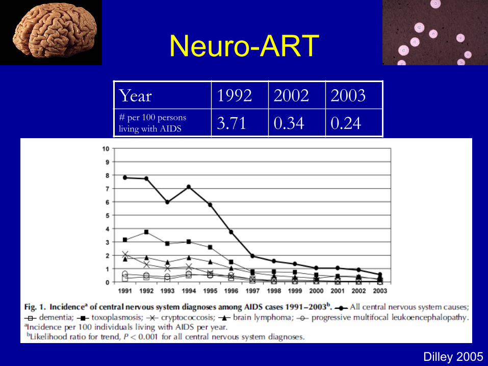
Dilley 2005
Year 1992 2002 2003# per 100 persons
living with AIDS 3.71 0.34 0.24
Neuro-ART
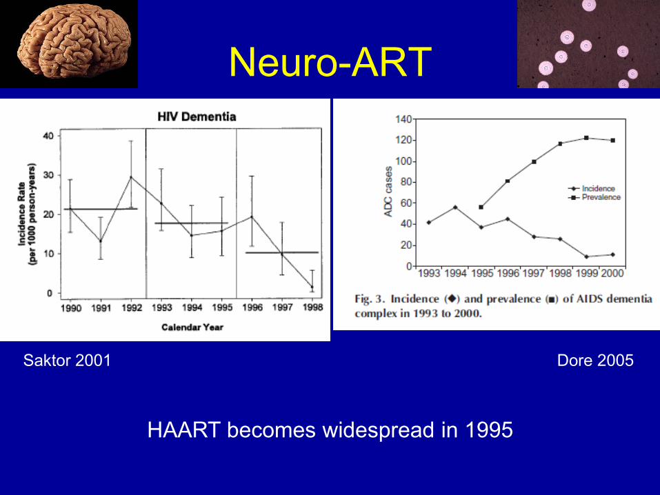
Saktor 2001 Dore 2005
Neuro-ART
HAART becomes widespread in 1995
Neuro-ART
• Weight of evidence suggests that control
of HIV in periphery = control of HIV in CNS
• To date, no convincing and reproducible
evidence that CNS-penetration matters if
regimen is effective in periphery
CNS Penetration Effectiveness Score
• Classification of ARVs into 3 categories
– 0 = low penetration
• Chemical properties with poor CNS penetration
• Unmeasurable in CSF or IC50 in CSF is low
• Clinical data for poor CNS penetration
– 0.5 = medium penetration
– 1 = high penetration
• cART CPE: sum of CPE for ARVs in
regimen Letendre 2008
Neuro-ART
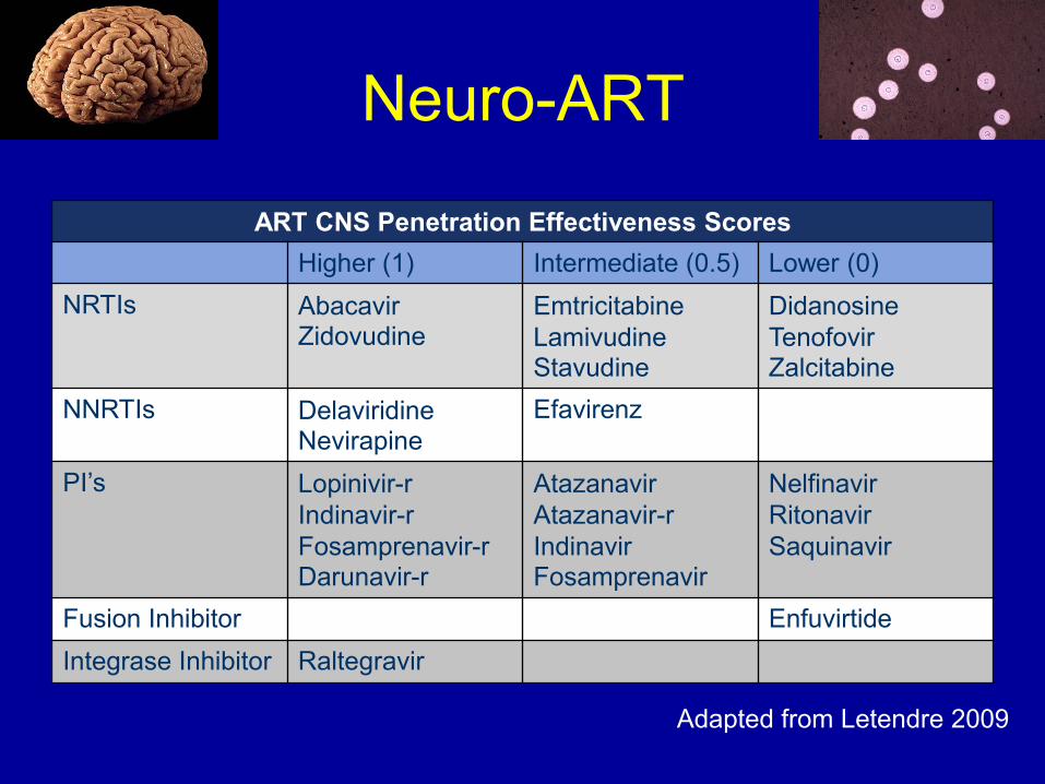
ART CNS Penetration Effectiveness Scores
Higher (1) Intermediate (0.5) Lower (0)
NRTIs AbacavirZidovudine
Emtricitabine
LamivudineStavudine
Didanosine
TenofovirZalcitabine
NNRTIs DelaviridineNevirapine
Efavirenz
PI’s Lopinivir-r
Indinavir-r
Fosamprenavir-rDarunavir-r
Atazanavir
Atazanavir-r
IndinavirFosamprenavir
Nelfinavir
Ritonavir
Saquinavir
Fusion Inhibitor Enfuvirtide
Integrase Inhibitor Raltegravir
Adapted from Letendre 2009
Neuro-ART
Neuro-ART
• In cross-sectional study, higher CPE
associated with lower CSH viral load
• Clinical outcomes have been mixed
– Some evidence of benefit in cognitive function
– Some evidence of no difference
– Most recently some evidence of worsening
cognitive function
• Limited by small observational studies,
incomplete toxicity data, poor
understanding of CSF to CNS relationship
Acknowledgements
“We must try to form and strengthen
synapses with other neurons (medical
colleagues) as much as possible, to make
the neural network stronger.”
-Nagagopal Venna
End
Case 2b
67M with a history of leukopenia dating to at
least 1989 presents with several weeks of
progressive confusion and gait problems.
PE: encephalopathic, left arm weakness.
MRI…
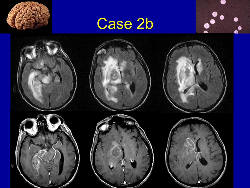
Case 2b
Case 2b
Diagnosis:
• Clinical course of subacute focal deficits
• MRI characteristic of PCNSL (mass effect,
patchy ring enhancement, cross corpus
callosum)
• CSF cytology insensitive but highly
specific
• CSF EBV PCR 80-98% sens, 88-100%
spec
Case 2b
Diagnosis:
• Metabolic imaging (SPECT, PET)
suggestive but not reliable
• Gold standard remains biopsy
• Should consider when Toxo IgG neg, no
response to Toxo Rx or presents while on
TMP-SMX
Case 2b
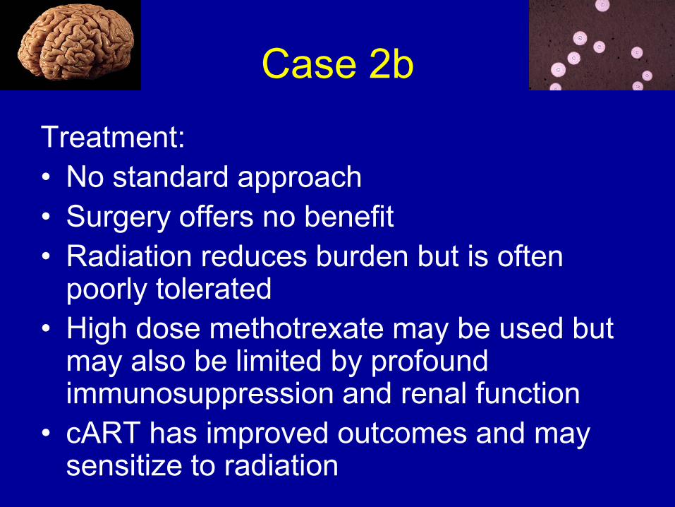
Treatment:
• No standard approach
• Surgery offers no benefit
• Radiation reduces burden but is often poorly tolerated
• High dose methotrexate may be used but may also be limited by profound immunosuppression and renal function
• cART has improved outcomes and may sensitize to radiation
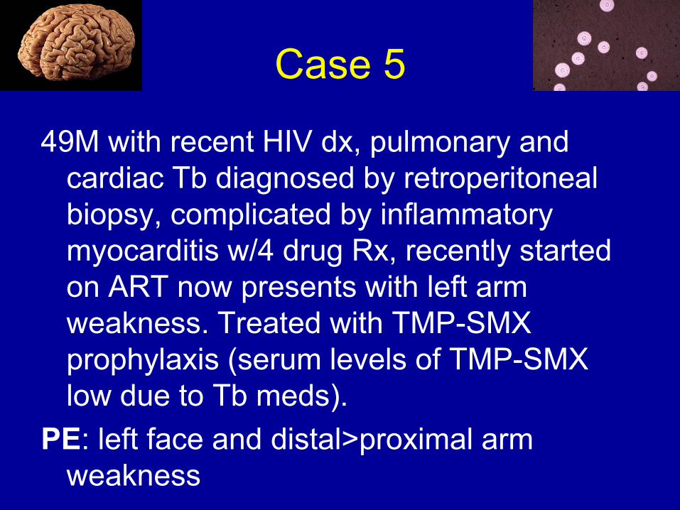
Case 5
49M with recent HIV dx, pulmonary and
cardiac Tb diagnosed by retroperitoneal
biopsy, complicated by inflammatory
myocarditis w/4 drug Rx, recently started
on ART now presents with left arm
weakness. Treated with TMP-SMX
prophylaxis (serum levels of TMP-SMX
low due to Tb meds).
PE: left face and distal>proximal arm
weakness
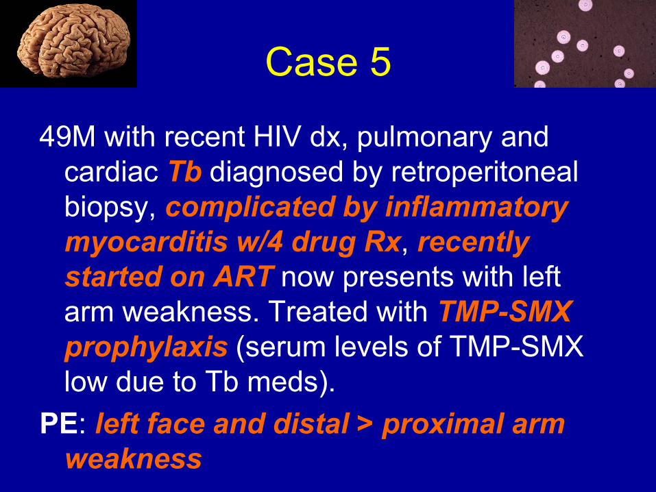
Case 5
49M with recent HIV dx, pulmonary and
cardiac Tb diagnosed by retroperitoneal
biopsy, complicated by inflammatory
myocarditis w/4 drug Rx, recently
started on ART now presents with left
arm weakness. Treated with TMP-SMX
prophylaxis (serum levels of TMP-SMX
low due to Tb meds).
PE: left face and distal > proximal arm
weakness
Case 5
Labs: CD4 44, viral load 961K, toxoplasma
IgM-/IgG+, CMV IgG+, Urine histo Ag -.
CSF: (for ? seizure) normal protein and
glucose with 3 WBC. CSF AFB and
mycobacterial culture, EBV and CMV
PCR, and crypto Ag were negative.
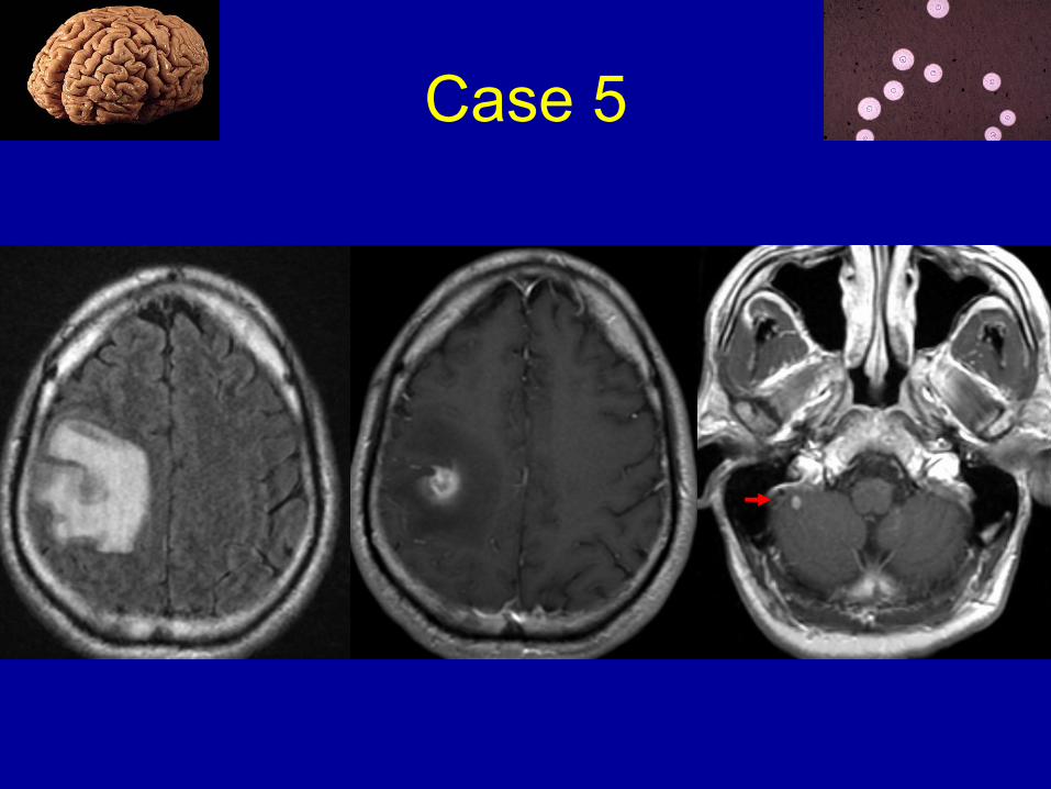
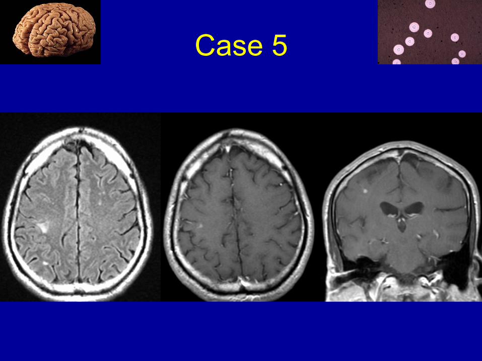
Baseline MRI (for ? seizure): right pre/post-
central T2 lesion, no enhancement.

Case 5

Case 5
Follow-up MRI at time of neurological
symptoms:
Case 5
Case 5
Due to concern for toxo, bactrim changed to
treatment dose sulfadiazine and
pyrimethamine. LP was deferred due to
concern for mass effect. Given his
concurrent use of rifabutin and bactrim,
serum sulfamethoxazole level was
checked and was low.
Baseline eval had shown serum toxo IgG+,
CSF EBV-, and systemic Tb as above.
Case 5
The plan was to give toxo Rx for two weeks, then reassess for improvement. Due to the development of continuous partial seizures, however, dexamethasone and antiepileptics were given. He improved over several days with increased strength in his left arm but developed myoclonus of the left wrist. He was continued on toxo and Tb treatment with slow steroid taper.
Follow-up MRI showed…
Case 5
Case 5
CNS tuberculosis vs. toxoplasmosis IRIS?
• Significant edema and previous Tb-related inflammation suggest CNS Tb IRIS
• Low sulfa level, + serum toxo IgG, and quick response could support toxoplasmosis
• While toxoplasmosis is the most common focal CNS OI in HIV, there are few reports of Toxo-IRIS (possibly because it resolves quickly with treatment)