Embed Size (px)
Citation preview
CEREBROVASCULAR DISEASES
By: Shifaa’ AlQa’qa’
Cerebrovascular diseases
• Brain disorders caused by pathologic processes involving blood vessels
• 3 pathogenic mechanisms
(1) thrombotic occlusion,
(2) embolic occlusion,
(3) vascular rupture/Hemorrhage
• Stroke:
is the clinical term for acute-onset neurologic deficits resulting from hemorrhagic or obstructive vascular lesions.
transient ischemic attack (TIA) ????
Hypoxia, Ischemia, and Infarction
• The brain may be deprived of oxygen by two general mechanisms:
Functional hypoxia: - low partial pressure of oxygen (e.g., high altitude), - impaired oxygen-carrying capacity (e.g., severe anemia, carbon monoxide poisoning), - inhibition of oxygen use by tissue (e.g., cyanide poisoning)
Ischemia: - Hypoperfusion ----- hypotension, vascular obstruction - transient or permanent
Global Cerebral Ischemia
• occur in the setting of severe systemic hypotension, usually when systolic pressures fall below 50 mm Hg, as in cardiac arrest, shock, and severe hypotension.
• Neurons are more susceptible to hypoxic injury than are glial cells
• The most susceptible neurons (vulnerable areas) are: - The pyramidal cells of the hippocampus and neocortex - Purkinje cells of the cerebellum
- In severe global cerebral ischemia, widespread neuronal
death occurs irrespective of regional vulnerability
• MORPHOLOGY:
the brain is swollen, wide gyri, narrowed sulci.
The cut surface shows poor demarcation between gray and white matter

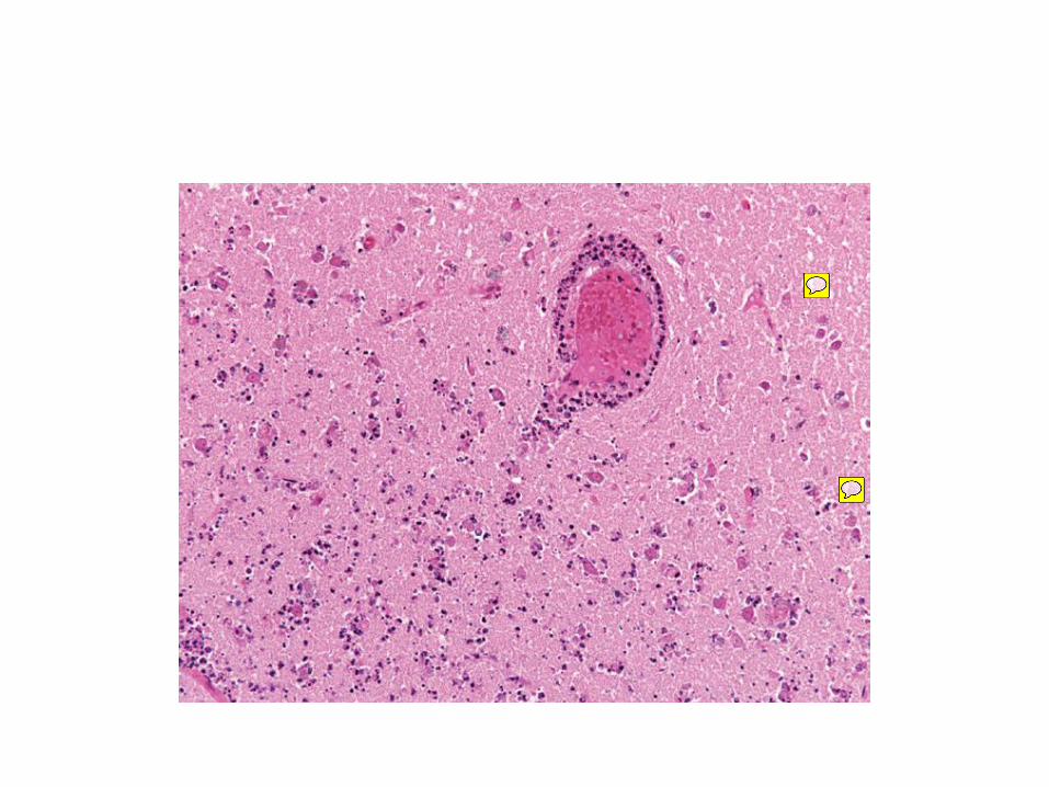
• Irreversible ischemic injury---- infarction Early changes (12 to 24 hours): - acute neuronal cell change - the reaction to tissue damage begins with infiltration by neutrophils Subacute changes (24 hours to 2 weeks) - necrosis of tissue - influx of macrophages - vascular proliferation, - reactive gliosis Repair (after 2 weeks) - removal of all necrotic tissue - Loss of organized CNS structure - gliosis
• The distribution of neuronal loss and gliosis in the neocortex typically is uneven with preservation of some layers and devastation of others—a pattern termed pseudolaminar necrosis.
• Border zone (“watershed”) infarcts:
- wedge shaped
- regions of the brain and spinal cord that lie at the most distal portions of arterial territories.
- In the cerebral hemispheres, the border zone between the anterior and the middle cerebral artery distributions is at greatest risk.
• The clinical outcome varies with the severity and duration of the insult:
Mild insult---- only a transient postischemic
confusional state, with eventual complete recovery
Severe insult ---- vegetative state, brain death--- mechanical ventilation---- brain autolysis
Focal Cerebral Ischemia
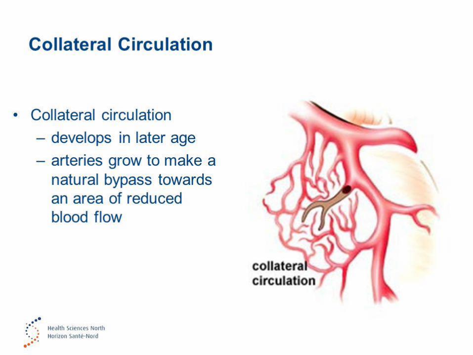
• Cerebral arterial occlusion ----- focal ischemia ------ infarction in the distribution of the compromised vessel.
• modified by collateral blood flow -------- the circle of Willis or cortical-leptomeningeal anastomoses
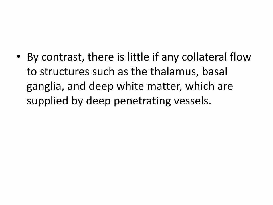
• By contrast, there is little if any collateral flow to structures such as the thalamus, basal ganglia, and deep white matter, which are supplied by deep penetrating vessels.
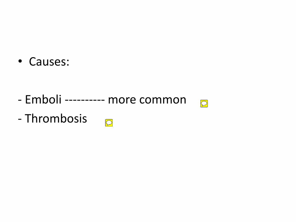
• Causes:
- Emboli ---------- more common
- Thrombosis

• Emboli:
- Cardiac mural thrombi (myocardial dysfunction, valvular disease, and atrial fibrillation)
- Arterial thromboemboli (atheromatous plaques within the carotid arteries or aortic arch)
- Venous emboli/paradoxical embolism (thromboemboli from deep leg veins and fat emboli)
• The territory of the middle cerebral artery, a direct extension of the internal carotid artery, is most frequently affected by embolic infarction.
• Emboli tend to lodge where vessels branch or in areas of stenosis, usually caused by atherosclerosis.
• Thrombosis:
- Thrombotic occlusions usually are superimposed on atherosclerotic plaques
- common sites are:
The carotid bifurcation,
The origin of the middle cerebral artery,
At either end of the basilar artery.
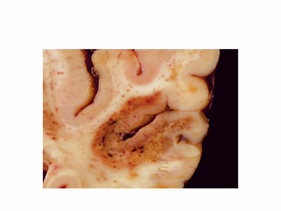
• Infarcts can be divided into two broad groups: Nonhemorrhagic infarcts--- Hemorrhagic infarcts--- result from reperfusion of ischemic tissue, either through collaterals or after dissolution of emboli ----- multiple, petechial hemorrhages Thrombolytic therapies????

• MORPHOLOGY: Macroscopic appearance of a nonhemorrhagic infarct: first 6 hours---no change 48 hours--- tissue is pale, soft, and swollen days 2 to 10--- the brain turns gelatinous and friable, and the boundary between normal and abnormal tissue becomes more distinct as edema resolves in the adjacent viable tissue. day 10 to week 3--- the tissue liquefies, eventually leaving a fluid-filled cavity lined by dark gray tissue, which gradually expands as dead tissue is resorbed
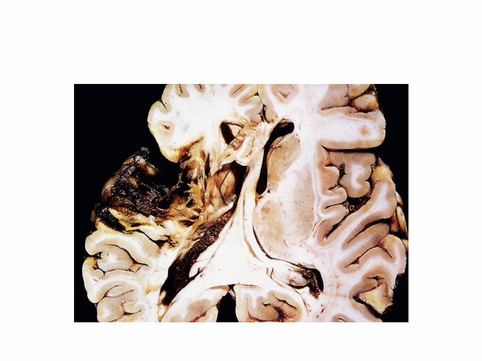
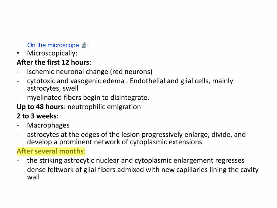
• Microscopically: After the first 12 hours: - ischemic neuronal change (red neurons) - cytotoxic and vasogenic edema . Endothelial and glial cells, mainly
astrocytes, swell - myelinated fibers begin to disintegrate. Up to 48 hours: neutrophilic emigration 2 to 3 weeks: - Macrophages - astrocytes at the edges of the lesion progressively enlarge, divide, and
develop a prominent network of cytoplasmic extensions After several months: - the striking astrocytic nuclear and cytoplasmic enlargement regresses - dense feltwork of glial fibers admixed with new capillaries lining the cavity
wall
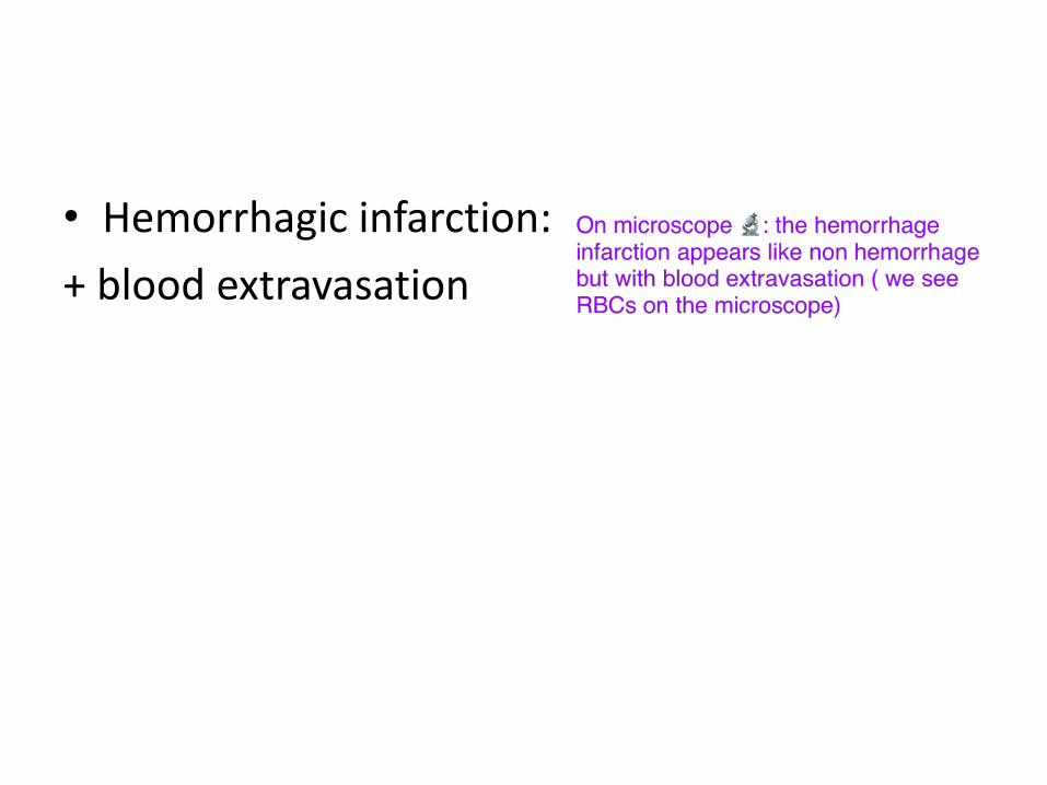
• Hemorrhagic infarction:
+ blood extravasation
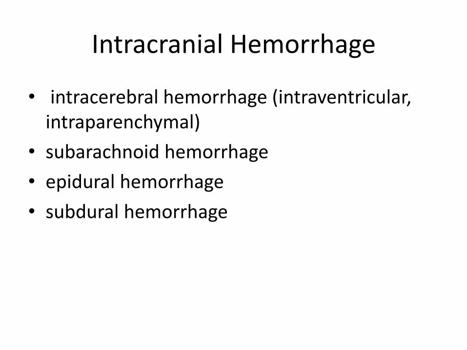
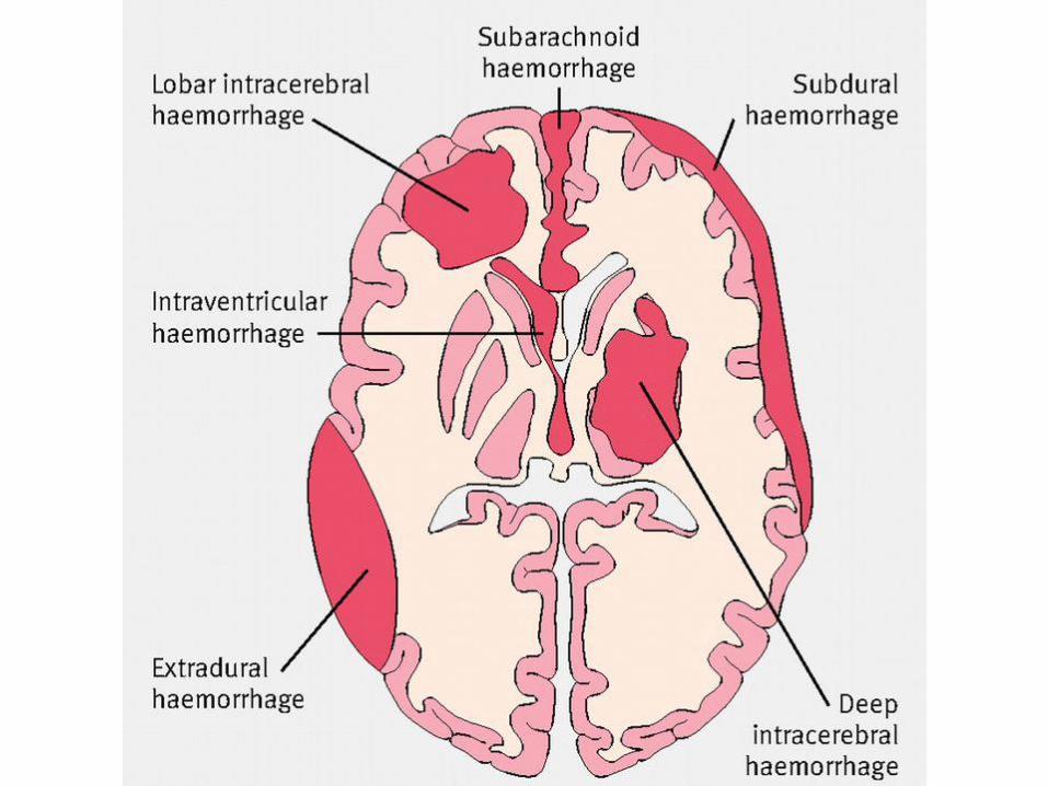
Intracranial Hemorrhage
• intracerebral hemorrhage (intraventricular, intraparenchymal)
• subarachnoid hemorrhage
• epidural hemorrhage
• subdural hemorrhage
Primary Brain Parenchymal Hemorrhage
• Spontaneous ----- nontraumatic
• peak incidence at about 60 years of age
• rupture of a small intraparenchymal vessels
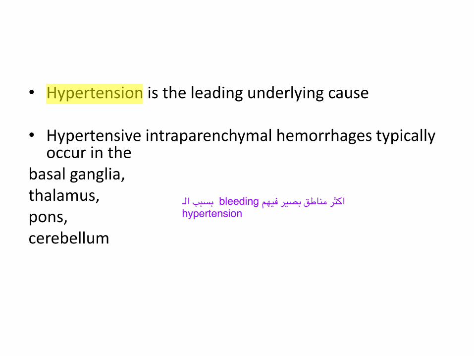
• Hypertension is the leading underlying cause
• Hypertensive intraparenchymal hemorrhages typically occur in the
basal ganglia, thalamus, pons, cerebellum
• it can affect small regions and be clinically silent
• can be clinically devastating when it affects large portions of the brain or extends into the ventricular system
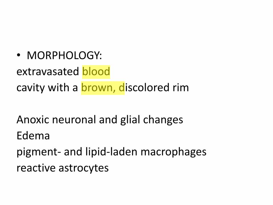
• MORPHOLOGY:
extravasated blood
cavity with a brown, discolored rim
Anoxic neuronal and glial changes
Edema
pigment- and lipid-laden macrophages
reactive astrocytes
Cerebral Amyloid Angiopathy
• is a disease in which amyloidogenic peptides (beta amyloid), typically the same ones found in Alzheimer disease, deposit in the walls of medium- and small-caliber meningeal and cortical vessels.
• Amyloid deposition weakens vessel walls and increases the risk of hemorrhages
• CAA-associated hemorrhages often occur in the lobes of the cerebral cortex (lobar hemorrhages) ----- Primary intraparenchymal hemorrhages
Hypertensive Cerebrovascular Disease
• Hypertension --- hyaline arteriolar sclerosis ----weakened wall---- vulnerable to rupture
• arteries and arterioles that supply:
the basal ganglia,
the hemispheric white matter,
and the brain stem
• In some instances, minute aneurysms (Charcot-Bouchard microaneurysms) form in vessels less than 300 μm in diameter.
• Pathologic brain processes are related to hypertension:
- Intracerebral hemorrhage
- Lacunar infarcts
- slit hemorrhage
- Acute hypertensive encephalopathy
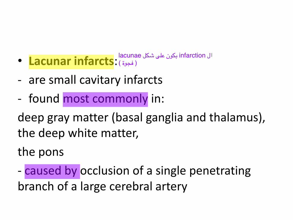
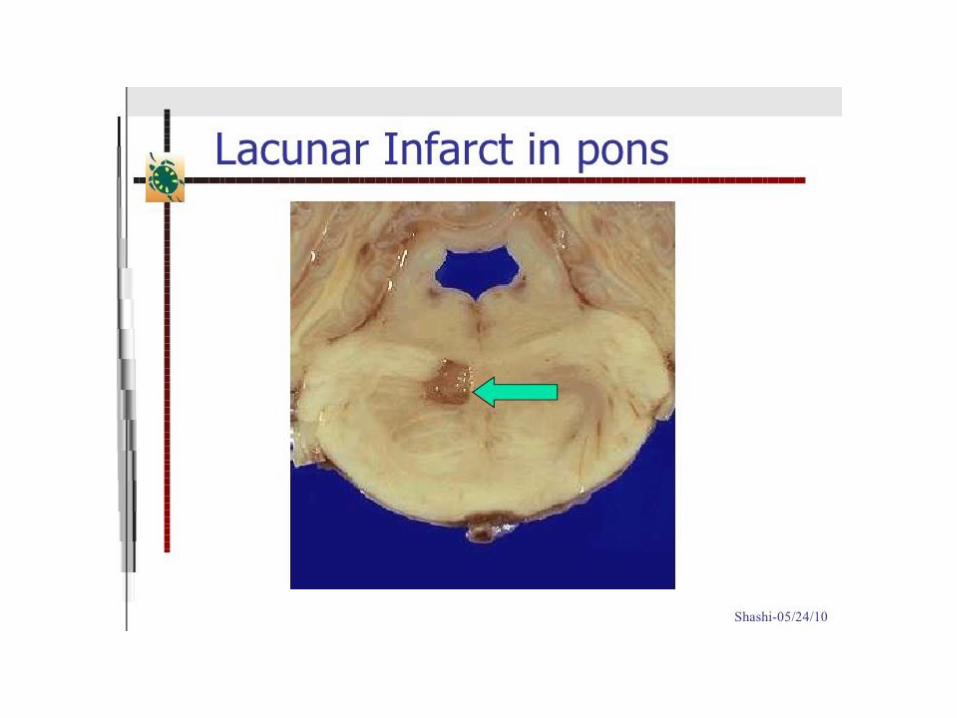
• Lacunar infarcts:
- are small cavitary infarcts
- found most commonly in:
deep gray matter (basal ganglia and thalamus), the deep white matter,
the pons
- caused by occlusion of a single penetrating branch of a large cerebral artery
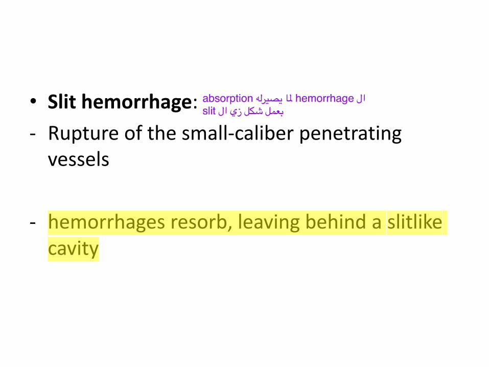
• Slit hemorrhage:
- Rupture of the small-caliber penetrating vessels
- hemorrhages resorb, leaving behind a slitlike cavity
• Acute hypertensive encephalopathy:
- sudden sustained rises in diastolic blood pressure to greater than 130 mm Hg
- increased intracranial pressure
- global cerebral dysfunction
headaches, confusion, vomiting, convulsions, coma
- brain edema,
- transtentorial or tonsillar herniation
- Petechiae and fibrinoid necrosis of arterioles in the gray and white matter may be seen microscopically

Vasculitis
• cause cerebral infarction
• Infectious
• systemic forms of vasculitis
• Primary angiitis of the CNS