Embed Size (px)
Citation preview
Folia Neuropathologica 2010; 48/4300
Cervical spinal tuberculosis
Teresa Wierzba-Bobrowicz1, Elżbieta Michalak2, Radosław Michalik3, Tomasz Stępień1
1Department of Neuropathology, Institute of Psychiatry and Neurology, Warsaw, Poland, 2Department of Pathology, Institute of Mother
and Child, Warsaw, Poland, 3Department of Neurosurgery, Institute of Psychiatry and Neurology, Warsaw, Poland
Folia Neuropathol 2010; 48 (4): 300-304
A b s t r a c t
Cervical spinal tuberculosis is a rare variant of extra-pulmonary tuberculosis. We present the case of Vietnamesewoman, aged 48, who was admitted to the Department of Neurosurgery because of a cervical spine (C7) compressionfracture. Several months earlier, the patient complained of neck pain and numbness of the hands. On physical exam-ination, the woman was subfebrile and complained of pain over the cervical spinal area. Neurological examinationrevealed no focal motor weakness. The roentgenograms of chest, pelvis and cranium were without patholo gicalchanges. Abdominal ultrasonography was normal. Radioisotope bone-scanning showed abnormal accumulation of iso-tope in the lower cervical region, thoracic vertebra, as well as in the articulations of knees and shoulders and in theleft tibial bone. An MRI scan revealed compression fracture of the C7 vertebral body with infiltration of paraspinal tis-sues at the vertebral column with indentation of osseous masses into the spinal canal. The lesion resembled neoplasmmetastasis. The neoplasm infiltrating vertebral body C7, two discs, C6-C7 and C7-Th1, and ligament were removed sur-gically. Neuropathological examination of the removed material showed typical granulomatous inflammation withcharacteristic infiltrate of lymphocytes, epithelioid macrophages and Langhans-type multi nucleated giant cells. Thespoligotyping method confirmed the presence of Mycobacterium tuberculosis complex in the specimens.
Key words: tuberculosis, cervical spine, compression fracture, spoligotyping.
Communicating author:
Teresa Wierzba-Bobrowicz, MD, PhD, Department of Neuropathology, Institute of Psychiatry and Neurology, Sobieskiego 9, 02-957 Warsaw,
Poland, e-mail: [email protected]
Case report
Introduction
Tuberculosis (tubercle bacillus – TB) is a majorglobal health problem. It is a communicable diseasecaused by Mycobacterium tuberculosis or Mycobac-terium bovis, including the attenuated BCG strain.
Every year eight million new cases are detectedand about three million people die from this diseaseworldwide. Extra-pulmonary TB may involve any organsystems and clinical symptoms are non-specific [3].Extra-pulmonary TB is more common in childhoodand in patients with HIV/AIDS [5,6,10,17].
Spinal tuberculosis is a rare variant of TB, com-prising less than 3% of cases. Skeletal tuberculosislesions may simulate primary or metastatic disease[2,15,19]. Lumbar and thoracic regions are often in -volved, whereas TB occurrence in the cervical spine is uncommon [18].
We report herein the case of a Vietnamese wo manwho had cervical spinal TB, initially treated as com-press ion fracture, and probably other inflammatoryfoci in the bone system.
Folia Neuropathologica 2010; 48/4 301
Case report
A 48-year-old Vietnamese woman was admitted tothe Department of Neurosurgery, Institute of Psychiatryand Neurology, because of a cervical spine (C7) com-pression fracture. Five months earlier, the patient com-plained of neck pain and numbness of the hands. Tenyears ago she had a total hysterectomy (the operationtook place in Vietnam; documentation was lacking).
On physical examination, the patient was subfebrileand complained of pain over the cervical spinal area.
Neurological examination revealed no focal motorweakness.
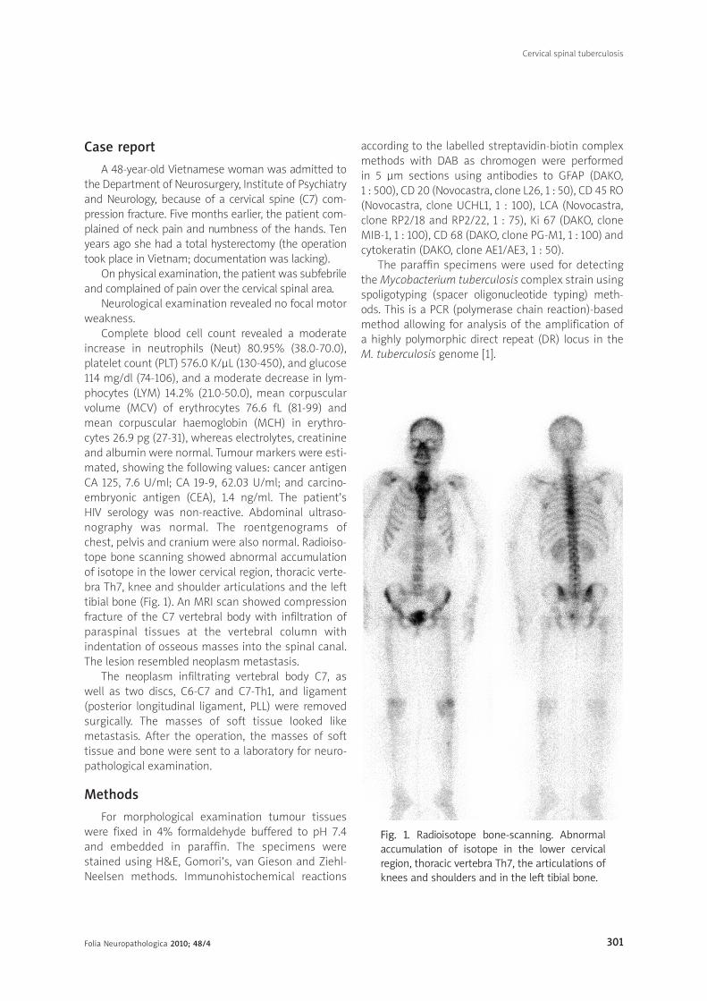
Complete blood cell count revealed a moderateincrease in neutrophils (Neut) 80.95% (38.0-70.0),platelet count (PLT) 576.0 K/µL (130-450), and glucose114 mg/dl (74-106), and a moderate decrease in lym-phocytes (LYM) 14.2% (21.0-50.0), mean corpuscularvolume (MCV) of erythrocytes 76.6 fL (81-99) andmean corpuscular haemoglobin (MCH) in erythro-cytes 26.9 pg (27-31), whereas electrolytes, creatinineand albumin were normal. Tumour markers were esti-mated, showing the following values: cancer antigenCA 125, 7.6 U/ml; CA 19-9, 62.03 U/ml; and carcino -embryonic antigen (CEA), 1.4 ng/ml. The patient’s HIV serology was non-reactive. Abdominal ultraso -nography was normal. The roentgenograms of chest, pelvis and cranium were also normal. Radioiso-tope bone scanning showed abnormal accumulationof isotope in the lower cervical region, thoracic verte-bra Th7, knee and shoulder articulations and the lefttibial bone (Fig. 1). An MRI scan showed compressionfracture of the C7 vertebral body with infiltration ofparaspinal tissues at the vertebral column withindentation of osseous masses into the spinal canal.The lesion resembled neoplasm metastasis.
The neoplasm infiltrating vertebral body C7, aswell as two discs, C6-C7 and C7-Th1, and ligament(posterior longitudinal ligament, PLL) were removedsurgically. The masses of soft tissue looked likemetastasis. After the operation, the masses of softtissue and bone were sent to a laboratory for neuro -pathological examination.
Methods
For morphological examination tumour tissueswere fixed in 4% formaldehyde buffered to pH 7.4and embedded in paraffin. The specimens werestained using H&E, Gomori’s, van Gieson and Ziehl-Neelsen methods. Immunohistochemical reactions
according to the labelled streptavidin-biotin complexmethods with DAB as chromogen were performed in 5 µm sections using antibodies to GFAP (DAKO, 1 : 500), CD 20 (Novocastra, clone L26, 1 : 50), CD 45 RO(Novocastra, clone UCHL1, 1 : 100), LCA (Novocastra,clone RP2/18 and RP2/22, 1 : 75), Ki 67 (DAKO, cloneMIB-1, 1 : 100), CD 68 (DAKO, clone PG-M1, 1 : 100) andcytokeratin (DAKO, clone AE1/AE3, 1 : 50).
The paraffin specimens were used for detectingthe Mycobacterium tuberculosis complex strain usingspoligotyping (spacer oligonucleotide typing) meth-ods. This is a PCR (polymerase chain reaction)-basedmethod allowing for analysis of the amplification of a highly polymorphic direct repeat (DR) locus in the M. tuberculosis genome [1].
Fig. 1. Radioisotope bone-scanning. Abnormalaccumulation of isotope in the lower cervicalregion, thoracic vertebra Th7, the articulations ofknees and shoulders and in the left tibial bone.
Cervical spinal tuberculosis
Folia Neuropathologica 2010; 48/4302
Results
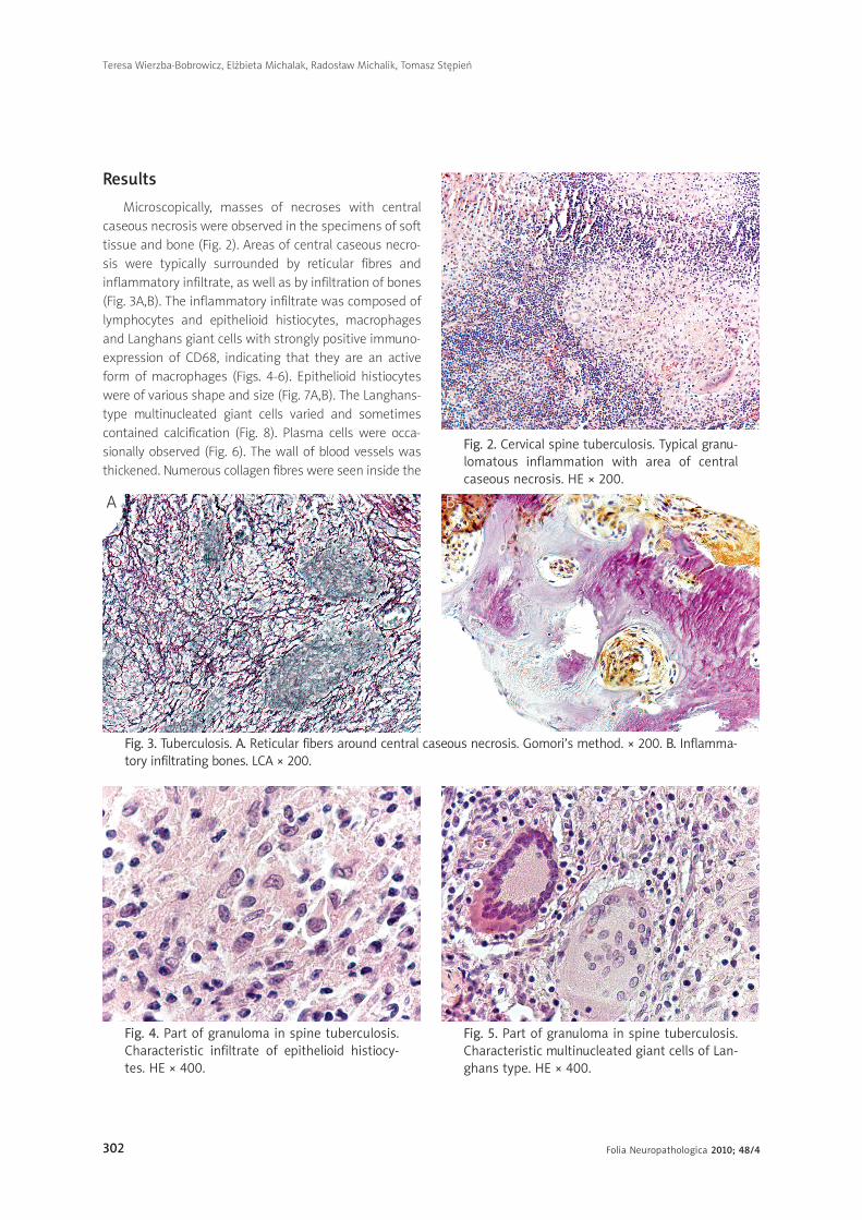
Microscopically, masses of necroses with centralcaseous necrosis were observed in the specimens of softtissue and bone (Fig. 2). Areas of central caseous necro-sis were typically surrounded by reticular fibres andinflammatory infiltrate, as well as by infiltration of bones(Fig. 3A,B). The inflammatory infiltrate was composed oflymphocytes and epithelioid histiocytes, macrophagesand Langhans giant cells with strongly positive immuno-expression of CD68, indicating that they are an activeform of macrophages (Figs. 4-6). Epithelioid histiocyteswere of various shape and size (Fig. 7A,B). The Langhans-type multinucleated giant cells varied and sometimescontained calcification (Fig. 8). Plasma cells were occa-sionally observed (Fig. 6). The wall of blood vessels wasthickened. Numerous collagen fibres were seen inside the
Fig. 3. Tuberculosis. A. Reticular fibers around central caseous necrosis. Gomori’s method. × 200. B. Inflam ma-tory infiltrating bones. LCA × 200.
A B
Fig. 4. Part of granuloma in spine tuberculosis.Characteristic infiltrate of epithelioid histiocy -tes. HE × 400.
Fig. 5. Part of granuloma in spine tuberculosis.Characteristic multinucleated giant cells of Lan -ghans type. HE × 400.
Fig. 2. Cervical spine tuberculosis. Typical granu -lomatous inflammation with area of centralcaseous necrosis. HE × 200.
Teresa Wierzba-Bobrowicz, Elżbieta Michalak, Radosław Michalik, Tomasz Stępień
Folia Neuropathologica 2010; 48/4 303
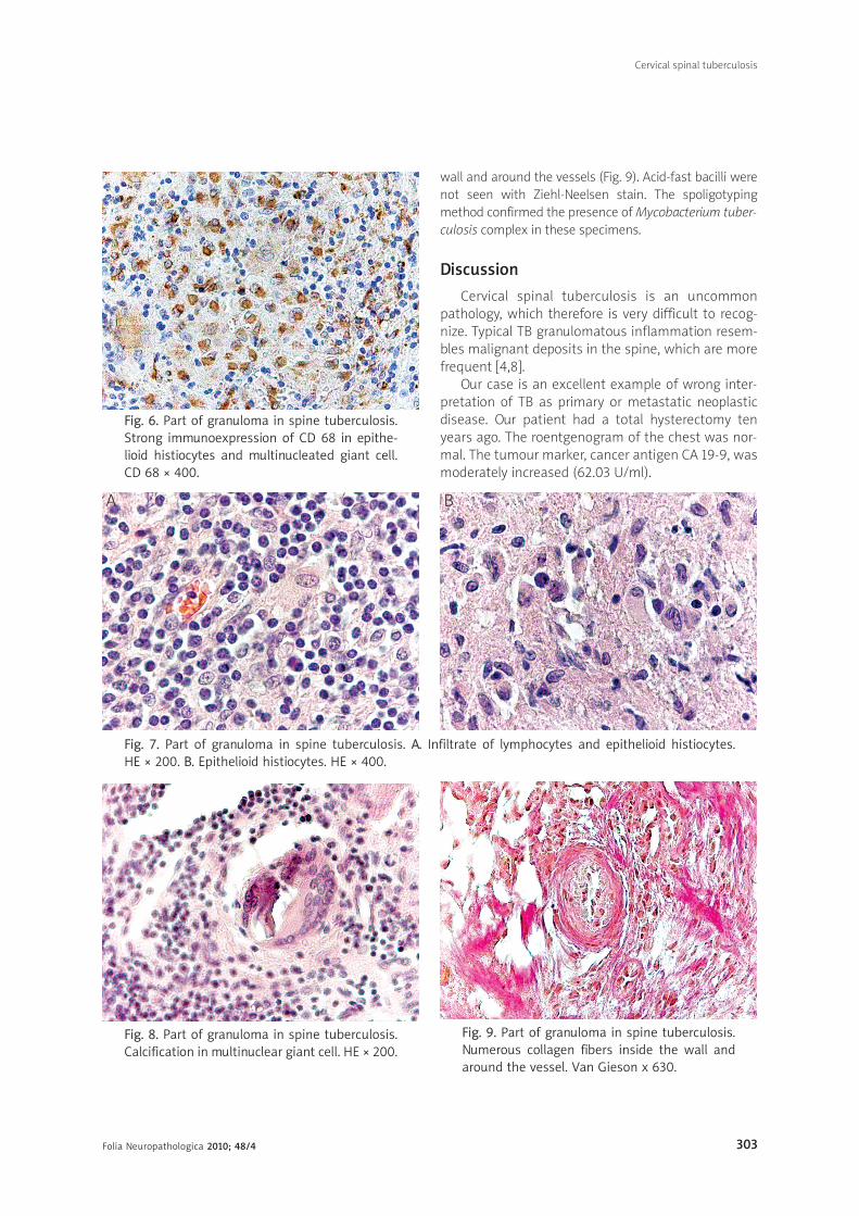
Fig. 8. Part of granuloma in spine tuberculosis.Calcification in multinuclear giant cell. HE × 200.
Fig. 7. Part of granuloma in spine tuberculosis. A. Infiltrate of lymphocytes and epithelioid histiocytes. HE × 200. B. Epithelioid histiocytes. HE × 400.
Fig. 9. Part of granuloma in spine tuberculosis.Numerous collagen fibers inside the wall andaround the vessel. Van Gieson x 630.
B
Fig. 6. Part of granuloma in spine tuberculosis.Strong immunoexpression of CD 68 in epithe-lioid histiocytes and multinucleated giant cell.CD 68 × 400.
A
wall and around the vessels (Fig. 9). Acid-fast bacilli werenot seen with Ziehl-Neelsen stain. The spoligotypingmethod confirmed the presence of Mycobacterium tuber-culosis complex in these specimens.
Discussion
Cervical spinal tuberculosis is an uncommonpathology, which therefore is very difficult to recog-nize. Typical TB granulomatous inflammation resem-bles malignant deposits in the spine, which are morefrequent [4,8].
Our case is an excellent example of wrong inter-pretation of TB as primary or metastatic neoplasticdisease. Our patient had a total hysterectomy tenyears ago. The roentgenogram of the chest was nor-mal. The tumour marker, cancer antigen CA 19-9, wasmoderately increased (62.03 U/ml).
Cervical spinal tuberculosis

Folia Neuropathologica 2010; 48/4304
Primarily, antigen CA 19-8 was determined in neo-plastic cells of the large intestine [7]. Nowadays, it isknown that the level of this marker increases in pan-creatic cancer, carcinoma of the stomach and cholan-gioma (70-100%). The correlation between the devel-opmental stage of this neoplasm and the CA 19-9level is also known. However, the specificity of testsbased on this antigen is rather limited because of itsincreased concentration, for example in the case ofpancreatitis and liver pathologies [12,16]. In our case,radioisotope bone scanning showed abnormal accu-mulation of isotope in two regions of the vertebra, aswell as in articulations and long bone, which suggestsa multifocal disease process. An MRI scan showedcompression fracture of the C7 vertebral body withinfiltration of paraspinal tissue at the vertebral col-umn with indentation of osseous masses into thespinal canal. The presented case was similar to thoseof bone and spinal neoplastic metastasis [4]. There-fore, other conditions such as TB, pyogenic infection,neurosarcoidosis, syphilis and fungal osteomyelitisshould also be taken into consideration [9,11,13,14].Neuropathological examination of the tissue re -moved surgically showed typical granulomatousinflammation with characteristic infiltrate of lym- phocytes, epithelioid macrophages and Langhanstype multinucleated giant cells. Unfortunately, acid-fast bacilli were not seen with the use of the Ziehl-Neelsen staining method. The spoligotyping methodwas applied to identify the pathogenic factor. Spo-li gotyping is a novel PCR-based method. This geno-typing technique allows one to analyse strain-depen-dent polymorphisms found in spacer sequencespresent within the direct repeat genomic region ofMycobacterium tuberculosis complex strains. The spo -ligotyping method confirmed the presence of Myco -bacte rium tuberculosis complex in our specimens.Myco bacterium tuberculosis complex consists of thefollowing bacilli pathogenic for humans: Mycobacte -rium (M.) tuberculosis, M. africanum, M. bovis, includ-ing BCC strain, and M. microti.
This group of bacilli is responsible for the develop-ment of the classic form of tuberculosis. Diagnosticprocedures of TB should include serological examina-tion, and neuropathological investigation of speci-mens obtained from the lesion, including the detec-tion of acid-fast bacilli with Ziehl-Neelsen staining, orDNA fragments of Mycobacterium tuberculosis com-plex, using spoligotyping.
References
1. Augustynowicz-Kopeć E, Jagielski T, Kozińska M, Zabost A, Zwol-ska Z. Znaczenie metody spoligotyping w epidemiologicznychdochodzeniach gruźlicy. Pneumol Alergol Pol 2007; 75: 22-31.
2. Biernat W. Metastatic tumours of the central nervous system –a pathological approach. Folia Neuropathol 2009; 47: 228-233.
3. Dass B, Puet TA, Watanakunakorn C. Tuberculosis of the spine(Pott’s disease) presenting as “compression fractures”. SpinalCord 2002; 40: 604-608.
4. Dima-Cozma C, Mitu F, Rezus E, Arhire O, Pectu I, Grigorias C,Banu C, Cozma S. Spinal tuberculosis or bone metastases? Casereport. Rev Med Chir Soc Med Nat lasi 2010; 114: 115-119.
5. Dolin PJ, Raviglione MC, Kochi A. Global tuberculosis incidenceand mortality during 1990-2000. Bull World Health Organ 1994;72: 213-220.
6. Gunasekera TMR, Karunathilake DH, Wickramasinghe EMNW,Jayaweera KAHM. A rare presentation of tuberculousosteomyelitis in childhood. Sri Lanka Journal of Child Health2008; 37: 61-62.
7. Jessup JM. Tumor markers – prognostic and therapeutic impli-cantions for colorectal carcinoma. Surg Oncol 1998; 7: 139-151.
8. Kolasa M, Jesionek-Kupnicka D, Kordek R, Kolasa P. Primaryspinal cord melanoma – a case report. Folia Neuropathol 2010;48: 212-216.
9. Lindhal S, Nymann RS, Brismar J. Imaging of tuberculosis. IV.Spinal manifestations in 63 patients. Acta Radiol 1996; 37: 506-511.
10. Mannepalli S, Mitchell-Samon L, Guzman N, Relan M, McCar -ter YS. Mycobacterium tuberculosis osteomyelitis in a patientwith human immunodeficiency virus/acquired immunodeficiencysyndrome (HIV/AIDS): a case report. Cases Journal 2010; 3: 67.
11. Mathew J, Tripathy P, Grewal S. Epidural tuberculosis involvingthe entire spine. Neurol Neurochir Pol 2009; 43: 470-474.
12. Paduch R, Klatka J. Markery nowotworowe. Onkol Pol 2003; 6:77-82.
13. Perronne C, Saba J, Behloul Z. Pyogenic and tuberculosisspondylodiskitis (vertebral osteomylitis) in 80 adult patients.Clin Infect Dis 1994; 19: 746-750.
14. Pertuiset E, Johann B, Liote F. Spinal tuberculosis in adults. A study of 103 cases in a developed country, 1980-1994. Medi-cine 1999; 78: 309-320.
15. Szpak GM, Lewandowska E, Schmidt-Sidor B, Passennik E,Modzelewska J, Stępień T, Zdaniuk G, Kulczycki J, Wierzba-Bobrowicz T. Giant cell ependymoma of the spinal cord andfourth ventricle coexisting with syringomyelia. Folia Neu-ropathol 2008; 46: 220-231.
16. Tannapfel A, Weber A. Tumor markers in squamous cell carcino-ma of the head neck: clinical effectiveness and prognostic val-ue. Eur Arch Otorhinolaryngol 2001; 258: 83-88.
17. Tripathi AK, Gupta N, Khanna M, Ahmad R, Tripathi P. Tuberculo-sis presenting as osteolytic soft tissue swellings of skull in HIVpositive patient: a case report. Indian J Tuberc 2007; 54: 193-195.
18. Wolf H, Siemińska A, Goszka-Wolska L, Pętlak A, Jassem E.Gruźlica kręgosłupa – trudności w rozpoznawaniu i leczeniu.Polska Medycyna Paliatywna 2004; 3: 71-74.
19. Velnar T, Smrkolj V, Cerovic O, Meglic NP, Tauscher G. An unusu-al case of high cervical spinal cord injury. Folia Neuropathol2010; 48: 134-138.
Teresa Wierzba-Bobrowicz, Elżbieta Michalak, Radosław Michalik, Tomasz Stępień


![Cervical Spinal Injuries and Risk Assessment€¦ · (55%) were cervical spine injuries [18]. Additionally, out of all the cervical spinal injuries incurred by patients in the United](https://img.pdfslide.net/doc/110x75/5f630ddb1e893b01604cd461/cervical-spinal-injuries-and-risk-assessment-55-were-cervical-spine-injuries.jpg)


![A Traumatic Cervical Epidural Hematoma that Showed Rapid · Cervical spinal epidural hematoma is rare, and most cases are caused by spontaneous bleeding [1]. Traumatic cervical spinal](https://img.pdfslide.net/doc/110x75/5d1b365088c993dc468c7296/a-traumatic-cervical-epidural-hematoma-that-showed-rapid-cervical-spinal-epidural.jpg)