Embed Size (px)
Citation preview
Cervical Transforaminal
Epidural injections
Pro-view
Richard Kendall, D.O.
Associate Professor, PM&R
Disclosures
• No financial disclosures
Overview of procedure
• Anatomical reasoning
• Physiological reasoning
• Risks/complications
• Benefits
• Techniques
– Flouro, CT, US
Why not interlaminar?
Cervical Epidemiology
• Radharkrishnan et al.
– Average annual age-adjusted incidence of
83.2 per 100 000 (0.08% of population)
• Men 107.3/100 000, Women 63.5/100 000
– Peak incidence between 50-54 years of age
– History of trauma/ physical exertion prior to
onset in just under 15%
– Order of decreasing frequency: C7 (31-81%),
C6 (19-25%), C8 (4-12%), C5 (2-14%)
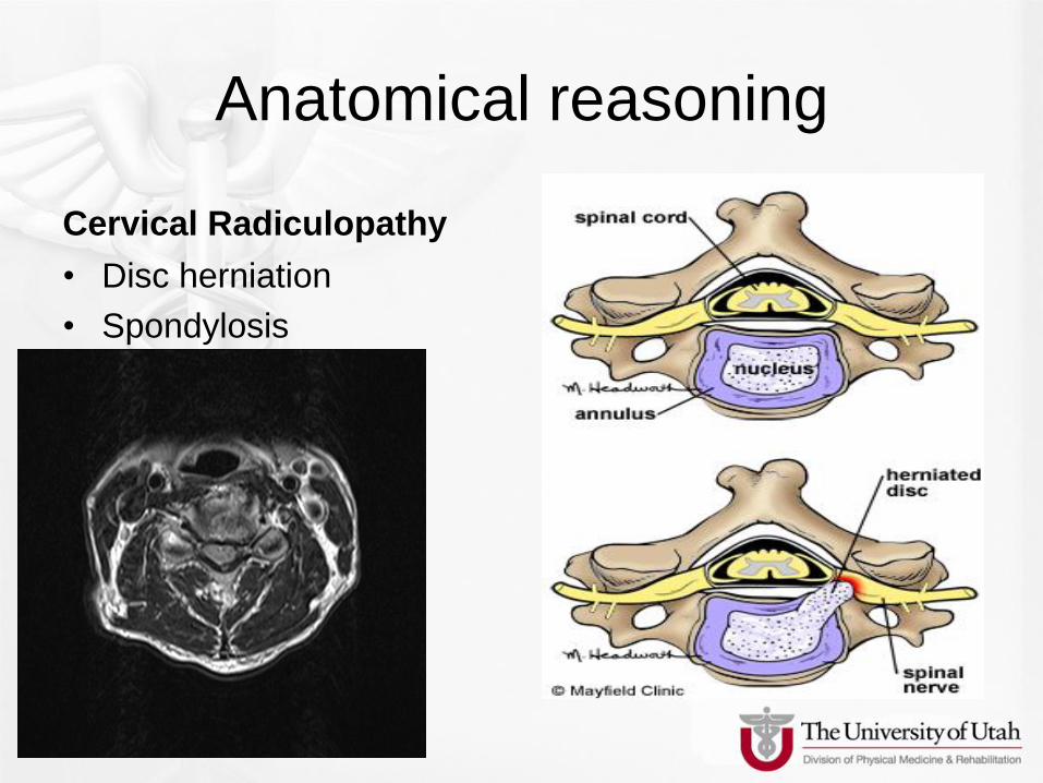
Anatomical reasoning
Cervical Radiculopathy
• Disc herniation
• Spondylosis
Indications
• Should have extensive trial or
medications, PT, activity modification
given the potential risks of ALL cervical
epidural injections.
• Scanlon GC, et al. Cervical transforaminal epidural steroid injections: more
dangerous than we think? Spine. 2007;32(11):1249-56.
• Abbassi A, et al. Complications of interlaminar Cervical Epidural Steroid Injections.
Spine 2007;32(19):2144-51
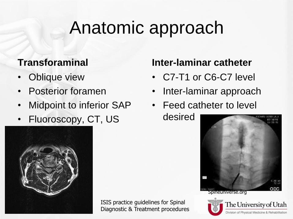
Anatomic approach
Transforaminal
• Oblique view
• Posterior foramen
• Midpoint to inferior SAP
• Fluoroscopy, CT, US
guided
• 2004.
Inter-laminar catheter
• C7-T1 or C6-C7 level
• Inter-laminar approach
• Feed catheter to level
desired
Spineuniverse.org
ISIS practice guidelines for Spinal Diagnostic & Treatment procedures
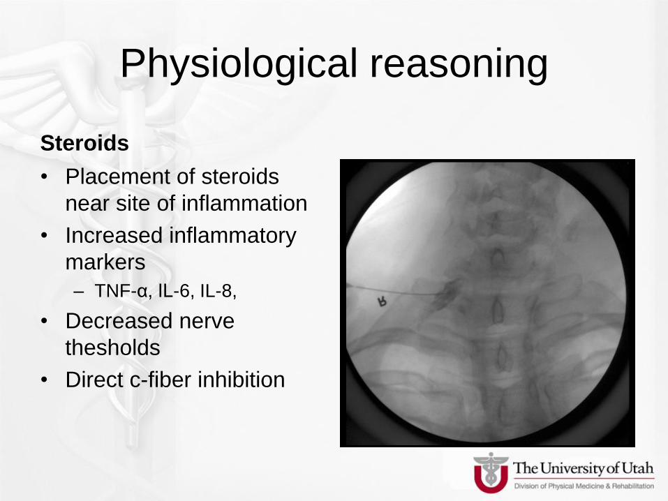
Physiological reasoning
Steroids
• Placement of steroids
near site of inflammation
• Increased inflammatory
markers
– TNF-α, IL-6, IL-8,
• Decreased nerve
thesholds
• Direct c-fiber inhibition
CTF ESI Outcome studies
Good
• Bush 1996
– N=68, 76% cured, 24% minimal pain, 39m f/u
• Berger 1999
– CT-guided, 59% improved
• Slipman 2000
– 60% excellent at 21m, 2.2 injections, 30% surgery
• Riew, 2000
– 60% cancel surgery, no difference steroid, bupivicaine
Poor
• Slipman- 2001
– Whiplash, nml imaging
– 14% good outcome
• Slipman- 2004
– Whiplash, foraminal
stenosis
– 20% good outcome
Outcome studies
• No direct comparisons CIL to CTF
• Lumbar studies show benefit for
radiculopathy with TF approach.
• Schaufele MK, et al. Pain Physician. 2006;9(4):361-66.
Risks
Bleeding
• Epidural Hematoma
– ASA closed claims database 1970-1999
• 40% of all chronic pain mgmt claims were epidurals
• Most common complication was nerve injury- 25%
– Pre-fluoroscopy, pre-ultrasound nerve blocks
• Spinal cord injury is leading cause of type of nerve injury in 1990’s. Epidurals 50% of these.
• ~1% of interlaminar epidurals hematoma. • Cheney FW. Nerve injury associated with anesthesia: A closed claims
analysis. Anesthesiology 1999,90:1062-9.
Bleeding
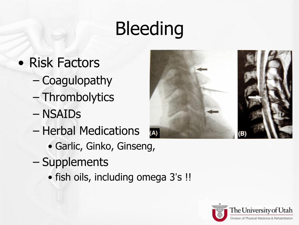
• Risk Factors
– Coagulopathy
– Thrombolytics
– NSAIDs
– Herbal Medications
• Garlic, Ginko, Ginseng,
– Supplements
• fish oils, including omega 3’s !!
Bleeding
• NSAIDs
– No significant risk or hematoma
– Minor Hemorrhagic complications
• Horlocker TT. Anest Analg 2002; 95: 1691-7
• Horlocker TT. Anest Analg 1995; 80: 303-9.
• Antiplatelet Meds (clopidrogrel, ticlopidine, asa/dipyridamole, dabigatran, rivaroxaban) – Increases risk of bleeding complications, stop 7 days prior
• Supplements/Herbals (MSM, garlic, ginseng, ginger, Fish oil*) – Concurrent use with other meds affecting clotting mechanisms increase
the risk of bleeding *- recommend stop 7 days in advance.
Bleeding
• Need to be aware of WHY patient is anticoagulated. – ISIS ASM 2009. Furman: Pt cancelled day of
appt, died unexpectedly stopping ASA 7 days prior.
• Stents: recommendations are for 1 yr anticoagulation plavix/asa- drug eluting, then ASA for life. Bare metal: 6 mo.
• ALWAYS consult with prescribing MD before stopping anticoagulants.
Infections
• Meningitis – Rarely Reported
• Morris 1994 • Daugherty. J Neurosurg 1978. 48: 1023-5. 2 case reports • Nelson 1973 • Civen 2006 Clin Infect dz: Serratia from compound betamethasone
– Dural Puncture
• Arachnoiditis (infectious and medication etiology) • Ryan 1981
• Abscess- most common organism Staph. Epi. – Major risk factor: Indwelling catheter – Second most common complication in ASA claims- 21% (nerve inj 25%) – Epidural abscess
• Hooten WM. Mayo Clin Proc 2004; 79: 682-6. • Huang RC. Spine 2004; 29 E7-9.
– Extra dural abscess • Gouke, British Journal of Anesthesia 1990; 65: 427-429 • Facet joint infections.
• Local infection
Procedure Specific Complications
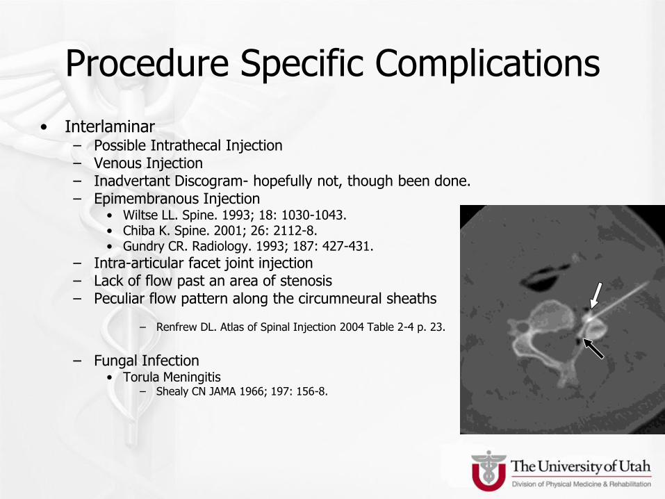
• Interlaminar – Possible Intrathecal Injection – Venous Injection – Inadvertant Discogram- hopefully not, though been done. – Epimembranous Injection
• Wiltse LL. Spine. 1993; 18: 1030-1043. • Chiba K. Spine. 2001; 26: 2112-8. • Gundry CR. Radiology. 1993; 187: 427-431.
– Intra-articular facet joint injection – Lack of flow past an area of stenosis – Peculiar flow pattern along the circumneural sheaths
– Renfrew DL. Atlas of Spinal Injection 2004 Table 2-4 p. 23.
– Fungal Infection
• Torula Meningitis – Shealy CN JAMA 1966; 197: 156-8.
Procedure Specific Complications
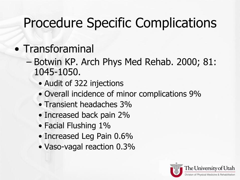
• Transforaminal – Botwin KP. Arch Phys Med Rehab. 2000; 81:
1045-1050. • Audit of 322 injections
• Overall incidence of minor complications 9%
• Transient headaches 3%
• Increased back pain 2%
• Facial Flushing 1%
• Increased Leg Pain 0.6%
• Vaso-vagal reaction 0.3%
Neuraxis Injury • Direct Mechanical Injury
– Spinal Cord – Spinal Nerve – Vascular Injury
• Anterior Spinal Artery Syndrome – Sudden painless onset of LE weakness – Variable sensory deficits, with relative preservation of
proprioception – Supplies Cauda Equina and anterior 2/3 of spinal cord
Procedure Specific Complications
• Transforaminal – Intrathecal/Subarachnoid injection (dural sleeve) – Intravascular injection
• Furman MB. Spine 2003; 28: 21-5. – Audit of 504 cervical injections-19% incidence of
intravascular injection
• Baker R. Pain 2002; 103: 211-5. – Presence of reinforcing arteries are more common in lower
cervical spine
– Brain or cord infarcts, vertebrobasilar infarcts, death. 78 cases reported by Scanlon et al. Spine 2007;32(11):1249-56
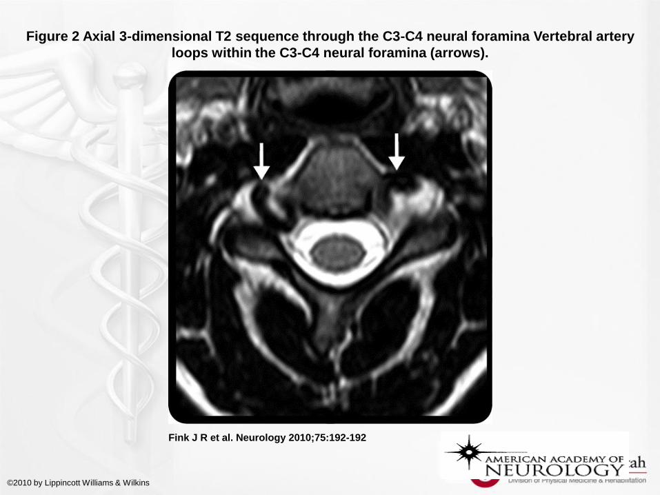
Figure 2 Axial 3-dimensional T2 sequence through the C3-C4 neural foramina Vertebral artery
loops within the C3-C4 neural foramina (arrows).
Fink J R et al. Neurology 2010;75:192-192
©2010 by Lippincott Williams & Wilkins
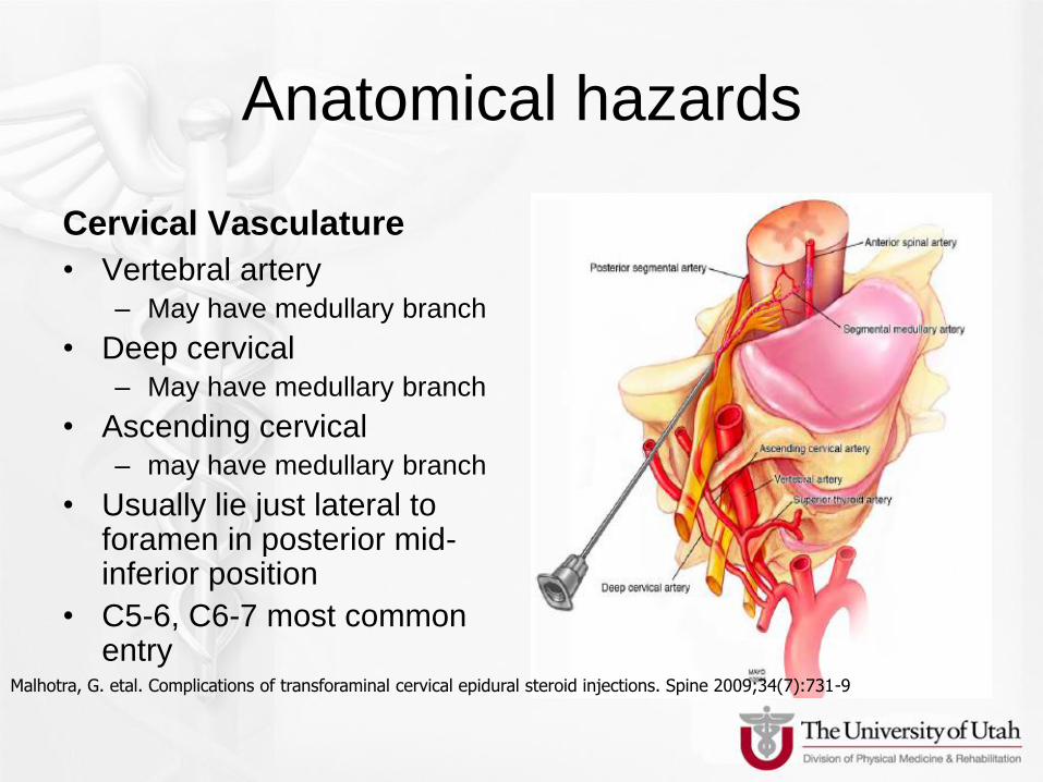
Anatomical hazards
Cervical Vasculature
• Vertebral artery
– May have medullary branch
• Deep cervical
– May have medullary branch
• Ascending cervical
– may have medullary branch
• Usually lie just lateral to foramen in posterior mid-inferior position
• C5-6, C6-7 most common entry
Malhotra, G. etal. Complications of transforaminal cervical epidural steroid injections. Spine 2009;34(7):731-9
Procedure Specific Complications
• Transforminal Cervical
• Decreasing Risk of Complications – Advance needle from Anterior Oblique approach
– Ensure needle remains over SAP along the posterior aspect of the intervertebral foramen
– Use AP view to adjust final needle depth into foramen
– Do not advance needle >50% across medial-lateral dimension of the foramen
– Use contrast under “live” fluoro. Leave tubing connected. Test dose, then steroid.
– Use of a non-particulate steroid? • Riew 2003- intravertebral a. injection
Needle choice
• Blunt tip needles
– Less likely to penetrate vascular?
• Trucath
– Still 10% vascular uptake with catheter.
– 3% unable to get to foramen – Kloth D, et al.Pain Physician 2011;14:285-293
Summary
• Try ALL non-interventional therapies first!!
• Use for radiculopathy ONLY. Not axial.
• Have extensive training, excellent
radiology.
• Low threshold to abort. Especially if
flashback seen in needle.
• Digital subtraction imaging, if available.
• Non-particulate steroid.
Safest Approach?
• Unclear, both have rare catastrophic
complications
• But then again….
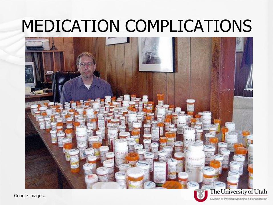
MEDICATION COMPLICATIONS
Google images.