Embed Size (px)
Citation preview
454454
17 ▼BLEEDING AND CLOTTINGDISORDERS
LAUREN L. PATTON, DDS
Dental health care workers are increasingly called upon to pro-vide quality dental care to individuals whose bleeding andclotting mechanisms have been altered by inherited oracquired diseases. This provides an opportunity for the den-tist who is trained in the recognition of oral and systemic signsof altered hemostasis to assist in the diagnosis of the underly-ing condition. A number of dental procedures result in the riskof bleeding that can have serious consequences, such as severehemorrhage or possibly death, for the patient with a bleedingdisorder. Safe dental care may require consultation with thepatient’s physician, systemic management, and dental treat-ment modifications.
Of the inherited coagulopathies, von Willebrand’s disease(vWD) is the most common. It results from deficiency of vonWillebrand’s factor (vWF) and affects about 0.8 to 1% of thepopulation.1 Hemophilia A, caused by coagulation factor (F)VIII deficiency, is the next most common, followed by hemo-philia B, a F IX deficiency. The age-adjusted prevalence ofhemophilia in six surveillance states in 1994 was 13.4 cases in100,000 males (10.5 for hemophilia A and 2.9 for hemophiliaB).2 Application to the US population resulted in an estimatednational prevalence of 13,320 cases of hemophilia A and 3,640cases of hemophilia B, with an incidence rate of 1 per 5,032 livemale births. Hemophilia A was predominant, accounting for79% of all hemophiliacs, and prevalence of disease severity was43% severe (< 1% F VIII), 26% moderate (1–5% F VIII), and31% with mild (6–30% F VIII) disease.2
Acquired coagulation disorders can result from drugactions or side effects, or underlying systemic disease. A strat-ified household sample of 4,163 community residents aged 65years or older living in a five-county area of North Carolinarevealed 51.7% to be taking one or more medications (aspirin,warfarin, dipyridamole, nonsteroidal anti-inflammatory drugs
▼ PATHOPHYSIOLOGYBasic Mechanisms of Hemostasis and Their InteractionsVascular PhasePlatelet PhaseCoagulation PhaseFibrinolytic Phase
▼ CLINICAL AND LABORATORY FINDINGSClinical ManifestationsClinical Laboratory Tests
▼ CLASSIFICATION OF BLEEDINGDISORDERSVessel Wall DisordersPlatelet Disorders Coagulation DisordersFibrinolytic Disorders
▼ IDENTIFICATION OF THE DENTALPATIENT WITH A BLEEDING DISORDER
▼ MANAGEMENTPlatelet DisordersHemophilias A and BVon Willebrand’s DiseaseDisease-Related Coagulopathies
▼ PROGNOSIS
▼ ORAL HEALTH CONSIDERATIONSOral FindingsDental Management
Bleeding and Clotting Disorders 455
[NSAIDs], or heparin) with the potential to alter hemostasis.3
The use of coumarin anticoagulants is increasing as a result oftheir demonstrated effectiveness in the treatment of atrial fib-rillation and venous thromboembolism, and control of throm-bosis in the presence of a mechanical heart valve.4
▼PATHOPHYSIOLOGY
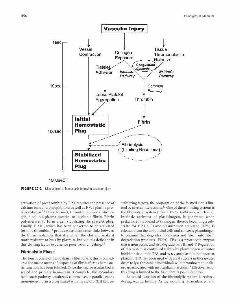
Basic Mechanisms of Hemostasis and TheirInteractionsInteraction of several basic mechanisms produces normalhemostasis. For clarity and understanding, these are presentedseparately. Hemostasis can be divided into four general phases:the vascular phase; the platelet phase; the coagulation cascadephase, consisting of intrinsic, extrinsic, and common path-ways; and the fibrinolytic phase. The first three phases are theprincipal mechanisms that stop the loss of blood following vas-cular injury. Briefly, when vessel integrity is disrupted, plateletsare activated, adhere to the site of injury, and form a plateletplug that reduces or temporarily arrests blood loss.5 The expo-sure of collagen and activation of platelets also initiates thecoagulation cascade, which leads to fibrin formation and thegeneration of an insoluble fibrin clot that strengthens theplatelet plug.5 Fibrinolysis is the major means of disposing offibrin after its hemostatic function has been fulfilled, and it canbe considered the rate-limiting step in clotting. It leads to fib-rin degradation by the proteolytic enzyme plasmin. As seen inFigure 17-1, multiple processes occur either simultaneously orin rapid sequence, such that, following almost immediate vas-cular contraction, platelets begin to aggregate at the woundsite. The coagulation cascade is underway within 10 to 20 sec-onds of injury, an initial hemostatic plug is formed in 1 to 3minutes, and fibrin has been generated and added to stabilizethe clot by 5 to 10 minutes.
Vascular PhaseAfter tissue injury, there is an immediate reflex vasoconstric-tion that may alone be hemostatic in small vessels. Reactantssuch as serotonin, histamine, prostaglandins, and other mate-rials are vasoactive and produce vasoconstriction of themicrovascular bed in the area of the injury.
Platelet PhaseWhen circulating platelets are exposed to damaged vascularsurfaces (in the presence of functionally normal vWF,endothelial cells, collagen or collagen-like materials, base-ment membrane, elastin, microfibrils, and other cellulardebris), platelets are activated to experience physical andchemical changes.6 These changes produce an environmentthat causes the platelets to undergo the aggregation-and-release phenomenon and form the primary vascular plugthat reduces blood loss from small blood vessels and capil-laries. These platelet plugs adhere to exposed basementmembranes. As this reaction is occurring, the release reac-tion is underway, involving the intracellular release of active
components for further platelet aggregation as well as pro-motion of the clotting mechanism. Adenosine diphosphate(ADP) is a potent nucleotide that activates and recruits otherplatelets in the area, immensely adding to the size of theplug. Platelet factor 3 (PF3) is the intracellular phospho-lipid that activates F X and subsequently results in the con-version of prothrombin to thrombin. Additionally, theplatelet plug, intermixed with fibrin and cellular compo-nents such as red and white cells, contracts to further reduceblood loss and to seal the vascular bed.
Coagulation PhaseThe generation of thrombin and fibrin the end product of thethird phase of hemostasis, the coagulation phase. This processinvolves multiple proteins, many of which are synthesized bythe liver (fibrinogen, prothrombin, Fs V,VII, IX, X, XI, XII, andXIII) and are vitamin K dependent (Fs II, VII, IX, and X). Theprocess of coagulation essentially involves three separate path-ways. It initially proceeds by two separate pathways (intrinsicand extrinsic) that converge by activating a third (common)pathway.
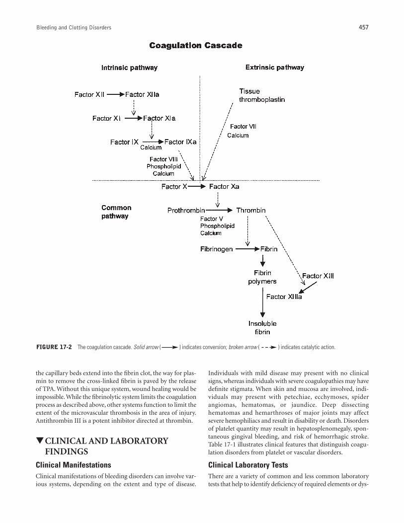
The blood clotting mechanism is the most studied unit; itwas outlined originally in 1903 by Markowitz as the pro-thrombin-to-thrombin and fibrinogen-to-fibrin conversionsystem. In 1964, the “cascade” or “waterfall” theory was pro-posed.7,8 It offered a useful device for understanding this com-plex system and its control, as well as the clinically importantassociated laboratory tests.
Figure 17-2 depicts the sequence of interactions between thevarious clotting factors following injury of tissue. The schemeof reaction is a bioamplification, in which a precursor is alteredto an active form, which, in turn, activates the next precursorin the sequence. Beginning with an undetectable biochemicalreaction, the coagulation mechanism results in a final explosivechange of a liquid to a gel. The major steps involve the conver-sion of a precursor protein to an “activated” form, which acti-vates another precursor protein, and so on down the cascade.The coagulation of blood also requires the presence of both cal-cium ions and phospholipid (or a phospholipid-containingmembrane fragment derived from blood platelets).
The intrinsic pathway is initiated when F XII is activated bysurface contact (eg, with collagen or subendothelium), and itinvolves the interaction of F XII and F XI. The next step ofintrinsic coagulation, the activation of F IX to F XIa, requiresa divalent cation.9 Once activated, F IXa forms a complex withF VIII, in a reaction that requires the presence of both calciumions and phospholipid, which, in turn, converts F X to an acti-vated form— F Xa.
The extrinsic pathway is initiated by the release of tissuethromboplastin, also called tissue factor, and does not requirecontact activation. Tissue thromboplastin binds to F VII in thepresence of calcium, and this complex is capable of activatingFs IX and X, linking the intrinsic and extrinsic pathways.
It is the activation of X that begins the common pathway.Once activated, F Xa converts prothrombin to thrombin ina reaction similar to the activation of F X by F IXa. The
456 Principles of Medicine
activation of prothrombin by F Xa requires the presence ofcalcium ions and phospholipid as well as F V, a plasma pro-tein cofactor.10 Once formed, thrombin converts fibrino-gen, a soluble plasma protein, to insoluble fibrin. Fibrinpolymerizes to form a gel, stabilizing the platelet plug.Finally, F XIII, which has been converted to an activatedform by thrombin,11 produces covalent cross-links betweenthe fibrin molecules that strengthen the clot and make itmore resistant to lysis by plasmin. Individuals deficient inthis clotting factor experience poor wound healing.12
Fibrinolytic PhaseThe fourth phase of hemostasis is fibrinolysis; this is consid-ered the major means of disposing of fibrin after its hemosta-tic function has been fulfilled. Once the microvascular bed issealed and primary hemostasis is complete, the secondaryhemostasis pathway has already commenced in parallel. As themonomeric fibrin is cross-linked with the aid of F XIII (fibrin-
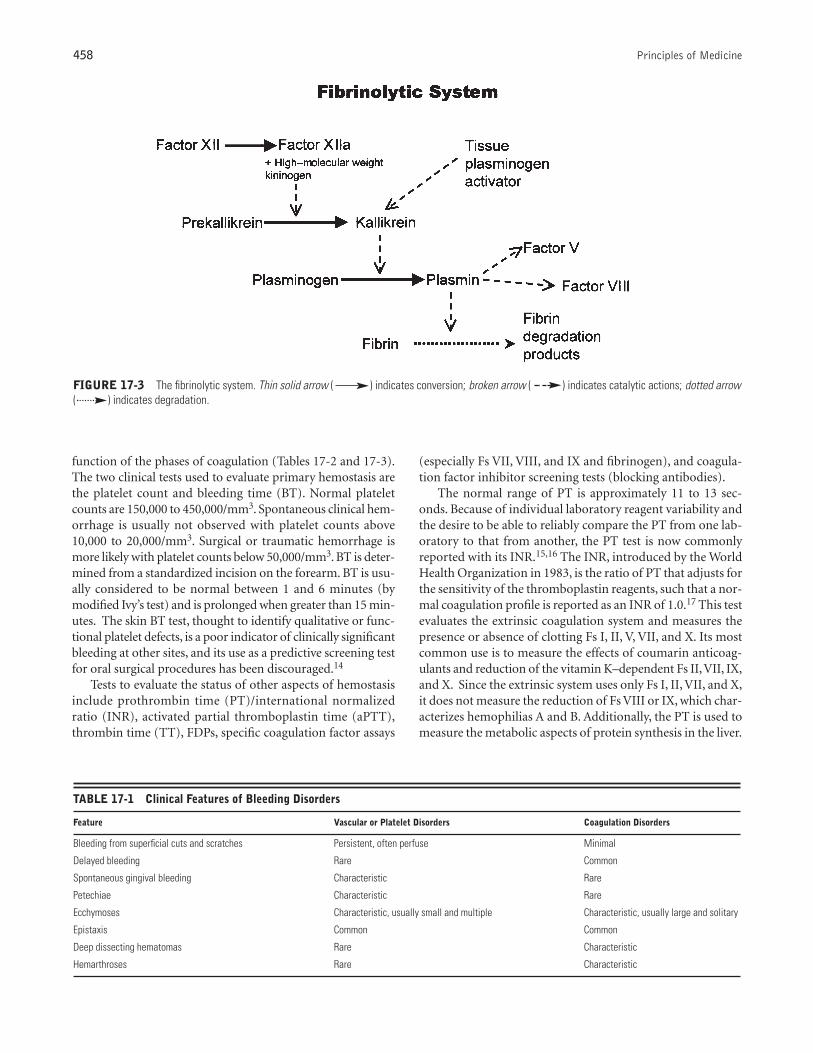
stabilizing factor), the propagation of the formed clot is lim-ited by several interactions.12 One of these limiting systems isthe fibrinolytic system (Figure 17-3). Kallikrein, which is anintrinsic activator of plasminogen, is generated whenprekallikrein is bound to kininogen, thereby becoming a sub-strate for F XIIa. Tissue plasminogen activator (TPA) isreleased from the endothelial cells and converts plasminogento plasmin that degrades fibrinogen and fibrin into fibrindegradation products (FDPs). TPA is a proteolytic enzymethat is nonspecific and also degrades Fs VIII and V. Regulationof this system is controlled tightly by plasminogen activatorinhibitor that limits TPA, and by α2-antiplasmin that restrictsplasmin. TPA has been used with great success in therapeuticdoses to lyse thrombi in individuals with thromboembolic dis-orders associated with myocardial infarction.13 Effectiveness ofthis drug is limited to the first 6 hours post infarction.
Extended function of the fibrinolytic system is realizedduring wound healing. As the wound is revascularized and
FIGURE 17-1 Mechanisms of hemostasis following vascular injury.
Bleeding and Clotting Disorders 457
the capillary beds extend into the fibrin clot, the way for plas-min to remove the cross-linked fibrin is paved by the releaseof TPA. Without this unique system, wound healing would beimpossible. While the fibrinolytic system limits the coagulationprocess as described above, other systems function to limit theextent of the microvascular thrombosis in the area of injury.Antithrombin III is a potent inhibitor directed at thrombin.
▼CLINICAL AND LABORATORYFINDINGS
Clinical ManifestationsClinical manifestations of bleeding disorders can involve var-ious systems, depending on the extent and type of disease.
Individuals with mild disease may present with no clinicalsigns, whereas individuals with severe coagulopathies may havedefinite stigmata. When skin and mucosa are involved, indi-viduals may present with petechiae, ecchymoses, spiderangiomas, hematomas, or jaundice. Deep dissectinghematomas and hemarthroses of major joints may affectsevere hemophiliacs and result in disability or death. Disordersof platelet quantity may result in hepatosplenomegaly, spon-taneous gingival bleeding, and risk of hemorrhagic stroke.Table 17-1 illustrates clinical features that distinguish coagu-lation disorders from platelet or vascular disorders.
Clinical Laboratory TestsThere are a variety of common and less common laboratorytests that help to identify deficiency of required elements or dys-
FIGURE 17-2 The coagulation cascade. Solid arrow ( ) indicates conversion; broken arrow ( ) indicates catalytic action.
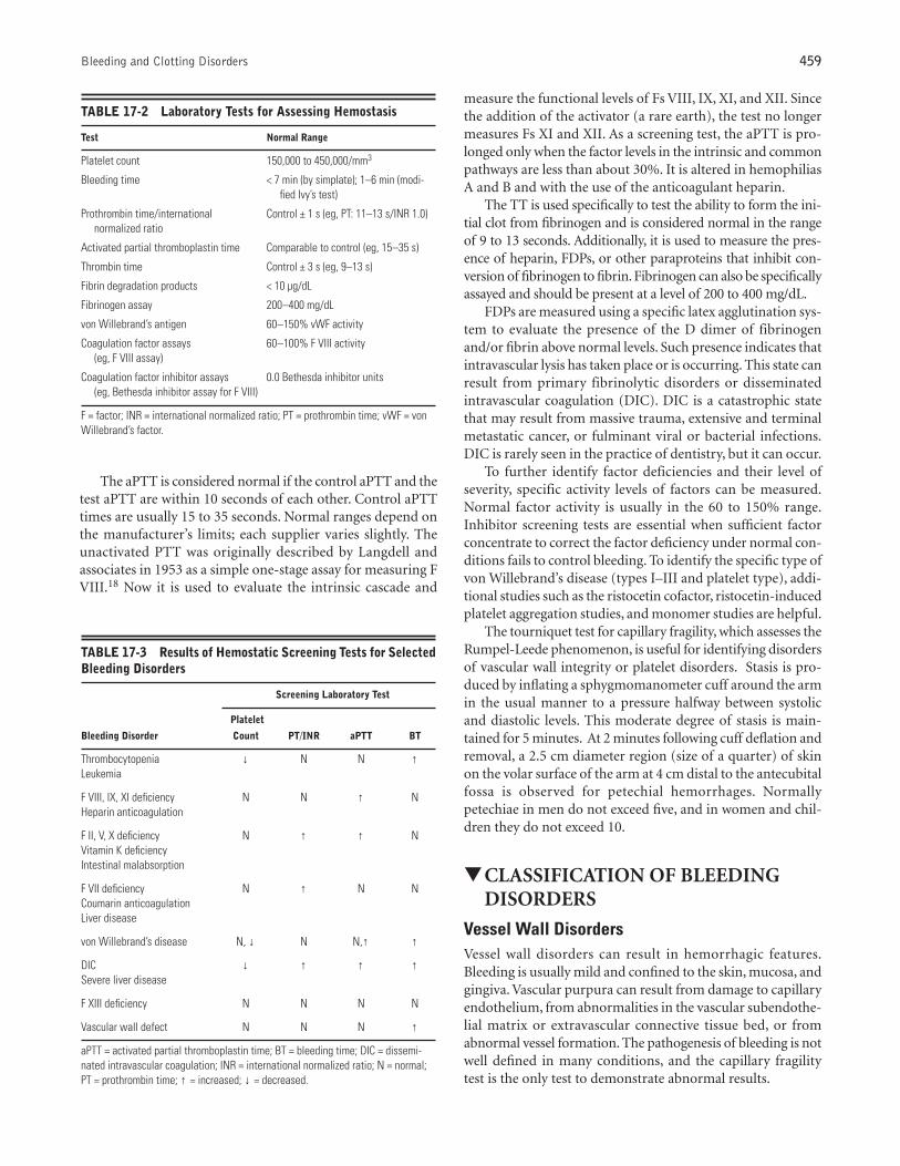
function of the phases of coagulation (Tables 17-2 and 17-3).The two clinical tests used to evaluate primary hemostasis arethe platelet count and bleeding time (BT). Normal plateletcounts are 150,000 to 450,000/mm3. Spontaneous clinical hem-orrhage is usually not observed with platelet counts above10,000 to 20,000/mm3. Surgical or traumatic hemorrhage ismore likely with platelet counts below 50,000/mm3. BT is deter-mined from a standardized incision on the forearm. BT is usu-ally considered to be normal between 1 and 6 minutes (bymodified Ivy’s test) and is prolonged when greater than 15 min-utes. The skin BT test, thought to identify qualitative or func-tional platelet defects, is a poor indicator of clinically significantbleeding at other sites, and its use as a predictive screening testfor oral surgical procedures has been discouraged.14
Tests to evaluate the status of other aspects of hemostasisinclude prothrombin time (PT)/international normalizedratio (INR), activated partial thromboplastin time (aPTT),thrombin time (TT), FDPs, specific coagulation factor assays
458 Principles of Medicine
(especially Fs VII, VIII, and IX and fibrinogen), and coagula-tion factor inhibitor screening tests (blocking antibodies).
The normal range of PT is approximately 11 to 13 sec-onds. Because of individual laboratory reagent variability andthe desire to be able to reliably compare the PT from one lab-oratory to that from another, the PT test is now commonlyreported with its INR.15,16 The INR, introduced by the WorldHealth Organization in 1983, is the ratio of PT that adjusts forthe sensitivity of the thromboplastin reagents, such that a nor-mal coagulation profile is reported as an INR of 1.0.17 This testevaluates the extrinsic coagulation system and measures thepresence or absence of clotting Fs I, II, V, VII, and X. Its mostcommon use is to measure the effects of coumarin anticoag-ulants and reduction of the vitamin K–dependent Fs II,VII, IX,and X. Since the extrinsic system uses only Fs I, II, VII, and X,it does not measure the reduction of Fs VIII or IX, which char-acterizes hemophilias A and B. Additionally, the PT is used tomeasure the metabolic aspects of protein synthesis in the liver.
TABLE 17-1 Clinical Features of Bleeding Disorders
Feature Vascular or Platelet Disorders Coagulation Disorders
Bleeding from superficial cuts and scratches Persistent, often perfuse Minimal
Delayed bleeding Rare Common
Spontaneous gingival bleeding Characteristic Rare
Petechiae Characteristic Rare
Ecchymoses Characteristic, usually small and multiple Characteristic, usually large and solitary
Epistaxis Common Common
Deep dissecting hematomas Rare Characteristic
Hemarthroses Rare Characteristic
FIGURE 17-3 The fibrinolytic system. Thin solid arrow ( ) indicates conversion; broken arrow ( ) indicates catalytic actions; dotted arrow( ) indicates degradation.
Bleeding and Clotting Disorders 459
The aPTT is considered normal if the control aPTT and thetest aPTT are within 10 seconds of each other. Control aPTTtimes are usually 15 to 35 seconds. Normal ranges depend onthe manufacturer’s limits; each supplier varies slightly. Theunactivated PTT was originally described by Langdell andassociates in 1953 as a simple one-stage assay for measuring FVIII.18 Now it is used to evaluate the intrinsic cascade and
measure the functional levels of Fs VIII, IX, XI, and XII. Sincethe addition of the activator (a rare earth), the test no longermeasures Fs XI and XII. As a screening test, the aPTT is pro-longed only when the factor levels in the intrinsic and commonpathways are less than about 30%. It is altered in hemophiliasA and B and with the use of the anticoagulant heparin.
The TT is used specifically to test the ability to form the ini-tial clot from fibrinogen and is considered normal in the rangeof 9 to 13 seconds. Additionally, it is used to measure the pres-ence of heparin, FDPs, or other paraproteins that inhibit con-version of fibrinogen to fibrin. Fibrinogen can also be specificallyassayed and should be present at a level of 200 to 400 mg/dL.
FDPs are measured using a specific latex agglutination sys-tem to evaluate the presence of the D dimer of fibrinogenand/or fibrin above normal levels. Such presence indicates thatintravascular lysis has taken place or is occurring. This state canresult from primary fibrinolytic disorders or disseminatedintravascular coagulation (DIC). DIC is a catastrophic statethat may result from massive trauma, extensive and terminalmetastatic cancer, or fulminant viral or bacterial infections.DIC is rarely seen in the practice of dentistry, but it can occur.
To further identify factor deficiencies and their level ofseverity, specific activity levels of factors can be measured.Normal factor activity is usually in the 60 to 150% range.Inhibitor screening tests are essential when sufficient factorconcentrate to correct the factor deficiency under normal con-ditions fails to control bleeding. To identify the specific type ofvon Willebrand’s disease (types I–III and platelet type), addi-tional studies such as the ristocetin cofactor, ristocetin-inducedplatelet aggregation studies, and monomer studies are helpful.
The tourniquet test for capillary fragility, which assesses theRumpel-Leede phenomenon, is useful for identifying disordersof vascular wall integrity or platelet disorders. Stasis is pro-duced by inflating a sphygmomanometer cuff around the armin the usual manner to a pressure halfway between systolicand diastolic levels. This moderate degree of stasis is main-tained for 5 minutes. At 2 minutes following cuff deflation andremoval, a 2.5 cm diameter region (size of a quarter) of skinon the volar surface of the arm at 4 cm distal to the antecubitalfossa is observed for petechial hemorrhages. Normallypetechiae in men do not exceed five, and in women and chil-dren they do not exceed 10.
▼CLASSIFICATION OF BLEEDINGDISORDERS
Vessel Wall DisordersVessel wall disorders can result in hemorrhagic features.Bleeding is usually mild and confined to the skin, mucosa, andgingiva. Vascular purpura can result from damage to capillaryendothelium, from abnormalities in the vascular subendothe-lial matrix or extravascular connective tissue bed, or fromabnormal vessel formation. The pathogenesis of bleeding is notwell defined in many conditions, and the capillary fragilitytest is the only test to demonstrate abnormal results.
TABLE 17-2 Laboratory Tests for Assessing Hemostasis
Test Normal Range
Platelet count 150,000 to 450,000/mm3
Bleeding time < 7 min (by simplate); 1–6 min (modi-fied Ivy’s test)
Prothrombin time/international Control ± 1 s (eg, PT: 11–13 s/INR 1.0)normalized ratio
Activated partial thromboplastin time Comparable to control (eg, 15–35 s)
Thrombin time Control ± 3 s (eg, 9–13 s)
Fibrin degradation products < 10 µg/dL
Fibrinogen assay 200–400 mg/dL
von Willebrand’s antigen 60–150% vWF activity
Coagulation factor assays 60–100% F VIII activity(eg, F VIII assay)
Coagulation factor inhibitor assays 0.0 Bethesda inhibitor units(eg, Bethesda inhibitor assay for F VIII)
F = factor; INR = international normalized ratio; PT = prothrombin time; vWF = vonWillebrand’s factor.
TABLE 17-3 Results of Hemostatic Screening Tests for SelectedBleeding Disorders
Screening Laboratory Test
Platelet
Bleeding Disorder Count PT/INR aPTT BT
Thrombocytopenia ↓ N N ↑Leukemia
F VIII, IX, XI deficiency N N ↑ NHeparin anticoagulation
F II, V, X deficiency N ↑ ↑ NVitamin K deficiencyIntestinal malabsorption
F VII deficiency N ↑ N NCoumarin anticoagulationLiver disease
von Willebrand’s disease N, ↓ N N,↑ ↑
DIC ↓ ↑ ↑ ↑Severe liver disease
F XIII deficiency N N N N
Vascular wall defect N N N ↑
aPTT = activated partial thromboplastin time; BT = bleeding time; DIC = dissemi-nated intravascular coagulation; INR = international normalized ratio; N = normal;PT = prothrombin time; ↑ = increased; ↓ = decreased.
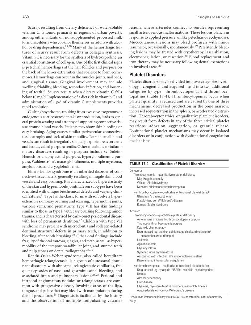
TABLE 17-4 Classification of Platelet Disorders
Congenital Thrombocytopenic—quantitative platelet deficiency
May-Hegglin anomalyWiskott-Aldrich syndromeNeonatal alloimmune thrombocytopenia
Nonthrombocytopenic—qualitative or functional platelet defectGlanzmann’s thrombastheniaPlatelet-type von Willebrand’s diseaseBernard-Soulier syndrome
Acquired Thrombocytopenic—quantitative platelet deficiency
Autoimmune or idiopathic thrombocytopenia purpuraThrombotic thrombocytopenia purpuraCytotoxic chemotherapy Drug-induced (eg, quinine, quinidine, gold salts, trimethoprim/
sulfamethoxazole, rifampin)LeukemiaAplastic anemiaMyelodysplasiaSystemic lupus erythematosus Associated with infection: HIV, mononucleosis, malariaDisseminated intravascular coagulation
Nonthrombocytopenic—qualitative or functional platelet defectDrug-induced (eg, by aspirin, NSAIDs, penicillin, cephalosporins)Uremia Alcohol dependencyLiver disease Myeloma, myeloproliferative disorders, macroglobulinemia Acquired platelet-type von Willebrand’s disease
HIV=human immunodeficiency virus; NSAIDs = nonsteroidal anti-inflammatorydrugs.
460 Principles of Medicine
Scurvy, resulting from dietary deficiency of water-solublevitamin C, is found primarily in regions of urban poverty,among either infants on nonsupplemented processed milkformulas, elderly who cook for themselves, or adults with alco-hol or drug dependencies.19,20 Many of the hemorrhagic fea-tures of scurvy result from defects in collagen synthesis.Vitamin C is necessary for the synthesis of hydroxyproline, anessential constituent of collagen. One of the first clinical signsis petechial hemorrhages at the hair follicles and purpura onthe back of the lower extremities that coalesce to form ecchy-moses. Hemorrhage can occur in the muscles, joints, nail beds,and gingival tissues. Gingival involvement may includeswelling, friability, bleeding, secondary infection, and loosen-ing of teeth.20 Scurvy results when dietary vitamin C fallsbelow 10 mg/d. Implementation of a diet rich in vitamin C andadministration of 1 g/d of vitamin C supplements providesrapid resolution.
Cushing’s syndrome, resulting from excessive exogenous orendogenous corticosteroid intake or production, leads to gen-eral protein wasting and atrophy of supporting connective tis-sue around blood vessels. Patients may show skin bleeding oreasy bruising. Aging causes similar perivascular connective-tissue atrophy and lack of skin mobility. Tears in small bloodvessels can result in irregularly shaped purpuric areas on armsand hands, called purpura senilis. Other metabolic or inflam-matory disorders resulting in purpura include Schönlein-Henoch or anaphylactoid purpura, hyperglobulinemic pur-pura, Waldenström’s macroglobulinemia, multiple myeloma,amyloidosis, and cryoglobulinemia.
Ehlers-Danlos syndrome is an inherited disorder of con-nective-tissue matrix, generally resulting in fragile skin bloodvessels and easy bruising. It is characterized by hyperelasticityof the skin and hypermobile joints. Eleven subtypes have beenidentified with unique biochemical defects and varying clini-cal features.21 Type I is the classic form, with soft velvety hyper-extensible skin, easy bruising and scarring, hypermobile joints,varicose veins, and prematurity. Type VIII has skin findingssimilar to those in type I, with easy bruising following minortrauma, and is characterized by early-onset periodontal diseasewith loss of permanent dentition.22 Children with type VIIsyndrome may present with microdontia and collagen-relateddentinal structural defects in primary teeth, in addition tobleeding after tooth brushing.23 Other oral findings includefragility of the oral mucosa, gingiva, and teeth, as well as hyper-mobility of the temporomandibular joint, and stunted teethand pulp stones on dental radiographs.24,25
Rendu-Osler-Weber syndrome, also called hereditaryhemorrhagic telangiectasia, is a group of autosomal domi-nant disorders with abnormal telangiectatic capillaries, fre-quent episodes of nasal and gastrointestinal bleeding, andassociated brain and pulmonary lesions.26,27 Perioral andintraoral angiomatous nodules or telangiectases are com-mon with progressive disease, involving areas of the lips,tongue, and palate that may bleed with manipulation duringdental procedures.28 Diagnosis is facilitated by the historyand the observation of multiple nonpulsating vascular
lesions, where arterioles connect to venules representingsmall arteriovenous malformations. These lesions blanch inresponse to applied pressure, unlike petechiae or ecchymoses.Mucocutaneous lesions may bleed profusely with minortrauma or, occasionally, spontaneously.29 Persistently bleed-ing lesions may be treated with cryotherapy, laser ablation,electrocoagulation, or resection.30 Blood replacement andiron therapy may be necessary following dental extractionsin involved areas.29
Platelet Disorders Platelet disorders may be divided into two categories by eti-ology—congential and acquired—and into two additionalcategories by type—thrombocytopenias and thrombocy-topathies (Table 17-4). Thrombocytopenias occur whenplatelet quantity is reduced and are caused by one of threemechanisms: decreased production in the bone marrow,increased sequestration in the spleen, or accelerated destruc-tion. Thrombocytopathies, or qualitative platelet disorders,may result from defects in any of the three critical plateletreactions: adhesion, aggregation, or granule release.Dysfunctional platelet mechanisms may occur in isolateddisorders or in conjunction with dysfunctional coagulationmechanisms.
Bleeding and Clotting Disorders 461
CONGENITAL PLATELET DISORDERS
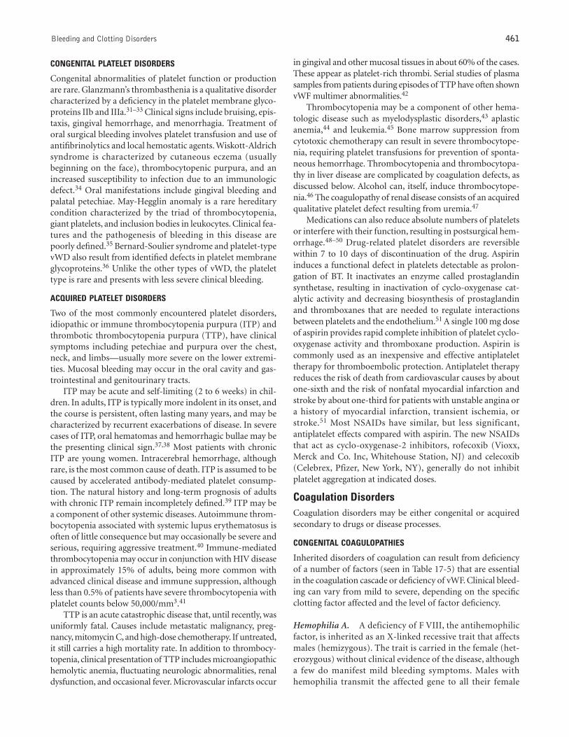
Congenital abnormalities of platelet function or productionare rare. Glanzmann’s thrombasthenia is a qualitative disordercharacterized by a deficiency in the platelet membrane glyco-proteins IIb and IIIa.31–33 Clinical signs include bruising, epis-taxis, gingival hemorrhage, and menorrhagia. Treatment oforal surgical bleeding involves platelet transfusion and use ofantifibrinolytics and local hemostatic agents. Wiskott-Aldrichsyndrome is characterized by cutaneous eczema (usuallybeginning on the face), thrombocytopenic purpura, and anincreased susceptibility to infection due to an immunologicdefect.34 Oral manifestations include gingival bleeding andpalatal petechiae. May-Hegglin anomaly is a rare hereditarycondition characterized by the triad of thrombocytopenia,giant platelets, and inclusion bodies in leukocytes. Clinical fea-tures and the pathogenesis of bleeding in this disease arepoorly defined.35 Bernard-Soulier syndrome and platelet-typevWD also result from identified defects in platelet membraneglycoproteins.36 Unlike the other types of vWD, the platelettype is rare and presents with less severe clinical bleeding.
ACQUIRED PLATELET DISORDERS
Two of the most commonly encountered platelet disorders,idiopathic or immune thrombocytopenia purpura (ITP) andthrombotic thrombocytopenia purpura (TTP), have clinicalsymptoms including petechiae and purpura over the chest,neck, and limbs—usually more severe on the lower extremi-ties. Mucosal bleeding may occur in the oral cavity and gas-trointestinal and genitourinary tracts.
ITP may be acute and self-limiting (2 to 6 weeks) in chil-dren. In adults, ITP is typically more indolent in its onset, andthe course is persistent, often lasting many years, and may becharacterized by recurrent exacerbations of disease. In severecases of ITP, oral hematomas and hemorrhagic bullae may bethe presenting clinical sign.37,38 Most patients with chronicITP are young women. Intracerebral hemorrhage, althoughrare, is the most common cause of death. ITP is assumed to becaused by accelerated antibody-mediated platelet consump-tion. The natural history and long-term prognosis of adultswith chronic ITP remain incompletely defined.39 ITP may bea component of other systemic diseases. Autoimmune throm-bocytopenia associated with systemic lupus erythematosus isoften of little consequence but may occasionally be severe andserious, requiring aggressive treatment.40 Immune-mediatedthrombocytopenia may occur in conjunction with HIV diseasein approximately 15% of adults, being more common withadvanced clinical disease and immune suppression, althoughless than 0.5% of patients have severe thrombocytopenia withplatelet counts below 50,000/mm3.41
TTP is an acute catastrophic disease that, until recently, wasuniformly fatal. Causes include metastatic malignancy, preg-nancy, mitomycin C, and high-dose chemotherapy. If untreated,it still carries a high mortality rate. In addition to thrombocy-topenia, clinical presentation of TTP includes microangiopathichemolytic anemia, fluctuating neurologic abnormalities, renaldysfunction, and occasional fever. Microvascular infarcts occur
in gingival and other mucosal tissues in about 60% of the cases.These appear as platelet-rich thrombi. Serial studies of plasmasamples from patients during episodes of TTP have often shownvWF multimer abnormalities.42
Thrombocytopenia may be a component of other hema-tologic disease such as myelodysplastic disorders,43 aplasticanemia,44 and leukemia.45 Bone marrow suppression fromcytotoxic chemotherapy can result in severe thrombocytope-nia, requiring platelet transfusions for prevention of sponta-neous hemorrhage. Thrombocytopenia and thrombocytopa-thy in liver disease are complicated by coagulation defects, asdiscussed below. Alcohol can, itself, induce thrombocytope-nia.46 The coagulopathy of renal disease consists of an acquiredqualitative platelet defect resulting from uremia.47
Medications can also reduce absolute numbers of plateletsor interfere with their function, resulting in postsurgical hem-orrhage.48–50 Drug-related platelet disorders are reversiblewithin 7 to 10 days of discontinuation of the drug. Aspirininduces a functional defect in platelets detectable as prolon-gation of BT. It inactivates an enzyme called prostaglandinsynthetase, resulting in inactivation of cyclo-oxygenase cat-alytic activity and decreasing biosynthesis of prostaglandinand thromboxanes that are needed to regulate interactionsbetween platelets and the endothelium.51 A single 100 mg doseof aspirin provides rapid complete inhibition of platelet cyclo-oxygenase activity and thromboxane production. Aspirin iscommonly used as an inexpensive and effective antiplatelettherapy for thromboembolic protection. Antiplatelet therapyreduces the risk of death from cardiovascular causes by aboutone-sixth and the risk of nonfatal myocardial infarction andstroke by about one-third for patients with unstable angina ora history of myocardial infarction, transient ischemia, orstroke.51 Most NSAIDs have similar, but less significant,antiplatelet effects compared with aspirin. The new NSAIDsthat act as cyclo-oxygenase-2 inhibitors, rofecoxib (Vioxx,Merck and Co. Inc, Whitehouse Station, NJ) and celecoxib(Celebrex, Pfizer, New York, NY), generally do not inhibitplatelet aggregation at indicated doses.
Coagulation DisordersCoagulation disorders may be either congenital or acquiredsecondary to drugs or disease processes.
CONGENITAL COAGULOPATHIES
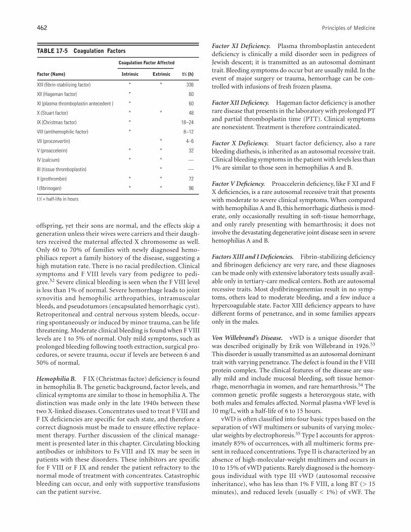
Inherited disorders of coagulation can result from deficiencyof a number of factors (seen in Table 17-5) that are essentialin the coagulation cascade or deficiency of vWF. Clinical bleed-ing can vary from mild to severe, depending on the specificclotting factor affected and the level of factor deficiency.
Hemophilia A. A deficiency of F VIII, the antihemophilicfactor, is inherited as an X-linked recessive trait that affectsmales (hemizygous). The trait is carried in the female (het-erozygous) without clinical evidence of the disease, althougha few do manifest mild bleeding symptoms. Males withhemophilia transmit the affected gene to all their female
462 Principles of Medicine
offspring, yet their sons are normal, and the effects skip ageneration unless their wives were carriers and their daugh-ters received the maternal affected X chromosome as well.Only 60 to 70% of families with newly diagnosed hemo-philiacs report a family history of the disease, suggesting ahigh mutation rate. There is no racial predilection. Clinicalsymptoms and F VIII levels vary from pedigree to pedi-gree.52 Severe clinical bleeding is seen when the F VIII levelis less than 1% of normal. Severe hemorrhage leads to jointsynovitis and hemophilic arthropathies, intramuscularbleeds, and pseudotumors (encapsulated hemorrhagic cyst).Retroperitoneal and central nervous system bleeds, occur-ring spontaneously or induced by minor trauma, can be lifethreatening. Moderate clinical bleeding is found when F VIIIlevels are 1 to 5% of normal. Only mild symptoms, such asprolonged bleeding following tooth extraction, surgical pro-cedures, or severe trauma, occur if levels are between 6 and50% of normal.
Hemophilia B. F IX (Christmas factor) deficiency is foundin hemophilia B. The genetic background, factor levels, andclinical symptoms are similar to those in hemophilia A. Thedistinction was made only in the late 1940s between thesetwo X-linked diseases. Concentrates used to treat F VIII andF IX deficiencies are specific for each state, and therefore acorrect diagnosis must be made to ensure effective replace-ment therapy. Further discussion of the clinical manage-ment is presented later in this chapter. Circulating blockingantibodies or inhibitors to Fs VIII and IX may be seen inpatients with these disorders. These inhibitors are specificfor F VIII or F IX and render the patient refractory to thenormal mode of treatment with concentrates. Catastrophicbleeding can occur, and only with supportive transfusionscan the patient survive.
Factor XI Deficiency. Plasma thromboplastin antecedentdeficiency is clinically a mild disorder seen in pedigrees ofJewish descent; it is transmitted as an autosomal dominanttrait. Bleeding symptoms do occur but are usually mild. In theevent of major surgery or trauma, hemorrhage can be con-trolled with infusions of fresh frozen plasma.
Factor XII Deficiency. Hageman factor deficiency is anotherrare disease that presents in the laboratory with prolonged PTand partial thromboplastin time (PTT). Clinical symptomsare nonexistent. Treatment is therefore contraindicated.
Factor X Deficiency. Stuart factor deficiency, also a rarebleeding diathesis, is inherited as an autosomal recessive trait.Clinical bleeding symptoms in the patient with levels less than1% are similar to those seen in hemophilias A and B.
Factor V Deficiency. Proaccelerin deficiency, like F XI and FX deficiencies, is a rare autosomal recessive trait that presentswith moderate to severe clinical symptoms. When comparedwith hemophilias A and B, this hemorrhagic diathesis is mod-erate, only occasionally resulting in soft-tissue hemorrhage,and only rarely presenting with hemarthrosis; it does notinvolve the devastating degenerative joint disease seen in severehemophilias A and B.
Factors XIII and I Deficiencies. Fibrin-stabilizing deficiencyand fibrinogen deficiency are very rare, and these diagnosescan be made only with extensive laboratory tests usually avail-able only in tertiary-care medical centers. Both are autosomalrecessive traits. Most dysfibrinogenemias result in no symp-toms, others lead to moderate bleeding, and a few induce ahypercoagulable state. Factor XIII deficiency appears to havedifferent forms of penetrance, and in some families appearsonly in the males.
Von Willebrand’s Disease. vWD is a unique disorder thatwas described originally by Erik von Willebrand in 1926.53
This disorder is usually transmitted as an autosomal dominanttrait with varying penetrance. The defect is found in the F VIIIprotein complex. The clinical features of the disease are usu-ally mild and include mucosal bleeding, soft tissue hemor-rhage, menorrhagia in women, and rare hemarthrosis.54 Thecommon genetic profile suggests a heterozygous state, withboth males and females affected. Normal plasma vWF level is10 mg/L, with a half-life of 6 to 15 hours.
vWD is often classified into four basic types based on theseparation of vWF multimers or subunits of varying molec-ular weights by electrophoresis.55 Type I accounts for approx-imately 85% of occurrences, with all multimeric forms pre-sent in reduced concentrations. Type II is characterized by anabsence of high-molecular-weight multimers and occurs in10 to 15% of vWD patients. Rarely diagnosed is the homozy-gous individual with type III vWD (autosomal recessiveinheritance), who has less than 1% F VIII, a long BT (> 15minutes), and reduced levels (usually < 1%) of vWF. The
TABLE 17-5 Coagulation Factors
Coagulation Factor Affected
Factor (Name) Intrinsic Extrinsic t1⁄2 (h)
XIII (fibrin-stabilizing factor) * * 336
XII (Hageman factor) * 60
XI (plasma thromboplastin antecedent ) * 60
X (Stuart factor) * * 48
IX (Christmas factor) * 18–24
VIII (antihemophilic factor) * 8–12
VII (proconvertin) * 4–6
V (proaccelerin) * * 32
IV (calcium) * * —
III (tissue thromboplastin) * —
II (prothrombin) * * 72
I (fibrinogen) * * 96
t 1⁄2 = half-life in hours

Bleeding and Clotting Disorders 463
fourth type is called pseudo- or platelet-type vWD, and it isa primary platelet disorder that mimics vWD. The increasedplatelet affinity for large multimers of vWF results primarilyin mucocutaneous bleeding. Due to familial genetic variants,wide variations occur in the patient’s laboratory profile overtime; therefore, diagnosis may be difficult.56 The uncoveringof all of the biochemical, physiologic, and clinical manifes-tations of vWD has held experts at bay for many years. Asearly as 1968, acquired vWD was noted to occur as a rarecomplication of autoimmune or neoplastic disease, associ-ated mostly with lymphoid or plasma cell proliferative dis-orders and having clinical manifestations that are similar tocongenital vWD.57
ANTICOAGULANT-RELATED COAGULOPATHIES
Heparin. Intentional anticoagulation is delivered acutelywith heparin or as chronic oral therapy with coumarin drugs.Indications for heparin therapy include prophylaxis or treat-ment for venous thromboembolism, including prophylaxis inmedical and surgical patients.58 Heparin is a potent anticoag-ulant that binds with antithrombin III to dramatically inhibitactivation of Fs IX, X, and XI, thereby reducing thrombin gen-eration and fibrin formation. The major bleeding complica-tions from heparin therapy are bleeding at surgical sites andbleeding into the retroperitoneum.
Heparin has a relatively short duration of action of 3 to4 hours, so is typically used for acute anticoagulation,whereas chronic therapy is initiated with coumarin drugs.For acute anticoagulation, intravenous infusion of 1,000units unfractionated heparin per hour, sometimes followinga 5,000-unit bolus, is given to raise the aPTT to 1.5 to 2 timesthe pre-heparin aPTT. Alternatively, subcutaneous injec-tions of 5,000 to 10,000 units of heparin are given every 12hours. Newer biologically active low-molecular-weightheparins administered subcutaneously once or twice dailyare less likely to result in thrombocytopenia and bleedingcomplications. Protamine sulfate can rapidly reverse the anti-coagulant effects of heparin.
Coumarin. Coumarin anticoagulants, which include war-farin and dicumarol (Coumadin, DuPont Pharmaceuticals,Wilmington, DE), are used for anticoagulation to preventrecurrent thrombotic phenomena (pulmonary embolism,venous thrombosis, stroke, myocardial infarction), to treatatrial fibrillation, and in conjunction with prosthetic heartvalves.59 They slow thrombin production and clot formationby blocking the action of vitamin K. Levels of vitaminK–dependent Fs II, VI, IX, and X (prothrombin complexproteins) are reduced. The anticoagulant effect of coumarindrugs may be reversed rapidly by infusion of fresh frozenplasma, or over the course of 12 to 24 hours by administra-tion of vitamin K. PT/INR is used to monitor anticoagula-tion levels. Therapeutic ranges, depending on the indica-tion for anticoagulation, vary from a PT of 18 to 30 seconds(INR of 1.5 to 4.0). Doses of 2.5 to 7.5 mg coumarin daily
typically are required to maintain adequate anticoagulation.Patients with paroxysmal atrial fibrillation and porcine heartvalves require minimal anticoagulation (INR target 1.5–2.0),venous thrombosis is managed with intermediate-rangecoagulation (INR 2.0–3.0), whereas mechanical prostheticheart valves and hypercoagulable states require more intenseanticoagulation (INR target 3.0–4.0).
Coumarin therapy requires continual laboratory monitor-ing, typically every 2 to 8 weeks, as fluctuations can occur. Ithas a longer duration of action, with coagulant activity inblood decreased by 50% in 12 hours and 20% in 24 hours oftherapy initiation. Coagulation returns to normal levels inapproximately 2 to 4 days following discontinuation ofcoumarin drugs. Coumarin therapy can result in bleedingepisodes that are sometimes fatal. Intramuscular injectionsare avoided in anticoagulated patients because of increasedrisk of intramuscular bleeding and hematoma formation.Coumarin drugs are particularly susceptible to drug interac-tions. Drugs that potentially increase coumarin potency (ie,elevate the INR) include metronidazole, penicillin, ery-thromycin, cephalosporins, tetracycline, fluconazole, keto-conazole, chloral hydrate, and propoxyphene; those thatreduce its potency (ie, decrease the INR) include barbiturates,ascorbic acid, dicloxacillin, and nafcillin.60 Additive hemosta-tic effect is seen when coumarin drugs are used in combina-tion with aspirin or NSAIDs.
DISEASE-RELATED COAGULOPATHIES
Liver Disease. Patients with liver disease may have a widespectrum of hemostatic defects depending upon the extent ofliver damage.61 Owing to impaired protein synthesis, impor-tant factors and inhibitors of the clotting and the fibrinolyticsystems are markedly reduced. Additionally, abnormal vitaminK–dependent factor and fibrinogen molecules have beenencountered. Thrombocytopenia and thrombocytopathy arealso common in severe liver disease. Acute or chronic hepato-cellular disease may display decreased vitamin K–dependentfactor levels, especially Fs II, VII, IX, and X and protein C,with other factors still being normal.
Vitamin K Deficiency. Vitamin K is a fat-soluble vitaminthat is absorbed in the small intestine and stored in the liver.It plays an important role in hemostasis. Vitamin K deficiencyis associated with the production of poorly functioning vita-min K–dependent Fs II, VII, IX, and X.62 Deficiency is rare butcan result from inadequate dietary intake, intestinal malab-sorption, or loss of storage sites due to hepatocellular disease.Biliary tract obstruction and long-term use of broad-spec-trum antibiotics, particularly the cephalosporins, can causevitamin K deficiency. Although there is a theoretic 30-day storeof vitamin K in the liver, severe hemorrhage can result inacutely ill patients in 7 to 10 days. A rapid fall in F VII levelsleads to an initial elevation in INR and a subsequent prolon-gation of aPTT. When vitamin K deficiency results in coagu-lopathy, supplemental vitamin K by injection restores theintegrity of the clotting mechanism.
464 Principles of Medicine
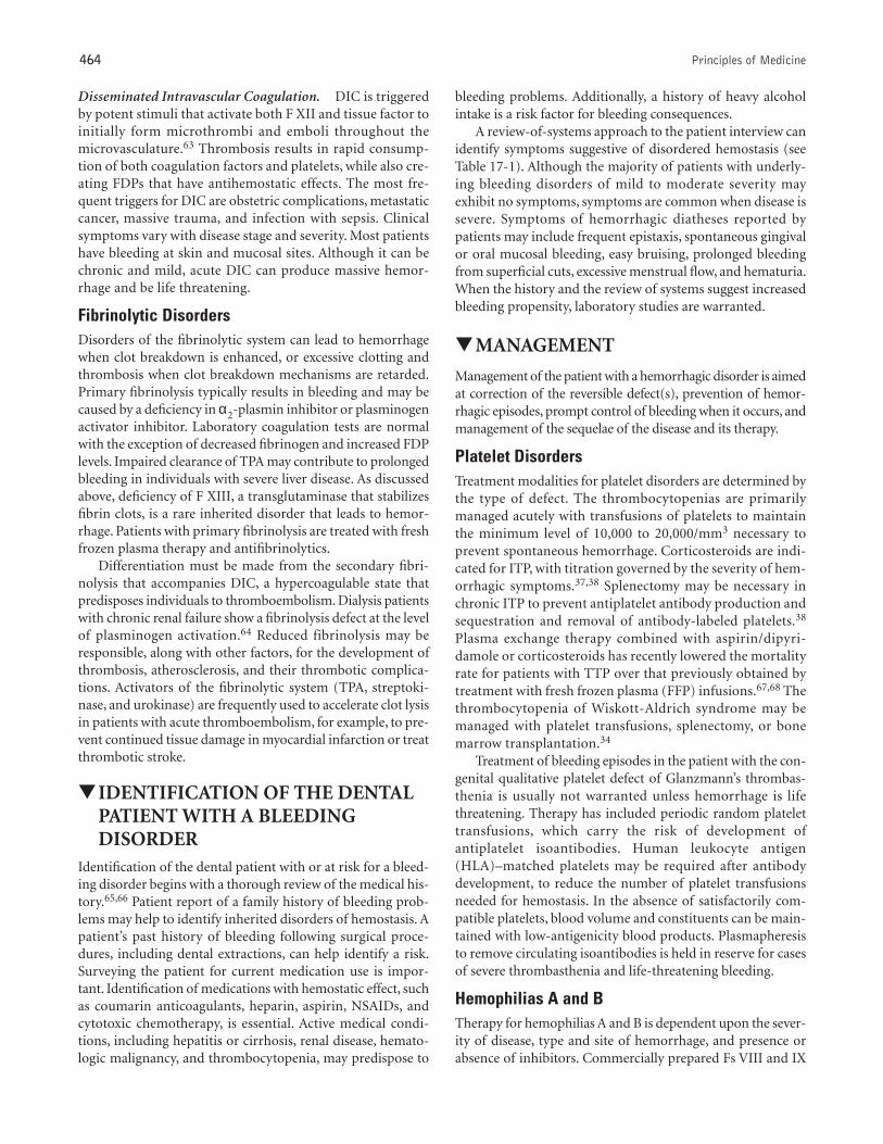
bleeding problems. Additionally, a history of heavy alcoholintake is a risk factor for bleeding consequences.
A review-of-systems approach to the patient interview canidentify symptoms suggestive of disordered hemostasis (seeTable 17-1). Although the majority of patients with underly-ing bleeding disorders of mild to moderate severity mayexhibit no symptoms, symptoms are common when disease issevere. Symptoms of hemorrhagic diatheses reported bypatients may include frequent epistaxis, spontaneous gingivalor oral mucosal bleeding, easy bruising, prolonged bleedingfrom superficial cuts, excessive menstrual flow, and hematuria.When the history and the review of systems suggest increasedbleeding propensity, laboratory studies are warranted.
▼MANAGEMENT
Management of the patient with a hemorrhagic disorder is aimedat correction of the reversible defect(s), prevention of hemor-rhagic episodes, prompt control of bleeding when it occurs, andmanagement of the sequelae of the disease and its therapy.
Platelet DisordersTreatment modalities for platelet disorders are determined bythe type of defect. The thrombocytopenias are primarilymanaged acutely with transfusions of platelets to maintainthe minimum level of 10,000 to 20,000/mm3 necessary toprevent spontaneous hemorrhage. Corticosteroids are indi-cated for ITP, with titration governed by the severity of hem-orrhagic symptoms.37,38 Splenectomy may be necessary inchronic ITP to prevent antiplatelet antibody production andsequestration and removal of antibody-labeled platelets.38
Plasma exchange therapy combined with aspirin/dipyri-damole or corticosteroids has recently lowered the mortalityrate for patients with TTP over that previously obtained bytreatment with fresh frozen plasma (FFP) infusions.67,68 Thethrombocytopenia of Wiskott-Aldrich syndrome may bemanaged with platelet transfusions, splenectomy, or bonemarrow transplantation.34
Treatment of bleeding episodes in the patient with the con-genital qualitative platelet defect of Glanzmann’s thrombas-thenia is usually not warranted unless hemorrhage is lifethreatening. Therapy has included periodic random platelettransfusions, which carry the risk of development ofantiplatelet isoantibodies. Human leukocyte antigen(HLA)–matched platelets may be required after antibodydevelopment, to reduce the number of platelet transfusionsneeded for hemostasis. In the absence of satisfactorily com-patible platelets, blood volume and constituents can be main-tained with low-antigenicity blood products. Plasmapheresisto remove circulating isoantibodies is held in reserve for casesof severe thrombasthenia and life-threatening bleeding.
Hemophilias A and BTherapy for hemophilias A and B is dependent upon the sever-ity of disease, type and site of hemorrhage, and presence orabsence of inhibitors. Commercially prepared Fs VIII and IX
Disseminated Intravascular Coagulation. DIC is triggeredby potent stimuli that activate both F XII and tissue factor toinitially form microthrombi and emboli throughout themicrovasculature.63 Thrombosis results in rapid consump-tion of both coagulation factors and platelets, while also cre-ating FDPs that have antihemostatic effects. The most fre-quent triggers for DIC are obstetric complications, metastaticcancer, massive trauma, and infection with sepsis. Clinicalsymptoms vary with disease stage and severity. Most patientshave bleeding at skin and mucosal sites. Although it can bechronic and mild, acute DIC can produce massive hemor-rhage and be life threatening.
Fibrinolytic DisordersDisorders of the fibrinolytic system can lead to hemorrhagewhen clot breakdown is enhanced, or excessive clotting andthrombosis when clot breakdown mechanisms are retarded.Primary fibrinolysis typically results in bleeding and may becaused by a deficiency in α2-plasmin inhibitor or plasminogenactivator inhibitor. Laboratory coagulation tests are normalwith the exception of decreased fibrinogen and increased FDPlevels. Impaired clearance of TPA may contribute to prolongedbleeding in individuals with severe liver disease. As discussedabove, deficiency of F XIII, a transglutaminase that stabilizesfibrin clots, is a rare inherited disorder that leads to hemor-rhage. Patients with primary fibrinolysis are treated with freshfrozen plasma therapy and antifibrinolytics.
Differentiation must be made from the secondary fibri-nolysis that accompanies DIC, a hypercoagulable state thatpredisposes individuals to thromboembolism. Dialysis patientswith chronic renal failure show a fibrinolysis defect at the levelof plasminogen activation.64 Reduced fibrinolysis may beresponsible, along with other factors, for the development ofthrombosis, atherosclerosis, and their thrombotic complica-tions. Activators of the fibrinolytic system (TPA, streptoki-nase, and urokinase) are frequently used to accelerate clot lysisin patients with acute thromboembolism, for example, to pre-vent continued tissue damage in myocardial infarction or treatthrombotic stroke.
▼IDENTIFICATION OF THE DENTALPATIENT WITH A BLEEDINGDISORDER
Identification of the dental patient with or at risk for a bleed-ing disorder begins with a thorough review of the medical his-tory.65,66 Patient report of a family history of bleeding prob-lems may help to identify inherited disorders of hemostasis. Apatient’s past history of bleeding following surgical proce-dures, including dental extractions, can help identify a risk.Surveying the patient for current medication use is impor-tant. Identification of medications with hemostatic effect, suchas coumarin anticoagulants, heparin, aspirin, NSAIDs, andcytotoxic chemotherapy, is essential. Active medical condi-tions, including hepatitis or cirrhosis, renal disease, hemato-logic malignancy, and thrombocytopenia, may predispose to
Bleeding and Clotting Disorders 465
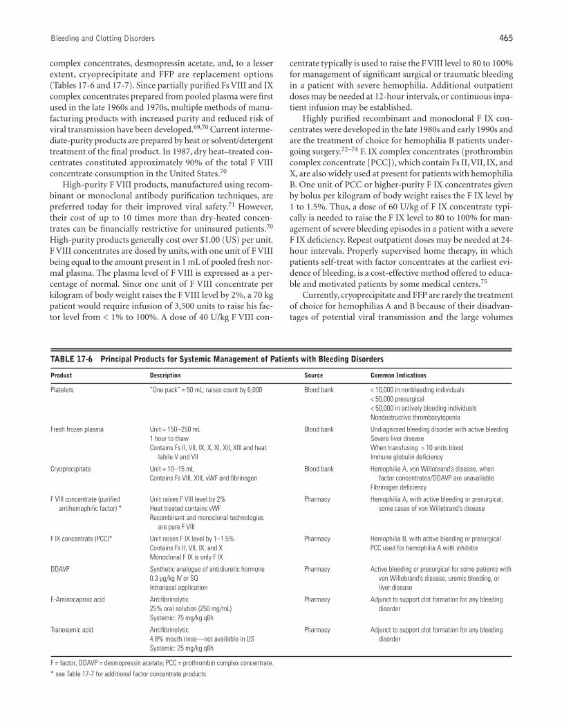
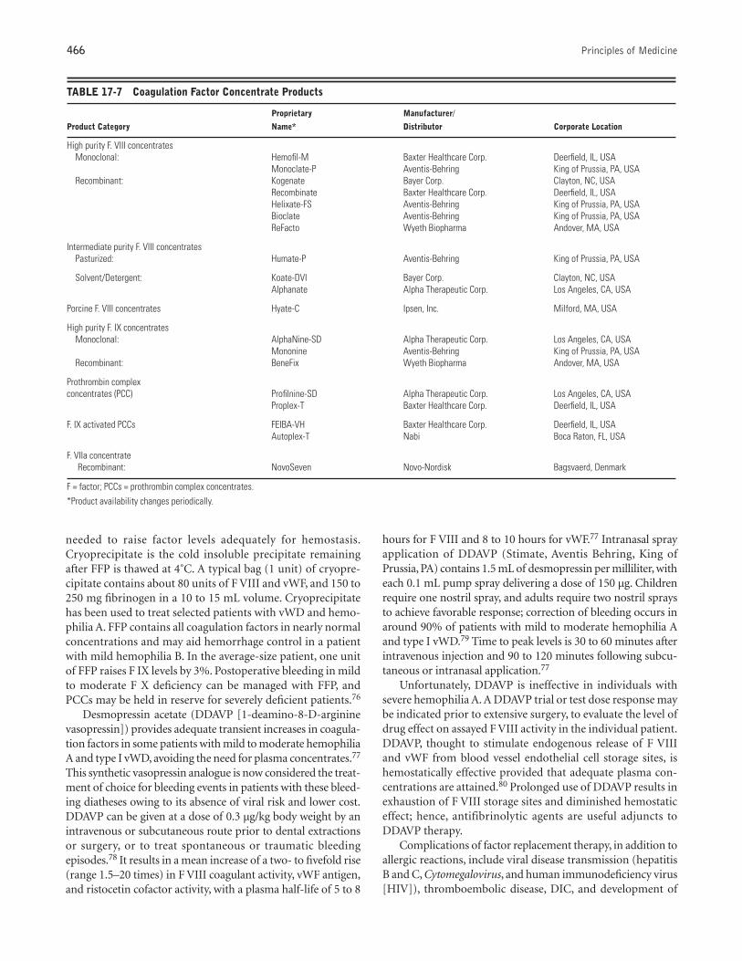
complex concentrates, desmopressin acetate, and, to a lesserextent, cryoprecipitate and FFP are replacement options(Tables 17-6 and 17-7). Since partially purified Fs VIII and IXcomplex concentrates prepared from pooled plasma were firstused in the late 1960s and 1970s, multiple methods of manu-facturing products with increased purity and reduced risk ofviral transmission have been developed.69,70 Current interme-diate-purity products are prepared by heat or solvent/detergenttreatment of the final product. In 1987, dry heat–treated con-centrates constituted approximately 90% of the total F VIIIconcentrate consumption in the United States.70
High-purity F VIII products, manufactured using recom-binant or monoclonal antibody purification techniques, arepreferred today for their improved viral safety.71 However,their cost of up to 10 times more than dry-heated concen-trates can be financially restrictive for uninsured patients.70
High-purity products generally cost over $1.00 (US) per unit.F VIII concentrates are dosed by units, with one unit of F VIIIbeing equal to the amount present in 1 mL of pooled fresh nor-mal plasma. The plasma level of F VIII is expressed as a per-centage of normal. Since one unit of F VIII concentrate perkilogram of body weight raises the F VIII level by 2%, a 70 kgpatient would require infusion of 3,500 units to raise his fac-tor level from < 1% to 100%. A dose of 40 U/kg F VIII con-
centrate typically is used to raise the F VIII level to 80 to 100%for management of significant surgical or traumatic bleedingin a patient with severe hemophilia. Additional outpatientdoses may be needed at 12-hour intervals, or continuous inpa-tient infusion may be established.
Highly purified recombinant and monoclonal F IX con-centrates were developed in the late 1980s and early 1990s andare the treatment of choice for hemophilia B patients under-going surgery.72–74 F. IX complex concentrates (prothrombincomplex concentrate [PCC]), which contain Fs II, VII, IX, andX, are also widely used at present for patients with hemophiliaB. One unit of PCC or higher-purity F IX concentrates givenby bolus per kilogram of body weight raises the F IX level by1 to 1.5%. Thus, a dose of 60 U/kg of F IX concentrate typi-cally is needed to raise the F IX level to 80 to 100% for man-agement of severe bleeding episodes in a patient with a severeF IX deficiency. Repeat outpatient doses may be needed at 24-hour intervals. Properly supervised home therapy, in whichpatients self-treat with factor concentrates at the earliest evi-dence of bleeding, is a cost-effective method offered to educa-ble and motivated patients by some medical centers.75
Currently, cryoprecipitate and FFP are rarely the treatmentof choice for hemophilias A and B because of their disadvan-tages of potential viral transmission and the large volumes
TABLE 17-6 Principal Products for Systemic Management of Patients with Bleeding Disorders
Product Description Source Common Indications
Platelets “One pack” = 50 mL; raises count by 6,000 Blood bank < 10,000 in nonbleeding individuals< 50,000 presurgical< 50,000 in actively bleeding individualsNondestructive thrombocytopenia
Fresh frozen plasma Unit = 150–250 mL Blood bank Undiagnosed bleeding disorder with active bleeding1 hour to thaw Severe liver diseaseContains Fs II, VII, IX, X, XI, XII, XIII and heat When transfusing > 10 units blood
labile V and VII Immune globulin deficiency
Cryoprecipitate Unit = 10–15 mL Blood bank Hemophilia A, von Willebrand’s disease, whenContains Fs VIII, XIII, vWF and fibrinogen factor concentrates/DDAVP are unavailable
Fibrinogen deficiency
F VIII concentrate (purified Unit raises F VIII level by 2% Pharmacy Hemophilia A, with active bleeding or presurgical; antihemophilic factor) * Heat treated contains vWF some cases of von Willebrand’s disease
Recombinant and monoclonal technologiesare pure F VIII
F IX concentrate (PCC)* Unit raises F IX level by 1–1.5% Pharmacy Hemophilia B, with active bleeding or presurgicalContains Fs II, VII, IX, and X PCC used for hemophilia A with inhibitorMonoclonal F IX is only F IX
DDAVP Synthetic analogue of antidiuretic hormone Pharmacy Active bleeding or presurgical for some patients with0.3 µg/kg IV or SQ von Willebrand’s disease, uremic bleeding, or Intranasal application liver disease
E-Aminocaproic acid Antifibrinolytic Pharmacy Adjunct to support clot formation for any bleeding25% oral solution (250 mg/mL) disorderSystemic: 75 mg/kg q6h
Tranexamic acid Antifibrinolytic Pharmacy Adjunct to support clot formation for any bleeding 4.8% mouth rinse—not available in US disorderSystemic: 25 mg/kg q8h
F = factor; DDAVP = desmopressin acetate; PCC = prothrombin complex concentrate.* see Table 17-7 for additional factor concentrate products.
466 Principles of Medicine
needed to raise factor levels adequately for hemostasis.Cryoprecipitate is the cold insoluble precipitate remainingafter FFP is thawed at 4˚C. A typical bag (1 unit) of cryopre-cipitate contains about 80 units of F VIII and vWF, and 150 to250 mg fibrinogen in a 10 to 15 mL volume. Cryoprecipitatehas been used to treat selected patients with vWD and hemo-philia A. FFP contains all coagulation factors in nearly normalconcentrations and may aid hemorrhage control in a patientwith mild hemophilia B. In the average-size patient, one unitof FFP raises F IX levels by 3%. Postoperative bleeding in mildto moderate F X deficiency can be managed with FFP, andPCCs may be held in reserve for severely deficient patients.76
Desmopressin acetate (DDAVP [1-deamino-8-D-argininevasopressin]) provides adequate transient increases in coagula-tion factors in some patients with mild to moderate hemophiliaA and type I vWD, avoiding the need for plasma concentrates.77
This synthetic vasopressin analogue is now considered the treat-ment of choice for bleeding events in patients with these bleed-ing diatheses owing to its absence of viral risk and lower cost.DDAVP can be given at a dose of 0.3 µg/kg body weight by anintravenous or subcutaneous route prior to dental extractionsor surgery, or to treat spontaneous or traumatic bleedingepisodes.78 It results in a mean increase of a two- to fivefold rise(range 1.5–20 times) in F VIII coagulant activity, vWF antigen,and ristocetin cofactor activity, with a plasma half-life of 5 to 8
hours for F VIII and 8 to 10 hours for vWF.77 Intranasal sprayapplication of DDAVP (Stimate, Aventis Behring, King ofPrussia, PA) contains 1.5 mL of desmopressin per milliliter, witheach 0.1 mL pump spray delivering a dose of 150 µg. Childrenrequire one nostril spray, and adults require two nostril spraysto achieve favorable response; correction of bleeding occurs inaround 90% of patients with mild to moderate hemophilia Aand type I vWD.79 Time to peak levels is 30 to 60 minutes afterintravenous injection and 90 to 120 minutes following subcu-taneous or intranasal application.77
Unfortunately, DDAVP is ineffective in individuals withsevere hemophilia A. A DDAVP trial or test dose response maybe indicated prior to extensive surgery, to evaluate the level ofdrug effect on assayed F VIII activity in the individual patient.DDAVP, thought to stimulate endogenous release of F VIIIand vWF from blood vessel endothelial cell storage sites, ishemostatically effective provided that adequate plasma con-centrations are attained.80 Prolonged use of DDAVP results inexhaustion of F VIII storage sites and diminished hemostaticeffect; hence, antifibrinolytic agents are useful adjuncts toDDAVP therapy.
Complications of factor replacement therapy, in addition toallergic reactions, include viral disease transmission (hepatitisB and C, Cytomegalovirus, and human immunodeficiency virus[HIV]), thromboembolic disease, DIC, and development of
TABLE 17-7 Coagulation Factor Concentrate Products
Proprietary Manufacturer/
Product Category Name* Distributor Corporate Location
High purity F. VIII concentratesMonoclonal: Hemofil-M Baxter Healthcare Corp. Deerfield, IL, USA
Monoclate-P Aventis-Behring King of Prussia, PA, USARecombinant: Kogenate Bayer Corp. Clayton, NC, USA
Recombinate Baxter Healthcare Corp. Deerfield, IL, USAHelixate-FS Aventis-Behring King of Prussia, PA, USABioclate Aventis-Behring King of Prussia, PA, USAReFacto Wyeth Biopharma Andover, MA, USA
Intermediate purity F. VIII concentratesPasturized: Humate-P Aventis-Behring King of Prussia, PA, USA
Solvent/Detergent: Koate-DVI Bayer Corp. Clayton, NC, USAAlphanate Alpha Therapeutic Corp. Los Angeles, CA, USA
Porcine F. VIII concentrates Hyate-C Ipsen, Inc. Milford, MA, USA
High purity F. IX concentratesMonoclonal: AlphaNine-SD Alpha Therapeutic Corp. Los Angeles, CA, USA
Mononine Aventis-Behring King of Prussia, PA, USARecombinant: BeneFix Wyeth Biopharma Andover, MA, USA
Prothrombin complex concentrates (PCC) Profilnine-SD Alpha Therapeutic Corp. Los Angeles, CA, USA
Proplex-T Baxter Healthcare Corp. Deerfield, IL, USA
F. IX activated PCCs FEIBA-VH Baxter Healthcare Corp. Deerfield, IL, USAAutoplex-T Nabi Boca Raton, FL, USA
F. VIIa concentrateRecombinant: NovoSeven Novo-Nordisk Bagsvaerd, Denmark
F = factor; PCCs = prothrombin complex concentrates.*Product availability changes periodically.
Bleeding and Clotting Disorders 467
antibodies to factor concentrates. Hepatitis B and non-A/non-B have been major causes of morbidity and mortality in thehemophiliac population, resulting in chronic active hepatitisand cirrhosis in a number of patients.52 More recently, hepati-tis C and HIV infection have become the most common trans-fusion-related infections in hemophiliacs. By the end of 1986,some centers reported that 80 to 90% of hemophiliacs treatedwith F VIII concentrates and around 50% of those who hadreceived F IX concentrates were HIV seropositive.81 Since 1986,with viral screening of donated plasma, there have been fewtransfusion-related HIV seroconversions.
Use of factor IX complex concentrate can result in throm-botic complications, such as deep venous thromboses, myocar-dial infarctions, pulmonary emboli, and DIC. Concurrent useof systemic antifibrinolytics with these products may increasethe risks. DIC is believed to occur as a consequence of high lev-els of activated clotting factors, such as Fs VIIa, IXa, and Xa,that cannot adequately be cleared by the liver.
Development of a F VIII or F IX inhibitor is a serious com-plication. These pathologic circulating antibodies of the IgGclass, which specifically neutralize F VIII or F IX procoagulantactivity, arise as alloantibodies in some patients with hemo-philia.82 Inhibitors develop in at least 10 to 15% of patientswith severe hemophilia A and less commonly in patients withhemophilia B.83,84 Development is related to exposure to fac-tor products and genetic predisposition.82,83 Inhibitor level isquantified by the Bethesda inhibitor assay and is reported asBethesda units (BU).
The inhibitor titer and responsiveness to further factorinfusion (responder-type) dictate which factor replacementtherapy should be used. Patients with inhibitors are classifiedaccording to titer level—low ( < 10 BU/mL) or high (> 10BU/mL)—and also by responder type.84 Low responders typ-ically maintain low titers with repeated factor concentrateexposure, whereas high responders show a brisk elevation intiter due to the amnestic response and are the most chal-lenging to manage.84,85 Patients with low inhibitor titers areusually low responders, and those with high titers are oftenhigh responders. Seventy-five percent of hemophilia Apatients with inhibitors are high responders, whereas only25% are low responders.84
For hemorrhages, hemophilia A patients with low-levellow-responding inhibitors are treated with F VIII concentratesin doses sufficient to raise plasma F VIII levels to the thera-peutic range. Critical hemorrhages in patients with high-responding inhibitors may be treated with large quantities ofporcine F VIII; however, routine hemorrhages are often man-aged initially with PCCs, which provoke anamnesis in a fewpatients.82 PCCs can bypass the F VIII inhibitor and are effec-tive about 50% of the time.86 Activated PCCs show slightlyincreased effectiveness (65–75%). Highly purified porcine F VIII product use can be advantageous in patients with lessthan 50 BU, since human F VIII inhibitors cross-react less fre-quently with porcine products.82 However, because of the riskof hemostatic failure, surgery should be performed under cov-erage of F VIII.87 Treatment of the patient with low-level
(< 10 BU) F IX inhibitors requires higher doses of F IX com-plex concentrates to achieve hemostasis. Developed in the early1990s, recombinant F VIIa is a novel product that provides analternative treatment option for patients with hemophilia A orB with inhibitors by enhancing the extrinsic pathway.88 It hasbeen proven to effectively control bleeding in patients withhigh-titer inhibitors.89,90
Several methods have demonstrated temporary removalof high-titer inhibitors in both hemophilia A and B. Exchangetransfusion or plasmapheresis produces a rapid transientreduction in antibody level, with a rate of 40 mL plasma perkilogram decreasing levels by half.91 Although laborious, itmay be attempted in cases of critical hemorrhage as an adjunctto high-dose F VIII concentrate therapy. Antibody removal byextracorporeal adsorption of the plasma to protein A-Sepharose or a specific F IX–Sepharose in columns has alsoshown promise in hemophilias A and B.92,93
Von Willebrand’s DiseaseTherapy for vWD depends on the type of vWD and the sever-ity of bleeding. Type I is treated preferentially with DDAVP asdescribed above. Intermediate-purity F VIII concentrates, FFP,and cryoprecipitate are held in reserve for DDAVP nonre-sponders.55 Types II and III require intermediate-purity F VIIIconcentrates, such as Humate-P or Koate-HS, or, rarely, cryo-precipitate or FFP. Bleeding episodes in patients with platelet-type vWD are usually controlled with platelet concentrateinfusions. Other therapy is used for site-specific bleeding, suchas estrogens or oral contraceptive agents for menorrhagia andlocal hemostatic agents and antifibrinolytics for dental proce-dures. Occasionally, circulating plasma inhibitors of vWF areobserved in multiply transfused patients with severe disease.Cryoprecipitate infusion can cause transient neutralization ofthis inhibitor.94
Disease-Related CoagulopathiesManagement of disease-related coagulopathies varies withhemostatic abnormality.
LIVER DISEASE
Hepatic disease that results in bleeding from deficient vitaminK–dependent clotting factors (Fs II, VII, IX, and X) may bereversed with vitamin K injections for 3 days, either intra-venously or subcutaneously. However, infusion of FFP may beemployed when more immediate hemorrhage control is nec-essary, such as prior to dental extractions.95 Cirrhotic patientswith moderate thrombocytopenia and functional plateletdefects may benefit from DDAVP therapy.96 Antifibrinolyticdrugs, if used cautiously, have markedly reduced bleeding andthus reduced need for blood and blood product substitution.61
RENAL DISEASE
In uremic patients, dialysis remains the primary preventiveand therapeutic modality used for control of bleeding,although it is not always immediately effective.97 Hemodialysisand peritoneal dialysis appear to be equally efficacious in
468 Principles of Medicine
improving platelet function abnormalities and clinical bleed-ing in the uremic patient. The availability of cryoprecipitate98
and DDAVP99 offers alternative effective therapy for patientswho require shortened bleeding times acutely in preparationfor urgent surgery. Conjugated estrogen preparations100 andrecombinant erythropoietin101 have also been shown to bebeneficial for uremic patients with chronic abnormal bleeding.
DISSEMINATED INTRAVASCULAR COAGULATION
Although somewhat controversial, active DIC is usually treatedinitially with intravenous unfractionated heparin or subcuta-neous low-molecular-weight heparin, to prevent thrombinfrom acting on fibrinogen, thereby preventing further clot for-mation.102–104 It is important to expeditiously identify andinstitute therapy for the underlying triggering disease or con-dition if long-term survival is to be a possibility. The dentistmay be called upon to provide a gingival or oral mucosalbiopsy specimen for histopathologic examination to confirmthe diagnosis of DIC by the presence of microthrombi in thevascular bed. Replacement of deficient coagulation factorswith FFP and correction of the platelet deficiency with platelettransfusions may be necessary for improvement or prophylaxisof the hemorrhagic tendency of DIC prior to emergency sur-gical procedures. Elective surgery is deferred due to the volatil-ity of the coagulation mechanism in these patients.
▼PROGNOSIS
Prognosis for patients with bleeding disorders depends onappropriate diagnosis and the ability to prevent and manageacute bleeding episodes. Individuals with mild or manageabledisease have a normal life expectancy, with morbidity relatingto bleeding episode frequency and severity. Acute DIC carriesthe highest risk of death by exsanguination. Individuals withsevere liver disease may succumb to rupture of esophagealvarices. Severe thrombocytopenia and other severe coagu-lopathies carry a higher risk of hemorrhagic stroke.
Advances in the treatment of hemophilia, from the use ofcryoprecipitate in the 1960s to the introduction of plasma-derived factor concentrates in the 1970s, have led to dramaticimprovement in quality of life and raised the lifespan for hemo-philiacs from 11 years in 1921 to 60 years in 1980.105 Viralinfections, such as hepatitis B, C, and G and HIV acquired frominfected blood products, have altered the prognosis for somepatients.106–110 As discussed above, before effective virucidalmethods were used in the manufacture of clotting-factor con-centrates in 1985, hemophiliacs were at a very high risk of con-tracting bloodborne viruses from factor concentrates thatexposed them to the plasma of thousands of donors. HIV sero-prevalence increased to 60 to 75% of patients with hemophilia(85–90% with severe hemophilia), with HIV-associated oppor-tunistic infections and neoplasms contributing substantially tothe morbidity and mortality of hemophiliacs.106,107,109 Oralmucosal diseases are common in hemophiliacs with HIV, par-ticularly in those with advanced immunosuppression,110,111
and are discussed in more detail in chapter ***. HIV protease
inhibitor–containing drug combinations that resulted inimproved health of some HIV-infected patients in the late1990s are showing significant clinical and laboratory benefitswhen used by HIV-infected hemophiliacs.112 Co-infection withviral hepatitis remains a challenge for the next decade.
▼ORAL HEALTH CONSIDERATIONS
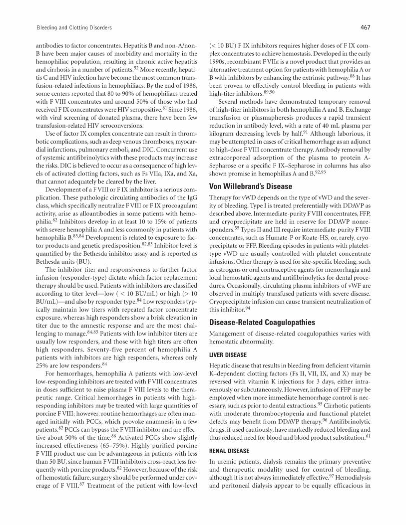
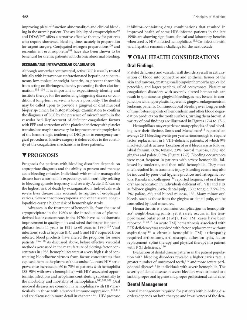
Oral FindingsPlatelet deficiency and vascular wall disorders result in extrava-sation of blood into connective and epithelial tissues of theskin and mucosa, creating small pinpoint hemorrhages, calledpetechiae, and larger patches, called ecchymoses. Platelet orcoagulation disorders with severely altered hemostasis canresult in spontaneous gingival bleeding, as may be seen in con-junction with hyperplastic hyperemic gingival enlargements inleukemic patients. Continuous oral bleeding over long periodsof time fosters deposits of hemosiderin and other blood degra-dation products on the tooth surfaces, turning them brown. Avariety of oral findings are illustrated in Figures 17-4 to 17-6.
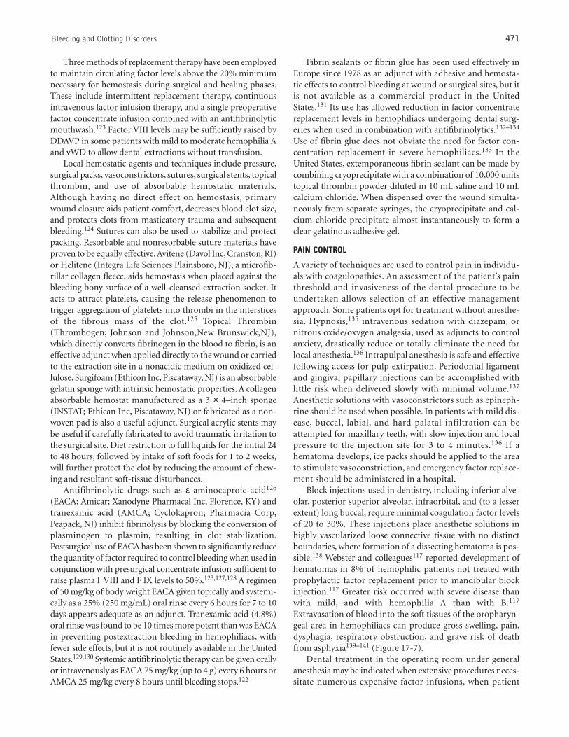
Hemophiliacs may experience many episodes of oral bleed-ing over their lifetime. Sonis and Musselman113 reported anaverage 29.1 bleeding events per year serious enough to requirefactor replacement in F VIII–deficient patients, of which 9%involved oral structures. Location of oral bleeds was as follows:labial frenum, 60%; tongue, 23%; buccal mucosa, 17%; andgingiva and palate, 0.5% (Figure 17-7). Bleeding occurrenceswere most frequent in patients with severe hemophilia, fol-lowed by moderate, and then mild hemophilia. They mostoften resulted from traumatic injury. Bleeding events may alsobe induced by poor oral hygiene practices and iatrogenic fac-tors. Kaneda and colleagues114 reported frequency of oral hem-orrhage by location in individuals deficient of F VIII and F IXas follows: gingiva, 64%; dental pulp, 13%; tongue, 7.5%; lip,7%; palate, 2%; and buccal mucosa, 1%. Many minor oralbleeds, such as those from the gingiva or dental pulp, can becontrolled by local measures.
Hemarthrosis is a common complication in hemophili-acs’ weight-bearing joints, yet it rarely occurs in the tem-poromandibular joint (TMJ). Two TMJ cases have beenreported.115,116 An acute TMJ hemarthrosis associated withF IX deficiency was resolved with factor replacement withoutaspiration;115 a chronic hemophilic TMJ arthropathyrequired arthrotomy, arthroscopic adhesion lysis, factorreplacement, splint therapy, and physical therapy in a patientwith F XI deficiency.116
Evaluation of dental disease patterns in the patient popula-tion with bleeding disorders revealed a higher caries rate, agreater number of unrestored teeth,117 and more severe peri-odontal disease118 in individuals with severe hemophilia. Theseverity of dental disease in severe bleeders was attributed to alack of proper oral hygiene and proper professional dental care.
Dental Management Dental management required for patients with bleeding dis-orders depends on both the type and invasiveness of the den-
Bleeding and Clotting Disorders 469
tal procedure and the type and severity of the bleeding disor-der. Thus, less modification is needed for patients with mildcoagulopathies in preparation for dental procedures antici-pated to have limited bleeding consequences. When signifi-cant bleeding is expected, the goal of management is to pre-operatively restore the hemostatic system to an acceptablerange, while supporting coagulation with adjunctive and/orlocal measures. For reversible coagulopathies, (eg, coumarinanticoagulation), it may be best to remove the causative agent
or treat the primary illness or defect in order to allow thepatient to return to a manageable bleeding risk for the dentaltreatment period. For irreversible coagulopathies, the missingor defective element may need to be replaced from an exoge-nous source to allow control of bleeding (eg, coagulation fac-tor concentrate therapy for hemophilia). Assessment of thecoagulopathy and delivery of appropriate therapy prior todental procedures is best accomplished in consultation witha hematologist.
FIGURE 17-4 A 36-year-old male with idiopathic thrombocytopenia pur-pura and a platelet count of 5,000/mm3. Supportive platelet transfusions andimmunoglobulin therapy were used to control bleeding. A, Labial and tongueecchymoses; B, palatal ecchymoses; C, buccal ecchymoses and fibrinous clot.
FIGURE 17-5 A 68-year-old female with acute myelogenous leukemia and a platelet count of 9,000/mm3. Platelet transfusion and ε-aminocaproic acidoral rinses were used to control bleeding. A, Buccal mucosa and palatal ecchymoses. B, Extrinsic stains on teeth from erythrocyte degradation followingcontinual gingival oozing.
A
B C
A B
470 Principles of Medicine
Platelet DisordersWhen medical management is unable to restore platelet countsto above the level of 50,000/mm3 required for surgical hemo-stasis, platelet transfusions may be required prior to dentalextractions or other oral surgical procedures. The therapeuti-cally expected increment in platelet count from infusion of oneunit of platelets is approximately 10,000 to 12,000/mm3. Sixunits of platelets are commonly infused at a time. Patients whohave received multiple transfusions may be refractory to ran-dom donor platelets as a result of alloimmunization. Theseindividuals may require single-donor apheresis or leukocyte-reduced platelets. Local hemostatic measures are also impor-tant. The thrombasthenic patient needing dental extractionsmay be successfully treated with the use of hemostatic measuressuch as microfibrillar collagen and antifibrinolytic drugs.32,33
Since the antiplatelet activity of aspirin remains for the 8-to 10-day lifetime of the affected platelets, avoidance of aspirinis recommended for 1 to 2 weeks prior to extensive oral surgi-cal procedures. Other NSAIDs have a similar but less pro-nounced antiplatelet effect. Adjunctive local hemostatic agentsare useful in preventing postoperative oozing when aspirin
therapy is in use at the time of minor oral surgery. When exten-sive surgery is emergently indicated, DDAVP can be used todecrease the aspirin-induced prolongation of the BT or totreat aspirin-related postoperative oozing, often eliminatingthe need for platelet infusion.119
Chemotherapy-associated oral hemorrhages, most fre-quently related to thrombocytopenia, are best managed bytransfusions of HLA-matched platelets and FFP, together withtopically applied clot-promoting agents.45 A pilot study sug-gests a possible benefit of DDAVP for the prevention or treat-ment of bleeding in patients with thrombocytopenia associ-ated with hematologic malignancy.120
Hemophilias A and B and Von Willebrand’sDisease
ORAL SURGICAL PROCEDURES
Oral surgical procedures have the greatest potential for hemor-rhage of all dental procedures. Hemorrhagic problems afterextractions have drastically declined over the last 20 years suchthat only an estimated 8% of hemophilic patients experienceone or more delayed bleeding episodes.121 Appropriate precau-tionary measures now allow surgery to be performed safely. Tomake certain that preoperative factor levels of at least 40 to 50%of normal activity have been obtained, transfusion recommen-dations generally aim for replacement of missing coagulationfactors to levels of 50 to 100% when single-bolus infusion is usedfor outpatient treatment. This provides greater assurance ofhemorrhage control, given the problems of possible failure offactor activity to rise as high as expected and variable plasmahalf-lives of 8 to 12 hours for F VIII and 18 to 24 hours for F IX.Additional postoperative factor maintenance may be indicatedfor extensive surgery. This can be accomplished by infusion offactor concentrates, DDAVP, cryoprecipitate, or FFP, dependingon the patient’s deficiency state. When postsurgical bleedingoccurs due to fibrinolysis, it commonly starts 3 to 5 days aftersurgery and can usually be controlled by local measures and useof antifibrinolytics. Continual oozing from unstable fibrinousclots may require their removal and the repacking of the extrac-tion socket with hemostatic agents.
Determination of factor replacement requirements forsurgical hemostasis and selection of plasma product or drugtherapy should be accomplished in consultation with thepatient’s hematologist. Canadian clinical practice guide-lines122 recommend replacement factor levels of 40 to 50% ofF VIII (dose 20–25 U/kg) and F IX (dose 40–50 U/kg), usedin conjunction with antifibrinolytics. Gingival or dentalbleeding unresponsive to antifibrinolytics requires 20 to 30%clotting F VIII or F IX.122 The level of factor activity requiredfor hemostasis varies in relation to local factors. Higher hemo-static factor levels are needed for large wound cavities createdby extraction of multiple or multirooted teeth, or when gin-gival inflammation, bleeding, tooth mobility, or apical lesionsare present.114 Kaneda and colleagues114 report that deficientfactor activity levels required for postextraction hemostasisvaried from 3.5 to 25% for deciduous teeth and 5.5 to 20% forpermanent teeth.114
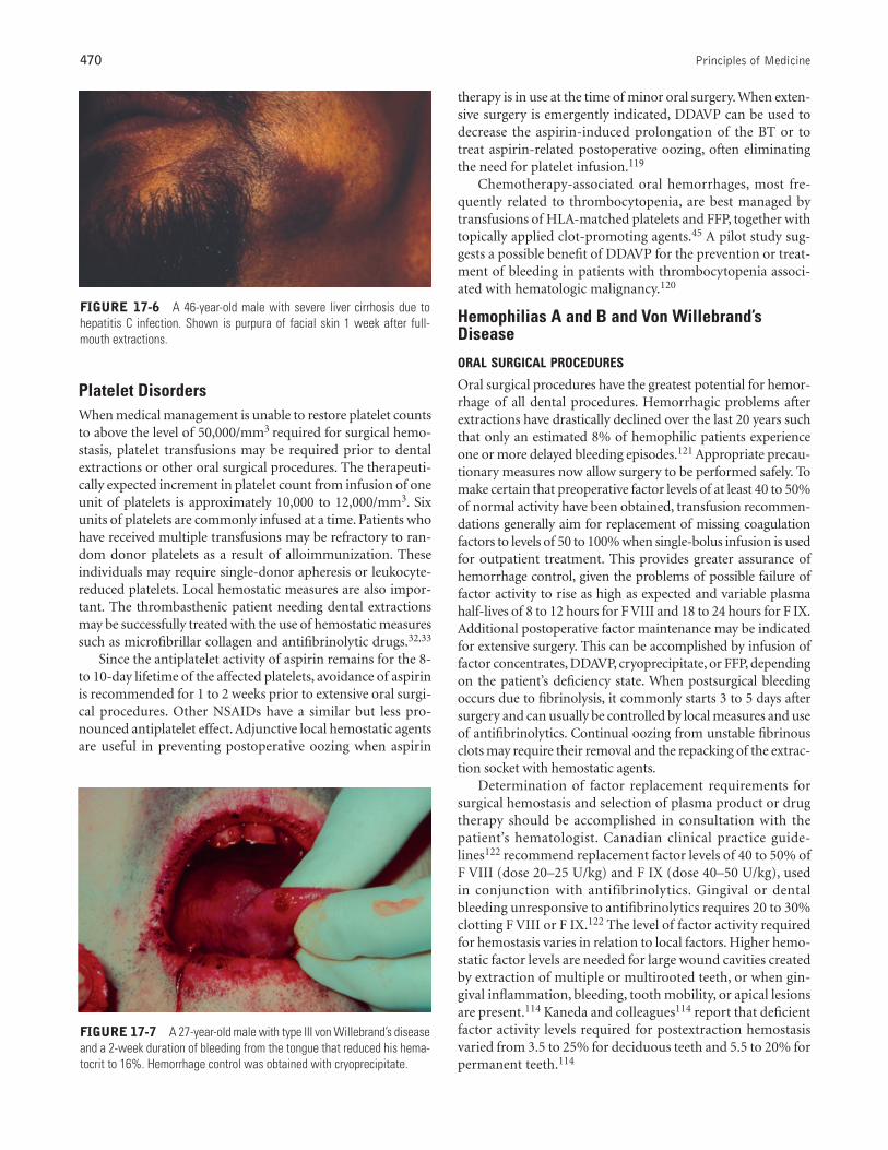
FIGURE 17-6 A 46-year-old male with severe liver cirrhosis due tohepatitis C infection. Shown is purpura of facial skin 1 week after full-mouth extractions.
FIGURE 17-7 A 27-year-old male with type III von Willebrand’s diseaseand a 2-week duration of bleeding from the tongue that reduced his hema-tocrit to 16%. Hemorrhage control was obtained with cryoprecipitate.
Bleeding and Clotting Disorders 471
Three methods of replacement therapy have been employedto maintain circulating factor levels above the 20% minimumnecessary for hemostasis during surgical and healing phases.These include intermittent replacement therapy, continuousintravenous factor infusion therapy, and a single preoperativefactor concentrate infusion combined with an antifibrinolyticmouthwash.123 Factor VIII levels may be sufficiently raised byDDAVP in some patients with mild to moderate hemophilia Aand vWD to allow dental extractions without transfusion.
Local hemostatic agents and techniques include pressure,surgical packs, vasoconstrictors, sutures, surgical stents, topicalthrombin, and use of absorbable hemostatic materials.Although having no direct effect on hemostasis, primarywound closure aids patient comfort, decreases blood clot size,and protects clots from masticatory trauma and subsequentbleeding.124 Sutures can also be used to stabilize and protectpacking. Resorbable and nonresorbable suture materials haveproven to be equally effective. Avitene (Davol Inc, Cranston, RI)or Helitene (Integra Life Sciences Plainsboro, NJ), a microfib-rillar collagen fleece, aids hemostasis when placed against thebleeding bony surface of a well-cleansed extraction socket. Itacts to attract platelets, causing the release phenomenon totrigger aggregation of platelets into thrombi in the intersticesof the fibrous mass of the clot.125 Topical Thrombin(Thrombogen; Johnson and Johnson,New Brunswick,NJ),which directly converts fibrinogen in the blood to fibrin, is aneffective adjunct when applied directly to the wound or carriedto the extraction site in a nonacidic medium on oxidized cel-lulose. Surgifoam (Ethicon Inc, Piscataway, NJ) is an absorbablegelatin sponge with intrinsic hemostatic properties. A collagenabsorbable hemostat manufactured as a 3 × 4–inch sponge(INSTAT; Ethican Inc, Piscataway, NJ) or fabricated as a non-woven pad is also a useful adjunct. Surgical acrylic stents maybe useful if carefully fabricated to avoid traumatic irritation tothe surgical site. Diet restriction to full liquids for the initial 24to 48 hours, followed by intake of soft foods for 1 to 2 weeks,will further protect the clot by reducing the amount of chew-ing and resultant soft-tissue disturbances.
Antifibrinolytic drugs such as ε-aminocaproic acid126
(EACA; Amicar; Xanodyne Pharmacal Inc, Florence, KY) andtranexamic acid (AMCA; Cyclokapron; Pharmacia Corp,Peapack, NJ) inhibit fibrinolysis by blocking the conversion ofplasminogen to plasmin, resulting in clot stabilization.Postsurgical use of EACA has been shown to significantly reducethe quantity of factor required to control bleeding when used inconjunction with presurgical concentrate infusion sufficient toraise plasma F VIII and F IX levels to 50%.123,127,128 A regimenof 50 mg/kg of body weight EACA given topically and systemi-cally as a 25% (250 mg/mL) oral rinse every 6 hours for 7 to 10days appears adequate as an adjunct. Tranexamic acid (4.8%)oral rinse was found to be 10 times more potent than was EACAin preventing postextraction bleeding in hemophiliacs, withfewer side effects, but it is not routinely available in the UnitedStates.129,130 Systemic antifibrinolytic therapy can be given orallyor intravenously as EACA 75 mg/kg (up to 4 g) every 6 hours orAMCA 25 mg/kg every 8 hours until bleeding stops.122
Fibrin sealants or fibrin glue has been used effectively inEurope since 1978 as an adjunct with adhesive and hemosta-tic effects to control bleeding at wound or surgical sites, but itis not available as a commercial product in the UnitedStates.131 Its use has allowed reduction in factor concentratereplacement levels in hemophiliacs undergoing dental surg-eries when used in combination with antifibrinolytics.132–134
Use of fibrin glue does not obviate the need for factor con-centration replacement in severe hemophiliacs.133 In theUnited States, extemporaneous fibrin sealant can be made bycombining cryoprecipitate with a combination of 10,000 unitstopical thrombin powder diluted in 10 mL saline and 10 mLcalcium chloride. When dispensed over the wound simulta-neously from separate syringes, the cryoprecipitate and cal-cium chloride precipitate almost instantaneously to form aclear gelatinous adhesive gel.
PAIN CONTROL
A variety of techniques are used to control pain in individu-als with coagulopathies. An assessment of the patient’s painthreshold and invasiveness of the dental procedure to beundertaken allows selection of an effective managementapproach. Some patients opt for treatment without anesthe-sia. Hypnosis,135 intravenous sedation with diazepam, ornitrous oxide/oxygen analgesia, used as adjuncts to controlanxiety, drastically reduce or totally eliminate the need forlocal anesthesia.136 Intrapulpal anesthesia is safe and effectivefollowing access for pulp extirpation. Periodontal ligamentand gingival papillary injections can be accomplished withlittle risk when delivered slowly with minimal volume.137
Anesthetic solutions with vasoconstrictors such as epineph-rine should be used when possible. In patients with mild dis-ease, buccal, labial, and hard palatal infiltration can beattempted for maxillary teeth, with slow injection and localpressure to the injection site for 3 to 4 minutes.136 If ahematoma develops, ice packs should be applied to the areato stimulate vasoconstriction, and emergency factor replace-ment should be administered in a hospital.
Block injections used in dentistry, including inferior alve-olar, posterior superior alveolar, infraorbital, and (to a lesserextent) long buccal, require minimal coagulation factor levelsof 20 to 30%. These injections place anesthetic solutions inhighly vascularized loose connective tissue with no distinctboundaries, where formation of a dissecting hematoma is pos-sible.138 Webster and colleagues117 reported development ofhematomas in 8% of hemophilic patients not treated withprophylactic factor replacement prior to mandibular blockinjection.117 Greater risk occurred with severe disease thanwith mild, and with hemophilia A than with B.117
Extravasation of blood into the soft tissues of the oropharyn-geal area in hemophiliacs can produce gross swelling, pain,dysphagia, respiratory obstruction, and grave risk of deathfrom asphyxia139–141 (Figure 17-7).
Dental treatment in the operating room under generalanesthesia may be indicated when extensive procedures neces-sitate numerous expensive factor infusions, when patient
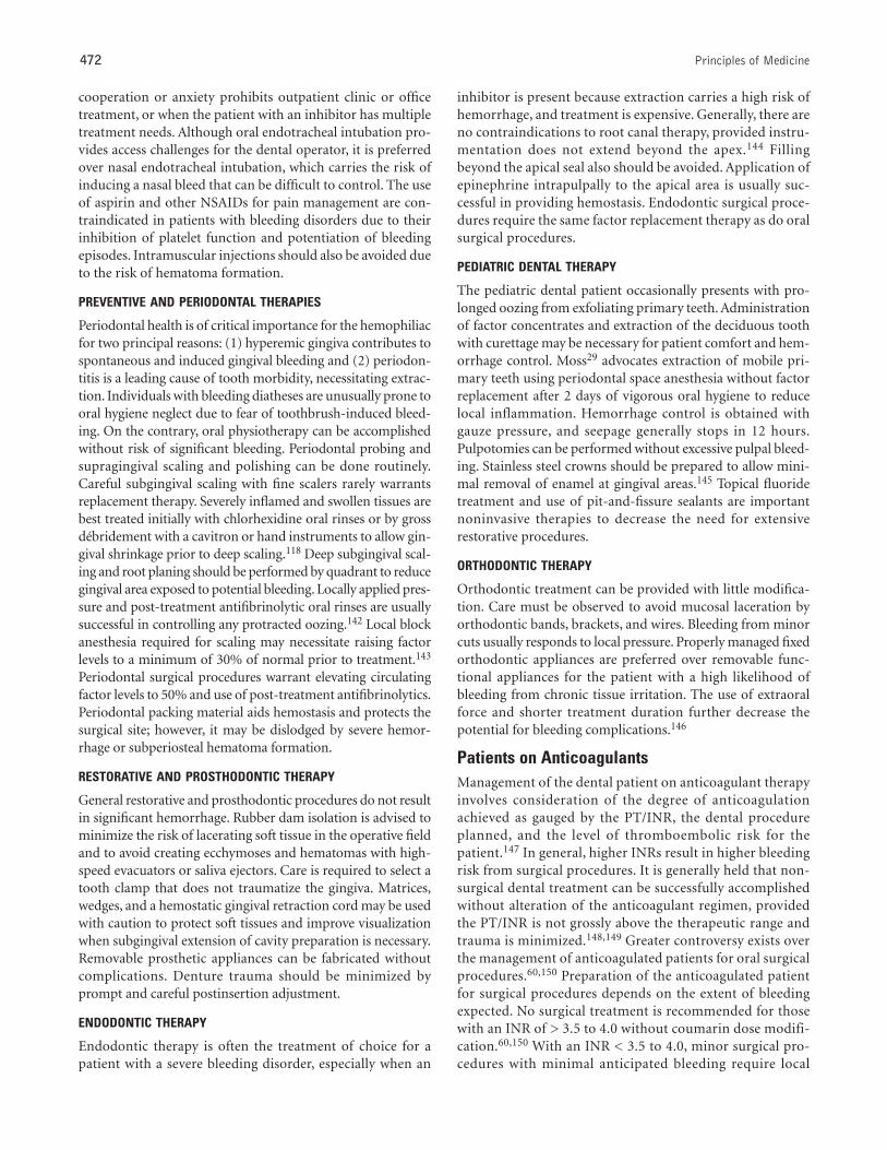
472 Principles of Medicine
cooperation or anxiety prohibits outpatient clinic or officetreatment, or when the patient with an inhibitor has multipletreatment needs. Although oral endotracheal intubation pro-vides access challenges for the dental operator, it is preferredover nasal endotracheal intubation, which carries the risk ofinducing a nasal bleed that can be difficult to control. The useof aspirin and other NSAIDs for pain management are con-traindicated in patients with bleeding disorders due to theirinhibition of platelet function and potentiation of bleedingepisodes. Intramuscular injections should also be avoided dueto the risk of hematoma formation.
PREVENTIVE AND PERIODONTAL THERAPIES
Periodontal health is of critical importance for the hemophiliacfor two principal reasons: (1) hyperemic gingiva contributes tospontaneous and induced gingival bleeding and (2) periodon-titis is a leading cause of tooth morbidity, necessitating extrac-tion. Individuals with bleeding diatheses are unusually prone tooral hygiene neglect due to fear of toothbrush-induced bleed-ing. On the contrary, oral physiotherapy can be accomplishedwithout risk of significant bleeding. Periodontal probing andsupragingival scaling and polishing can be done routinely.Careful subgingival scaling with fine scalers rarely warrantsreplacement therapy. Severely inflamed and swollen tissues arebest treated initially with chlorhexidine oral rinses or by grossdébridement with a cavitron or hand instruments to allow gin-gival shrinkage prior to deep scaling.118 Deep subgingival scal-ing and root planing should be performed by quadrant to reducegingival area exposed to potential bleeding. Locally applied pres-sure and post-treatment antifibrinolytic oral rinses are usuallysuccessful in controlling any protracted oozing.142 Local blockanesthesia required for scaling may necessitate raising factorlevels to a minimum of 30% of normal prior to treatment.143
Periodontal surgical procedures warrant elevating circulatingfactor levels to 50% and use of post-treatment antifibrinolytics.Periodontal packing material aids hemostasis and protects thesurgical site; however, it may be dislodged by severe hemor-rhage or subperiosteal hematoma formation.
RESTORATIVE AND PROSTHODONTIC THERAPY
General restorative and prosthodontic procedures do not resultin significant hemorrhage. Rubber dam isolation is advised tominimize the risk of lacerating soft tissue in the operative fieldand to avoid creating ecchymoses and hematomas with high-speed evacuators or saliva ejectors. Care is required to select atooth clamp that does not traumatize the gingiva. Matrices,wedges, and a hemostatic gingival retraction cord may be usedwith caution to protect soft tissues and improve visualizationwhen subgingival extension of cavity preparation is necessary.Removable prosthetic appliances can be fabricated withoutcomplications. Denture trauma should be minimized byprompt and careful postinsertion adjustment.
ENDODONTIC THERAPY
Endodontic therapy is often the treatment of choice for apatient with a severe bleeding disorder, especially when an
inhibitor is present because extraction carries a high risk ofhemorrhage, and treatment is expensive. Generally, there areno contraindications to root canal therapy, provided instru-mentation does not extend beyond the apex.144 Fillingbeyond the apical seal also should be avoided. Application ofepinephrine intrapulpally to the apical area is usually suc-cessful in providing hemostasis. Endodontic surgical proce-dures require the same factor replacement therapy as do oralsurgical procedures.
PEDIATRIC DENTAL THERAPY
The pediatric dental patient occasionally presents with pro-longed oozing from exfoliating primary teeth. Administrationof factor concentrates and extraction of the deciduous toothwith curettage may be necessary for patient comfort and hem-orrhage control. Moss29 advocates extraction of mobile pri-mary teeth using periodontal space anesthesia without factorreplacement after 2 days of vigorous oral hygiene to reducelocal inflammation. Hemorrhage control is obtained withgauze pressure, and seepage generally stops in 12 hours.Pulpotomies can be performed without excessive pulpal bleed-ing. Stainless steel crowns should be prepared to allow mini-mal removal of enamel at gingival areas.145 Topical fluoridetreatment and use of pit-and-fissure sealants are importantnoninvasive therapies to decrease the need for extensiverestorative procedures.
ORTHODONTIC THERAPY
Orthodontic treatment can be provided with little modifica-tion. Care must be observed to avoid mucosal laceration byorthodontic bands, brackets, and wires. Bleeding from minorcuts usually responds to local pressure. Properly managed fixedorthodontic appliances are preferred over removable func-tional appliances for the patient with a high likelihood ofbleeding from chronic tissue irritation. The use of extraoralforce and shorter treatment duration further decrease thepotential for bleeding complications.146
Patients on AnticoagulantsManagement of the dental patient on anticoagulant therapyinvolves consideration of the degree of anticoagulationachieved as gauged by the PT/INR, the dental procedureplanned, and the level of thromboembolic risk for thepatient.147 In general, higher INRs result in higher bleedingrisk from surgical procedures. It is generally held that non-surgical dental treatment can be successfully accomplishedwithout alteration of the anticoagulant regimen, providedthe PT/INR is not grossly above the therapeutic range andtrauma is minimized.148,149 Greater controversy exists overthe management of anticoagulated patients for oral surgicalprocedures.60,150 Preparation of the anticoagulated patientfor surgical procedures depends on the extent of bleedingexpected. No surgical treatment is recommended for thosewith an INR of > 3.5 to 4.0 without coumarin dose modifi-cation.60,150 With an INR < 3.5 to 4.0, minor surgical pro-cedures with minimal anticipated bleeding require local
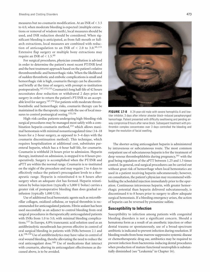
Bleeding and Clotting Disorders 473
measures but no coumarin modification. At an INR of < 3.5to 4.0, when moderate bleeding is expected (multiple extrac-tions or removal of wisdom teeth), local measures should beused, and INR reduction should be considered. When sig-nificant bleeding is anticipated, as from full-mouth or full-arch extractions, local measures are combined with reduc-tion of anticoagulation to an INR of < 2.0 to 3.0.60,151
Extensive flap surgery or multiple bony extractions mayrequire an INR of < 1.5.60
For surgical procedures, physician consultation is advisedin order to determine the patient’s most recent PT/INR leveland the best treatment approach based on the patient’s relativethromboembolic and hemorrhagic risks. When the likelihoodof sudden thrombotic and embolic complications is small andhemorrhagic risk is high, coumarin therapy can be discontin-ued briefly at the time of surgery, with prompt re-institutionpostoperatively.147,152,153 Coumarin’s long half-life of 42 hoursnecessitates dose reduction or withdrawal 2 days prior tosurgery in order to return the patient’s PT/INR to an accept-able level for surgery.147,154 For patients with moderate throm-boembolic and hemorrhagic risks, coumarin therapy can bemaintained in the therapeutic range with the use of local mea-sures to control postsurgical oozing.155,156
High-risk cardiac patients undergoing high-bleeding-risksurgical procedures may be managed most safely with a com-bination heparin-coumarin method,153 which allows maxi-mal hemostasis with minimal nonanticoagulated time (14–18hours for a 2-hour surgery, as opposed to 3–4 days with thecoumarin discontinuation method). This technique, whichrequires hospitalization at additional cost, substitutes par-enteral heparin, which has a 4-hour half-life, for coumarin.Coumarin is withheld 24 hours prior to admission. Heparintherapy, instituted on admission, is stopped 6 to 8 hours pre-operatively. Surgery is accomplished when the PT/INR andaPTT are within the normal range. Coumarin is re-institutedon the night of the procedure and may require 2 to 4 days toeffectively reduce the patient’s procoagulant levels to a ther-apeutic range. Heparin is reinstituted 6 to 8 hours aftersurgery when an adequate clot has formed. Heparin reinsti-tution by bolus injection (typically a 5,000 U bolus) carries agreater risk of postoperative bleeding than does gradual re-infusion (typically 1,000 U/h).
Use of additional local hemostatic agents such as microfib-rillar collagen, oxidized cellulose, or topical thrombin is rec-ommended for anticoagulated patients. Fibrin sealant has beenused successfully as an adjunct to control bleeding from oralsurgical procedures in therapeutically anticoagulated patientswith INRs from 1.0 to 5.0, with minimal bleeding complica-tions.157 In Europe, 4.8% tranexamic acid solution used as anantifibrinolytic mouthwash has proven effective in control oforal surgical bleeding in patients with INRs between 2.1 and4.8.158,159 Use of antifibrinolytics may have value in control oforal wound bleeding, thereby alleviating the need to reduce theoral anticoagulant dose.160 Use of medications that interactwith coumarin, altering its anticoagulant effectiveness as dis-cussed above, is to be avoided.
The shorter-acting anticoagulant heparin is administeredby intravenous or subcutaneous route. The most commonoutpatient use of subcutaneous heparin is for the treatment ofdeep venous thrombophlebitis during pregnancy,161 with thegoal being regulation of the aPTT between 1.25 and 1.5 timescontrol. In general, oral surgical procedures can be carried outwithout great risk of hemorrhage when local hemostatics areused in a patient receiving heparin subcutaneously; however,on consultation, the patient’s physician may recommend with-holding the scheduled injection immediately prior to the oper-ation. Continuous intravenous heparin, with greater hemor-rhagic potential than heparin delivered subcutaneously, isdiscontinued 6 to 8 hours prior to surgery to allow adequatesurgical hemostasis. If a bleeding emergency arises, the actionof heparin can be reversed by protamine sulfate.
Susceptibility to InfectionSusceptibility to infection among patients with congenitalbleeding disorders is not a significant concern. Should ahematoma form as a result of an anesthetic injection or otherdental trauma or spontaneously, use of a broad-spectrumantibiotic is indicated to prevent infection during resolution. Ifbleeding results from bone marrow suppressive systemic diseaseor chemotherapeutic drug use, antibiotics may be required toprevent infection from bacteremia-inducing dental procedureswhen production of mature functional neutrophils is substan-tially diminished (see “Leukemia” in Chapter 16).
FIGURE 17-8 A 24-year-old male with severe hemophilia A and low-titer inhibitor, 3 days after inferior alveolar block–induced parapharyngealhemorrhage. Patient presented with difficulty swallowing and pending air-way compromise 8 hours after nerve block. Subsequent treatment with pro-thrombin complex concentrates over 3 days controlled the bleeding andbegan the resolution of facial swelling.
474 Principles of Medicine
Ability to Withstand CarePatients with bleeding disorders, appropriately prepared pre-operatively, are generally as able to withstand dental care aswell as unaffected individuals. Consultation with the patient’sphysician is recommended for guidance on medical manage-ment required for higher-risk surgical dental procedures.Because of the expense of some medical managementapproaches to severe bleeding disorders (eg, coagulation factorreplacement for severe hemophiliacs), the coumarin with-drawal–heparinization approach to extractions for patients athigh risk of thrombosis, and the bleeding risk to the patient,long treatment sessions may be required to maximize treat-ment accomplishments while minimizing the risk and cost.
▼REFERENCES
1. Rodeghiero F, Castaman G, Dini E. Epidemiological investiga-tion of the prevalence of von Willebrand’s disease. Blood1987;692:454–9.
2. Soucie JM, Evatt B, Jackson D, et al. Occurrence of hemophiliain the United States. Am J Hematol 1998;59:288–94.
3. Lewis IK, Hanlon JT, Hobbins MJ, et al. Use of medicationswith potential oral adverse drug reactions in community-dwelling elderly. Spec Care Dent 1993;13:171–6.
4. Becker RC, Ansell J. Antithrombotic therapy: an abbreviatedreference for clinicians. Arch Intern Med 1995;155:149–61.
5. Davie EW, Fujikawa K, Kisiel W. The coagulation cascade: ini-tiation, maintenance, and regulation. Biochemistry1991;30:10363–70.
6. Mustard JF, Packman MA. Factors influencing platelet func-tion: adhesion, release, and aggregation. Pharmacol Rev1970;22:97–187.
7. Davie EW, Ratnoff OD. Waterfall sequence for intrinsic bloodclotting. Science 1964;145:1310–2.
8. MacFarlane RG. An enzyme cascade in the blood clottingmechanism and its function as a biochemical amplifier. Nature1964;202:498–9.
9. Kingdon HS, Davie EW. Further studies on the activation offactor IX by activated factor XI. Thromb Diath Haemorrh(Stuttg) 1965;Suppl 17:15–22.
10. Bajaj SP, Butkowski RJ, Mann KG. Prothrombin fragments—Ca2+ binding and activation kinetics. J Biol Chem 1975;250:2150–6.
11. Lorand L. Fibrinoligase: the fibrin-stabilizing factor system ofblood plasma. Ann N Y Acad Sci 1972;202:6–30.
12. Duckert F. Documentation of the plasma factor XIII deficiencyin man. Ann N Y Acad Sci 1972;202:190-–9.
13. Verstraete M, Bleifeld W, Brower RW, et al. Double-blind ran-domized trial of intravenous tissue-type plasminogen activa-tor versus placebo in acute myocardial infarction. Lancet1985;2:965–9.
14. De Rossi SS, Glick M. Bleeding time: an unreliable predictor ofclinical hemostasis. J Oral Maxillofac Surg 1996;54:1119–20.
15. Meehan S, Schmidt MC, Mitchell PF. The international nor-malized ratio as a measure of anticoagulation: significance forthe management of the dental outpatient. Spec Care Dent1997;17:94–6.
16. Smith RE. The INR: a perspective. Semin Thromb Hemost1997;23:547–9.
17. Stern R, Karlis V, Kinney L, et al. Using the international nor-malized ratio to standardize prothrombin time. J Am DentAssoc 1997;128:1121–2.
18. Langdell RD, Wagner RH, Brinkhous KM. Effect of antihe-mophilic factor on one-stage clotting tests. J Lab Clin Med1953;41:637–47.
19. Reuler JB, Broudy VC, Cooney TG. Adult scurvy. JAMA1985;253:805–7.
20. Touyz LZ. Oral scurvy and periodontal disease. J Can DentAssoc 1997;63:837–45.
21. De Paepe A. The Ehlers-Danlos syndrome: a heritable collagendisorder as cause of bleeding. Thromb Haemost 1996;75:379–86.
22. Nelson DL, King RA. Ehlers-Danlos syndrome type VIII. J AmAcad Dermatol 1981;5:297–303.
23. Ooshima T, Abe K, Kohno H, et al. Oral manifestations ofEhlers-Danlos syndrome type VII: histological examinationof a primary tooth. Pediatr Dent 1990;12;102–6.
24. Barabas GM, Barabas AP. The Ehlers-Danlos syndrome. Areport of the oral and haematological findings in nine cases. BrDent J 1967;123:473–9.
25. Welbury RR. Ehlers-Danlos syndrome: historical review, reportof two cases in one family and treatment needs. J Dent Child1989;56:220–4.
26. Guttmacher AE, Marchuk DA, White RI Jr. Hereditary hem-orrhagic telangiectasia. N Engl J Med 1995;333:918–24.
27. Haitjema T, Westermann CJ, Overtoom TT, et al. Hereditaryhemorrhagic telangiectasia (Osler-Weber-Rendu disease): newinsights in pathogenesis, complications, and treatment. ArchIntern Med 1996;156:714–9.
28. Christensen GJ. Nosebleeds may mean something much moreserious: an introduction to HHT. J Am Dent Assoc1998;129:635–7.
29. Moss SJ. Newer approaches to dental therapy. Ann N Y AcadSci 1975;240:259–62.
30. Flint SR, Keith O, Scully C. Hereditary hemorrhagic telang-iectasia: family study and review. Oral Surg Oral Med OralPathol 1988;66:440–4.
31. Bisch FC, Bowen KJ, Hanson BS, et al. Dental considerationsfor a Glanzmann’s thrombasthenia patient: case report. JPeriodontol 1996;67:536–40.
32. Perkin RF, White GC, Webster WP. Glanzmann’s thrombas-thenia. Report of two oral surgical cases using a new microfib-rillar collagen preparation and EACA for hemostasis. Oral SurgOral Med Oral Pathol 1979;47:36–9.
33. Jackson MR, MacPhee MJ, Drohan WN, et al. Fibrin sealant:current and potential clinical applications. Blood CoagulFibrinolysis 1996;7:737–46.
34. Boraz RA. Dental considerations in the treatment of Wiskott-Aldrich syndrome: report of case. ASDC J Dent Child 1989;56:225–7.
35. Noris P, Spedini P, Belletti S, et al. Thrombocytopenia, giantplatelets, and leukocyte inclusion bodies (May-Hegglin anom-aly): clinical and laboratory findings. Am J Med 1998;104:355–60.
36. Clemetson KJ, Clemetson JM. Molecular abnormalities inGlanzmann’s thrombasthenia, Bernard-Soulier syndrome, andplatelet-type von Willebrand’s disease. Curr Opin Hematol1994;1:388–93.
37. James WD, Guirty CC, Grote WR. Acute thrombocytopeniapurpura. Oral Surg Oral Med Oral Pathol 1984;57:149–51.
Bleeding and Clotting Disorders 475
38. Fotos PG, Graham WL, Bowers DC, et al. Chronic autoim-mune thrombocytopenia purpura. A 3-year case study. OralSurg Oral Med Oral Pathol 1983;55:564–7.
39. George JN, Raskob GE. Idiopathic thrombocytopenic pur-pura: a concise summary of the pathophysiology and diagno-sis in children and adults. Semin Hematol 1998;35:5–8.
40. Keeling DM, Isenberg DA. Haematological manifestations ofsystemic lupus erythematosus. Blood Rev 1993;7:199–207.
41. Patton LL. Hematologic abnormalities among HIV-infectedpatients. Associations of significance for dentistry. Oral SurgOral Med Oral Pathol Oral Radiol Endod 1999;88:561–7.
42. Moake JL, Chow TW. Thrombotic thrombocytopenic pur-pura: understanding a disease no longer rare. Am J Med Sci1998;316:105–19.
43. Heaney ML, Golde DW. Myelodysplasia. N Engl J Med 1999;340:1649–60.
44. Valdez IH, Patton LL. Aplastic anemia: current concepts anddental management. Spec Care Dent 1990;10:185–9.
45. Dreizen S, McCredie KB, Keating MJ. Chemotherapy-associ-ated oral hemorrhages in adults with acute leukemia. OralSurg Oral Med Oral Pathol 1984;57:494–8.
46. Heiman JL. Alcohol-induced thrombocytopenia: review of theliterature and report of case. J Am Dent Assoc 1972;85:1358–61.
47. Zachee P, Vermylen J, Boogaerts MA. Hematologic aspects ofend-stage renal failure. Ann Hematol 1994;69:33–40.
48. George JN, Raskob GE, Shah SR. Drug-induced thrombocy-topenia: a systematic review of published case reports. AnnIntern Med 1998;129:886–90.
49. Barak S, Shaked Y, Bar ZG, et al. Drug-induced post-surgicalhaemorrhage resulting from trimethoprim-sulphamethoxa-zole. A case report. Int J Oral Maxillofac Surg 1989;18:206–7.
50. Laskin JL. Oral hemorrhage after the use of quinidine: reportof case. J Am Dent Assoc 1974;88:137–9.
51. Wood AJJ. Aspirin as an antiplatelet drug. N Engl J Med1994;330:1287–94.
52. White GC II, Blatt PM, McMillan CW. Medical complicationsof hemophilia. South Med J 1980;73:155–60.
53. De Gopegui RR, Feldman BF. Von Willebrand’s disease. CompHaematol Int 1997;7:187–96.
54. Federici AB. Diagnosis of von Willebrand disease. Haemophilia1998;4:654–60.
55. Johnson RS, Heldt LV, Keaton WM. Diagnosis and treatmentof von Willebrand’s disease. J Oral Maxillofac Surg1987;45:608–12.
56. Batlle J, Torea J, Rendal E, et al. The problem of diagnosing vonWillebrand’s disease. J Intern Med 1997;740:121–8.
57. Tefferi A, Nichols WL. Acquired von Willebrand disease: con-cise review of occurrence, diagnosis, pathogenesis, and treat-ment. Am J Med 1997;103:536–40.
58. Hirsch J, Warkentin TE, Rasche R, et al. Heparin and low-mol-ecular-weight heparin: mechanisms of action, pharmacoki-netics, dosing considerations, monitoring, efficacy, and safety.Chest 1998;114:489–510S.
59. Hirsch J, Dalen JE, Anderson DR, et al. Oral anticoagulants:mechanism of action, clinical effectiveness, and optimal ther-apeutic range. Chest 1998;114:445–69S.
60. Herman WW, Konzelman JL Jr, Sutley SH. Current perspec-tives on dental patients receiving coumarin anticoagulant ther-apy. J Am Dent Assoc 1997;128:327–35.
61. Mammen EF. Coagulation defects in liver disease. Med ClinNorth Am 1994;78:545–54.
62. Shearer MJ. Vitamin K. Lancet 1995;345:229–34.63. Levi M, Ten Cate H. Disseminated intravascular coagulation.
N Engl J Med 1999;341:586–92.64. Opatrny K Jr. Hemostasis disorders in chronic renal failure.
Kidney Int Suppl 1997;62:S87–9.65. Thomson PJ, Langton SG. Persistent haemorrhage following
dental extractions in patients with liver disease: two caution-ary tales. Br Dent J 1996;180:141–4.
66. Sallah S, Kato G. Evaluation of bleeding disorders. A detailedhistory and laboratory tests provide clues. Postgrad Med1998;103:209–18.
67. Rock GA, Shumak KH, Buskard NA, et al. Comparison ofplasma exchange with plasma infusion in the treatment ofthrombotic thrombocytopenia purpura. N Engl J Med1991;325:393–7.
68. Bell WR, Braine HG, Ness PM, et al. Improved survival inthrombotic thrombocytopenic purpura–hemolytic uremicsyndrome. Clinical experience in 108 patients. N Engl J Med1991;325:398–403.
69. Brinkhous KM, Shanbrom E, Roberts HR, et al. A new high-potency glycine-precipitated antihemophilia factor (AHF)concentrate. JAMA 1968;205:613–7.
70. Pierce GF, Lusher JM, Brownstein AP, et al. The use of purifiedclotting factor concentrates in hemophilia. Influence of viralsafety, cost, and supply on therapy. JAMA 1989;261:3434–8.
71. Schwartz RS, Abildgaard CF, Aledort LM, et al. Human recom-binant DNA-derived antihemophilic factor (factor VIII) in thetreatment of hemophilia A. N Engl J Med 1990;323:1800–5.
72. White GC II, Beebe A, Nielsen B. Recombinant factor IX.Thromb Haemost 1997;78:261–5.
73. Goldsmith JC, Kasper CK, Blatt PM, et al. Coagulation factorIX: successful surgical experience with a purified factor IXconcentrate. Am J Hematol 1992;40:210–5.
74. Djulbegovic B,Marasa M,Pesto A,et al.Safety and efficacy of puri-fied factor IX concentrate and antifibrinolytic agents for dentalextractions in hemophilia B. Am J Hematol 1996;51:168–70.
75. Levine PH. Efficacy of self-therapy in hemophilia. A study of72 patients with hemophilia A and B. N Engl J Med 1974;291:1381–4.
76. Eastman JR, Triplett DA, Nowakowski AR. Inherited factor Xdeficiency: presentation of a case with etiologic and treat-ment considerations. Oral Surg Oral Med Oral Pathol 1983;56:461–6.
77. Mannucci PM. Desmopressin (DDAVP) in the treatment ofbleeding disorders: the first 20 years. Blood 1997;90:2515–21.
78. Saulnier J, Marey A, Horellou M-H, et al. Evaluation of desmo-pressin for dental extractions in patients with hemostatic dis-orders. Oral Surg Oral Med Oral Pathol 1994;77:6–12.
79. Semeritis SV, Aledort LM. Desmopressin nasal spray for hemo-philia A and type I von Willebrand disease. Ann Intern Med1997;126:744–5.
80. Mannucci PM, Pareti FI, Ruggeri ZM, et al. 1-Deamino-8-arginine vasopressin: a new therapeutic approach to the man-agement of haemophilia and von Willebrand’s disease. Lancet1977;I:869–72.
81. Ragni MV, Winkelstein A, Kingsley L, et al. 1986 update ofHIV seroprevalence, seroconversion, AIDS incidence, andimmunologic correlates of HIV infection in patients withhemophilia A and B. Blood 1987;70:786–90.
82. Kasper CK. Complications of hemophilia A treatment: factorVIII inhibitors. Ann N Y Acad Sci 1991;614:97–105.
476 Principles of Medicine
83. Roberts HR, Cromartie R. Overview of inhibitors to factorVIII and IX. Prog Clin Biol Res 1984;150;1–18.
84. Aledort L. Inhibitors in hemophilia patients: current statusand management. Am J Hematol 1994;47:208–17.
85. Durham TM, Hodges ED, Harper J, et al. Management of trau-matic oral-facial injury in the hemophiliac patient withinhibitor: case report. Pediatr Dent 1993;15:282–7.
86. Lusher JM, Shapiro SS, Palasczk JE, et al. Efficacy of pro-thrombin-complex concentrates in hemophiliacs with anti-bodies to factor VIII. A multicenter therapeutic trial. N Engl JMed 1980;303:421–5.
87. Blatt PM, Pearsall AH, Givhan EG, et al. Haemostatic failure ofprothrombin complex concentrates during elective dental pro-cedure. Thromb Haemost 1980;42:1604–6.
88. Lusher J, Ingerslev J, Roberts H, et al. Clinical experience withrecombinant factor VIIa. Blood Coagul Fibrinolysis 1998;9:119–28.
89. McPherson J, Teague L, Lloyd J, et al. Experience with recom-binant factor VIIa in Australia and New Zealand. Haemostasis1996;26:109–17.
90. Ciavarella N, Schiavoni M, Valenzano E, et al. Use of recombi-nant factor VIIa (NovoSeven) in the treatment of two patientswith type III von Willebrand’s disease and an inhibitor againstvon Willebrand factor. Haemostasis 1996;26:150–4.
91. Francesconi M, Korninger C, Thaler E, et al. Plasmapheresis:its value in the management of patients with antibodies to fac-tor VIII. Haemostasis 1982;11:79–86.
92. Nilsson IM, Sundqvist S-B, Freiburghaus C. Extracorporealprotein A–sepharose and specific affinity chromatography forremoval of antibodies. Prog Clin Biol Res 1984;150:225–41.
93. Freiburghaus C, Berntorp E, Ekman M, et al. Immuno-adsorption for removal of inhibitors: update on treatments inMalmo-Lund between 1980 and 1995. Haemophilia 1998;4:16–20.
94. Stratton RD, Wagner RH, Webster WP, et al. Antibody natureof circulating inhibitor of plasma von Willebrand factor. ProcNatl Acad Sci U S A 1975;72:4167–71.
95. Spector I, Corn M, Ticktin HE. Effect of plasma transfusionson the prothrombin time and clotting factors in liver disease.N Engl J Med 1966;275:1032–7.
96. Mannucci PM. Desmopressin (DDAVP) for treatment of dis-orders of hemostasis. Prog Hemostat Thromb 1986;8:19–45.
97. Jubelirer SJ. Hemostatic abnormalities in renal disease. Am JKidney Dis 1985;5:219–25.
98. Janson PA, Jubelirer SJ, Weinstein JM, et al. Treatment of thebleeding tendency in uremia with cryoprecipitate. N Engl JMed 1980;303:1318–22.
99. Mannucci PM, Remuzzi G, Pusineri F, et al. Deamino-8-D-arginine vasopressin shortens the bleeding time in uremia. NEngl J Med 1983;308:8–12.
100. Liu YK, Kosfeld RE, Marcum SG. Treatment of uraemic bleed-ing with conjugated oestrogen. Lancet 1984;2:887–90.
101. Moia M, Vizzotto L, Cattaneo M, et al. Improvement in thehaemostatic defect of uraemia after treatment with recombi-nant human erythropoietin. Lancet 1987;2:1227–9.
102. Falace DA, Kelly DE. Disseminated intravascular coagulationand fibrinolysis as a cause of postextraction hemorrhage.Report of a case. Oral Surg Oral Med Oral Pathol 1976;41:718–25.
103. Shikimori M, Oka T. Disseminated intravascular coagulationsyndrome. Int J Oral Surg 1985;14:451–5.
104. De Jonge E, Levi M, Stoutenbeek CP, et al. Current drug treat-ment strategies for disseminated intravascular coagulation.Drugs 1998;55:767–77.
105. Larsson S. Life expectancy of Swedish haemophiliacs,1831–1980. Br J Haematol 1985;59:593–602.
106. Telfer MC. Clinical spectrum of viral infections in hemophilicpatients. Hematol Oncol Clin North Am 1992;6:1047–56.
107. Eyster M, Diamondstone L. Natural history of hepatitis C virusinfection in multitransfused hemophiliacs: effect of coinfec-tion with human immunodeficiency virus. The Multi-centerHaemophilia Cohort Study. J Acquir Immun Defic Syndr1993;6:602–10.
108. Wilde JT, Ahmed MM, Collingham KE, et al. Hepatitis G virusinfection in patients with bleeding disorders. Br J Haematol1997;99:285–8.
109. Lee CA, Sabin CA, Phillips AN, et al. Morbidity and mortalityfrom transfusion-transmitted disease in haemophilia [letter].Lancet 1995;345:1309.
110. Bolski E, Hunt R. The prevalence of AIDS-associated orallesions in a cohort of patients with hemophilia. Oral Surg OralMed Oral Pathol 1988;65:406–10.
111. Ficarra G, Chiodo M, Morfini M, et al. Oral lesions amongHIV-infected hemophiliacs. A study of 54 patients.Haematologica 1994;79:148–53.
112. Merry C, McMahon C, Ryan M, et al. Successful use of pro-tease inhibitors in HIV-infected haemophilia patients. Br JHaematol 1998;101:475–9.
113. Sonis AL, Musselman RJ. Oral bleeding in classic hemophilia.Oral Surg Oral Med Oral Pathol 1982;53:363–6.
114. Kaneda T, Shikimori I, Watanabe F, et al. The importance oflocal hemostatic procedures in dental extractions and oralmucosal bleeding of hemophiliac patients. Int J Oral Surg1981;10:266–71.
115. Kaneda T, Nagayama M, Ohmori M, et al. Hemarthrosis of thetemporomandibular joint in a patient with hemophilia B:report of case. J Oral Surg 1979;37:513–4.
116. Nishioka GJ, Van Sickels JE, Tilson HB. Hemophilic arthropa-thy of the temporomandibular joint: review of the literature,a case report, and discussion. Oral Surg Oral Med Oral Pathol1988;65:145–50.
117. Webster WP, Roberts HR, Penick GD. Dental care of patientswith hereditary disorders of blood coagulation. In: RatnoffOD, editor. Treatment of hemorrhagic disorders. New York:Harper & Row; 1968. p. 93–110.
118. Webster WP, Courtney RM. Diagnosis and treatment of peri-odontal disease in the hemophiliac. In: Proceedings, DentalHemophilia Institute, January, 1968. The National HemophiliaFoundation, U.S.A.
119. Kobrinsky NL, Israeles ED, Gerrard JM, et al. Shortening ofbleeding time by 1-deamino-8-d-arginine vasopressin in var-ious bleeding disorders. Lancet 1984;I:1145–8.
120. Castaman G, DiBona E, Schiavotto C, et al. Pilot study on thesafety and efficacy of desmopressin for the treatment or pre-vention of bleeding in patients with hematologic malignancy.Haematologica 1997;82:584–7.
121. Vinckier F, Vermylen J. Dental extractions in hemophilia:reflections on 10 years’ experience. Oral Surg Oral Med OralPathol 1985;59:6–9.
122. Association of Hemophilia Clinic Directors of Canada. Clinicalpractice guidelines. Hemophilia and von Willebrand’s disease:2. Management. Can Med Assoc J 1995;153:147–57.
Bleeding and Clotting Disorders 477
fibrinolysis with tranexamic acid. J Periodontal Res 1988;23:72–4.
143. Michaelides PL. Outpatient management of periodontalpatients with hemophilia. Int J Periodont Restorative Dent1983;3:65–73.
144. Keila S, Kaufman A, Itckowitch D. Uncontrolled bleeding dur-ing endodontic treatment as the first symptoms for diagnos-ing von Willebrand’s disease. Oral Surg Oral Med Oral Pathol1990;69:243–6.
145. White GE. Medical review—factor VIII deficiency andpedodontics. J Pedodont 1979;3:176–92.
146. van Venrooy JR, Proffit WR. Orthodontic care for medicallycompromised patients: possibilities and limitations. J Am DentAssoc 1985;111:262–6.
147. Mulligan R, Weitzel KG. Treatment management of the patient receiving anticoagulant drugs. J Am Dent Assoc1988;117:479–83.
148. Rooney TP. General dentistry during continuous anticoagula-tion therapy. Oral Surg Oral Med Oral Pathol 1983;56:252–5.
149. Benoliel R, Leviner E, Katz J, et al. Dental treatment for patientson anticoagulant therapy: prothrombin time value—what dif-ference does it make? Oral Surg Oral Med Oral Pathol 1986;62:149–51.
150. Wahl MJ. Myths of dental surgery in patients receiving anti-coagulant therapy. J Am Dent Assoc 2000;131:77–80.
151. Beirne OR, Koehler JR. Surgical management of patients onwarfarin sodium. J Oral Maxillofac Surg 1996;54:1115–8.
152. Ziffer AM, Scopp IW, Beck J, et al. Profound bleeding after den-tal extractions during dicumarol therapy. N Engl J Med1957;256:351–3.
153. Roser SM, Rosenbloom B. Continued anticoagulation in oralsurgery procedures. Oral Surg Oral Med Oral Pathol1975;40:448–57.
154. Mulligan R. Response to anticoagulant withdrawal. J Am DentAssoc 1987;115:435–8.
155. Waldrep AC, McKelvey LE. Oral surgery for patients on anti-coagulant therapy. J Oral Surg 1968;26:374–80.
156. Bailey BMW, Fordyce AM. Complications of dental extrac-tions in patients receiving warfarin anticoagulant therapy. Acontrolled clinical trial. Br Dent J 1983;155:308–10.
157. Bodner L, Weinstein JM, Baumgartner AK. Efficacy of fibrinsealant in patients on various levels of oral anticoagulantundergoing oral surgery. Oral Surg Oral Med Oral Pathol OralRadiol Endod 1998;86:421–4.
158. Ramstrom G, Sindet-Pedersen S, Hall G, et al. Prevention ofpostsurgical bleeding in oral surgery using tranexamic acidwithout dose modification of oral anticoagulants. J OralMaxillofac Surg 1993;51:1211–6.
159. Sindet-Pedersen S, Ramstrom G, Bernvil S, et al. Hemostaticeffect of tranexamic acid mouthwash in anticoagulant-treatedpatients undergoing oral surgery. N Engl J Med 1989;320:840–3.
160. Souto JC, Oliver A, Zuazu-Jausoro I, et al. Oral surgery in anti-coagulated patients without reducing the dose of oral antico-agulant: a prospective randomized study. J Oral MaxillofacSurg 1996;54:27–32.
161. Belfiglio EJ. The heparinized dental patient. Gen Dent1991;39:38–9.
123. Webster WP, McMillan CW, Lucas ON, et al. Dental manage-ment of the bleeder patient. A comparative review of replace-ment therapy. In: Ala F, Denson KWE, editors. Haemophilia.Amsterdam: Excerpta Medica; 1973. p. 33–7.
124. Stajcic Z, Baklaja R, Elezovic I, et al. Primary wound closure inhaemophiliacs undergoing dental extractions. Int J OralMaxillofac Surg 1989;18:14–6.
125. Evans BE. Local hemostatic agents (and techniques). Scand JHaematol 1984;33:417–22.
126. Lucas ON, Albert TW. Epsilon aminocaproic acid in hemo-philiacs undergoing dental extractions: a concise review. OralSurg Oral Med Oral Pathol 1981;51:115–20.
127. Walsh PN, Rizza CR, Matthews JM, et al. Epsilon-aminocaproic acid therapy for dental extractions inhaemophilia and Christmas disease: a double blind controlledtrial. Br J Haematol 1971;20:463–75.
128. Walsh PN, Rizza CR, Evans BE, et al. The therapeutic role ofepsilon aminocaproic acid (EACA) for dental extractions inhemophiliacs. Ann N Y Acad Sci 1975;240:267–76.
129. Forbes CD, Barr RD, Reid G, et al. Tranexamic acid in controlof haemorrhage after dental extraction in haemophilia andChristmas disease. Br Med J 1972;2:311–3.
130. Tavenner RWH. Use of tranexamic acid in control of haemor-rhage after extraction of teeth in haemophilia and Christmasdisease. Br Med J 1972;2:314–5.
131. Sugar AW. The management of dental extractions in cases ofthrombasthenia complicated by the development of isoanti-bodies to donor platelets. Oral Surg Oral Med Oral Pathol1979;48:116–9.
132. Rakocz M, Mazar A, Varon D, et al. Dental extractions inpatients with bleeding disorders. The use of fibrin glue. OralSurg Oral Med Oral Pathol 1993;75:280–2.
133. Martinowitz U, Schulman S. Fibrin sealant in surgery ofpatients with a hemorrhagic diathesis. Thromb Hemost1995;74:486–92.
134. Martinowitz U, Schulman S, Horoszowski H, et al. Role of fib-rin sealants in surgical procedures on patients with hemosta-tic disorders. Clin Orthop 1996;328:65–75.
135. Lucas ON. The use of hypnosis in hemophilia dental care. AnnN Y Acad Sci 1975;240:263–6.
136. Geffner I, Porteous JR. Haemorrhage and pain control in con-servative dentistry for haemophiliacs. Br Dent J 1981;151:256–8.
137. Spuller RL. Use of the periodontal ligament injection in den-tal care of the patient with hemophilia-a clinical evaluation.Spec Care Dent 1988;8:28–9.
138. Nazif M. Local anesthesia for patients with hemophilia. J DentChild 1970;37:79–84.
139. Archer WH, Zubrow HJ. Fatal hemorrhage following regionalanesthesia for operative dentistry in a hemophiliac. Oral SurgOral Med Oral Pathol 1954;7:464–70.
140. Leatherdale RAI. Respiratory obstruction in haemophilicpatients. Br Med J 1960;II:1316–20.
141. Bogdan CJ, Strauss M, Ratnoff OD. Airway obstruction inhemophilia (factor VIII deficiency): a 28-year institutionalreview. Laryngoscope 1994;104:789–94.
142. Sindet-Pedersen S, Stenbjerg S, Ingerslev J. Control of gin-gival hemorrhage in hemophilic patients by inhibition of