Embed Size (px)
Citation preview

100
Oral cavity cancer is the sixth leading cause of cancerworldwide. In the United States alone, there are over21,500 oral carcinomas diagnosed each year, and6,000 Americans die of oral cancer each year.1 Theincidence of oral carcinoma varies throughout theworld, with estimates exceeding 40 in 100,000 inparts of France, Southeast Asia, Hungary and Singa-pore.2 Thus oral cancer is a major cause of morbidityworldwide. Ninety percent of oral malignancies aresquamous cell carcinomas and therefore are the prin-cipal topic of this chapter. However, the treatment ofsome other oral malignancies, like sarcoma andminor salivary gland carcinoma, is also primarily sur-gical excision, and the surgical principles are applica-ble to the treatment of these other tumors as well.
The etiology of oral cancer is exposure to car-cinogens in tobacco and the tumor-promoting effectsof alcohol. Ninety percent of the risk of oral cancerin the United States is directly attributable to smok-ing.3 Tobacco smoke and alcohol are synergistic intheir carcinogenic effects in the oral cavity. The rel-ative risk of oral cancer for heavy smokers is 7 timesthat of nonsmokers. The risk for heavy drinkers is 6times that of nondrinkers. The risk for patients abus-ing both alcohol and tobacco is 38 times that ofthose who abstain from both.4 Chewing tobacco andbetel quid also increase the risk of oral cancer.5
Chronic carcinogen exposure creates a field effectand the entire mucosa of the upper aerodigestivetract is at risk for malignancy in smokers anddrinkers.6 After successful treatment of oral cancer,the risk of a second primary cancer is 3.7 percent peryear and increases to 24 percent at ten years.7 Ces-sation of alcohol and tobacco exposure reduces therisk of second aerodigestive carcinoma.
5
Oral Cavity CancerJAY O. BOYLE, MDELLIOT W. STRONG, MD, FACS
Chronic carcinogen exposure causes mucosalcells to acquire genetic abnormalities. When thesegenetics abnormalities result in the activation ofproto-oncogenes and the inactivation of tumor-sup-pressive genes, the cells may be afforded a growthadvantage. Dysregulated proliferation due to aber-rant cell cycle control leads to clonal populations ofpremalignant, genetically abnormal mucosal cells.These populations of cells have a high tendency toaccumulate additional genetic abnormalities due togenomic instability. Genomic instability is the resultof rapid cell cycling with decreased genomic sur-veillance, decreased capacity to repair geneticdefects and ineffective signalling of apoptosis orprogrammed cell death. In these cell populations therate of accumulation of acquired genetic abnormali-ties increases logarithmically with time.8
Eventually these abnormal cells acquire themalignant phenotype in which they lose normal dif-ferentiation, invade the basement membrane,become locally destructive, and metastasize region-ally or distantly. These cells evade immune surveil-lance of the body, produce angiogenic factors allow-ing ingrowth of blood vessels, and become clinicalcarcinomas.
ANATOMY
The critical functions normally accomplished by oraltissues include articulation of speech, facial expres-sion and cosmesis, respiration, mastication, degluti-tion, and taste. Ablative surgery for oral cancerremoves the malignant tumor en bloc with a marginof normal tissue, and the integrity of functionallyimportant structures is not violated unnecessarily.

Oral Cavity Cancer 101
upper lip possesses two peaks forming a “cupid’sbow” where the filtrum ascends to the columella ofthe nasal septum.
The orbicularis oris muscle receives motor inner-vation from the marginal and buccal branches of thefacial nerve and performs a sphincteral function tomaintain oral competence and to facilitate articula-tion of speech. This muscle has many attachmentsfrom other muscles of facial expression that elevateand depress the lips. Of clinical importance is theinnervation of the depressor anguli oris muscle bythe marginal mandibular branch of the facial nerve.
Sensation of the lower lip is provided by the men-tal nerve, the terminal segment of the alveolar branchof the mandibular division of the trigeminal nerve.The nerve exits the mental foramen of the mandiblenear the root of the canine tooth. Paresthesia of thechin suggests extensive mandible invasion and infe-rior alveolar nerve involvement by oral carcinoma.
The anterior two-thirds of the tongue is called theoral or mobile tongue and is bounded posteriorly bythe V-shaped line of the circumvallate papillae. Pos-terior to this line is the base of tongue, which is partof the oropharynx. The oral tongue has ventral anddorsal surfaces. The mucosa of the tongue is simplestratified squamous epithelium with interspersedpapillae or taste buds of four morphologies: fili-form, foliate, fungiform, and circumvallate.
The tongue is comprised of intrinsic and extrinsicmuscles. The intrinsic muscles are arranged in verticaland horizontal fascicles that allow the mobile tongueto change shape and consistency. There are three pairsof extrinsic muscles that provide mobility of thetongue: genioglossus, hyoglossus, and styloglossus.Protrusion of the tongue is primarily accomplished bythe action of the genioglossus muscle which originatesfrom the mandibular tubercles on the lingual surfaceof the arch of the mandible, and inserts diffusely intothe substance of the intrinsic musculature on each sideof the tongue. The motor supply to the intrinsic andextrinsic tongue muscles is the hypoglossal nerve (CNXII), which exits the skull through its own hypoglos-sal canal and courses laterally and anteriorly betweenthe external and internal carotid arteries, immediatelyinferior to the occipital artery.
The sensation of the tongue is supplied by the lin-gual nerve, a branch of the mandibular division of the
The oral cavity is bounded anteriorly by the skinand the vermilion border of the upper and lower lips.The oral cavity extends posteriorly to the circumval-late papillae of the tongue, the junction of the hardand soft palates, and the anterior faucial arch. Thetonsil, soft palate and posterior one-third of thetongue are oropharyngeal structures and are not con-sidered in our discussion of the oral cavity. Laterallythe oral cavity is bounded by the buccal mucosa.
The oral cavity is divided into the following sub-sites: the lip, anterior two-thirds of the tongue, floorof mouth, gingiva, retromolar trigone, buccalmucosa, and hard palate (Figure 5–1). Figure 5–2demonstrates the distribution of oral tumors by sub-site. Tumors of different subsites demonstrate dis-tinct clinical behavior.
The lip is a common site of skin cancer. The lay-ers of the lip, from external to internal, include theepidermis, dermis, subcutaneous tissue, the orbicu-laris oris and attached musculature, the oral submu-cosa and the oral mucosa. The oral submucosa con-tains minor salivary glands, copious lymphaticvessels, blood vessels and sensory nerves. The lip issupplied by the labial arteries and veins, which arebranches of the facial vessels. The generous lym-phatics of the lower lip cross and drain bilaterally tolevel I nodes of the submental and submandibulartriangles. Pre- or post-facial nodes lie anterior andposterior to the facial vessels in the superior aspectof the submandibular triangle and are potential sitesof metastasis of lip cancers. Lymphatic channels ofthe upper lip respect the midline and drain to sub-mandibular, periparotid, or preauricular nodes. The
Figure 5–1. Diagram of the oral cavity and subsites.

102 CANCER OF THE HEAD AND NECK
trigeminal nerve (CN V3). The lingual nerve alsotransports parasympathetic fibers from the chordatympani branch of the facial nerve to the sub-mandibular ganglion. The blood supply to the tongueis derived from the paired lingual arteries.
The lymphatic drainage of the tongue begins in arich submucosal plexus, which may drain bilaterallywhen lesions approach the midline, the tip, or espe-cially the base of the tongue. Tumors of the lateralmid-tongue drain predictably to the ipsilateral lymphnodes. The first echelon nodes for lesions of the tipinclude the submental nodes. The lateral and ventraltongue lesions metastasize to submandibular orjugulodigastric nodes while the base of tonguedrains to the jugulodigastric and deep jugular nodes.Lesions of the anterior tongue may metastasizedirectly to the low jugular lymph nodes (level IV) ofthe neck.
The buccal mucosa lines the lateral oral cavityand blends with the gingiva superiorly and inferiorlyand with the retromolar trigone posteriorly. Themucosa is pierced by the Stensen’s duct of theparotid gland at the papilla adjacent to the secondmaxillary molar tooth.
The gingiva consists of thick keratinized mucosawith deep rete pegs and submucosal adherence tothe periosteum. The mucosa covers the alveolarprocesses of the mandible and the maxilla.
The mandible possesses lingual and buccal cor-tices which envelop cancellous bone, dental sockets,and the mandibular canal transmitting the mandibu-lar vessels and nerves (branch of CN V3). Themandibular surface is innervated by branches of thelingual and mental nerves while the maxillary sur-
face is innervated by alveolar branches of the secondand third divisions of the 5th cranial (trigeminal)nerve.
The retromolar trigone is that portion of adherentkeratinized mucosa covering the ascending ramus ofthe mandible from the third mandibular molar to themaxillary tubercle. It represents the area betweenthe buccal mucosa laterally and the anterior tonsillarpillar medially and posteriorly. Tumors of this smallregion spread readily to the adjacent mandibularbone, alveolar foramen, masticator space, oropha-ryngeal tonsil, floor of mouth and base of tongue.
The hard palate lies within the horseshoe shapeof the maxillary alveolar process. Keratinized adher-ent mucosa covers the palatal bone, which is dividedinto the primary and secondary bony palate. The pri-mary palate consists of the palatal processes of themaxillary bones and represents the premaxilla ante-rior to the incisive foramen. The secondary palate ismade up of the horizontal processes of the L-shapedpalatine bones. On the posterior hard palate, near themaxillary second or third molar, are found thegreater and lesser palatine foramina which transmittheir respective vessels and nerves which are the ter-minal branches of the sphenopalatine vessels(branches of the internal maxillary artery) andnerves (branches of V2). Anteriorly, the midline inci-sive foramen near the incisors transmits the terminalbranches of the nasociliary nerve and vessels to sup-ply the primary palate region. Lymphatic drainage ofthe palate includes the deep jugular chain as well asthe retropharyngeal nodes. Anterior lesions maymetastasize to pre-vascular facial lymph nodes ofthe submandibular region.
Figure 5–2. Distribution of oral cancers by subsite. A selected series of cases presentingto the head and neck service of Memorial Sloan-Kettering Cancer Center, New York.

Oral Cavity Cancer 103
The floor of the mouth is a soft thin layer of U-shaped mucosa overlying the insertion of the mylohy-oid muscle laterally, the hyoglossus muscle medially,and the insertion of the genioglossus muscle anteri-orly. It covers the sublingual salivary glands, sub-mandibular (Wharton’s) duct, and the lingual nerve.The blood supply is from the lingual vessels. Its lym-phatic plexus is copious and drains bilaterally in themidline. The lymphatic drainage patterns include thesubmental and bilateral submandibular nodes, as wellas the ipsilateral jugulodigastric nodes posteriorly.
DIAGNOSIS
The diagnostic evaluation of a patient with oral car-cinoma consists of the history and the physicalexamination, histopathologic tissue diagnosis, andimaging—when indicated.
The clinical history begins with the present ill-ness and includes the duration and location of symp-toms such as non-healing ulcer, mass in the oral cav-ity or neck, pain, bleeding, and any symptoms ofcranial nerve deficits. A thorough exploration of thepatient’s past medical and surgical history, and thereview of systems yield operative risk data. A thor-ough history of etiologic risk factors for squamouscarcinomas not only reflects the patient’s relativerisk of malignancy but also suggests factors thataffect the patient’s overall health, fitness for surgeryand emotional state. Current and distant abuse oftobacco and alcohol are critical factors and may beunderreported by the patient. In many parts of theworld the use of oral chews (“pan,” betel nuts, etc.)is the chief etiologic factor.9 These may containtobacco, slaked lime and other irritants and may beretained in the oral cavity nearly constantly. Anoccupational exposure to heavy metals such asnickel,10 and previous radiation exposure to headand neck are other important risk factors of head andneck cancer that are elicited in the history.
The social history impacts strongly on thepatient’s ability to comply with and tolerate treat-ment and rehabilitation programs, and these issuesare resolved during the treatment planning phase.The family history reflects any familial tendenciestoward malignant disease and completes the histori-cal data.
A complete examination of the head and neck isperformed to assess the precise location and extentof the primary tumor, identify regionally metastaticdisease and to rule out multiple primary malignan-cies. Grossly, the earliest cancers may present asnonulcerous white or red patches. More advancedoral squamous cell carcinomas (SCC) present asmucosal lesions, although occasionally an SCC maypresent as predominantly submucosal with little orno mucosal involvement. Firm submucosal lesionsare often minor salivary gland neoplasms. SCC maybe ulcerative and invasive, fungating and exophyticor both (Figure 5–3). They may arise within prema-lignant lesions such as leukoplakia or erythroplakia.The following characteristics of the lesion should bedocumented: appearance and character, location,size in centimeters, texture to palpation, mobility,proximity to surrounding structures—especiallybone, and the estimated palpable thickness (superfi-cial vs. deeply infiltrating).
Figure 5–3. A, An exophytic lesion involving the right lateral bor-der of the tongue. B, An endophytic lesion of the right lateral borderof the tongue.
A
B
104 CANCER OF THE HEAD AND NECK
Trismus suggests ominous pterygoid and masti-cator space involvement. The condition of the denti-tion should be noted as tumors may, as the first sign,displace or loosen teeth. The distance from thetumor to the mandible and the mobility of the lesionin relation to the mandible are critical elements indetermining the management of perimandibularcancers. A complete examination of the cranialnerves is performed, emphasizing sensation over thechin for mandibular nerve deficit, tongue mobilityfor hypoglossal nerve deficit, facial nerve function,palatal elevation and gag reflex, and function of theaccessory nerve. A mirror or a flexible or rigid tele-scope is needed to document vocal cord mobilityand to ensure that no lesions exist in the oropharynx,nasopharynx, endolarynx, and visible hypopharynx.Small lesions of the hypopharynx may only be visi-ble by direct examination under anesthesia with therigid laryngoscopes.
The neck should be thoroughly palpated formetastatic disease in the nodal groups at risk, and forother abnormalities of the great vessels and the thy-roid gland which might impact treatment. Masses ofthe neck should be measured in centimeters, charac-terized for site (level), mobility, consistency, skininvolvement, and proximity to vital structures. Nor-mal neck structures commonly mistaken for metasta-tic masses include: the transverse process of C2 in thejugulodigastric region of thin patients, the scalenemuscles, a tortuous carotid artery, a carotid aneurysm,a prominent carotid bulb, a cervical rib, and ptoticsubmandibular glands.
A complete general physical examination shouldbe performed emphasizing the cardiovascular andpulmonary systems, which are commonly abnormalin this oral cancer population. The systemic effects ofmalnutrition or excessive alcohol intake should alsobe noted.
The history and physical examination with orwithout adjunctive imaging and histopathologic tis-sue diagnosis are sufficient to plan and execute sur-gical treatment for many patients with oral cancer.However, some patients will benefit from examina-tion under anesthesia including direct palpation withor without biopsy, laryngoscopy, esophagoscopy,and bronchoscopy. The indications for examinationunder anesthesia include an inadequate assessment
of the extent of the disease by history and physicalexamination and imaging, or the presence of symp-toms referable to the trachea, larynx, hypopharynxand esophagus that need endoscopic assessment. Itis not cost-effective screening to perform panen-doscopy on all patients with head and neck can-cer.11–13 Symptoms suggesting lesions of the trachea,larynx, hypopharynx, or esophagus include: dyspha-gia, odynophagia, pain, hoarseness, hemoptysis orstridor. A careful history and meticulous head andneck exam is necessary to identify these lesions.
Evaluation of the deep extent of oral cancer oftenrequires the use of imaging modalities. Imaging stud-ies, however, will not adequately identify the superfi-cial mucosal extent of disease, which must be estab-lished by visualization, palpation and biopsy. Plainradiographs such as panorex, dental films or a sub-mental occlusal film may demonstrate gross boneinvolvement but do not show early cortical invasion.
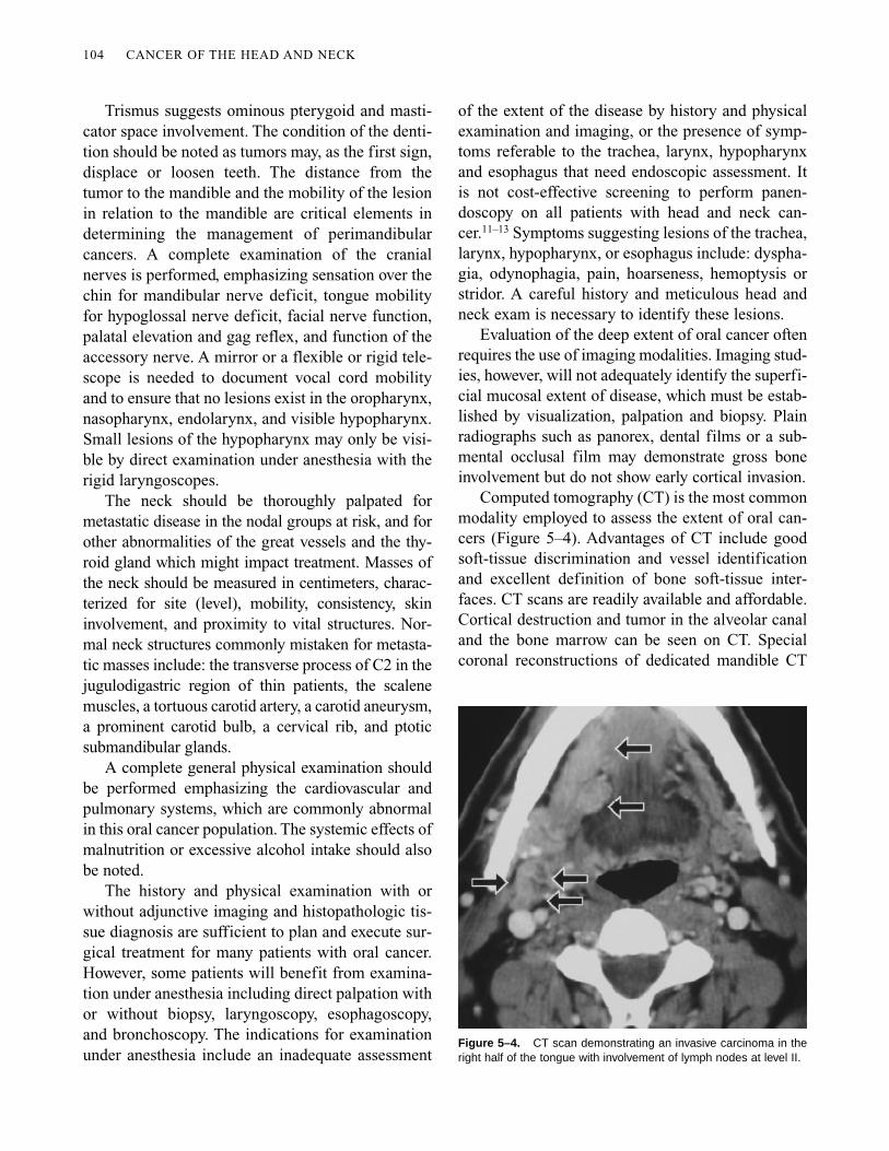
Computed tomography (CT) is the most commonmodality employed to assess the extent of oral can-cers (Figure 5–4). Advantages of CT include goodsoft-tissue discrimination and vessel identificationand excellent definition of bone soft-tissue inter-faces. CT scans are readily available and affordable.Cortical destruction and tumor in the alveolar canaland the bone marrow can be seen on CT. Specialcoronal reconstructions of dedicated mandible CT
Figure 5–4. CT scan demonstrating an invasive carcinoma in theright half of the tongue with involvement of lymph nodes at level II.

Oral Cavity Cancer 105
irreparably while sparing the normal tissue. Eithermodality is effective in controlling early oral carci-nomas, but the use of both modalities in combina-tion is necessary to control locally advanced disease.The role of chemotherapy alone in localized diseaseis palliative. Currently, distantly metastatic diseaseis incurable but can often be effectively palliatedwith chemotherapy and radiation.
Treatment choices are best made after consider-ing tumor factors, patient factors and resources fac-tors. Tumor factors include subsite, T stage, Nstage, histologic characteristics, endophytic vs. exo-phytic morphology, and proximity to bone. Patientfactors include the patient’s age, co-morbidities, con-venience, rehabilitation potential, and the patient’swishes. Resource factors include the availability of awell-trained surgeon or radiotherapist with a dedi-cated interest in head and neck cancer, availabilityof advanced hardware for the planning and deliveryof radiation, and the availability of funds to pay forthe treatment.
The mainstay of treatment of early oral cancer issurgery. External beam radiation therapy alone can beeffective for some early superficial lesions of thetongue or floor of mouth but sequelae of xerostomia
scans (Dentascan) is particularly helpful in imagingthe mandible. CT scans of the oral cavity should becombined with neck CT to assess for suspicious sub-clinical metastatic nodes. Axial and coronal viewswith bone and soft-tissue windows with contrastfrom the orbital floor to the base of the tongue aswell as axial views of the neck are obtained. Disad-vantages of CT scanning include radiation exposure,possible contrast dye sensitivity, dental amalgaminterference, difficult positioning for coronal views,and no direct sagittal views.
Compared to CT scanning, magnetic resonanceimaging (MRI) offers enhanced soft-tissue discrimi-nation, excellent skull base and CNS assessment,sagittal views, and no radiation exposure (Figure5–5). Disadvantages are that the examination takeslonger, is more expensive, is poorly tolerated bysome, and the black signal of bone makes corticalbone abnormalities difficult to see. An experimentalimaging modality with promise is positron emissiontomographic scanning. Positron emission tomogra-phy (PET) is a nuclear medicine study that demon-strates the difference in metabolism of radiolabeledglucose molecules between normal and malignanttissues. The clinical usage of this modality is cur-rently not well-defined, but will likely aid in thediagnosis of recurrent and metastatic lesions.14
The appropriate metastatic evaluation of the patientwith oral carcinoma is chest radiographs and serumliver function tests. The routine use of CT scanning ofthe chest, abdomen, and brain, or radionuclide bonescanning to evaluate oral cancer patients is not cost-effective and should be discouraged.
When all of the data from the history, physicalexamination, biopsy, imaging, and metastatic work-up are available, the tumor is staged according to theAJCC staging system (Table 5–1).
TREATMENT GOALS AND ALTERNATIVES;FACTORS AFFECTING CHOICE
OF TREATMENT
The surgeon’s goal is complete removal of allcells of the primary tumor and any cancer cells inregional lymph nodes, while preserving the integrityof uninvolved structures. Similarly, the radiothera-pist endeavors to damage the abnormal cells
Figure 5–5. A T2N0 squamous carcinoma of the left lateral borderof the tongue seen infiltrating the superficial musculature of thetongue on an MRI scan.

106 CANCER OF THE HEAD AND NECK
tion include decreased cost, decreased time of treat-ment, the generation of a surgical specimen for exam-ination of potential prognostic features and, in someinstances, an opportunity to sample the regional clin-ically negative nodes for occult disease. Advantagesof radiation therapy for early lesions are preservationof tissue and no need for general anesthetic.
Advanced T3 and T4 lesions are best treated witha combination of surgery and radiation therapy.Improvement in locoregional control of advancedoral cancer is attributable to the addition of postop-erative radiation.15,16
Brachytherapy can sometimes be employed fororal cancers (especially tumors of the tongue) utiliz-ing after-loading catheters.17 However, resection ofsmall lesions is usually simpler and less morbid, andsurgery followed by radiation is more appropriate fortreating the large volume T3 or T4 lesion. Closeproximity of the tumor to the mandible, complex sur-face anatomy, and uncertainty of the tumor marginsare tumor factors that also limit use of brachytherapyfor oral cavity cancers. Tumors of the oral cavity arepoorly responsive to traditional organ sparingapproaches combining either sequential or concomi-tant chemotherapy and radiation therapy. The controlrates for oral cavity cancers using these regimens arethe lowest of all head and neck sites.18 Chemotherapyalone for oral cavity cancers is palliative. While somecomplete clinical responses can be obtained, they arenot durable. Preoperative chemotherapy for oral can-cers is usually not helpful because adequate resectionmargins do not shrink with the clinical response ofthe tumor. Studies show that microscopic tumor fociexist where previous gross tumor has been shrunkenby chemotherapy treatment. It is therefore not ordi-narily possible to reduce the extent of surgical resec-tion and the morbidity of oral cancer surgery bytumor shrinkage with preoperative chemotherapy.19
It is important that all head and neck cancerpatients and their cases be discussed in a multi-modality treatment conference setting to insureappropriate management.
SURGICAL TREATMENT
Most patients with oral cancer are in their fifth toseventh decades of life with a history of tobacco and
and mandible irradiation, and long duration andexpense of treatment make radiation a poor choice.Also, bone involvement by oral cancer limits theeffectiveness of external beam radiation, so lesions ofthe gingiva and hard palate are best treated withsurgery due to the close proximity of bone and thehigh incidence of bone invasion. Advantages ofsurgery for T1 and T2 oral cancer compared to radia-
Table 5–I. UICC/AJCC STAGING SYSTEM FOR ORAL CANCER
Primary Tumor (T)TX Primary tumor cannot be assessedT0 No evidence of primary tumorTis Carcinoma in situT1 Tumor 2 cm or less in greatest dimensionT2 Tumor more than 2 cm but not more than 4 cm in greatest
dimensionT3 Tumor more than 4 cm in greatest dimensionT4 Tumor (lip) invades adjacent structures (eg, through cortical
bone, tongue, skin of neck) Tumor (oral cavity) invades adjacent structures (eg, through cortical bone, into deep [extrinsic] muscle of tongue, maxillary sinus, skin)
Regional Lymph Nodes (N)NX Regional lymph nodes cannot be assessedN0 No regional lymph node metastasisN1 Metastasis in a single ipsilateral lymph node, 3 cm or less
in greatest dimensionN2 Metastasis in a single ipsilateral lymph node, more than
3 cm but not more than 6 cm in greatest dimension; or inmultiple ipsilateral lymph nodes, none more than 6 cm in greatest dimension; or in bilateral or contralateral lymph nodes, none more than 6 cm in greatest dimensionN2a Metastasis in single ipsilateral lymph node more than
3 cm but not more than 6 cm in greatest dimensionN2b Metastasis in multiple ipsilateral lymph nodes, none
more than 6 cm in greatest dimensionN2c Metastasis in bilateral or contralateral lymph nodes,
none more than 6 cm in greatest dimensionN3 Metastasis in a lymph node more than 6 cm in
greatest dimension
Distant Metastasis(M)MX Presence of distant metastasis cannot be assessedM0 No distant metastasisM1 Distant metastasis
Stage GroupingStage 0 Tis N0 M0Stage I T1 N0 M0Stage II T2 N0 M0Stage III T3 N0 M0
T1 N1 M0T2 N1 M0T3 N1 M0
Stage IV T4 N0 MOT4 N1 M0Any T N2 M0Any T N3 M0Any T Any N M1
Oral Cavity Cancer 107
alcohol abuse, and therefore warrant preoperativeclearance by the patient’s medical doctor, a cardiol-ogist, and/or an anesthesiologist prior to surgery. Allpatients require preoperative chest radiographs,EKG, CBC and blood chemistry evaluation.
A significant portion of oral cancer patients willpresent in the malnourished state due to odynopha-gia or alcoholism. The malnourished patient will notwithstand aggressive surgical and postoperativeradiation treatment without complications. For thisreason, preoperative nutritional support should beconsidered for patients with weight loss greater than10 percent of body weight and those with low serumalbumin. The benefits of 2 or 3 weeks of enteralfeeding supplementation, to place the patient in apositive nitrogen balance, outweigh the risk of treat-ment delay in these patients. Nasogastric tube place-ment is the most common route of enteral supple-mentation. However, if patients require significantcancer resection, complex reconstruction, postoper-ative radiation therapy and extended swallowingrehabilitation, the temporary placement of a gastros-tomy tube is safe and well-tolerated. The benefits ofconsistent nutrition and hydration during treatmentand rehabilitation cannot be overemphasized.20
In patients with oral cancer undergoing generalanesthesia, the management of the airway is theresponsibility of both the surgeon and the anesthesi-ologist. Preoperative communication and preparationare critical. Patients may be difficult to orally intubatedue to trismus, hemorrhage, or tumor bulk, and thepresence of the oral endotracheal tube may interferewith the resection. The appropriate solution is nasalintubation with or without flexible fiberoptic naso-pharyngoscopic guidance. Another option for airwaymanagement is preoperative tracheostomy under localanesthetic. In the event of an airway emergency thesurgeon must be prepared to secure a surgical airwayvia cricothyroidotomy or an emergent tracheostomy.This must be performed within minutes of desatura-tion to prevent anoxic brain injury or cardiac arrest.
Similarly, postoperative airway management iscritical to safe surgery of the oral cavity. Indicationsfor tracheostomy after oral cancer surgery include:(1) the anticipation of significant postoperativeedema of the pharynx, floor of the mouth or the baseof the tongue, (2) a significant risk of postoperative
hemorrhage, (3) the presence of any bolster or otheraspiratable dressing material, (4) pre-existing pul-monary disease or obstructive sleep apnea, or thesimultaneous operation or compromise of the nasalairway, and (5) the need for frequent endotrachealsuctioning or ventilation support.
The anesthetist should be instructed to avoid par-alyzing agents if nerve stimulators are to be used tohelp identify motor nerves. Also, fluid overloadshould be avoided in oral cancer cases. Patientsundergoing head and neck surgeries have adecreased need for intraoperative fluid replacement,compared to patients undergoing abdominal surg-eries of similar duration. This is due to less thirdspacing of fluid and less insensate losses of fluid inhead and neck cases compared to abdominal cases.
Management of Leukoplakia
During the carcinogenic process, some abnormalclonal populations of mucosal cells form clinicalpremalignant lesions. These are manifested asleukoplakia or erythroplakia. Leukoplakias are com-mon lesions in smokers and patients with a previoushistory of head and neck cancer. These are alsonoted in patients without heavy carcinogenic expo-sure. In general, dysplastic leukoplakia should betreated while lesions harboring only hyperplasia andhyperkeratosis may be observed. Clinical character-istics of lesions suggesting the presence of dysplasiainclude large size, tongue or floor of the mouth loca-tion, red color, friability, and the patient’s prior his-tory of oral cancer or dysplasia. It should be empha-sized that any lesion which is red or red-speckled(erythroplakia) is of the highest risk for dyplasia orcarcinoma and should be biopsied.
Treatment of dysplastic leukoplakia is generallysurgical. While the vitamin A analogue isotretinoin(13 cis-retinoic acid or Accutane™) has been shownto be effective in the treatment of dysplastic oralleukoplakia, most lesions will recur after therapy hasbeen stopped and many patients do not tolerate themucocutaneous toxicity of isotretinoin treatment. 21,22
Small dysplastic leukoplakia lesions may be eas-ily excised in the office under local anesthesia withmillimeter margins. All excised leukoplakia shouldbe submitted for histopathologic assessment. Laser

108 CANCER OF THE HEAD AND NECK
excision of oral leukoplakias can also be accom-plished with good hemostasis and little tissue reac-tion.23 Other treatment options for leukoplakiainclude destruction by electrodesiccation, andcryotherapy with liquid nitrogen. Local recurrenceis common and occurs in up to one-third of cases.24
SURGERY FOR ORAL CANCER
The lip is the most common site for oral cancer. It isusually considered separately from cancers of otheroral subsites as it behaves more like skin cancer. Itoccurs in sun-exposed surfaces and more commonlyon the lower lip than upper lip. It is usually diag-nosed early due to bleeding and a visible ulcer.Large lesions may rarely invade the mandible or themental nerve and foramen.
T1 and T2 lesions are usually cured by wedgeresection of the lip with primary closure, (Figure5–6) although primary radiation therapy is alsohighly effective. Large T3 or T4 lesions requireresection of involved tissues, bilateral upper neckdissections, complex reconstruction, and postopera-tive radiation therapy.
Depending on the size of the mouth and the pres-ence of dentition, up to 50 percent of the lower lipcan be resected and closed primarily in three layerswith care to close the orbicularis oris musclesecurely. Re-approximation of the vermilion line isimportant cosmetically. If greater than 40 to 50 per-cent of the lip is to be resected an Abbe or Estlanderlip switch reconstruction borrows lip tissue from thenormal upper lip. Karapandzic advancement flapscan also be useful for large lip reconstructions.25
Free flap reconstruction is sometimes necessary butalways inferior cosmetically and functionally due tothe lack of orbicularis oris function and difficultywith commissure reconstruction.26
The anterior two-thirds of the tongue is the mostcommon site of primary lesions accounting for 40percent of oral cancers. Most malignancies occur onthe lateral borders and ventral surface but are occa-sionally confined to the tip or the dorsum. Evensmall lesions of the oral tongue are visible and usu-ally symptomatic, so oral tongue lesions tend to pre-sent in earlier stages: 37 percent stage I, 34 percentstage II, 21 percent stage III, and 8 percent stage IV.27
Tongue cancer may spread along the mucosalsurface to involve the floor of the mouth and themandible, or the oropharynx, or it may spread bydeep invasion between muscle fascicles which offerlittle resistance to tumor spread (Figure 5–7). It is
Figure 5–-6. A to C, Wedge resection of lower lip carcinoma withprimary closure in layers.
C
B
B

Oral Cavity Cancer 109
easy to underestimate the deep extension of tonguetumors and great care should be exercised to takemore than 1 cm cuff of normal tongue musculatureas the margin of surgical resection. The midlineraphe of the tongue does not provide any substantialresistance to tumor spread for lesions approachingor crossing the midline.
Peroral resection is the most common approachfor T1 and T2 lesions of the oral tongue (Figure5–8). A partial glossectomy is easily performedusing electrocautery to maximize hemostasis. A 1 to1.5 cm margin of normal tongue tissue is maintainedin all dimensions, and both visual assessment andpalpation of the tongue guide the resection. Intraop-erative margin specimens for frozen section aretaken with a scalpel to minimize cautery artifact.Resection can be performed with a carbon dioxidelaser. Whenever feasible, the resection is planned ina transverse wedge fashion. Intraoperative frozensections of the margins are mandatory. The defect ofa partial glossectomy is closed in the horizontaldirection causing a foreshortening of the tongue.The appearance and function after horizontal clo-sure are excellent. This is preferable to a longitudi-nal closure, which results in a thin pointed tongue.The size and the extent of the tumor will determinethe orientation of the resection.
For many T2 and T3 oral tongue tumors, and forany sized tumor in the posterior portion of thetongue or floor of mouth, the mandibulotomyapproach provides the exposure required to performan oncologically sound resection. The low morbidityof paramedian mandibulotomy is always preferredto the poor visualization and inadequate assessment
Figure 5–7. The anatomical routes of spread of oral tongue cancer.
Figure 5–8. A to C, Peroral wedge excision and primary closure of a T1 cancer of the tongue with horizontal closure.
A B C

110 CANCER OF THE HEAD AND NECK
of the deep and posterior margins that result frominappropriate peroral excision.28,29 In addition, themajority of these patients benefit from staging elec-tive supraomohyoid neck dissection, which providesthe neck exposure needed for the mandibulotomyapproach (Figure 5–9).
This procedure begins with elective supraomohy-oid or modified radical neck dissection, in which theskin and muscle flaps of the neck are raised exposingthe lower border of the mandible. The floor of themouth is exposed via the submandibular triangle.Next the lower lip splitting incision is performed. Thevermilion border is marked to ensure accuraterealignment, and the lip is split sharply in the midlineand connected with the anterior extent of the neckincision. The periosteum of the mandible is left undis-turbed while the soft tissues of the lip and cheek areelevated to identify and preserve the mental nerve.The gingival mucosa and periosteum are incised atthe mandibulotomy site anterior to the mental fora-men and lateral to the insertion of the digastric mus-cle. The cut is planned either between the lateralincisor and the canine tooth, or directly through theroot of a lateral incisor tooth that is extracted. Cutsbetween tooth roots may damage both roots and bothteeth may be lost subsequently. Prior to performingthe bone cut, the 4-hole reconstructive plates for thelateral and inferior margin of the mandible aremolded and the screw holes drilled to ensure accuraterealignment. The cut is performed at right angles tothe alveolar ridge and angled 45 degrees anteriorlybelow the tooth roots for better stabilization. Takingcare to avoid the lingual nerve, the floor of mouth
mucosa and mylohyoid muscles are divided posteri-orly up to the anterior tonsillar pillar, and one cen-timeter from the medial aspect of the mandible.28
Appropriate tumor resection is performed through theexposure thus provided (Figure 5–10).
The reconstruction requires the floor of mouthincision to be closed in layers, and the bone re-approximated with the preformed plates and screws(Figure 5–11). The lip is closed meticulously in threelayers with attention to the orbicularis oris muscleand the exact apposition of the vermilion border. Thebest reconstructive options for partial and hemiglos-sectomy are primary horizontal closure if the defectis not too large, or free flap reconstruction. Otheroptions include closure by secondary intention, split-thickness skin grafting and pedicled flaps. After largeresections of the oral tongue, patients require speechand swallowing therapy for functional recovery.
Every effort should be made to achieve negativemargins with the initial resection. Intraoperative pos-itive frozen-section margins in tongue surgery signif-icantly reduce local control and survival, even whenadditional resection and ultimately negative frozenand permanent sections are obtained.30 When intra-operative positive frozen sections occur it reflects atumor biology that is more invasive and aggressivethan is estimated by the surgeon and thus warrantsconsideration of postoperative radiation therapy.
Final histopathology report of margins may showfoci of premalignant change, carcinoma in situ(CIS), close surgical margins (less than 5 mm) or thepresence of microscopic foci of invasive cancer. Thepresence of any of these findings at the surgical mar-
Figure 5–9. The mandibulotomy approach to tumors of the posterior oral cavity.

Oral Cavity Cancer 111
gin increases the risk of local recurrence twofold,and significantly increases the mortality from oralcancers. Any of these histologic findings suggest arole for postoperative radiation therapy.31,32
Early tongue cancers demonstrate occult spread tothe cervical lymph nodes in 20 to 30 percent of cases.The frequency of metastasis is related to the T stageand depth of invasion of tongue cancers. Increasing Tstage correlates with increasing incidence of metasta-tic disease. A depth of invasion by tongue cancer ofgreater than 5 mm is associated with an increasedincidence of occult metastasis.33 Tumor depth greaterthan 2 mm is correlated with significantly lower sur-
vival and control of disease in the neck. In a study ofearly staged cancers of the tongue and floor of themouth, the 5-year survival of patients with thinlesions was greater than 95 percent, while survival ofpatients with thick lesions was less than 80 percent,regardless of T stage (Table 5–2).34 An appreciation oftumor depth can aid in the decision to perform elec-tive neck dissection. Except for oral cancers less than2 mm thick, all early staged oral cancer patientsshould receive elective supraomohyoid neck dissec-tion (SOHND). On the other hand, elective radiother-apy to the neck should be employed if radiation ther-apy is the treatment selected for the primary tumor.
Survival after treatment for tongue cancers hasimproved over the last 15 years, due to the use ofcombined modality treatment for advanced disease,and the aggressive treatment of the neck in earlystage disease. Franceschi reported 5-year survival of82 percent for patients treated between 1978 and1987 with stage I and II disease and 49 percent for
Table 5–2. THICKNESS OF ORAL CANCER PREDICTS
SURVIVAL AND TREATMENT FAILURE
Tumor 5-year Disease Treatment Thickness Specific Survival (%) Failure (%)
< 2mm 97 22–8mm 83
45>8mm 65
Data from Spiro RH, et al. Predictive value of tumor thickness in squamouscacinoma confined to the tongue and floor of the mouth. Am J Surg1986,152:345–50.
Figure 5–11. The mandibular osteotomy is fixed using miniplateswhich provide accurate dental occlusion, and stability.
Figure 5–10. The mandibulotomy approach to tumors of the posterior oral cavity. Biplane fixation ofthe mandible is necessary.

112 CANCER OF THE HEAD AND NECK
stage III and IV disease. These are improvementsover the survival rates in their experience from theperiod 1967 to 1978 (Figure 5–12).27
The floor of the mouth is the second most com-mon subsite accounting for 20 percent of oral can-cers. Due to its dependent location, carcinogens maypool in the floor of mouth leading to high rates ofcancer. Because of the small size of this area, floor ofthe mouth lesions often extend to involve the tongueand the mandibular gingiva. The size distribution offloor of mouth cancers at the time of diagnosis is 30percent T1, 37 percent T2, 19 percent T3, and 14 per-cent T4.28 Forty-one percent of patients present withregional neck metastasis, and micrometastases areidentified histologically in 17 percent of electiveneck dissection specimens. Of all treatment failures,21 percent recur locally, 37 percent recur in the neckand 29 percent at both sites. Staging elective suprao-mohyoid neck dissection is appropriate for all butvery superficial T1 lesions of the floor of the mouth,and bilateral staging neck dissection is indicated formidline lesions. Finally, survival for floor of mouthlesions is 88 percent, 80 percent, 66 percent, and 32percent for disease of stages I to IV respectively.35
Because of the frequent involvement of themandible by floor of the mouth tumors, managementof the mandible is an important aspect of planningresections of the floor of the mouth. The key clinicalquestion is: does the mandible require resection, and ifso how much—the periosteum, a marginal mandibularresection or a segmental resection? Management ofthe mandible depends on the lesion’s proximity to themandible, whether the mandible is dentate or edentu-lous, the degree of atrophy of the alveolar ridge,whether the mandible has been irradiated, and whetherthere is mandibular invasion.
Historically, a segmental mandibular resectionwas often performed not only for bone involvementby cancer, but also to accomplish a monobloc resec-tion of the primary carcinoma with cervical lymphnodes. It was incorrectly presumed that lymphaticsfrom the oral cavity passed through the periosteumof the mandible to the neck and that in-transit metas-tasis could be resected with the mandible. The ele-gant histologic work of Marchetta and col-leagues36,37 has conclusively demonstrated that thelymphatic drainage of the tongue and floor of mouth
does not pass through the mandibular periosteumnor through the substance of the mandible.
An additional advantage to routine mandibularresection in decades past was improved access andvisualization of oral cancers. However, the morbidityand reconstructive challenges of segmentalmandibulectomy led surgeons to reconsider the indi-cations for this procedure, and to explore the possibil-ity of partial-thickness mandibular resections. In theseprocedures only the alveolar ridge and/or the lingualplate of the mandible is resected (marginalmandibulectomy), and the inferior alveolar artery andnerve and the continuity of the mandibular arch arespared. In order to justify the oncologic soundness ofmarginal mandibulectomy, studies were undertaken tounderstand the routes of invasion and spread of cancerin the mandible. The results allow a rational approachto management of the mandible in oral cancer.
McGregor and colleagues have reported that theprimary route of SCC invasion of the edentulousmandible is through cortical deficiencies of theocclusal surface of the bone.38 The route of spread inthe dentate nonirradiated mandible is via the toothsockets, and the presence of teeth is a relative barrierto tumor infiltration. Also, the dentate mandible hasa greater height from the floor of the mouth thandoes the edentulous mandible due to the resorptionof the alveolar ridge after tooth loss. Therefore,tumors of the floor of the mouth must advance far-ther up the gingival mucosa to reach the occlusal
Figure 5–12. Graph demonstrating improved survival in tongue can-cer from the years 1967 through 1978 to the years 1978 through 1987.Data from Franceschi et al. Improved survival in the treatment of squa-mous carcinoma of the oral tongue. Am J Surg 1993;166:360–5.

Oral Cavity Cancer 113
surface of the dentate mandible than the edentulousmandible (see Figure 5–13).
Cancer invasion of the irradiated mandibleoccurs not only through the occlusal surface but alsodirectly through cortical bone of other surfaces.39
This suggests the loss of barrier function of theperiosteum after irradiation.
In both radiated and nonirradiated mandibles, thespread of squamous carcinoma within the cancel-lous bone is generally directed inferiorly toward theinferior alveolar nerve canal. Brown and colleaguesreported that the early phase of mandibular invasionis erosive and that this phase progresses to an infil-trative phase as the depth of invasion increases.40 Inthe histologic studies by McGregor and McDonald,tumor spread proximally and distally within the can-cellous bone of the mandible was observed to be nofarther than 5 mm beyond the region of overlyingsoft-tissue involvement, suggesting that a 5 to 10mm bony margin, beyond the extent of the soft-tis-sue tumor, is oncologically sound.40 On the otherhand, invasion of the alveolar canal by oral cancerallows extensive perineural spread. By this route,disease can travel distally or proximally to the skullbase, but does not tend to seed the bone along thecourse of the nerve or form skip lesions. Invasion ofthe ramus via the body of the bone occurs readily,especially in the irradiated mandible.41
With these principles in mind, one can develop arational approach to management of the mandible in
oral carcinoma. Because the dentate mandible is rel-atively resistant to cancer infiltration by adjacentlesions, marginal resection of the alveolar ridgeand/or the lingual plate, sparing the alveolar artery, issometimes acceptable treatment for disease in prox-imity to the bone. First, the proximity of the tumor isassessed by observation, palpation, and by CT scan ifthe lesion is fixed to the bone. If the tumor is greaterthan 1 cm away from the bone, then no mandibleresection is needed. If the tumor is less than 1 cmfrom the mandible, then a marginal resection of themandible will ensure 1 cm margins. If the tumorinvolves the gingival mucosa and the periosteumwithout clinical or radiologic evidence of cortical orcancellous bone involvement, then a marginal resec-tion of the mandible is satisfactory, because any sub-clinical bone involvement is likely to be localized tothe alveolar process. If the tumor is fixed to theocclusal surface with clinical or radiologic evidenceof cortical or cancellous bone involvement, then asegmental resection is performed because, once theocclusal cortex is breached, there is no barrier to thevertical spread of tumor through cancellous bone tothe alveolar canal. Totsuka and colleagues publishedstudies showing that marginal mandibular resectionwas safe for some tumors with minimal gross boneinvasion if there was a histologically “expansive”rather than “infiltrative” pattern of invasion. How-ever, this pattern of invasion was not readily pre-dictable based on radiographic findings.42,43
Figure 5–13. The routes of spread of oral cancer to the mandible in the dentate and eden-tulous mandible.

114 CANCER OF THE HEAD AND NECK
When extensive involvement of the cancellousbone is noted, the alveolar nerve must be assessed byfrozen section, and further resection along thecourse of the nerve to the inferior alveolar foramen,the mental foramen, or the skull base is considered.
Contraindications to marginal resection of themandible include gross involvement of the cortical orcancellous bone of the mandible, inability to pre-serve the inferior alveolar artery, significant resorp-tion of the mandible—suggesting very thin andweak residual bone, a previously irradiated mand-ible, and cancer abutting the mandible on more thantwo surfaces.
Small T1 lesions that are 1 cm from the mandibleare amenable to wide local excision via the peroralapproach. This is easier in the edentulous patient dueto better visualization. The mucosal margin is at leastone centimeter. The deep margin is just below thesublingual salivary gland for superficial lesions.Wharton’s duct may be ligated, but the authors prefer
to reroute the duct to the posterior edge of the resec-tion if the submandibular gland is not resected in theneck dissection. Caution is taken to identify and pre-serve the uninvolved branches of the lingual nerve asanesthesia of the tip of the tongue will result fromtheir sacrifice. Small defects may be closed primar-ily but many can be allowed to granulate and heal bysecondary intention. A split-thickness skin graft is anexcellent reconstruction for small defects in the floorof mouth that expose the mylohyoid muscle.
Excision of small lesions of the floor of themouth may require local resection with en bloc mar-ginal resection of the mandible (Figure 5–14).44 Thismay be accomplished via a peroral approach. Themucosal and soft-tissue excision is left attached tothe mandible, the extraction of teeth at sites of alve-olar cuts is performed, and the bone cuts are per-formed with the sagittal saw and ultra-thin blades.Smooth cuts rather than right-angle cuts are favoredto evenly distribute forces of mastication and pre-
Figure 5–14. Resection of floor of the mouth cancer with marginal mandibulectomy.

Oral Cavity Cancer 115
vent subsequent fractures. When the dentatemandible is encroached upon by tumor at the lingualplate only, a vertical partial mandibular resectioncan be accomplished using the tooth roots as the ver-tical plane of resection. The related teeth areextracted and the right-angle saw blade is used toresect only the lingual plate, exercising caution topreserve the alveolar artery. Elective or therapeuticneck dissection improves the exposure of the lowermandible. The specimen is delivered en bloc. Theresulting defects of the floor of the mouth and themandible can be left to granulate, however mucosaladvancement flaps or a split-thickness skin graft canoften close these defects well.45,46
Most T3 and T4 floor of mouth cancers requireextended local resections including partial glossec-tomy or a segmental mandibular resection, which isperformed through a lower lip splitting incision and alower cheek flap exposure (Figure 5–15). The mentalnerve is sacrificed. An elective or therapeutic neck dis-
section is always indicated in surgical treatment oflarge floor of the mouth tumors and this provides goodinferior exposure for the resection of the mandible.Tooth extractions and gingival mucosal incisions arethen performed. Mandibular reconstruction platesmay be pre-bent and screw holes drilled. With the soft-tissue portion of the tumor well defined and protected,the bone cuts are performed with the sagittal saw andthe specimen removed en bloc, often with the neckspecimen attached as well.44 A frozen-section assess-ment of the alveolar nerve is prudent. Reconstructionof lateral mandibular defects with reconstructionplates or free bone grafts requires excellent soft-tissuecoverage with myocutaneous flaps although the fail-ure rate is 50 percent. Exposure of reconstructionplates used for anterior arch reconstructionapproaches 100 percent. The free tissue transfer offibula with attached muscle and skin is the state of theart reconstruction for large composite resections,especially when the anterior arch is involved.
Figure 5–15. Segmental resection of the mandible through a lower cheek flap approach for a largefloor of the mouth carcinoma with gross mandibular bone involvement.

116 CANCER OF THE HEAD AND NECK
An alternative surgical approach to T3 and T4floor of the mouth lesions that do not require seg-mental mandibular resection is the transcervicalpull-through procedure.47 The primary tumor speci-men is delivered into the neck with or without mar-ginal mandibular resection. Bilateral upper neck dis-sections are usually performed. If necessary amarginal or lingual plate resection of the mandible isaccomplished. The remaining soft-tissue attach-ments to the mandible, including the mylohyoidmuscles, are divided and the oral contents deliveredinferiorly into the neck. This provides good visual-ization of the tumor for the remainder of the soft-tis-sue resection. It is critical that the oral tissues areproperly re-suspended and that the remaining extrin-sic tongue musculature is appropriately attached tothe mandible for postoperative swallowing function.
Maxillary and mandibular gingival lesions areoften reported in the literature together as gingivallesions. Surgically, lesions of the mandibular gingivaand retromolar trigone are similar and will be dis-cussed together. Lesions of the maxillary gingiva aresurgically similar to those of the hard palate and sothese two subsites will subsequently be addressed.
Three-quarters of gingival lesions involve themandibular alveolus and one-quarter involve themaxillary alveolus. A report of 283 mandibular alve-olar lesions from Memorial Sloan-Kettering CancerCenter showed the distribution of these primarytumors to be 30 percent T1, 48 percent T2, 17 percentT3 and 11 percent T4.48 Only 5 percent were resectedwithout bone, 32 percent were amenable to marginalresection and 63 percent required segmental boneresection. Local recurrence was 25 percent when themandible was initially involved with tumor. Occultneck metastasis was found in only 6 of 107 electiveradical neck dissections, indicating a low incidenceof occult neck disease compared to tumors of othersubsites of the oral cavity. Staging elective suprao-mohyoid neck dissection is indicated for T2 or largerlesions in conjunction with segmental mandibularresection. Five-year survival for all alveolar cancerswas 77 percent stage I, 70 percent stage II, 42 percentstage III, and 24 percent stage IV.
Overholt49 reviewed the M.D. Anderson Hospitalexperience of 155 mandibular alveolar lesions anddetermined that parameters affecting local control
and survival were: size greater than 3 cm, boneinvolvement, and positive surgical margins. As dis-cussed above, marginal mandibular resection isappropriate for periosteal involvement and segmen-tal resection indicated when the cortical bone isinvolved with cancer.
Peroral wide local resection with marginalmandibular resection can be performed for smallerlesions, while segmental resection requires lip split-ting incision and lower cheek flap elevation asdescribed previously.
Tumors of the retromolar trigone occur with adisproportionately high frequency considering thesmall surface area (Figure 5–16). Fifteen percent oforal cancers occur in the retromolar trigone. Thissite is difficult to assess clinically because of its pos-terior location, mucosal irregularity, small area, andvisual interference by the dentition. Trismus, if pre-sent, may also inhibit the examination, and is indica-tive of pterygoid involvement. Retromolar lesionsare relatively difficult to treat because they spreadearly to deep structures such as the ascending ramusof the mandible, pterygoid muscles, the masticatorspace, and the skull base. Another avenue of localspread is the foramen of the inferior alveolar nerveinto the ramus of the mandible. Tumor may alsospread proximally along the perineurium or withinthe nerve to the trigeminal ganglion and the CNS.Surgical access to this region is challenging. Boneresection is nearly always indicated, and recurrenceis difficult to diagnose.
Figure 5–16. An exophytic lesion in the retromolar trigone.

Oral Cavity Cancer 117
The peroral approach to the retromolar region israrely satisfactory due to the posterior location of thetrigone and the necessity for bone resection since theperiosteum or bone is usually involved with cancer.Often a segmental resection of the ascending ramusof the mandible and part of the pharynx is necessary.Mandibular rim resection of the molar alveolus andthe ascending ramus of the mandible is acceptablefor small lesions without gross bony involvement.Marginal mandibular resection of the ascendingramus through the open mouth is not satisfactory.
If bone is grossly involved by clinical or imagingevaluation, then segmental resection of the ramusand/or body is required. If superficial involvementof the molar alveolus or ascending ramus is identi-fied, then conservative segmental mandibular resec-tion sparing the condyle and a posterior strut ofascending ramus is satisfactory bone resection.41
Cuts are performed through the mandibular notchand the coronoid process (Figure 5–17). The alveo-lar foramen and nerve are included in the resection.Intraoperatively, a frozen-section margin on thisnerve is important. Assessment of the superiorextent of the disease, including the coronoid processof the mandible, the temporalis muscle, maxillarytubercle, masticator space, and pterygomaxillaryspace is necessary. Marginal resection of the maxil-
lary alveolus or partial maxillectomy may be neces-sary if they are involved with cancer.
Soft-tissue reconstruction with primary closure,or healing by secondary intention over the retromo-lar trigone is occasionally satisfactory. Posteriorly-based buccal mucosal random-pattern rotationalflaps, soft palate, tongue, and masseter muscle flapsare all described for this area. The need for thin tis-sue here suggests an advantage for skin grafting andradial forearm free flap reconstructions. If the ramusis sacrificed, a bulkier pectoralis pedicled flap maycover reconstructive hardware but tends to pull infe-riorly with time. The excellent bone stock of thefibular free flap is detracted from by its associationwith bulky muscle, and variable survival of the over-lying skin paddle. It is infrequent today that a lateralmandibular defect is left unreconstructed, but thisdefect is tolerated well by many patients and itallows easier assessment of the region for recur-rence. Defects that are small, posterior and occur inthe edentulous patient are tolerated well.
Carcinomas of the hard palate (Figure 5–18) andupper alveolus are relatively uncommon, accountingfor 10 percent of oral cancers,50 except in areas ofSoutheast Asia where reverse smoking is prac-ticed.51 In the United States, carcinoma of the hardpalate is only half as common as carcinoma of thesoft palate52,53 and carcinoma of the maxillary alve-olar ridge is only one-third as common as carcinomaof the mandibular alveolar ridge.48 These areas arelined with adherent keratinized mucosa, which pro-
Figure 5–17. Bone cuts for marginal and conservative segmentalresections of the mandibular ramus and body.
Figure 5–18. Squamous cell carcinoma of the hard palate.

118 CANCER OF THE HEAD AND NECK
vides protection from the trauma of mastication, andmay provide relative protection of the basal nucleifrom the effects of carcinogens.
Histologically, carcinomas of the upper alveolarridge are nearly all squamous cell carcinomas, butup to one-third of hard palate cancers are of minorsalivary gland origin.54 In contrast to squamous cellcarcinomas, palatal minor salivary gland tumors areoften submucosal masses rather than ulcerative orfungating mucosal lesions. Kaposi’s sarcoma can beseen on the hard palate of patients with acquiredimmunodeficiency syndrome. Malignant melanomaof the oral cavity, while rare, occurs most frequentlyon the palate.
Lesions of the maxillary alveolar ridge are symp-tomatic, thus allowing early diagnosis. Eighty-twopercent of maxillary alveolar ridge carcinomas areT1 or T2 at the time of diagnosis, and 86 percent areN0.48 Palatal carcinomas tend to be larger whendiagnosed but only 13 percent have regional metas-tases when diagnosed.49 The presence of regionalmetastases to the neck or locally advanced diseasedecreases 5-year survival from approximately 70percent to approximately 30 percent.53
Surgery is the treatment of choice for cancer ofthe maxillary alveolus and the hard palate, and it isfrequently necessary to resect periosteum and bonein order to ensure an adequate margin. The mucosaand the underlying periosteum are fused in thisregion forming a mucoperiosteum. Invasive carcino-mas of this area frequently involve the periosteum orthe underlying bone, thus reducing the effectivenessof primary radiation therapy for these lesions. How-ever, postoperative radiation therapy for aggressiveminor salivary gland malignancies or advancedsquamous carcinoma is recommended.54,55
T1 and T2 lesions may be amenable to peroralwide local excision with resection of the involvedmucoperiosteum and usually the underlying bone.Mucosal incisions are performed with electro-cautery, allowing a 1 cm margin of normal tissue.Teeth are extracted at the osteotomy sites, and bonycuts are performed with an oscillating saw. Manysmall defects granulate well and close by secondaryintention. Primary closure is usually not possibledue to the immobility of the surrounding adherentmucosa. A posteriorly-based buccal mucosa flap
Figure 5–20. The Weber-Ferguson incision and its subciliary andbrow extensions for maxillectomy.
Figure 5–19. Bone cuts for subtotal maxillectomy preserving theinferior orbital rim and floor of the orbit.

Oral Cavity Cancer 119
with random blood supply is effective in closing lat-eral palatal and alveolar defects. A flap of the palatalmucoperiosteum, based on the greater palatineartery in the posterior aspect of the hard palate, canbe rotated to cover a small defect, and the donor siteleft to granulate or be skin grafted. If the nasal andantral mucosa are intact after the resection, the oraldefect can be closed with a local flap with a low riskof oronasal fistula formation.
T3 or T4 cancers with invasion of the maxillaryantrum or nasal cavity often require partial or subto-tal maxillectomy. Advanced lesions invade the nasalcavity, maxillary sinus, the pterygomaxillary space,pterygoid plates and skull base. T3 and T4 cancersrequiring subtotal maxillectomy (preservation of theinfraorbital rim and floor of the orbit ) (Figure 5–19)need exposure via an extended Weber-Ferguson inci-sion and an upper cheek flap for maximum exposure(Figure 5–20), or exposure via the midface deglovingapproach. A midface degloving approach providesexcellent exposure for anterior lesions involving thelower maxilla and nasal cavity bilaterally without anyexternal incisions, but superior exposure is limitedabove the orbital rim.56,57 After the exposure isobtained, the soft-tissue cuts and dental extractionsare performed as needed. Alveolar cuts should bemade through the sockets of extracted teeth and notbetween them. This allows good bony support for theremaining teeth that will bear considerable forcesfrom dental rehabilitation. The following cuts are per-formed using the oscillating saw: (1) from the lateralmaxillary wall to the infraorbital rim preserving thelatter, (2) from the infraorbital rim to the nasal cavitythrough the lacrimal fossa, (3) from the nasal cavitythrough the alveolar ridge, and (4) through the hardpalate (see Figure 5–19). The remaining cuts are thelateral nasal wall cut, which joins the lacrimal cut tothe nasopharynx using a thin osteotome and Mayoscissors, and finally the posterior cut. The posteriorcut is performed only after all other aspects of themaxilla are freed. This is because significant bleedingcan occur from the pterygoid venous plexus and theinternal maxillary artery after this cut is performed.The expedient removal of the specimen and promptpacking of the maxillectomy defect are necessary toadequately control bleeding. The final cut is madeanterior to the pterygoid plates if the posterior wall of
the antrum is not involved, and posterior to the ptery-goid plates if the posterior wall and the pterygomax-illary space are involved with cancer. The cut is madeusing a curved osteotome and the heavy curved Mayoscissors under palpation guidance with cognizance ofthe proximity of the internal carotid artery in the deepaspect of the parapharyngeal space, and the apex ofthe orbit superomedially. Hemostasis is obtained, thelacrimal sac is tacked open with chromic sutureand/or the lacrimal duct cannulated with silastic tub-ing, and the cavity is skin grafted. The graft is sup-ported by packing with xeroform gauze, which is sup-ported by a preformed dental obturator that is wiredto the remaining maxillary teeth or the alveolar bone.
Only rarely is total maxillectomy (including theorbital rim and floor) or radical maxillectomy(including orbital exenteration) necessary for oralcavity cancers.
REHABILITATION
The rehabilitation of function after oral surgery is acritical element in effective oral cancer surgery. Aftermajor oral resections the patients need rehabilitationof speech, swallowing, dentition and mastication aswell as cosmesis. This process is best accomplishedin a multidisciplinary environment which include thehead and neck surgeons, plastic surgeons, speech andlanguage therapists, nurses, dentists, prosthodontistsand oral and maxillofacial surgeons.
Perhaps the most important element of rehabili-tation is optimizing the patient’s resection andreconstruction at the time of surgery. While theoncologic soundness of the tumor resection must notbe compromised for functional reasons, neithershould excessive resection of uninvolved soft tissue,nerve or bone be performed. Whenever oncologi-cally possible, preservation of the hypoglossal, lin-gual and mental nerves should be attempted. Gentlehandling of tissues, hemostasis, and obliteration ofdead space are general principles of surgery whichshould be adhered to. This, in combination with anti-septic preparation of the oral cavity preoperativelyand the use of perioperative antibiotics, may reduceinflammation and improve healing and reduce scartissue formation, which will tend to maximize post-operative function.
120 CANCER OF THE HEAD AND NECK
Reconstruction of oral defects after ablativesurgery is critical for oral rehabilitation. Perhaps themost important advance in head and neck surgery inthe last 15 years has been the safe and effective useof free tissue transfer for reconstruction. Free tissuetransfer techniques now allow the excellent recon-struction of the mandible, skin, and mucosa of theoral cavity. Bone flaps from the fibula, iliac crestand scapula are available to the reconstructive sur-geon. Soft tissue from the radial forearm, lateralarm, trapezius, rectus abdominis and other sites pro-vide vascularized, nonirradiated soft tissue forreconstructive purposes. It is clear that the appropri-ate use of these reconstructive tissues has dramati-cally improved the functional outcome of oral can-cer patients. They should be employed whenevernecessary. Adequate reconstruction of the mandibu-lar arch, and soft tissues of the tongue and floor ofmouth will significantly increase the likelihood ofacceptable speech and swallowing after major oralcavity cancer surgery.
Rehabilitation of swallowing after oral cavitysurgery is important. Swallowing can be divided intothe preparation phase, the oral phase and the pharyn-geal phase. Oral cavity surgery impacts most on thepreparatory phase and the oral phase. The preparatoryphase of swallowing begins with lubrication of thefood bolus by saliva. This is impaired when pre- orpostoperative radiation therapy is employed. Signifi-cant xerostomia results in the majority of irradiatedpatients. The xerostomia significantly limits the typesand consistencies of food that can be swallowed. Mostpatients with oral cavity radiation require frequentsips of water to maintain moisture and liquid to washdown the food at mealtimes. One experimental strat-egy to try to limit xerostomia is to use a salivary glandprotectant such as Salagen (pilocarpinehydrochlonde) during radiation. The benefit of Sala-gen™ is not yet proven and it is contraindicated in thepresence of coronary artery disease. Amifostine isapproved for the prevention of radiation-inducedxerostomia. It is not widely used. A number of prepa-rations are marketed for xerostomia but are not supe-rior to water for the majority of patients.
Mastication is critical to an effective preparatoryphase of swallowing. Certainly the quality and quan-tity of the teeth are important for mastication. Mas-
tication requires intact sensation of the dentition,gingiva, tongue and buccal mucosa, and intact motorfunction of the hypoglossal nerve for tongue muscu-lature, the facial nerve for oral competence and thethird division of the trigeminal nerve for buccinatorfunction. This combination of sensory and motorfunctions allows the food to be kept in the plane ofthe molars without biting the soft tissues.
Continuity of the mandibular arch provides greatadvantage for mastication. However, a patient with asegmental defect of the body of the mandible canfrequently masticate some foods satisfactorily.Occasionally a guide plane prosthesis is helpful tomaximize occlusion of the teeth in a patient with alateral mandible defect. These guide plane prosthe-ses help overcome the deviation of the mandible tothe resected side from the unopposed action of theintact contralateral pterygoids. An unreconstructeddefect of the anterior mandible is uncommon today.This defect will prohibit mastication of solids andpatients will tolerate no more than a puréed diet. Thecombination of poor mastication, swallowing,speech and articulation, cosmetic defect and oralincompetence makes the anterior mandibular archdefect something to be avoided in almost every cir-cumstance. The oral preparatory phase of swallow-ing can also be inhibited by trismus, which is com-mon after surgery and/or irradiation of the posteriororal cavity and oropharynx.
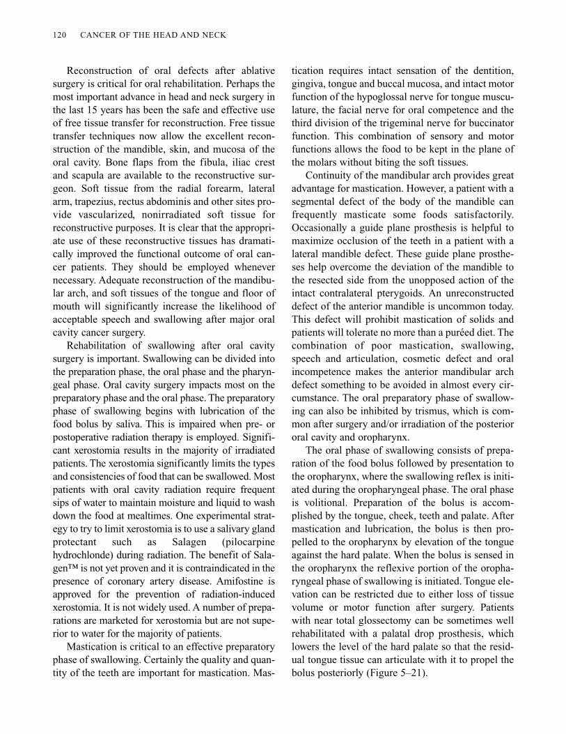
The oral phase of swallowing consists of prepa-ration of the food bolus followed by presentation tothe oropharynx, where the swallowing reflex is initi-ated during the oropharyngeal phase. The oral phaseis volitional. Preparation of the bolus is accom-plished by the tongue, cheek, teeth and palate. Aftermastication and lubrication, the bolus is then pro-pelled to the oropharynx by elevation of the tongueagainst the hard palate. When the bolus is sensed inthe oropharynx the reflexive portion of the oropha-ryngeal phase of swallowing is initiated. Tongue ele-vation can be restricted due to either loss of tissuevolume or motor function after surgery. Patientswith near total glossectomy can be sometimes wellrehabilitated with a palatal drop prosthesis, whichlowers the level of the hard palate so that the resid-ual tongue tissue can articulate with it to propel thebolus posteriorly (Figure 5–21).

Oral Cavity Cancer 121
The oral prosthodontist plays a critical role in therehabilitation of swallowing after oral cancer treat-ment. The proper number and quality of teeth andtheir alignment can be restored by maxillary and/orgingival dentures. After resection of the maxilla orhard palate, a dental obturator to cover the oro-antraland oronasal fistulae is necessary for swallowingwithout nasal regurgitation (Figure 5–22). Patientswith large maxillary defects can attain excellentfunctional results with an obturator. For defects ofthe soft palate, dysphagia due to nasal regurgitation,hyponasal speech and difficulty with articulation ofspeech sounds, an obturator with a nasopharyngealbulb is effective in minimizing nasal regurgitationand improving hyponasal speech. The bulb is prop-erly positioned in the nasopharynx articulating withthe posterior pharyngeal wall at the prominence ofthe body of C2, allowing the remainder of the softpalate to seal off the nasopharynx during swallowing(Figure 5–23).
Osseointegrated implants are an importantadvance in oral rehabilitation. If adequate bonestock exists, titanium posts can be placed in a multi-
staged process and the ingrowth of healthy bone intoand around the implants results in a very securefoundation for oral prostheses.58 Osseointegrativeimplants can be placed in fibula free flap recon-structions of the mandible after the healing andremoval of the fibula fixation hardware (Figure5–24). Osseointegrative implants should be avoidedin the atrophic edentulous mandible especially after
A
B
C
B
A
Figure 5–21. A, Patient with poor tongue mobility with a palataldrop prosthesis in place. B, Palatal drop prosthesis.
Figure 5–22. A, Maxillectomy defect with split-thickness skin graft.B, Prosthesis in place. C, Prosthesis.

122 CANCER OF THE HEAD AND NECK
radiation. Osseointegration can also be utilizedeffectively for external fixation of cosmetic prosthe-ses after extended surgery for oral cavity cancer,which includes soft tissues of the face. It is impor-tant for the patient’s rehabilitation that they have anacceptable cosmetic appearance in public.
Many patients benefit from evaluation and ther-apy by certified speech and swallowing therapists.
They can often recommend exercises for the artic-ulation of speech and can help both the patient andprosthodontist to optimize prostheses and to rec-ommend alternative methods of phoneme forma-tion.59 Patients with significant resections of thelips, maxilla, tongue and palate will often benefitfrom speech therapy.
Speech and swallowing therapists can also helpimprove swallowing in patients who have undergoneoral surgery.60 A modified barium swallow under flu-oroscopic observation by a radiologist and a speechtherapist may be helpful diagnostically.61 From thisstudy, abnormalities of mastication, bolus preparationand bolus presentation of the oropharynx can beobserved and studied frequently from this data.Strategies for improved function can be devised andtaught to the patient and exercises implemented.Accompanying abnormalities of the pharyngealphase of swallowing can also be diagnosed. Based on
C
B
A
Figure 5–23 A, Soft palate defect after surgical resection and freeflap reconstruction of the lateral pharyngeal wall. B, Prosthesis inplace. C, The nasopharyngeal bulb prosthesis.
B
A
Figure 5–24. A, Panorex of osseointegrated implants in the ante-rior and right lateral aspects of a fibula free flap reconstruction of themandible. B, The prosthesis in place.

Oral Cavity Cancer 123
the clinical findings and the modified barium swal-low, therapists can also suggest optimal consistencies,and temperatures of food that can be best managed.
Consultation with a trained nutritionist with expe-rience in treating head and neck cancer patients isessential to provide patients with information andsuggestions regarding optimal foods to maintain abalanced nutrition within the patient’s consistencyrestrictions. Patients with impaired oral function risknutritional deficiency unless an appropriately varieddiet is maintained. Many patients benefit from pre-pared commercial supplements, which are formulatedspecifically as a balanced diet. Some patients maysubsist on liquid dietary supplements alone, while themajority benefit from regular foods as tolerated withadditional dietary supplements as needed. Nearly anyeveryday food can be puréed with liquid in a blenderand drunk. Patients should be weighed frequently inthe postoperative period to monitor for weight loss.Supplemental tube feeding may be necessary whilethe patient is relearning swallowing.
Members of the rehabilitation team must educatethe oral cancer patient regarding oral hygiene. Teethbrushing and fluoride treatments should be done atleast twice daily. The patient should perform thesefluoride treatments at home regularly using moldeddental trays. Patients with post-radiation xerostomiarequire frequent sips of water, and may benefit fromsialagogues such as lozenges or chewing gum; how-ever it is critical that these be sugar free as the risk ofcaries is dramatically increased after radiation treat-ment. All patients with impaired oral function shouldbe instructed to cleanse the oral cavity after eating.This may involve simple rinsing with water or salinesolution or irrigation with a hanging bag and warmsaline solution. Reconstruction flaps with skin liningthe oral cavity may require frequent brushing toeliminate accumulated skin debris and sometimestrimming of the hair growing on the skin flaps is nec-essary for patient comfort and to decrease the adher-ence of food. Reconstructive flaps that have beenirradiated no longer grow hair. Mouth washes, whichcontain alcohol, should be avoided as they dry thetissues and cause burning and discomfort. Normalsaline or saline with bicarbonate of soda is preferred.Successful oral rehabilitation after oral cancersurgery requires a dedicated team of specialists
working together. Each can contribute significantlytoward the rehabilitation of speech, swallowing andappearance of the oral cancer patient.
SEQUELAE, COMPLICATIONS AND THEIR MANAGEMENT
Complications can be minimized by appropriate pre-operative evaluation including medical cardiologyand anesthesia consultation as indicated. Since themajority of oral cancer patients are elderly, many willhave significant co-morbidities which need assess-ment, diagnosis or intervention prior to, or after,surgery. A preoperative medical evaluation is recom-mended for all patients over the age of 60 regardlessof their health status. Routine preoperative testingshould screen for previously undiagnosed majororgan diseases and should consist of at least a preop-erative chest radiograph, serum tests of renal andhepatic function and electrocardiography. Patients innegative nitrogen balance due to poor nutritionwould be considered for a nasogastric feeding tubeplacement and several weeks of nutritional therapyprior to surgery. Properly selected patients shouldhave a low incidence of major complications.62
The most common complications after oralsurgery are wound related. The excellent blood sup-ply to the oral cavity helps to ensure good healing ofsoft tissues and to resolve infection. Careful surgicaltechnique can help to minimize complications. It isimportant to handle tissues atraumatically, avoidexcessive char from electrocautery, observe carefulhemostasis, obliterate any dead spaces and to mini-mize bacteria colony counts by gentle antiseptic pre-operative preparation and copious irrigation withsaline with or without antibiotics. Careful techniquesof closure will help to minimize postoperative woundcomplications. Closure under tension should beavoided, especially of an irradiated tissue. Separatesuture layers of muscle and mucosa should be per-formed. Oral wounds closed by primary intentionwill heal best. Many oral lesions will granulate wellover several days to several weeks. Skin grafts can behelpful but are frequently lost when placed overmobile surfaces or directly over cortical bone. Anyexposed bone or cartilage in the oral cavity will leadto granulation tissue formation and delay of healing.

124 CANCER OF THE HEAD AND NECK
Obviously carious or infected teeth should beremoved at the time of surgery. Twenty-four hours ofIV antibiotics, initiated at least 1 hour prior tosurgery, may help to reduce the wound infection rate.
Other intraoperative measures to decrease com-plications include consideration of procedure dura-tion to minimize the time of general anesthesia. Judi-cious intraoperative use of crystalloid will preventpostoperative complications of fluid overload. Insen-sate fluid loss in oral cavity surgery is significantlyless than in abdominal surgery, which results in alower requirement of intravenous fluid for oral cavitysurgery patients than for abdominal surgery patients.
Postoperative management impacts significantlyon complications of oral surgery. Aggressive oralirrigation should begin on the first day of the surgery.It should be accomplished with normal saline or nor-mal saline and bicarbonate of soda solution in hang-ing irrigation bags or via compressed air-sprayer.
Major systemic complications are uncommon inoral cavity surgery. Cardiopulmonary complicationsoccur due to pre-existing co-morbidities, the physi-ologic stress of surgery and fluid overload. Respira-tory complications such as pneumonia can be mini-mized by appropriate early mobilization and the useof sequential compression devices, and carefulobservation for aspiration of liquids. Due to earlymobilization, oral cavity cancer patients rarely suf-fer from deep venous thrombosis (DVT) or pul-monary embolism, however, immobilized patientsshould be placed on appropriate DVT prophylaxis,such as subcutaneous heparin or sequential com-pression devices.63
The majority of wound complications will healwith aggressive cleansing and infection control.Management of co-morbidities, such as diabetesmellitus, malnutrition and hypothyroidism, in orderto maximize wound healing is critical. Poor healingor a persistent oral cutaneous fistula may result fromthe presence of a foreign body such as hardware,non-absorbable suture or sequestered bone. Persis-tent or recurrent tumor must be ruled out by biopsyin any non-healing wound after oral cancer surgery.The frequency, complexity and duration of woundcomplications are greater in the irradiated patient.
In summary, the incidence of major complica-tions in oral cancer surgery can be minimized by
appropriate patient selection, preoperative evalua-tion, meticulous technique and appropriate postop-erative care.
OUTCOMES AND FUNCTION
Outcomes in oral cavity surgery may be divided intosurvival and functional outcomes. Five-year survivalrates for early (T1 and T2) oral cancers are reportedto be in the 70 to 90 percent range. In all head andneck sites, the presence of metastatic nodes to theneck decreases the survival by 50 percent. Five-yearsurvival for patients with stage IV disease, espe-cially with bulky or bilateral lower neck metastases,is less then 20 percent.27
In resectable stage III and stage IV tumors with N0or N1 disease, 5-year survival has been increased tothe 50 to 60 percent range by the aggressive additionof postoperative radiation therapy.15,16 With improvedlocal control rates a higher percentage of deaths aredue to distant disease and second primary carcinomasrather than from uncontrolled locoregional disease.
Factors that predict survival of oral cancerpatients are low T stage, low N stage, low overallstage, and the absence of significant co-morbidities.While the study of the molecular genetics of oralcancer is rapidly evolving, there are currently nomolecular markers which have been shown to pre-dict survival in head and neck cancer patients inlarge prospectively gathered series. It is howeverlikely that in the next several years valid molecularmarkers will be developed which can predict tumorbehavior, response to surgical and non-surgicaltreatment and patient survival rates.
Functional outcomes for surgery for early oralcancers is excellent. It is rare for patients to suffersignificant loss of speech and swallowing functionafter surgical resection for T1 or T2 lesions. Evenlarge T2 lesions of the tongue rehabilitate extremelywell due to the plasticity of the tongue as well as itsgood blood supply, copious sensory innervation andthe presence of intact musculature. Over 6 to 12months, the patients invariably find dramaticimprovement in articulation of speech sounds, mas-tication and swallowing. Aggressive and appropriaterehabilitation with speech and swallowing therapyand prosthodontics is critical to these results.

Oral Cavity Cancer 125
With increasing volumes of resected tissue, func-tional outcomes diminish. Tissues impacting moston function include tongue muscle, hypoglossalnerve, lingual nerve, anterior mandibular arch, andsoft palate. When extensive or multiple resections ofthe above structures are undertaken for advanceddisease, patient function may be poor even with themost advanced reconstructive and rehabilitativetechniques. Despite improvements in postoperativefunction attributed to free flap reconstruction, thedegree of coordination of motor and sensory func-tion necessary for good oral function cannot beattained with the current technology. These patientsare gastrostomy tube-dependent and speak poorly.Xerostomia from oral radiation therapy and trismusfrom surgery or radiation are also factors that candramatically compromise function.
REFERENCES
1. Landis SH, Murray T, Bolden S, Wingo PA. Cancer statistics,1999. CA Cancer J Clin 1999;49:8–31.
2. Cancer epidemiology and prevention. 2nd ed. New York:Oxford University Press; 1999.
3. Shopland DR, Eyre HJ, Pechacek TF. Smoking-attributablecancer mortality in 1991: is lung cancer now the leadingcause of death among smokers in the United States? JNatl Cancer Inst 1991;83:1142–8.
4. Blot WJ, McLaughlin JK, Winn DM, et al. Smoking anddrinking in relation to oral and pharyngeal cancer. CancerRes 1988;48:3282–7.
5. Mahale A, Saranath D. Microsatellite alterations on chromo-some 9 in chewing tobacco-induced oral squamous cellcarcinomas from India. Oral Oncol 2000;36:199–206.
6. Slaughter DP, Southwick HW, Smejkal J. “Field canceriza-tion” in oral stratified squamous epithelium. Cancer1953;5:963–8.
7. Day GL, Blot WJ. Second primary tumors in patients withoral cancer. Cancer 1992;70:14–9.
8. Califano J, van der Reit P, Westra W, et al. Genetic progres-sion model for head and neck cancer: implications forfield cancerization. Cancer Res 1996;56:2488–92.
9. Chen YK, Huang HC, Lin LM, Lin CC. Primary oral squa-mous cell carcinoma: an analysis of 703 cases in southernTaiwan. Oral Oncol 1999;35:173–9.
10. Sunderman FW Jr, Morgan LG, Andersen A, et al.Histopathology of sinonasal and lung cancers in nickelrefinery workers. Ann Clin Lab Sci 1989;19:44–50.
11. Benninger MS, Enrique RR, Nichols RD. Symptom-directedselective endoscopy and cost containment for evaluationof head and neck cancer. Head Neck 1993;15:532–6.
12. Shaha AR, Hoover EL, Mitrani M, et al. Synchronicity, mul-ticentricity, and metachronicity of head and neck cancer.Head Neck Surg 1988;10:225–8.
13. Hordijk GJ, Bruggink T, Ravasz LA. Panendoscopy: a valu-
able procedure? Otolaryngol Head Neck Surg 1989;101:426–8.
14. Hanasono MM, Kunda LD, Segall GM, et al. Uses and limita-tions of FDG positron emission tomography in patients withhead and neck cancer. Laryngoscope 1999;109:880–5.
15. Vikram B, Strong EW, Shah JP, Spiro R. Failure at the pri-mary site following multimodality treatment in advancedhead and neck cancer. Head Neck Surg 1984;6:720–3.
16. Vikram B, Strong EW, Shah JP, Spiro R. Failure in the neckfollowing multimodality treatment for advanced head andneck cancer. Head Neck Surg 1984;6:724–9.
17. Rudoltz MS, Perkins RS, Luthmann RW, et al. High-dose-ratebrachytherapy for primary carcinomas of the oral cavityand oropharynx. Laryngoscope 1999;109:1967–73.
18. Wolf GT, Forastiere A, Ang K, et al. Workshop report: organpreservation strategies in advanced head and neck can-cer—current status and future directions. Head Neck1999;21:689–93.
19. Schuller DE, Metch B, Stein DW, et al. Preoperativechemotherapy in advanced resectable head and neck can-cer: final report of the Southwest Oncology Group.Laryngoscope 1988;98:1205–11.
20. Williams EF III, Meguid MM. Nutritional concepts and con-siderations in head and neck surgery. Head Neck 1989;11:393–9.
21. Lotan R. Retinoids and chemoprevention of aerodigestivetract cancers. Cancer Metastasis Rev 1997;16:349–56.
22. Hong WK, Endicott J, Itri LM, et al. 13-cis-retinoic acid inthe treatment of oral leukoplakia. N Engl J Med1986;315:1501–5.
23. White JM, Chaudhry SI, Kudler JJ, et al. Nd:YAG and CO2
laser therapy of oral mucosal lesions. J Clin Laser MedSurg 1998;16:299–304.
24. Schoelch ML, Sekandari N, Regezi JA, Silverman S Jr. Lasermanagement of oral leukoplakias: a follow-up study of 70patients. Laryngoscope 1999;109:949–53.
25. Hamilton MM, Branham GH. Concepts in lip reconstruction.Otolaryngol Clin N Am 1997;30:593–606.
26. Cordeiro PG, Santamaria E. Primary reconstruction of com-plex midfacial defects with combined lip- switch proce-dures and free flaps. Plast Reconstr Surg 1999;103:1850–6.
27. Franceschi D, Gupta R, Spiro RH, Shah JP. Improved sur-vival in the treatment of squamous carcinoma of the oraltongue. Am J Surg 1993;166:360–5.
28. Spiro RH, Gerold FP, Shah JP, et al. Mandibulotomyapproach to oropharyngeal tumors. Am J Surg 1985;150:466–9.
29. Spiro RH, Gerold FP, Strong EW. Mandibular “swing”approach for oral and oropharyngeal tumors. Head NeckSurg 1981;3:371–8.
30. Scholl P, Byers RM, Batsakis JG, et al. Microscopic cut-through of cancer in the surgical treatment of squamouscarcinoma of the tongue. Prognostic and therapeuticimplications. Am J Surg 1986;152:354–60.
31. Loree TR, Strong EW. Significance of positive margins inoral cavity squamous carcinoma. Am J Surg 1990;160:410–4.
32. Looser KG, Shah JP, Strong EW. The significance of “posi-tive” margins in surgically resected epidermoid carcino-mas. Head Neck Surg 1978;1:107–11.

126 CANCER OF THE HEAD AND NECK
47. Stanley RB. Mandibular lingual releasing approach to oraland oropharyngeal carcinomas. Laryngoscope 1984;94:596–600.
48. Soo KC, Spiro RH, King W, et al. Squamous carcinoma ofthe gums. Am J Surg 1988;156:281–5.
49. Overholt SM, Eicher SA, Wolf P, Weber RS. Prognostic fac-tors affecting outcome in lower gingival carcinoma.Laryngoscope 1996;106:1335–9.
50. Petruzzelli GJ, Myers EN. Malignant neoplasms of the hardpalate and upper alveolar ridge. Oncology 1994;8:43–8.
51. Reddy CR. Carcinoma of hard palate in India in relation toreverse smoking of chuttas. J Natl Cancer Inst 1974;53:615–9.
52. Ratzer ER, Schweitzer RJ, Frazell EL. Epidermoid carci-noma of the palate. Am J Surg 1970;119:294–7.
53. Evans JF, Shah JP. Epidermoid carcinoma of the palate. AmJ Surg 1981;142:451–5.
54. Chung CK, Rahman SM, Constable WC. Malignant salivarygland tumors of the palate. Arch Otolaryngol 1978;104:501–4.
55. Kovalic JJ, Simpson JR. Carcinoma of the hard palate. J Oto-laryngol 1993;22:118–20.
56. Casson PR, Bonanno PC, Converse JM. The midface deglov-ing procedure. Plast Reconstr Surg 1974;53:102–3.
57. Price JC, Holliday MJ, Johns ME, et al. The versatile midfacedegloving approach. Laryngoscope 1988;98:291–5.
58. Marx RE, Morales MJ. The use of implants in the recon-struction of oral cancer patients. Dent Clin N Am 1998;42:177–202.
59. Fletcher SG. Speech production following partial glossec-tomy. J Speech Hear Disord 1988;53:232–8.
60. Logemann JA. Rehabilitation of the head and neck cancerpatient. Semin Oncol 1994;21:359–65.
61. Logemann JA. Role of the modified barium swallow in man-agement of patients with dysphagia. Otolaryngol HeadNeck Surg 1997;116:335–8.
62. Sessions RB, Hudkins C. Complications of surgery of theoral cavity. In: Eisele DW, editor. Complications in headand neck surgery. Baltimore: Mosby; 1993. p.218–22.
63. Lowry JC. Thromboembolic disease and thromboprophylaxisin oral and maxillofacial surgery: experience and practice.Br J Oral Maxillofac Surg 1995;33:101–6.
33. Fukano H, Matsuura H, Hasegawa Y, Nakamura S. Depth ofinvasion as a predictive factor for cervical lymph nodemetastasis in tongue carcinoma. Head Neck 1997;19:205–10.
34. Spiro RH, Huvos AG, Wong GY, et al. Predictive value oftumor thickness in squamous carcinoma confined to thetongue and floor of the mouth. Am J Surg 1986;152:345–50.
35. Shaha AR, Spiro RH, Shah JP, Strong EW. Squamous carci-noma of the floor of the mouth. Am J Surg 1984;148:455–9.
36. Marchetta FC, Sako K, Murphy JB. The periosteum of themandible and intraoral carcinoma. Am J Surg 1971;122:711–3.
37. Marchetta FC, Sako K, Badillo J. Periosteal lymphatics of themandible and intraoral carcinoma. Am J Surg 1964;108:505–7.
38. McGregor IA, MacDonald DG. Spread of squamous cell car-cinoma to the nonirradiated edentulous mandible—a pre-liminary report. Head Neck Surg 1987;9:157–61.
39. McGregor AD, MacDonald DG. Routes of entry of squa-mous cell carcinoma to the mandible. Head Neck Surg1988;10:294–301.
40. Brown JS, Browne RM. Factors influencing the patterns ofinvasion of the mandible by oral squamous cell carci-noma. Int J Oral Maxillofac Surg 1995;24:417–26.
41. McGregor AD, MacDonald DG. Patterns of spread of squa-mous cell carcinoma to the ramus of the mandible. HeadNeck 1993;15:440–4.
42. Totsuka Y, Usui Y, Tei K, et al. Results of surgical treatmentfor squamous carcinoma of the lower alveolus: segmentalvs. marginal resection. Head Neck 1991;13:114–20.
43. Totsuka Y, Usui Y, Tei K, et al. Mandibular involvement bysquamous cell carcinoma of the lower alveolus: analysisand comparative study of histologic and radiologic fea-tures. Head Neck 1991;13:40–50.
44. Shaha AR. Marginal mandibulectomy for carcinoma of thefloor of the mouth. J Surg Oncol 1992;49:116–9.
45. Schramm VL Jr, Myers EN. Skin grafts in oral cavity recon-struction. Arch Otolaryngol 1980;106:528–32.
46. Schramm VL Jr, Johnson JT, Myers EN. Skin grafts and flapsin oral cavity reconstruction. Arch Otolaryngol 1983;109:175–7.