Embed Size (px)
Citation preview

JSM Dentistry
Cite this article: Agar NJM, Patel RS (2014) Early Detection, Causes and Screening of Oral Cancer. JSM Dent 2(3): 1039.
Central
*Corresponding authorRajan Patel, Department of Otorhinolaryngology Head and Neck Surgery, Auckland City Hospital, New Zealand, Tel: 649367000; Email:
Submitted: 08 April 2014
Accepted: 22 May 2014
Published: 22 June 2014
ISSN: 2333-7133
Copyright© 2014 Patel et al.
OPEN ACCESS
Keywords•Oral squamous cell carcinoma•Screening•Early detection•Prevention
Review Article
Early Detection, Causes and Screening of Oral CancerNJM Agar and RS Patel*Department of Otorhinolaryngology Head and Neck Surgery, New Zealand
Abstract
Oral cavity Squamous Cell Carcinoma (SCC) is common, and despite its relative ease of detection, patients continue to present with late stage disease. The World Health Organisation (WHO) has urged member states to involve primary care givers (dentists and general practitioners) in increasing early referral for suspicious oral lesions.
Tobacco and alcohol consumption remain the two major risk factors for oral SCC. A brief screening history to identify high-risk individuals, followed bya simplebut thorough oral examination is the best tool available for screening for oral SCC. This is both cost effective and reduces mortality when applied to patients in high-risk groups.
ABBREVIATIONSSCC: Squamous Cell Carcinoma
INTRODUCTIONOral cavity Squamous Cell Carcinoma (SCC) is common.
Whilstthe oral cavity is easily accessible for examination, and althoughtumors of this site cause symptoms at a relatively early stage, a large majority of patients continue to present with late stage disease. Despite readily and widely available treatment,oral cancer carries an overall disease specificrelative mortality of 49% [1].
In 2007 the World Health Assembly passed a resolution on oral health, urging all member states to “take steps to ensure that prevention of oral cancer is an integral part of national cancer control programs, and to involve oral health professionals or primary health care personnel with relevant training in oral health in detection, early diagnosis and treatment [2].” Despite the multitude of tools marketed to aid in the early diagnosis of oral cancer, there is no general population based screening method shown to reduce mortality associated with oral cancer [3]. Dentists examine their patient’s oral cavity during almost every consultation and are in a unique position to both promote primary preventative measures to high risk groups, and aid in early diagnosis and referral of suspicious oral lesions.
Spectrum of disease
SCC’s make up approximately 90% of tumors in the oral cavity, with adenocarcinoma/minor salivary tumors accounting for 5%, verrucous carcinoma and lymphoma 2% each and the remainder being uncommon sarcomas or odontogenic tumours.SCC will be discussed for the purposes of this review.
Epidemiology
Oral SCC is the 6th most common cancer globally and its incidence is increasing [4]. The burden of oral cavity SCC varies significantly with cultural risk-taking behaviors worldwide. India, Pakistan, Sri Lanka and Bangladesh have the highest incidence with up to 25% of all new cancers affecting the oral cavity [5], compared with 6% in France and 3% in the UK [6]. The age adjusted incidence is reported from approximately 3.4 to 13.8 per 100,000. Males are more often affected than females by a ratio of 1.5 : 1 [7]. A rising incidence has been noted in patients under 45yrs of age [8], with approximately 6% of oral cancers now occurring in this age group compared with 3% in 1973 [9]. The average period for which a patient is aware of an oral lesion prior to bringing it to the attention of their doctor is 3 months [10]. Unfortunately, over 60% of patients present with stage III or stage IV disease [11], and after treatment can expect only a 45% and 32% five-year survival respectively.
Risk factors
Smoking and alcohol consumption are powerful and synergistic risk factors for the development of oral SCC. Heavy drinkers and smokers have 38 times the risk of abstainers from both products [12]. 20pack-years seems to be the threshold at which a significantly increased risk of cancer is imparted [13,14], and the risk reduces back to baseline 10years after cessation [15]. Betel nut chewing and reverse smoking have a strong causal relationship particularly with buccal and hard palate subsites respectively - this accounts for the extremely high rates of oral SCC in countries where these behaviors are entrenched. UV sunlight is a clear aetiologic factor in lip SCC, which predominantly affects Caucasian males.
The role of Human Papilloma Virus (HPV) has been well
Special Issue on
Oral Cancer
Patel et al. (2013)Email:
JSM Dent 2(3): 1039 (2014) 2/5
Central
established in SCC of the oropharynx [16], andwhile a link has been shown to exist with oral cancer also [17], further evidence is required to confirm it as a strong causal factor for the oral cavity. Immunocompromised individuals have a higher rate of nearly all malignancies and oral SCC is no exception. A number of genetic conditions carry an increased predisposition [18] – these are listed as part of Table 1. Chronic inflammation has long been purported to increase the risk of oral cancer – this may be the way in which candidiasis, syphilis, and chronic trauma from poor dentition contribute to a slight increase in risk of oral cancer. Poor oral hygiene has been linked to increased carcinogenesis, the exact mechanism is unclear but it may be related to carcinogenic effects from the high burden of polymicrobial oral flora [19].
Potentially malignant disorders
Use of the term ‘Premalignant Lesion’ is now discouraged in favour of the phrase “Potentially Malignant disorders [21].” The lesions of most relevance are erythroplakia, leukoplakia, oral lichen planus and submucous fibrosis.
Erythroplakia is defined as a ”fiery red patch”. These lesions are often symptomatic, have a degree of increased vascularity, and carry a high risk of harbouring dysplasia. All erythroplakic or leukoerythroplakic lesions should be referred for biopsy and or excision. Should mild or moderate dysplasia be confirmed on biopsy, its risk of malignant transformation is 10.3%, with high-grade dysplasia and carcinoma insitu carrying a 24.1% risk [22].
Leukoplakia is usually asymptomatic and is defined either as a “white plaque that will not rub off” or “a white plaque of questionable risk, having excluded other known diseases or disorders that carry no increased risk for cancer”. Confounding benign causes of a white plaque include a frictional lesion from habitual trauma or cheek biting, linea alba (normal white streaks bilaterally along the occlusal line), and leukoedema amongst others [23]. The prevalence of Leukoplakia has been estimated at 2% [24], although the true rate when the latter of the two above definitions is applied is likely to be a little lower at a more modest 0.5%. The annual malignant transformation rate is estimated to be from 0.3% [25] to 1%. It is generally accepted that referral for biopsy or excision is warranted for these lesions.
Lichen Planus is an oral autoimmune condition with a number of morphological variants including reticular (fine white lacy lines), erosive (shallow ulcers), atrophic (thinned erythematous
mucosa), and bullous (fluid filled vesicles). It can be symptomatic typically with a burning feeling or hypersensitivity of the affected mucosa. There remains debate as to its status as a potentially malignant condition. It is generally accepted that its risk of malignant transformation is below 1% per year [26]. A patient with lesions such as these should be referred to a specialist for biopsy.
Submucous Fibrosis causes progressive trismus due to fibrosis of the connective tissues of the cheeks. It is strongly associated with betel nut/tobacco chewing and it is likely that the use of these carcinogens give it an association with oral cancer. The rate of malignant transformation is estimated at 0.5% per year [27].
Screening/early detection
Screening for a disease implies the application of a test to an asymptomatic population with the aim of detecting disease at an early stage. This disease must be a significant public health issue, and the natural history and management of the disease must be well understood such that improved outcomes can be expected when treated at an early stage. Furthermore the test itself and any morbidity associated with further investigation of false positives must be acceptable [28].
The established screening test for oral SCC is clinical examination - with biopsy and histopathological assessment being the gold standard to confirm the diagnosis. Oral cavity examination has been demonstrated in a meta-analysis by Downer et al [29] to have a sensitivity of 0.84 and a specificity of 0.97. For comparison they note that both mammography and cervical screening programmesshare an approximatesensitivity and specificity of 0.8 and 0.98 respectively. Oral examination is simple, takes only a few minutes, requires minimal equipment, is non-invasive and can be performed by a wide range of health professionals.
For widespread structured screening to be instituted it must be shown to not only be of value in reducing morbidity and mortality, but also to be cost effective. Sankaranarayanan et al [30] published a hallmark paper in the Lancet in 2005 in which they assessed their oral cancer screening programme when applied to a prospective cohort of over 87,000 individuals over the age of 35.They found no significant effect on the disease specific mortality when applied to the general population. However, subgroup analysis of smokers and/or drinkers showed a reduction in mortality of 43% and 22% in men and women respectively. A subsequent paper [31] found this approach to be cost effective - albeit in India where there is a high rate of oral cancer. There are no similar trials in a low prevalence society,howeversimulation modeling has shown that a screening oral examination in high-risk individuals in a western population may also be cost effective [32].
The question still remains as to which group of health professionals should be undertaking oral cancer screens? An interesting paper by MacPherson et al [33] surveyed 357 general medical practitioners (GP’s) and 331 general dental practitioners to assess their knowledge of, and their self-perceived ability to diagnose oral cancer or precancerous lesions. 58% of Dentists claimed to examine opportunistically for signs of oral cancer
Major Risk Factors:Smoking Alcohol Betel Nut (buccal/retromolar trigone) UV light (lip) Reverse smoking (hard palate)Minor Risk Factors:HPVImmune deficiencyMicronutrient deficiency (low fruit and vegetable intake)Syphilis, Chronic candidiasisPrior radiation exposurePeriodontal diseasePoor oral hygiene [20]
Genetic: Fanconi’s anaemia, dyskeratosis congenita, xeroderma pigmentosa, plummer-vinson syndrome, patterson-kelly brown syndrome, scleroderma, diabetes.
Table 1: Risk Factors for Oral SCC.
Patel et al. (2013)Email:
JSM Dent 2(3): 1039 (2014) 3/5
Central
compared with GP’s who overwhelmingly only examined if a symptom was raised. 37% of dentists felt confident in their ability to diagnose these lesions, compared to only 15% of GP’s. A lack of specific education was cited as an important barrier to improving performance. Unfortunately while dentists are both more confident and more likely to examine the oral cavity of their patients, the high risk population are infrequent attenders to general dental practices [34]. Heavy smokers and drinkers from low socio-economic backgrounds tend to only present for dental care in the context of a dental abscess or severely carious teeth often with a corresponding florid gingivitis, whichmay impede or confound lesion detection. Furthermore, older patients who have a higher risk of cancer are frequently edentulous and have no cause to see a dentist. Another major barrier for these patients is the cost of dental care, which is self-funded by patients in many countries.
How to perform a screening oral examination
Oncological examination of the oral cavity is simple, cost free, non-invasive and should be within the skill set of all GP’s and dentists. Table 2 gives a brief account of equipment required and areas to examine.Palpation is of particular relevance- with bulky/firm lesions being of much higher concern than a soft lesion with identical texture to the adjacent tissues.
The areas easily overlooked are the lateral tongue, glossotonsillar sulcus and the floor of mouth as these areas require the patient to actively move their tongue to the contralateral cheek and also require active retraction of the tongue with a depressor to allow visualization.Table 3 lists the key red flag clinical features on history and Table 4 highlightssuspicious signs that should trigger referral Figures 1, 2 and 3.
Adjuncts
Many adjuncts to clinical examination for oral SCC exist. However as eluded to by Lingen et alxi in their excellent position paper of the American Academy of Oral and Maxillofacial Pathology, despite the “tantalizing implication that such technologies may improve detection of oral cancers and pre-cancers beyond conventional oral examination alone” none have been proven to do so.
Equipment required: Good lighting (ideally a head light) Tongue depressor +/- dental mirror Gloves to allow palpation of lesionsAssess the oral cavity subsites systematically:Both lips from vermilion to gingivo-labial sulcusBuccal mucosa Gingiva Retromolar Trigone Hard Palate Floor of Mouth and glossotonsillar sulcus* Tongue
- Dorsum (opposed to hard palate)- Ventral surface* (opposed to floor of mouth)- Lateral tongue*
Soft Palate and Tonsil fossae (strictly speaking these are oropharynx sites but should be included in a screening oncological oral examination)
Table 2: Oncological Oral cavity Examination. *areas easily overlooked.
The high risk patient for Oral SCCMale in 50’s to 60sExposure to tobacco, alcohol, betel nut. Low socioeconomic groupHistory of prior oral SCC (3-7% incidence per annum of 2nd primary)Immunocompromised
Table 3: High risk patient factors.
Red Flags – trigger for referralNon healing lesion >2 weeks Ulcer or mass with raised heaped up margins, puckering/tethering of surrounding tissuesPain or numbness/tingling associated with a persistent lesion Red lesion(erythroplasia) or Red-White lesion (leuko-erythroplakia)Unexplained loose tooth or non-healing extraction socket.Neck mass
Table 4: ExaminationRed Flags.
Figure 1 An early (T1) lateral tongue SCC. Firm to palpation, central ulceration, heaped edge.
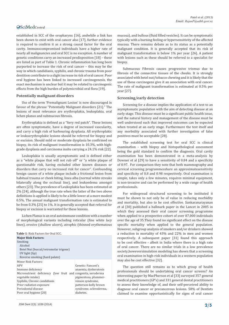
Figure 2 An early (T1) lateral tongue SCC. Firm to palpation, central leukoplakia andsurrounding tethering.
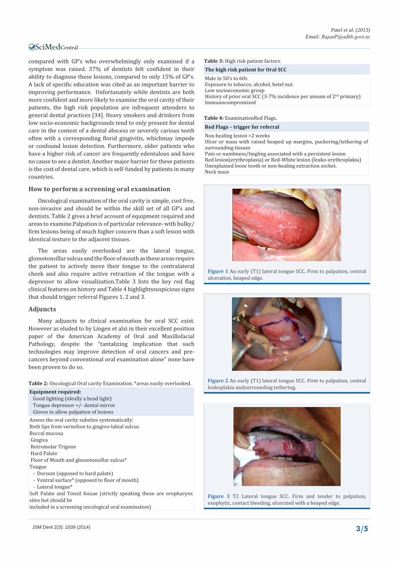
Figure 3 T2 Lateral tongue SCC. Firm and tender to palpation, exophytic, contact bleeding, ulcerated with a heaped edge.
Patel et al. (2013)Email:
JSM Dent 2(3): 1039 (2014) 4/5
Central
Toludine Blue is a topical dye, which is concentrated in cells with abundant nucleic acids, and has been used for decades on the cervix to aid in identification and demarcation of mucosal abnormalities. There is a large volume of literature assessing its role in the oral cavity, with mixed results. While it is generally accepted that Toludine Blue staining has a high sensitivity for detecting carcinoma, its sensitivity for identifying dysplasia is poor (sensitivity approximately 50% [11]) and has a low specificity with most oral lesions benign or otherwise taking up the dye to some degree. As a somewhat subjective guide to clinicians it is felt that carcinomas are likely to stain a deep royal blue, whereas benign lesions (leukoplakia’s, leukodema, lichen planus etc) are more often a pale blue [35]. In their systematic review, Gray et al [36] concluded that “the high rate of false positive stains and the low specificity in staining dysplasia likely outweigh the potential benefits of any additional cancers detected”.On balance this adjunctive measure offers benefits in targeting lesions to biopsy. However, it has only thoroughly been assessed for use in the hands of oral specialists, and only on lesions already identified by a clinical examination.
Brush Biopsy(OralCDx) utilises a stiff cytology brush to sample cells from the surface and basal layer of a lesion. When sent back to the provider’s laboratory it will yield either a positive, atypical or negative result.Its main role is in sampling lesions that on clinical grounds are felt likely to be innocuous [37,38]. Lingen concludes that “this tool may be beneficial in the patient with multiple lesions throughout their oral cavity” who is unlikely to accept a scalpel biopsy of them all, or “in the non-compliant patient who is unlikely to come back for a follow-up exam or accept an immediate referral to an oral surgeon”.
Multiple optical detection systems have been developed and marketed for the detection of oral premalignant lesions and oral cancers. These include tissue reflectance tools such as the ViziLite [39] which prepare the mucosa with an acetic acid mouthwash, tissue auto fluorescence technologies such as the VELscope [40], and Narrow Band Imaging [41] to name but a few. On the whole in comparison to standard oral examination, these technologies are expensive to set up, have a significant learning curve,and can betime consuming and labour intensive to use. While these techniques often have a high sensitivity for detecting premalignant and malignant oral lesions, their specificity is typically poor and again these systems should be reserved for the oral specialist with little or no role in the primary care setting.
CONCLUSIONOral SCC occurs typically in males from a low socioeconomic
background who smoke and consume alcohol. Patients unfortunately continue to present with late stage disease. An oral screening examination is a simple non-invasive test to apply, has a comparable sensitivity and specificity to that of the well established cervical and breast cancer screening programs, and is felt to probably be cost effective when applied to high-risk individuals in western society.
In the absence of any formal screening program being introduced, dentists and GP’s can best serve these high-risk patients by performing regular opportunistic oral examination and educating these patients to increase their awareness of the early signs and symptoms of oral cancer. Oral medicine
specialists, Otorhinolaryngologists and Oral maxillofacial surgeons must improve efforts to educate Dentists and GP’s to ensure they feel adequately equipped and supported to perform this role.If a suspicious lesion is found, immediate referral for further investigation and subsequent treatment is warranted.
REFERENCES1. AJCC Cancer staging manual 7th edition. 2010; 33.
2. World Health Organization Oral health: action plan for promotion and integrated disease prevention (60th world health assembly resolution A16), Geneva, WHO. 2007.
3. Speight PM, Downer MC, Zakrzewska J. Screening for oral cancer and precancer. A report of the UK working group on screening for oral cancer and precancer. Community Dent Health. 1993; 10: 1–89.
4. Ferlay J, Pisani P, Parkin DM. GLOBOCAN 2002. Cancer incidence, mortality and prevalence worldwide. IARC Cancer Base (2002 estimates). Lyon: IARC Press. 2004.
5. IARC. Cancer incidence in five continents. vol 1X.
6. Brocklehurst P, Kujan O, Glenny AM, Ogden G, Shepherd S, Glenny AM. et al. Screening programmes for the early detection and prevention of oral cancer. Cochrane Database of Systematic Reviews. 2010.
7. Warnakulasuriya S. Global epidemiology of oral and oropharyngeal cancer. Oral Oncol. 2009; 45: 309-316.
8. Warnakulasuriya S, Mak V, Möller H. Oral cancer survival in young people in South East England. Oral Oncol. 2007; 43: 982-986.
9. Llewellyn CD, Johnson NW, Warnakulasuriya KA. Risk factors for squamous cell carcinoma of the oral cavity in young people--a comprehensive literature review. Oral Oncol. 2001; 37: 401-418.
10. McGurk M, Chan C, Jones J, O’regan E, Sherriff M. Delay in diagnosis and its effect on outcome in head and neck cancer. Br J Oral Maxillofac Surg. 2005; 43: 281-284.
11. Lingen MW, Kalmar JR, Karrison T, Speight PM. Critical evaluation of diagnostic aids for the detection of oral cancer. Oral Oncol. 2008; 44: 10-22.
12. Blot WJ, McLaughlin JK, Winn DM, Austin DF, Greenberg RS, Preston-Martin S. Smoking and drinking in relation to oral and pharyngeal cancer. Cancer Res. 1988; 48: 3282-3287.
13. Hashibe M, Brennan P, Benhamou S, Castellsague X, Chen C, Curado MP. Alcohol drinking in never users of tobacco, cigarette smoking in never drinkers, and the risk of head and neck cancer: pooled analysis in the International Head and Neck Cancer Epidemiology Consortium. J Natl Cancer Inst. 2007; 99: 777-789.
14. IARC Working Group on the Evaluation of Carcinogenic Risks to Humans. Tobacco smoke and involuntary smoking. IARC Monogr Eval Carcinog Risks Hum. 2004; 83: 1-1438.
15. Warnakulasuriya S, Sutherland G, Scully C. Tobacco, oral cancer, and treatment of dependence. Oral Oncol. 2005; 41: 244-260.
16. Ang KK, Harris J, Wheeler R, Weber R, Rosenthal DI, Nguyen-Tân PF. Human papillomavirus and survival of patients with oropharyngeal cancer. N Engl J Med. 2010; 363: 24-35.
17. Scully C. Oral cancer; the evidence for sexual transmission. Br Dent J. 2005; 199: 203-207.
18. Prime SS, Thakker NS, Pring M, Guest PG, Paterson IC. A review of inherited cancer syndromes and their relevance to oral squamous cell carcinoma. Oral Oncol. 2001; 37: 1-16.
19. Bloching M, Reich W, Schubert J, Grummt T, Sandner A. The influence

Patel et al. (2013)Email:
JSM Dent 2(3): 1039 (2014) 5/5
Central
Agar NJM, Patel RS (2014) Early Detection, Causes and Screening of Oral Cancer. JSM Dent 2(3): 1039.
Cite this article
of oral hygiene on salivary quality in the Ames Test, as a marker for genotoxic effects. Oral Oncol. 2007; 43: 933-939.
20. Zheng TZ, Boyle P, Hu HF, Duan J, Jian PJ, Ma DQ, et al. Dentition, oral hygiene, and risk of oral cancer: a case-control study in Beijing, People’s Republic of China. Cancer Causes Control. 1990; 1: 235–241.
21. Warnakulasuriya S, Johnson NW, van der Waal I. Nomenclature and classification of potentially malignant disorders of the oral mucosa. J Oral Pathol Med. 2007; 36: 575-580.
22. Mehanna HM, Rattay T, Smith J, McConkey CC. Treatment and follow-up of oral dysplasia - a systematic review and meta-analysis. Head Neck. 2009; 31: 1600-1609.
23. van der Waal I. Potentially malignant disorders of the oral and oropharyngeal mucosa; terminology, classification and present concepts of management. Oral Oncol. 2009; 45: 317-323.
24. Petti S. Pooled estimate of world leukoplakia prevalence: a systematic review. Oral Oncol. 2003; 39: 770-780.
25. Gupta PC, Mehta FS, Daftary DK, Pindborg JJ, Bhonsle RB, Jalnawalla PN. Incidence rates of oral cancer and natural history of oral precancerous lesions in a 10-year follow-up study of Indian villagers. Community Dent Oral Epidemiol. 1980; 8: 283-333.
26. Gandolfo S, Richiardi L, Carrozzo M, et al. Risk of oral squamous cell carcinoma in 402 patients with oral lichen planus: a follow-up study in an Italian population. Oral Oncol. 2004; 40:77–83.
27. Murti PR, Bhonsle RB, Pindborg JJ, Daftary DK, Gupta PC, Mehta FS. Malignant transformation rate in oral submucous fibrosis over a 17-year period. Community Dent Oral Epidemiol. 1985; 13: 340-341.
28. Wilson JM, Jungner YG. Principles and practice of screening for disease. Public health paper number 34. WHO, Geneva, Switzerland. 1968.
29. Downer MC, Moles DR, Palmer S, Speight PM. A systematic review of test performance in screening for oral cancer and precancer. Oral Oncol. 2004; 40: 264-273.
30. Sankaranarayanan R, Ramadas K, Thomas G, Muwonge R, Thara S, Mathew B. Effect of screening on oral cancer mortality in Kerala, India:
a cluster-randomised controlled trial. Lancet. 2005; 365: 1927-1933.
31. Subramanian S, Sankaranarayanan R, Bapat B, Somanathan T, Thomas G, Mathew B. Cost-effectiveness of oral cancer screening: results from a cluster randomized controlled trial in India. Bull World Health Organ. 2009; 87: 200-206.
32. Speight PM, Palmer S, Moles DR, Downer MC, Smith DH, Henriksson M. The cost-effectiveness of screening for oral cancer in primary care. Health Technol Assess. 2006; 10: 1-144, iii-iv.
33. Macpherson LM, McCann MF, Gibson J, Binnie VI, Stephen KW. The role of primary healthcare professionals in oral cancer prevention and detection. Br Dent J. 2003; 195: 277-281.
34. Haughney MG, Devennie JC, Macpherson LM, Mason DK. Integration of primary care dental and medical services: a three-year study. Br Dent J. 1998; 184: 343-347.
35. Gandolfo S, Pentenero M, Broccoletti R, Pagano M, Carrozzo M, Scully C. Toluidine blue uptake in potentially malignant oral lesions in vivo: clinical and histological assessment. Oral Oncol. 2006; 42: 89-95.
36. Gray MGL, Burls A, Elley K. The clinical effectiveness of toluidine blue dye as an adjunct to oral cancer screening in general dental practice. A West Midlands Development and Evaluation Service Report. 2000.
37. Fist S. The oral brush biopsy: separating fact from fiction. Oral Surg Oral Med Oral Pathol Oral Radiol Endod. 2003; 96: 654-655.
38. Eisen D. Brush biopsy ‘saves lives’. J Am Dent Assoc. 2002; 133: 688, 690, 692.
39. Kerr AR, Sirois DA, Epstein JB. Clinical evaluation of chemiluminescent lighting: an adjunct for oral mucosal examinations. J Clin Dent. 2006; 17: 59-63.
40. De Veld DC, Witjes MJ, Sterenborg HJ, Roodenburg JL. The status of in vivo autofluorescence spectroscopy and imaging for oral oncology. Oral Oncol. 2005; 41: 117-131.
41. Watanabe A, Tsujie H, Taniguchi M, Hosokawa M, Fujita M, Sasaki S. Laryngoscopic detection of pharyngeal carcinoma in situ with narrowband imaging. Laryngoscope. 2006; 116: 650-654.