Embed Size (px)
DESCRIPTION
Â
Citation preview
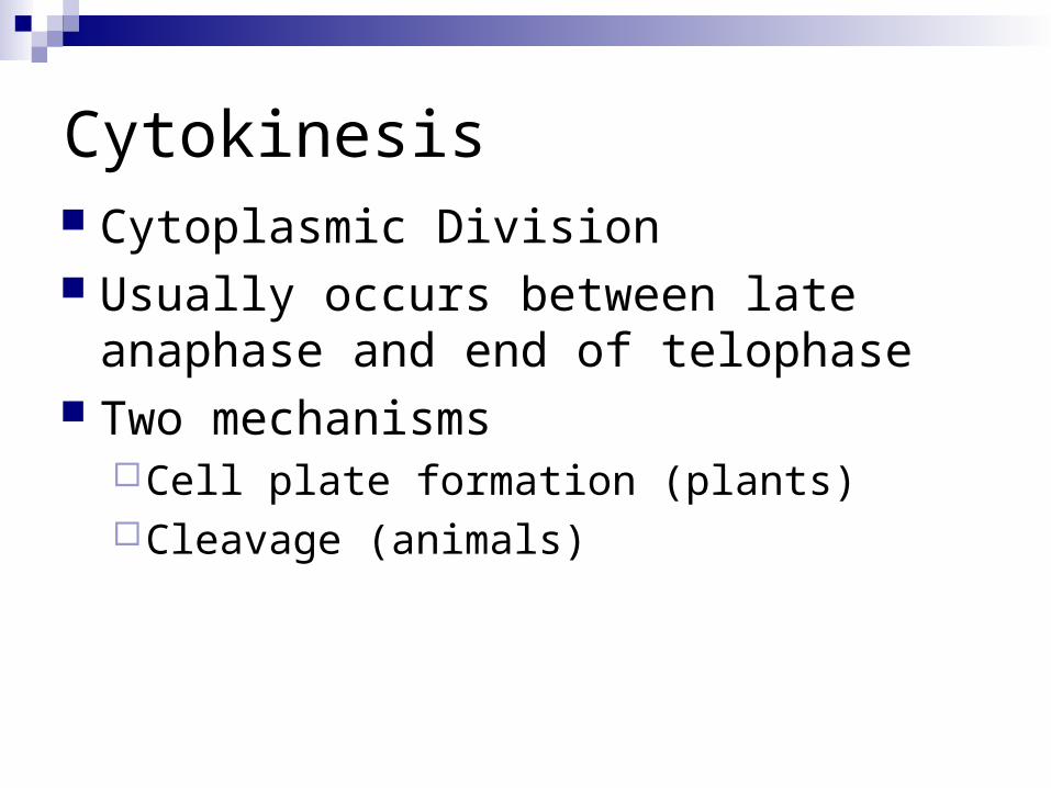
Cell Division and Mitosis
Chapter 9
Prokaryotic Cell Division
Bacterial cells reproduce by Binary Fission Much simpler process than in eukaryotic
organisms (why?) Begins with DNA replication (why?); each copy
moves to opposite sides of cell Followed by elongation of cell, and formation of
a septum (separation) between the two halves, forming two new cells
Results in two cells that are identical (clones) of original cells
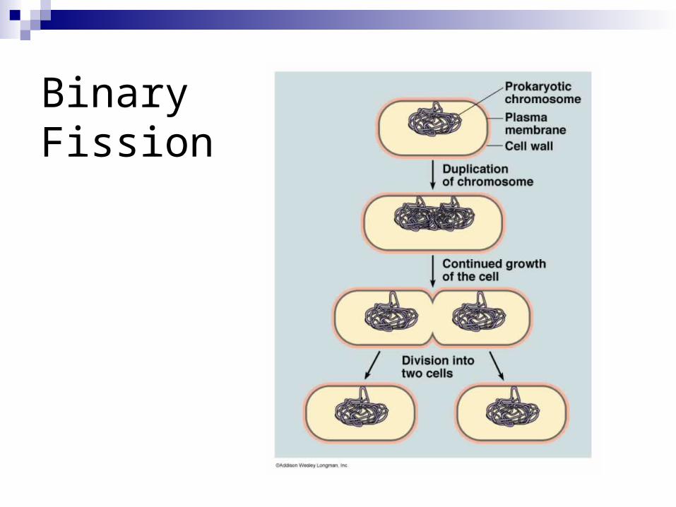
Binary Fission
Eukaryotic Cell Division
Two formsMitosis
grow, replace dead or worn out cells, or to repair wounds
Asexual reproduction in fungi, protists, some plants/animals
Meiosis Sexual reproduction
DNA and Cell Division
During cell division, the genetic material DNA, needs to be copied and divided between the two new cells
DNA in cells is divided into long chains called chromosomes (“volumes” of DNA)
Chromosome DNA is wrapped around proteins called histones to organize it
Nucleosome: unit of DNA wrapped around histones
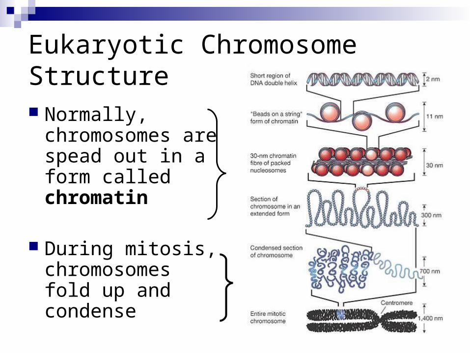
Eukaryotic Chromosome Structure Normally,
chromosomes are spead out in a form called chromatin
During mitosis, chromosomes fold up and condense
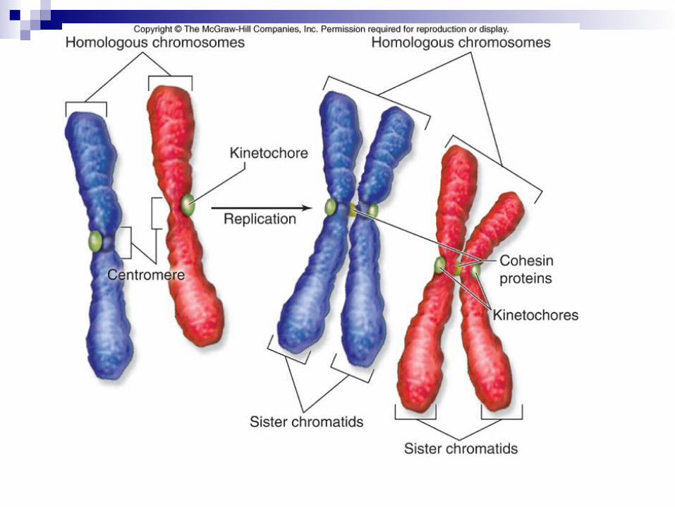
Eukaryotic ChromosomesChromosomes must be replicated before cell
division.-Replicated chromsomes are connected to
each other at their kinetochores-cohesin – complex of proteins holding
replicated chromosomes together-sister chromatids: 2 copies of the
chromosome within the replicated chromosome
Chromosome structure
Normally chromosomes are spread out & not identifiable (chromatin)
At the start of mitosis they condense & take the form shown
The replicated chromosomes stay together and are called sister chromatids
Sister chromatids are attached at the centromere by proteins called cohesins
The other side of the centromeres contain other proteins called kinetochore
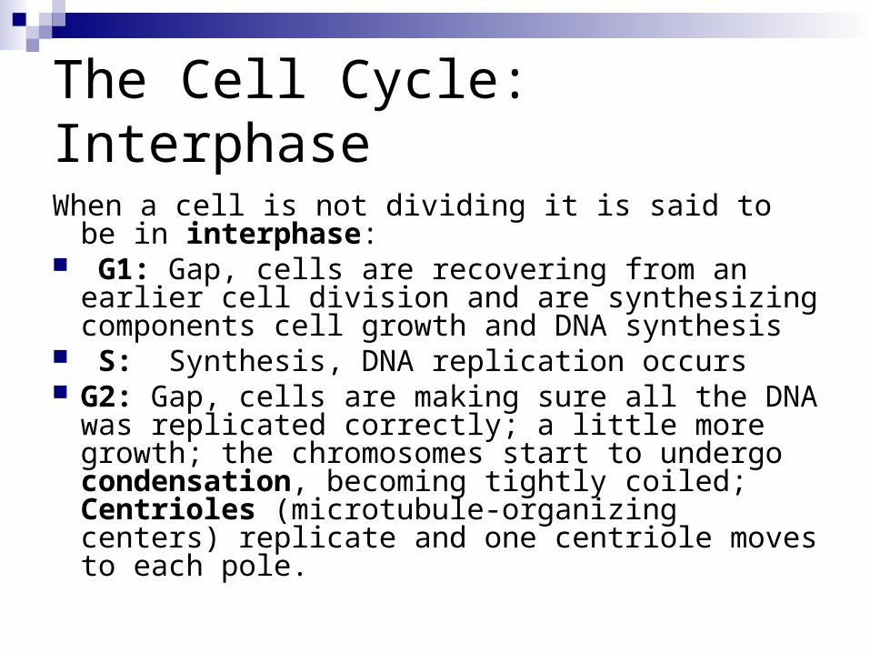
The Cell Cycle: Interphase
When a cell is not dividing it is said to be in interphase:
G1: Gap, cells are recovering from an earlier cell division and are synthesizing components cell growth and DNA synthesis
S: Synthesis, DNA replication occurs G2: Gap, cells are making sure all the DNA was
replicated correctly; a little more growth; the chromosomes start to undergo condensation, becoming tightly coiled; Centrioles (microtubule-organizing centers) replicate and one centriole moves to each pole.
The Cell Cycle: Cell Division
Mitosis (M Phase)Nuclear Division
Cytokinesis (C phase)Cytoplasmic Division
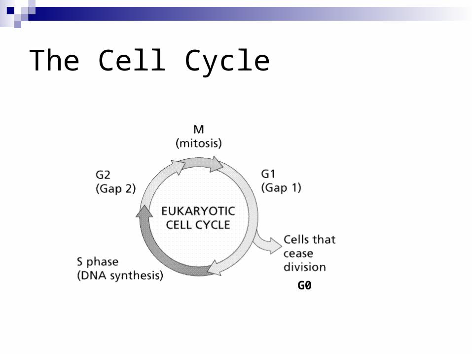
G0
The Cell Cycle
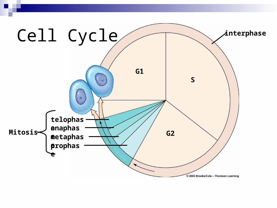
G1S
G2Mitosis
telophaseanaphasemetaphaseprophase
interphaseCell Cycle
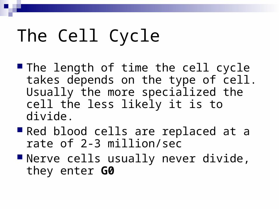
The Cell Cycle
The length of time the cell cycle takes depends on the type of cell. Usually the more specialized the cell the less likely it is to divide.
Red blood cells are replaced at a rate of 2-3 million/sec
Nerve cells usually never divide, they enter G0
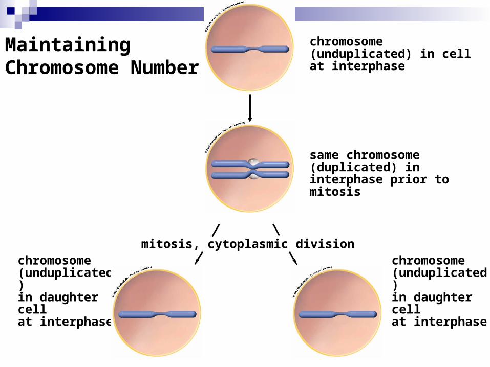
Maintaining Chromosome Number
mitosis, cytoplasmic divisionchromosome (unduplicated) in daughter cell at interphase
chromosome (unduplicated) in daughter cell at interphase
chromosome (unduplicated) in cell at interphase
same chromosome (duplicated) in interphase prior to mitosis
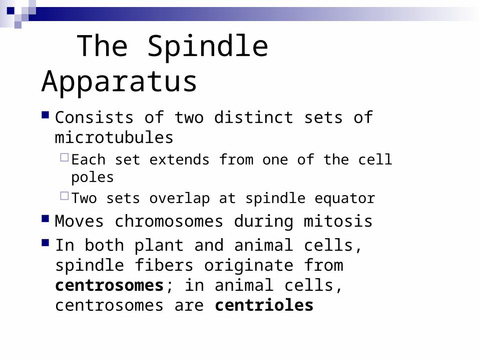
The Spindle Apparatus
Consists of two distinct sets of microtubulesEach set extends from one of the cell polesTwo sets overlap at spindle equator
Moves chromosomes during mitosis In both plant and animal cells, spindle fibers
originate from centrosomes; in animal cells, centrosomes are centrioles
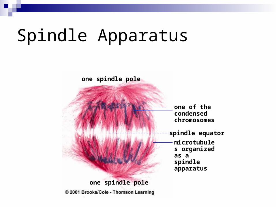
one spindle pole
one of the condensed chromosomes
spindle equatormicrotubules organized as a spindle apparatus
one spindle pole
Spindle Apparatus
Mitosis
Nuclear Division
Dividing up the genetic material (DNA)
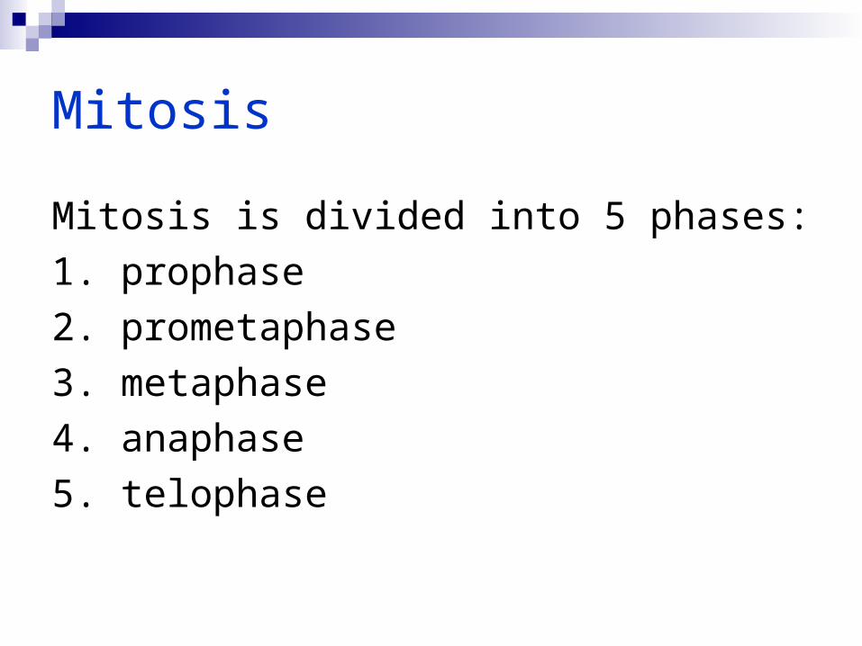
Mitosis
Mitosis is divided into 5 phases:1. prophase2. prometaphase3. metaphase4. anaphase5. telophase
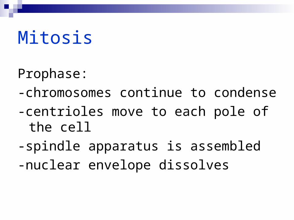
Mitosis
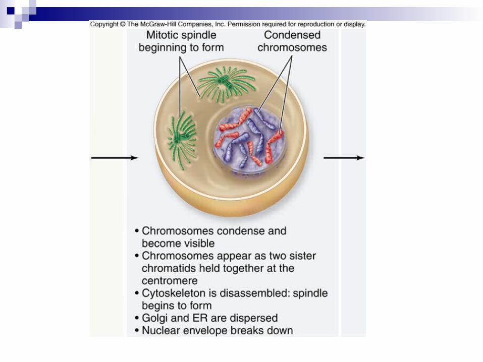
Prophase:-chromosomes continue to condense-centrioles move to each pole of the cell -spindle apparatus is assembled-nuclear envelope dissolves
Mitosis
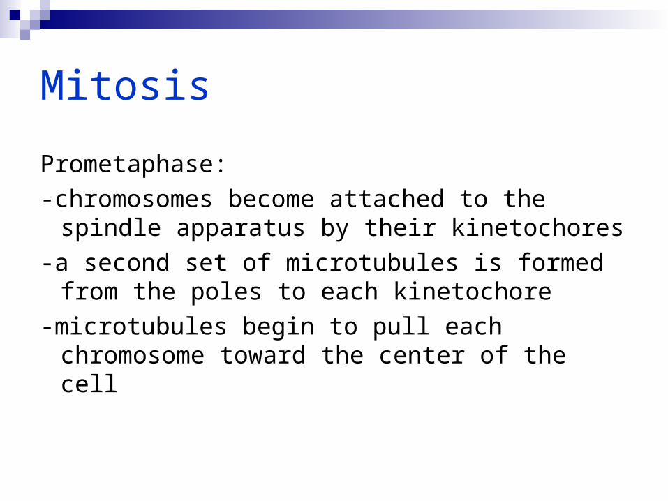
Prometaphase:-chromosomes become attached to the
spindle apparatus by their kinetochores-a second set of microtubules is formed from
the poles to each kinetochore-microtubules begin to pull each
chromosome toward the center of the cell
Mitosis
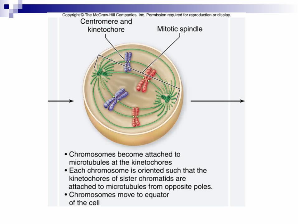
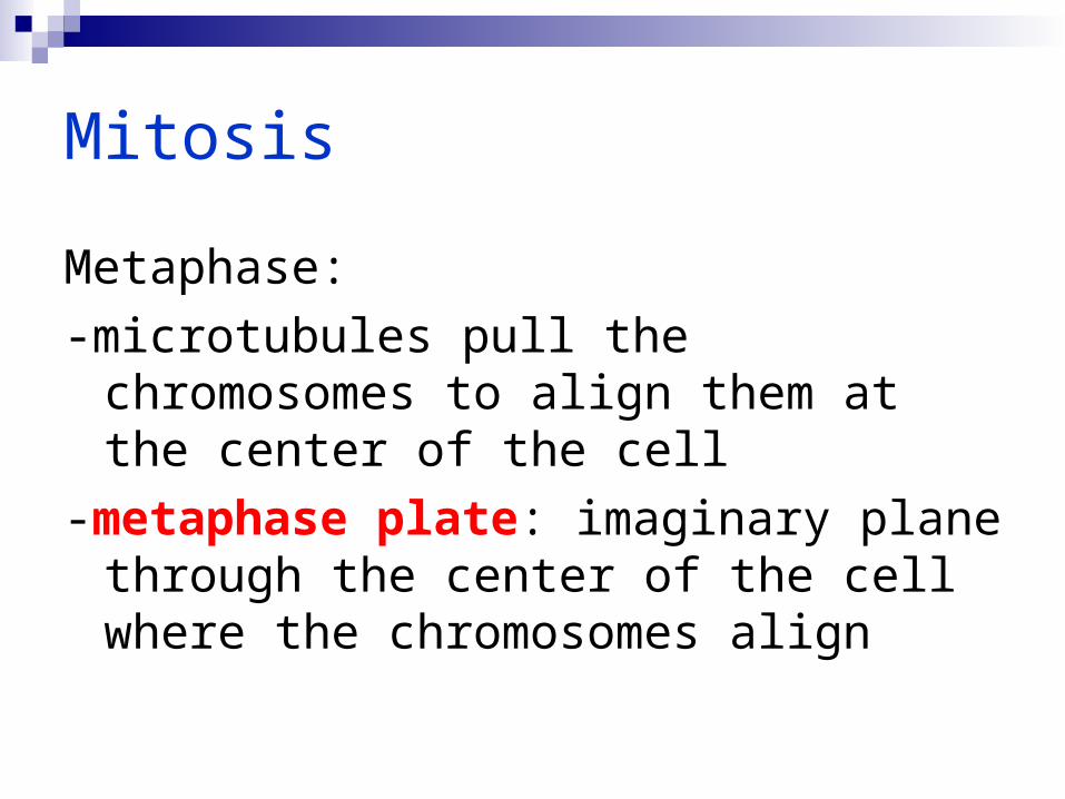
Metaphase:-microtubules pull the chromosomes to align
them at the center of the cell-metaphase plate: imaginary plane through
the center of the cell where the chromosomes align

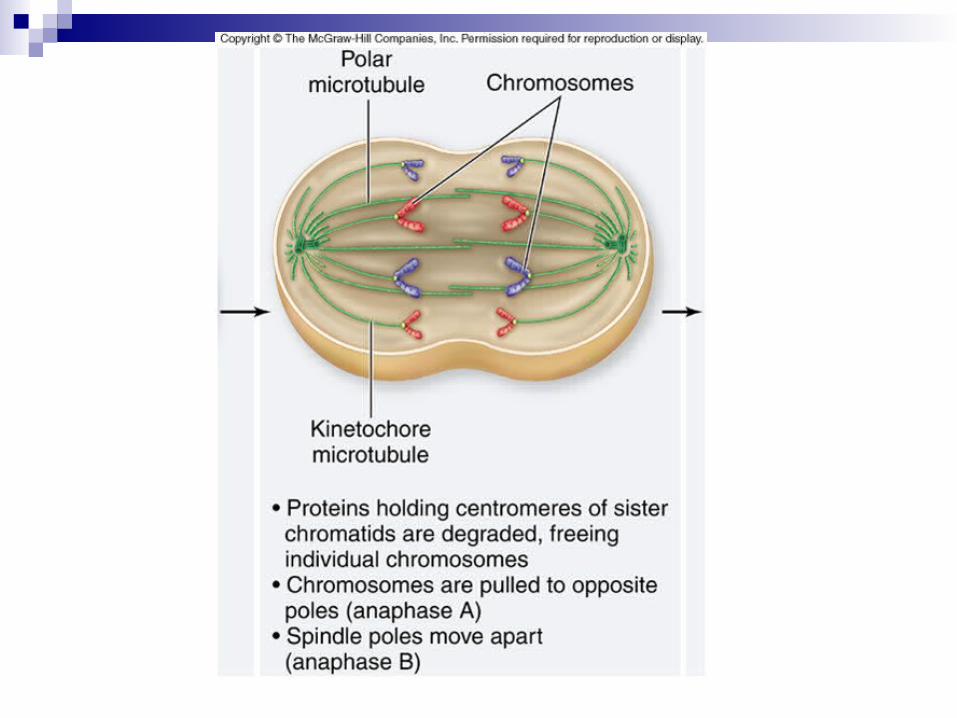
Mitosis
Anaphase:-removal of cohesin proteins causes the
centromeres to separate-microtubules pull sister chromatids toward the
poles-in anaphase A the kinetochores are pulled apart-in anaphase B the poles move apart
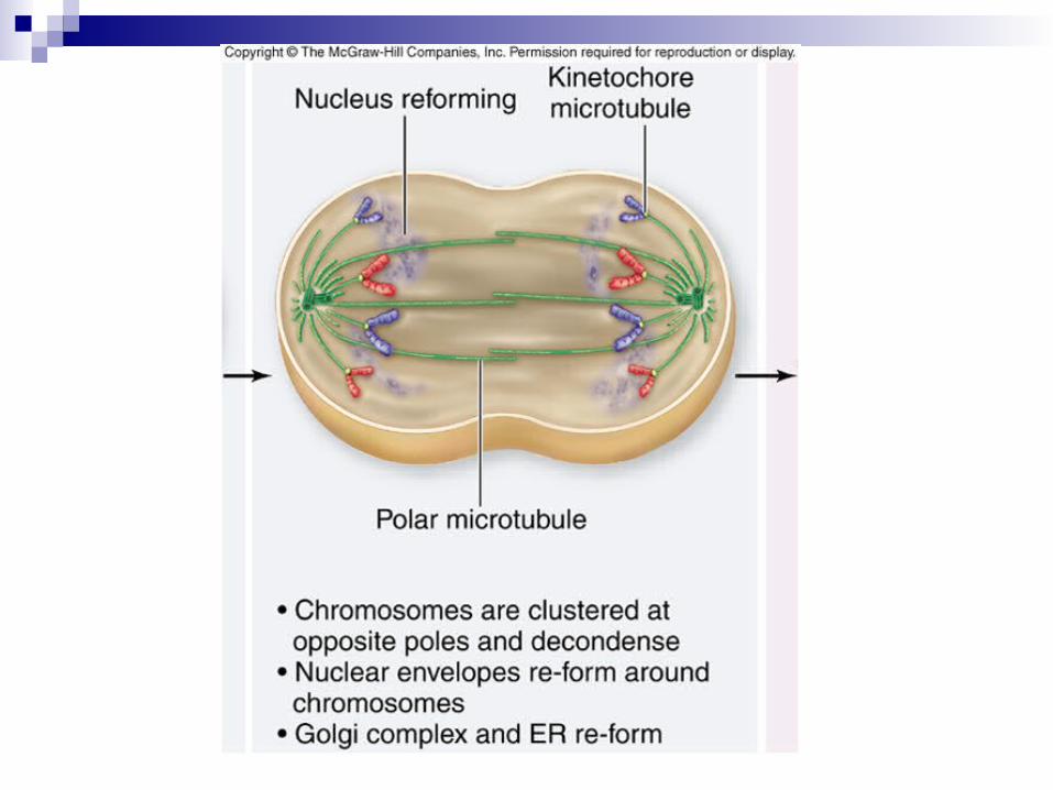
Mitosis
Telophase:-spindle apparatus disassembles-nuclear envelope forms around each set of
sister chromatids-chromosomes begin to uncoil-nucleolus reappears in each new nucleus
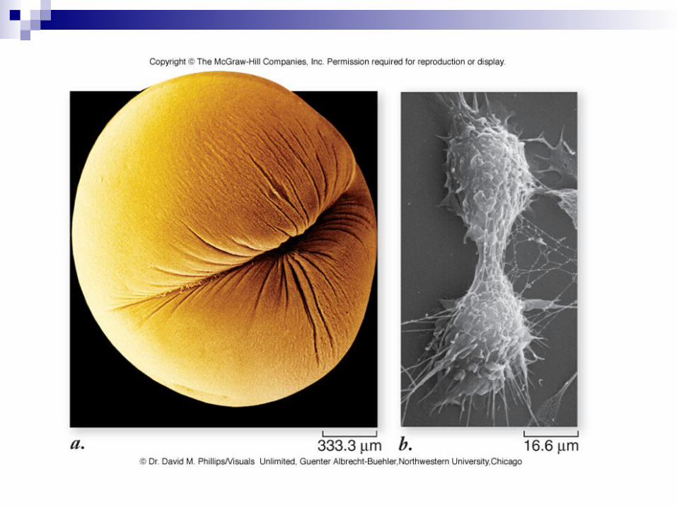
Cytokinesis Cytoplasmic Division Usually occurs between late anaphase
and end of telophase Two mechanisms
Cell plate formation (plants)Cleavage (animals)
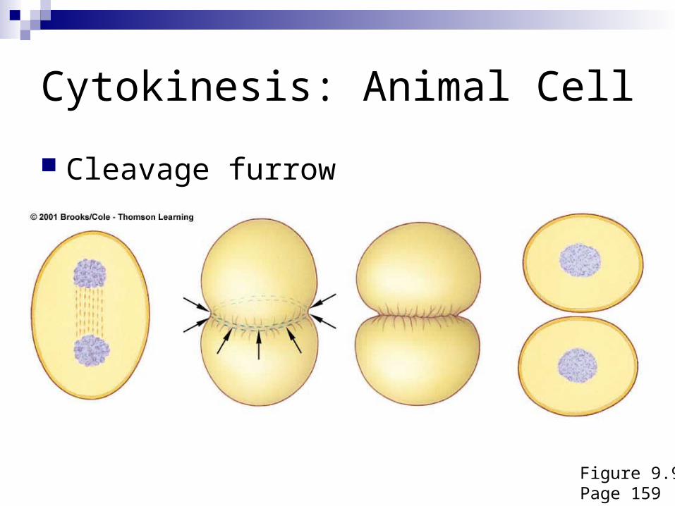
Cytokinesis: Animal Cell
Figure 9.9Page 159
Cleavage furrow
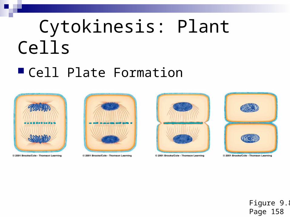
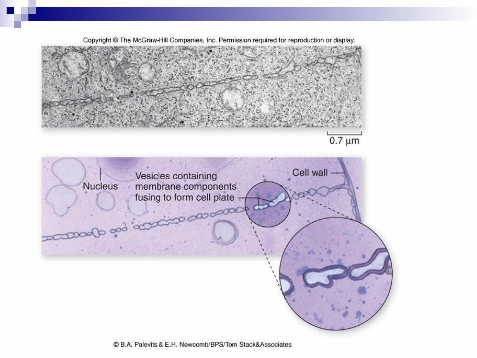
Cytokinesis: Plant Cells
Figure 9.8Page 158
Cell Plate Formation
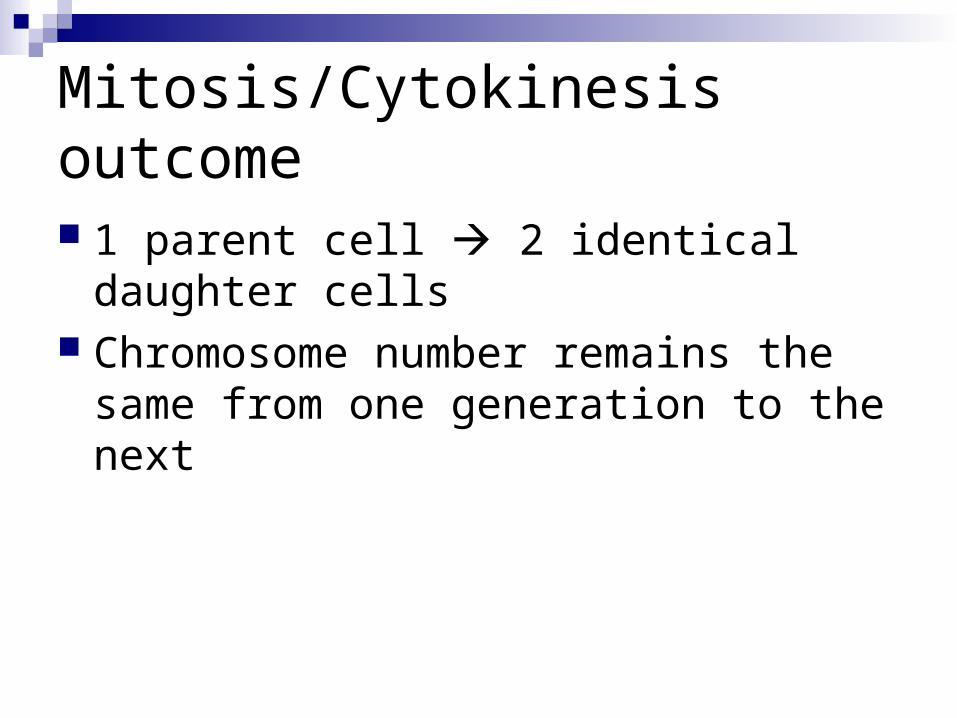
Mitosis/Cytokinesis outcome
1 parent cell 2 identical daughter cells Chromosome number remains the same
from one generation to the next
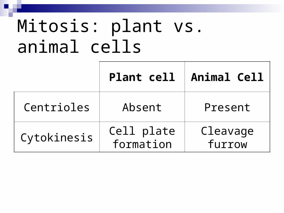
Mitosis: plant vs. animal cells
Plant cell Animal Cell
Centrioles Absent Present
Cytokinesis Cell plate formation
Cleavage furrow
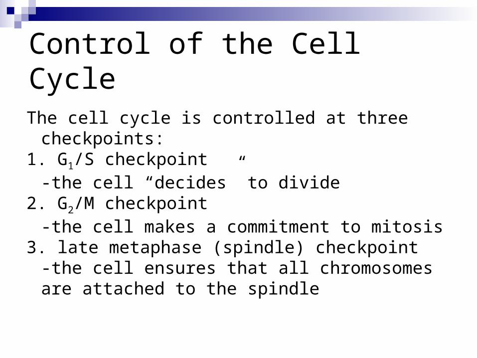
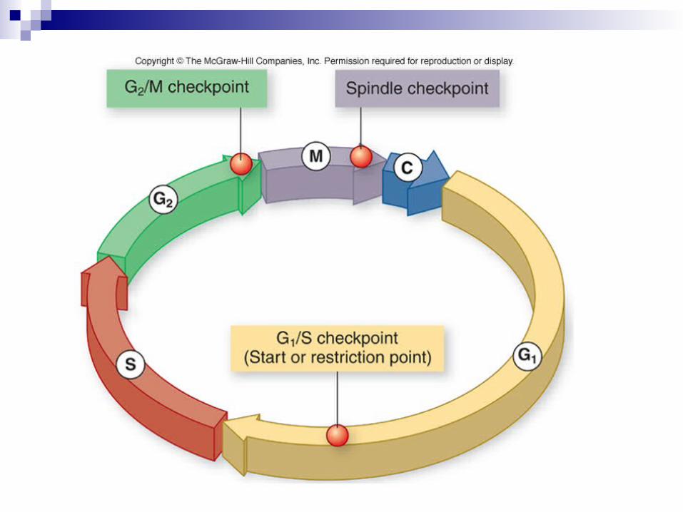
Control of the Cell Cycle
The cell cycle is controlled at three checkpoints:1. G1/S checkpoint
-the cell “decides” to divide2. G2/M checkpoint
-the cell makes a commitment to mitosis3. late metaphase (spindle) checkpoint
-the cell ensures that all chromosomes are attached to the spindle
Control of the Cell Cycle
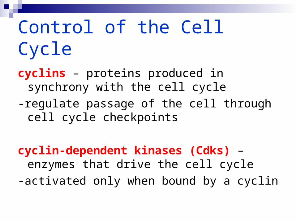
cyclins – proteins produced in synchrony with the cell cycle
-regulate passage of the cell through cell cycle checkpoints
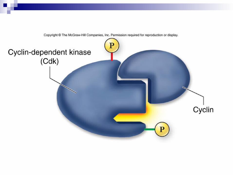
cyclin-dependent kinases (Cdks) – enzymes that drive the cell cycle
-activated only when bound by a cyclin
Control of the Cell Cycle
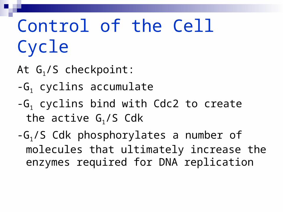
At G1/S checkpoint:
-G1 cyclins accumulate
-G1 cyclins bind with Cdc2 to create the active G1/S Cdk
-G1/S Cdk phosphorylates a number of molecules that ultimately increase the enzymes required for DNA replication
Control of the Cell CycleAt the spindle checkpoint:-the signal for anaphase to proceed is
transmitted through anaphase-promoting complex (APC)
-APC activates the proteins that remove the cohesin holding sister chromatids together
Control of the Cell Cycle
Growth factors:-can influence the cell cycle-trigger intracellular signaling systems-can override cellular controls that otherwise
inhibit cell divisionplatelet-derived growth factor (PDGF)
triggers cells to divide during wound healing
Control of the Cell Cycle
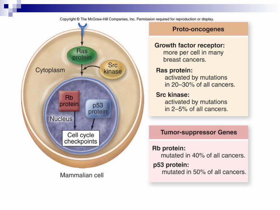
Cancer is a failure of cell cycle control.
Two kinds of genes can disturb the cell cycle when they are mutated:
1. tumor-suppressor genes2. proto-oncogenes
Control of the Cell Cycle
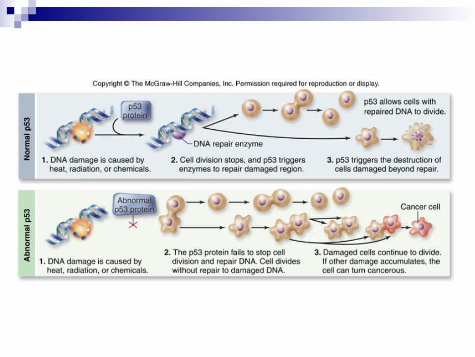
Tumor-suppressor genes:-prevent the development of many cells
containing mutations-for example, p53 halts cell division if
damaged DNA is detected-p53 is absent or damaged in many
cancerous cells
Control of the Cell Cycle
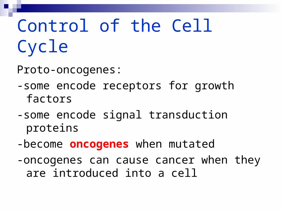
Proto-oncogenes:-some encode receptors for growth factors-some encode signal transduction proteins-become oncogenes when mutated-oncogenes can cause cancer when they
are introduced into a cell