Embed Size (px)
Citation preview
Food Structure Food Structure
Volume 4 Number 2 Article 3
1985
Changes in Typical Organelles in Developing Cotyledons of Changes in Typical Organelles in Developing Cotyledons of
Soybean Soybean
K. Saio
K. Kondo
T. Sugimoto
Follow this and additional works at: https://digitalcommons.usu.edu/foodmicrostructure
Part of the Food Science Commons
Recommended Citation Recommended Citation Saio, K.; Kondo, K.; and Sugimoto, T. (1985) "Changes in Typical Organelles in Developing Cotyledons of Soybean," Food Structure: Vol. 4 : No. 2 , Article 3. Available at: https://digitalcommons.usu.edu/foodmicrostructure/vol4/iss2/3
This Article is brought to you for free and open access by the Western Dairy Center at DigitalCommons@USU. It has been accepted for inclusion in Food Structure by an authorized administrator of DigitalCommons@USU. For more information, please contact [email protected].
FOOD MICROSTRUCTURE, Vol. 4 (1985), pp. 191-198 SEM Inc., AMF O'Hare (Chicago) , IL 60666-0507 U.S.A.
0730-5419/85$1.00+.05
CHANGES IN TYPICAL ORGANELLES IN DEVELOPING COTYLEDONS OF SOYBEAN
K. Saio, K. Kondo* and T. Sugimoto
National Food Research Institute, 2-l-2 Kannondai, Yatabe, Tsukuba, Ibaraki, Japan 305
*Nagano State Laboratory of Food Technology, 205 Nishibanba, Kurita, Nagano-shi, Japan 380
Abstract
Soybean cotyledonary cells harvested every 5-10 days at 15 to 60 days after flowering (OAF), were investigated by means of light and transmission electron microscopy. In the early developing stages (15-20 OAF) most of the cells were occupied by large, centrally located vacuoles while the cytoplasm was restricted to a thin layer against the cell wall and contained numerous ribosomes, mitochondria, plastids, small amounts of endoplasmic reticulum (ER) and minute lipid bodies. At about 25 OAF, spherical organelles which contained protein, lipid and sugar (PLS bodies), appeared and then increased in number and in size. Vacuoles had protein deposits lining the inner surfaces of the tonoplasts. At 30-35 OAF, the PLS bodies lost their characteristic ciccular shape because of invaginations containing cytoplasmic materials and small vacuoles . Such transformed PLS bodies, fragmented vacuoles and protein bodies became d ifficult to distinguish from eac h other. At this stage, the cytoplasm contained abundant rough ER with cisternae, dictyosomes , lipid bodies and plastids . At 35-40 OAF, protein bodies which vari ed in size and shape were observed in ce lls a long with lipid bodies and rough ER. At about 40 OAF the plastids reached maximum size and number and then most disappeared . In the final stage ( 55-60 OAF) , protein bodies became homogeneous in electron density with completion of protein accumulation.
Initial paper received April 16 1984 Manuscript received June 22 1985 Direct inquiries to K. Saio Telephone number: 81-2975-6-8051
Key words: Soybean, protein body, PLS body, lipid body, plastid, starch granule, endoplasmic reticulum, ripening, light microscope, transmission e l ec tron microscope
797
Introduction
Protein body ontology during seed development has been investigated by many researchers. There are excellent reviews dealing with this problem (Millerd, 1975, Oieckert and Oieckert, 1976, Pernollet, 1978). A theory that protein is biosynthesized by polysomes attached to the exterior surface of protein body membranes (Burr and Burr, 1976) seems to lack sufficient experimental evidence. According to Adler and MUntz ( 1983) two different mechanisms of protein body generation have been proposed: l) Protein bodies derive from the large vacuoles in which proteins are deposited ( Oieckert and Oieckert, 1976, Bergfeld et al., 1980). These vacuolAs seem to be transformed into protein bodies by fragmentation (Craig et al., 1980). 2) Protein bodies originate from the endoplasmic reticulum (ER), where the storage protein polypeptides are discharged from membrane-bound polysomes into the lumen of the rough ER (Higgins and Spencer, 1981, Weber et al. , 1980) . Most previous work on protein body formation was done using maize, wheat, mustard, castor bean, common bean, broad bean and cowpea. There is surprisingly little research on generation of protein bodies in soybeans (Bils and Howell, 1963, Yoo and Chrispeels, 1980, Norby et al., 1984), compared with the importance of soybeans as a protein resource. However, Kaizuma and coworkers (Kaizuma and Kasai, 1981, Sato et al., 1982) recently found organelles which contained nucleic acids (RNA and DNA), lipid and protein in the middle stage of development of soybean cotyledonary cel l s and have designated them RNA bodies or nucleo-globules. They also presented evidence that the isolated nucleo-globules could synthesize storage protein based on studies of incorporation of radioactive leucine (Kaizuma and Sato, 1983). In order to more clearly understand the process of formation of protein bodies in soybean cotyledons, we have conducted a detailed microscopic study, concentrating on special stages of development. Observations are also presented on other organelles that accompany protein body formation such as plastids, lipid bodies and rough ER.
K. Saio, K. Kondo and T. Sugimoto
Materials and Methods
Plant material Soybeans used were from the 1983 crop, var.
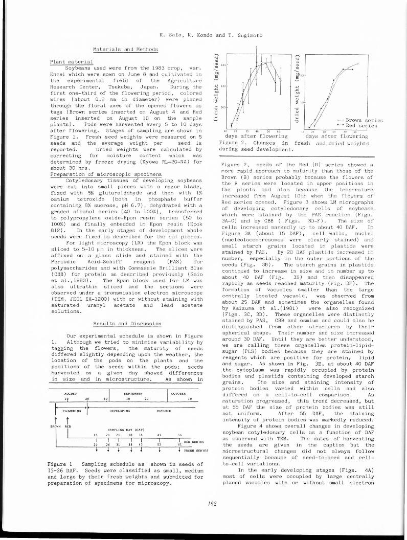
Enrei which were sown on June 8 and cultivated in the experimental field of the Agriculture Research Center, Tsukuba, Japan. During the first one-third of the flowering period, colored wires (about 0.2 mrn in diameter) were placed through the floral axes of the opened flowers as tags (Brown series inserted on August 4 and Red series inserted on August 10 on the sample plants). Pods were harvested every 5 to 10 days after flowering. Stages of sampling are shown in Figure 1. Fresh seed weights were measured on 5 seeds and the average weight per seed is reported. Dried weights were calculated by correcting for moisture content which was determined by freeze drying (Kyowa RL-20-NA) for about 30 hrs. Preparation of microscopic specimens
Cotyledonary tissues of developing soybeans were cut into small pieces with a razor blade, fixed with 5% glutara ldehyd e and then with 1% osmium tetroxide (both in phosphate buffer containing 5% sucrose, pH 6.7), dehydrated with a graded alcohol series (40 to 100%), transferred to polypropylene oxide-Epon resin series (50 to 100%) and finally embedded in Epon res in ( Epok 812). In the early stages of development whole seeds were fixed as described for the cut pieces .
For light microscopy (LM) the Epon block was sliced to 5-10 ~m in thickness. The slices were affixed on a glass slide and stained with the Periodic Acid-Schiff reagent (PAS) for polysaccharides and with Commassie Brilliant Blue (CBB) for protein as described previously (Saio et al., 1983). The Epon block used for LM was also ultrathin sliced and the sections were observed under a transmission electron microscope (TEM, JEOL EX-1200) with or without staining with saturated uranyl acetate and lead acetate solutions.
Results and Discussion
Our experimental schedule is shown in Figure 1. Although we tried to minimize variability by tagging the flowers, the maturity of seeds differed slightly depending upon the weather, the location of the pods on the plants and the positions of the seeds within the pods; seeds harvested on a given day showed differences in size and in microstructure. As shown in
Figure 1 Sampling schedule as shown in seeds of 15-26 DAF. Seeds were classified as small, medium and large by their fresh weights and submitted for preparation of specimens for microscopy.
,....... "d Q) Q) (/J -b[)
5
,....... "d Q) Q) (/J -! '""
..c: 200 (/J Q)
H lH
792
~ Brown series ------. Red series
30 so days af t er flowering days after flowering
Figure 2 . Changes in fresh and dried weights during seed development.
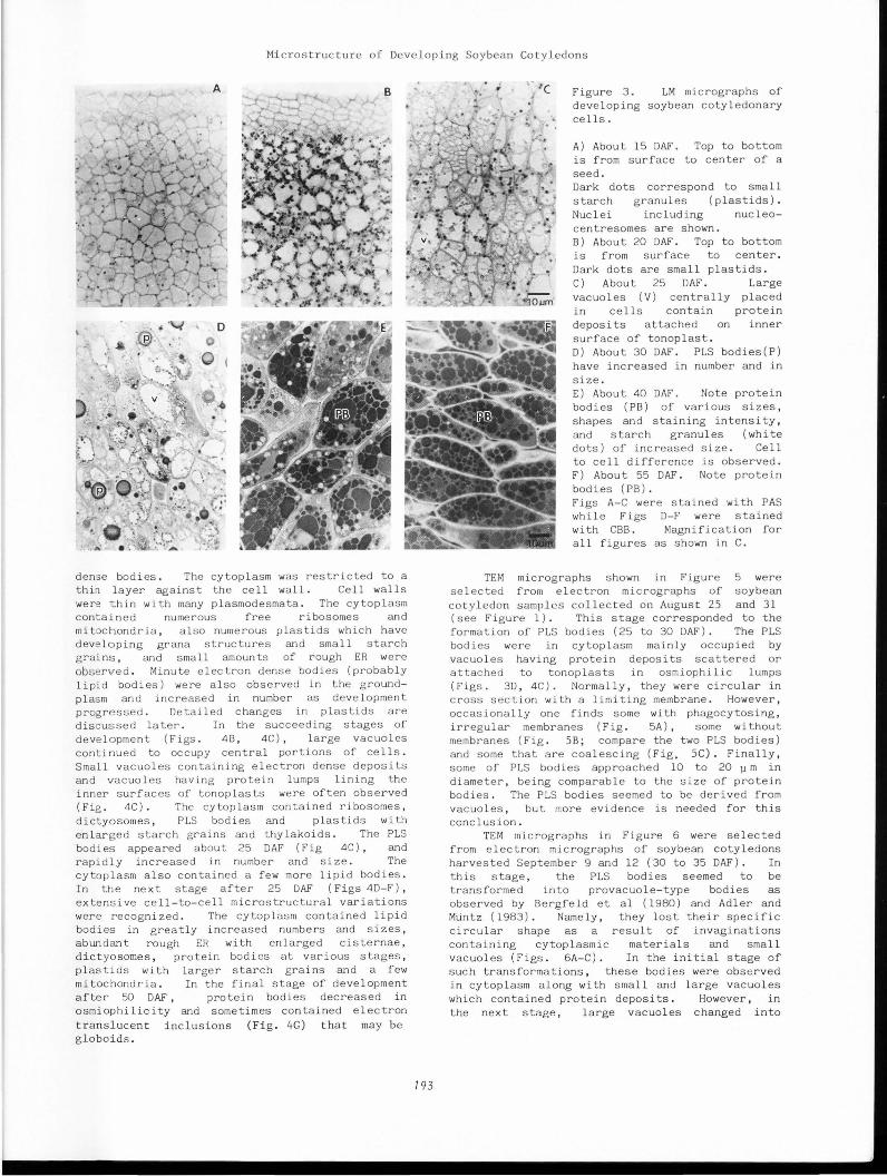
Figure 2 , seeds of the Red ( R) series showed a more rapid approach to maturity than those of the Brown (B) series probably because the flowers of the R series were located in upper positions in the plants and also because the temperature increased from August lOth when the flowers of Red series opened. Figure 3 shows LM micrographs of developing cotyledonary cells of soybeans which were stained by the PAS reaction (Figs. 3A-C) and by CBB ( Figs. 30-F) . The size of cells increased markedly up to about 40 OAF. In Figure 3A (about 15 OAF), cell walls, nuclei (nucleolocentresomes were c l early stained) and small starch grains located in plastids were stained by PAS. By 20 OAF plastids increased in number, especially in the outer portions of the seeds (Fig. 3B). The starch grains in plastids continued to increase in size and in number up to about 40 OAF (Fig. 3E) and then disappeared rapidly as seeds reached maturity (Fig. 3F). The formation of vacuoles smaller than the large centrally located vacuole, was observed from about 25 OAF and some times the organelles found by Kaizuma et al. ( 1981) were also recognized (Figs. 3C, 30) . These organelles were distinctly stained by PAS, CBB and osmium and could also be distinguished from other structures by their spherical shape . Their number and size increased around 30 OAF. Until they are better understood, we are calling these organelles protein-lipidsugar ( PLS) bodies because they are stained by reagents which are positive for protein, lipid and sugar. As shown in Fig. 3E, at about 40 OAF the cytoplasm was rapidly occupied by protein bodies and plastids containing developed starch grains. The size and staining intensity of protein bodies varied within cells and also differed on a cell-to-cell comparison. As maturation progressed, this trend decreased, but at 55 OAF the size of protein bodies was still not uniform. After 55 OAF, the staining intensity of protein bodies was markedly reduced.
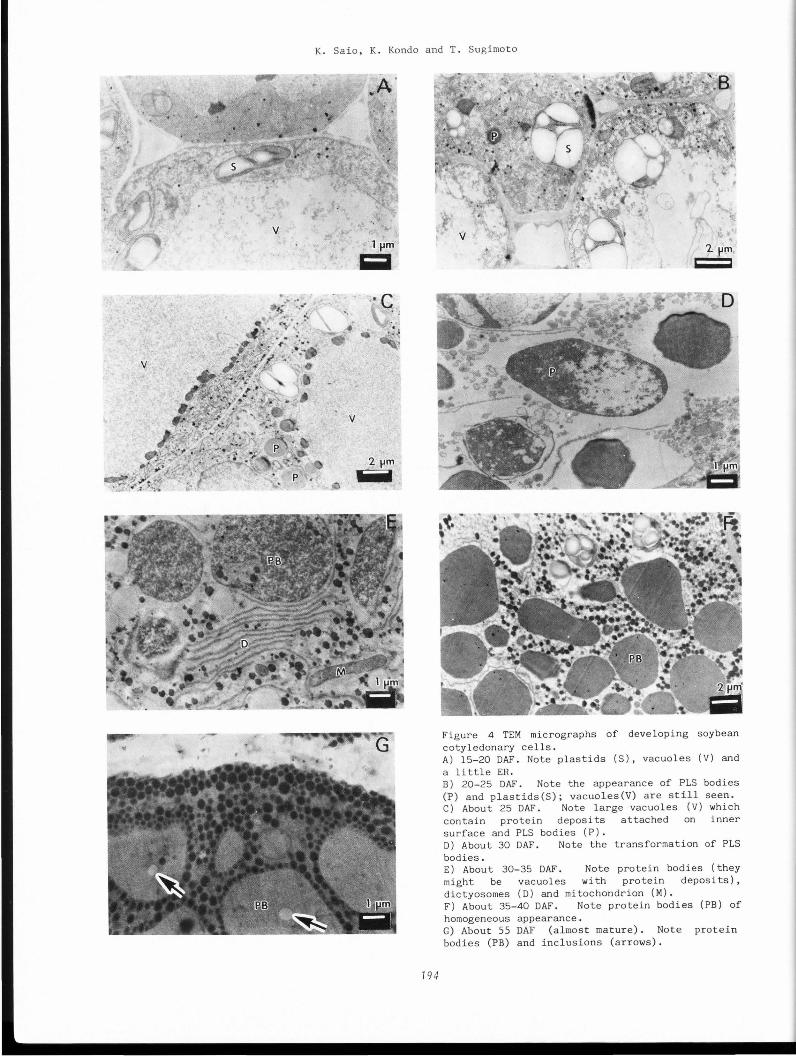
Figure 4 shows overall changes in developing soybean cotyledonary cells as a function of OAF as observed with TEM. The dates of harvesting the seeds are given in the caption but the microstructural changes did not always follow sequentially because of seed-to-seed and cellto-cell variations.
In the early developing stages (Figs. 4A) most of cells were occupied by large centrally placed vacuoles with or without small electron
Microstructure of Developing Soybean Cotyledons
dens e bodies. The cytoplasm was restricted to a thi:1 layer against the cell wall. Cell walls were ~hin with many plasmodesmata. The cytoplasm con t a i ned numerous free ribosomes and mitochondria, also numerous plas tids which have deve loping grana structures and small starch grains, and small amounts of rough ER were observed. Minute electron dense bodies (probably lipid bodies) were also observed in the groundplasm and increased in number as development progressed. Detailed changes in plastids are discus sed later. In the succeeding stages of development (Figs. 4B, 4C), large vacuoles continued to occupy central portions of cells. Small vacuoles containing electron dense deposits and vacuoles having protein lumps lining the inner surfaces of tonoplasts were often observed (Fig. 4C). The cytoplasm contained ribosomes, dictyosomes, PLS bodies and plastids with enlarged starch grains and thylakoids. The PLS bodies appeared about 25 OAF (Fig 4C), and rapidly increased in number and size. The cytoplasm also contained a few more lipid bodies. In the next stage after 25 OAF (Figs 40-F), extensive cell-to-cell microstructural variations were recognized . The cytoplasm contained lipid bodies in greatly increased numbers and sizes, abundant rough ER with enlarged cisternae, dictyosomes, protein bodies at various stages, plastids with larger starch grains and a few mitochondria. In the final stage of development after 50 OAF, protein bodies decreased in osmiophilicity and sometimes contained electron translucent inclusions (Fig. 4G) that may be globoids .
793
Figure 3. LM micrographs of developing soybean cotyledonary cells.
A) About 15 OAF. Top to bottom is from surface to center of a seed. Dark dots correspond to small starch granules (plastids). Nuclei including nucleocentresomes are shown. B) About 20 OAF. Top to bottom is from surface to center. Dark dots are small plastids. C) About 25 OAF. Large vacuoles (V) centrally placed in cells contain protein deposits attached on inner surface of tonoplast. D) About 30 OAF. PLS bodies(P) have increased in number and in size. E) About 40 OAF. Note protein bodies (PB) of various sizes, shapes and staining intensity, and starch granules (white dots) of increased size. Cell to cell difference is observed. F) About 55 OAF. Note protein bodies ( PB) . Figs A-C were stained with PAS while Figs 0-F were stained with CBB. Magnification for all figures as shown in C.
TEM micrographs shown in Figure 5 were selected from electron micrographs of soybean cotyledon samples collected on August 25 and 31 (see Figure l). This stage corresponded to the formation of PLS bodies (25 to 30 OAF). The PLS bodies were in cytoplasm mainly occupied by vacuoles having protein deposits scattered or attached to tonoplasts in osmiophilic lumps (Figs. 30, 4C). Normally, they were circular in cross section with a limiting membrane. However, occasionally one finds some with phagocytosing, irregular membranes (Fig. 5A), some without membranes (Fig. SB; compare the two PLS bodies) and some that are coalescing (Fig. SC). Finally, some of PLS bodies approached 10 to 20 Jl m in diameter, being comparable to the size of protein bodies. The PLS bodies seemed to be derived from vacuoles, but more evidence is needed for this conclusion.
TEM micrographs in Figure 6 were selected from electron micrographs of soybean cotyledons harvested September 9 and 12 (30 to 35 OAF). In this stage, the PLS bodies seemed to be transformed into provacuole-type bodies as observed by Bergfeld et al ( 1980) and Adler and Muntz (1983). Namely, they lost their specific circular shape as a result of invaginations containing cytoplasmic materials and small vacuoles (Figs. 6A-C). In the initial stage of such transformations, these bodies were observed in cytoplasm along with small and large vacuoles which contained protein deposits. However, in the next stage, large vacuoles changed into
K. Saio, K. Kondo and T. Sugimoto
2 pm liiil
794
Figure 4 TEM micrographs of developing soybean cotyledonary cells. A) 15-20 OAF. Note plastids (S), vacuoles (V) and a little ER. B) 20-25 OAF. Note the appearance of PLS bodies (P) and plastids(S); vacuoles(V) are still seen. C) About 25 OAF. Note large vacuoles (V) which contain protein deposits attached on inner surface and PLS bodies (P). D) About 30 OAF. Note the transformation of PLS bodies. E) About 30-35 OAF. Note protein bodies (they might be vacuoles with protein deposits), dictyosomes (D) and mitochondrion (M). F) About 35-40 OAF. Note protein bodies (PB) of homogeneous appearance. G) About 55 OAF (almost mature). Note protein bodies (PB) and inclusions (arrows).

Microstructure of Developing Soybean Cotyledons
795
Figure 5 TEM micrographs of PLS body formation A) PLS body with phagosytosing irregular membrane. B) Right PLS body has limiting membrane but left one does not. C) Coalescence of PLS bodies and protein deposits. Bar of each micrograph equals 500 nm. Figure 6. TEM micrographs of PLS body transformation. (below) A) Note PLS bodies which lose their circular shape and become heterogenous. B) PLS body invaginates cytoplasmic materials. Note rough ER with enlarged cisternae. C) PLS body invaginates. D) Bodies cannot be distinguished from vacuoles with protein deposits and developing protein bodies. Magnification for all figures as in Fig. 6D.
K. Saio, K. Kondo and T. Sugimoto
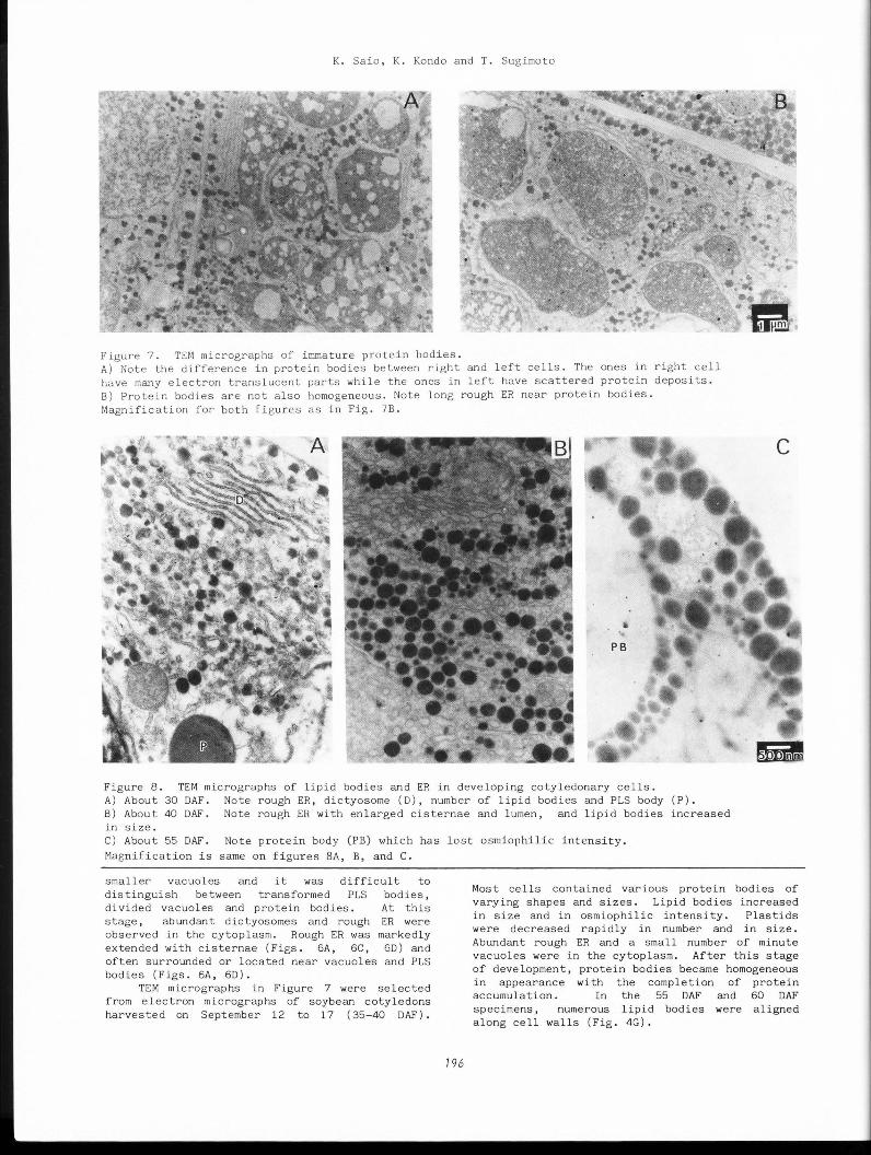
Figure 7. TD~ micrographs of irnrna ture protein bodies. A) Note the difference in protein bodies between right and left cel l s. The ones in right cell have many electron translucent parts while the ones in l eft have scattered protein deposits. B) Protein bodies are not also homogeneous. Note long rough ER near protein bodies. Magnification for both figu r es as in Fig. 7B.
Figure 8. TEM micrographs of lipid bodies and ER in developing cotyledonary cells. A) About 30 DAF. Note roughER, dictyosome (D), number of lipid bodies and PLS body (P). B) Abou t 40 DAF. Note roughER with enlarged cisternae and lumen, and lipid bodies increased in size. C) About 55 DAF. Note protein body (PB) which has lost osmiophilic intensity.
Magnification is same on figures SA, B, and C.
smaller vacuoles and it was difficult to distinguish between transformed PLS bodies, divided vacuoles and protein bodies. At this stage, abundant dictyosomes and rough ER were observed in the cytoplasm. Rough ER was markedly extended with cisternae (Figs. 6A, 6C, 6D) and often surrounded or located near vacuoles and PLS bodies (Figs. 6A, 6D).
TEM micrographs in Figure 7 were selected from electron micrographs of soybean cotyledons harvested on September 12 to 17 ( 35-40 DAF).
196
Most cells contained various protein bodies of varying shapes and sizes. Lipid bodies increased in size and in osmiophilic intensity. Plastids were decreased rapidly in number and in size. Abundant rough ER and a small number of minute vacuoles were in the cytoplasm. After this stage of development, protein bodies became homogeneous in appearance with the completion of protein accumulation. In the 55 DAF and 60 DAF specimens, numerous lipid bodies were aligned along cell walls (Fig. 4G).
Microstructure of Developing Soybean Cotyledons
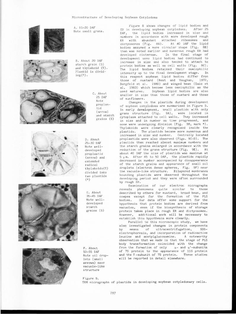
A. 15-20 OAF Note small grana.
B. About 20 OAF starch grain (S) and thylakoid (T) . Plastid is dividing(;'<).
C. About 25 OAF Note prop lastid (arrow) and starch grains (S)
D. About 25-30 OAF Note welldeveloped prop las tid (arrow) and extended radical thylakoids(T) divided into two plastids (*)
E. About 35-45 OAF Note welldeveloped starch grains (S)
F. About 50-55 OAF Note oil droplets (small arrows) near vacuole-like structure
Figure 9.
Fig ure 8 shows changes of lipid bodies and ER in developing soybean cotyledons. After 25 OAF , the lipid bodies increased in size and numbers in accordance with more developed rough ER with abundant attached ribosomes and dictyosomes (Fig. 8 A). At 40 OAF the lipid bodies assumed a more circular shape (Fig. 8B) than was noted earlier and numerous rough ER had developed cisternae. In the final stage of development some lipid bodies had continued to increase in size and also tended to attach to protein bodies as we ll as cell walls (Fig. 8C) . The lipid bodies retained their osmiophilic intensity up to the final development stage. In t his respect soybean lipid bodies differ from those of mustard (Rest and Vaughan, 1972, Bergfeld et al. 1980) and winged bean ( Saio et al , 1983) which become less osmiophilic as the seed matures. Soybean lipid bodies are also smaller in size than those of mustard and those of sunflowers .
Changes in the plastids during development of soybean cotyledons are su~marized in Figure 9. In early development, small plastids with only grana structure (Fig. 9A), were located in cytoplasm attached to cell walls. They increased in size and in number as time progressed, and some were undergoing division (Fig. 9B, mark*). Thylakoids were clearly recognized inside the plastids. The plastids became more numerous and increased in size and number. Centrally located proplastids were also observed (Figs. 9C-D). The plastids then reached almost maximum numbers and the starch grains enlarged in accordance with the reduction of the grana structure (Fig. 9E). At about 40 OAF the size of plastids was maximum at 5 JJ m. After 45 to 50 OAF, the plastids rapidly decreased in number accompanied by disappearance of the starch grains and appearance of small oil droplets (electron dense spheres, Fig. 9F) near the vacuole-like structure. Bilayered membranes bounding plastids were observed throughout the developing period and they were often surrounded by rough ER.
Examination of our electron micrographs reveals phenomena quite similar to those described by others for mustard, broad bean, and cowpea except for the formation of the PLS bodies. Our data offer some support for the hypothesis that protein bodies are derived from vacuoles, even if the biosynthesis of storage protein takes place in rough ER and dictyosomes. However, additional work will be necessary to establish this hypothesis more clearly.
Para llel to this microscopic study, we have also investigated changes in protein components by means of ultracentrifugation, SDSelectrophoresis, and incorporation of radioactive leucine and acetylglucosamine. A noteworthy observation that we made is that the stage of PLS body transformation coincided with the change from the formation of only a- and a'-subunits of 7S protein to the appearance of llS protein and the S -subunit of 7S protein . These studies will be reported in detail elsewhere.
TEM micrographs of plastids in developing soybean cotyledonary cells.
797
K. Saio, K. Kondo and T. Sugimoto
Acknowledgement
The authors thank Dr. I. Watanabe, Agriculture Forestry and Fi s heri e s Research Council Secre tari a t, for hi s c ultivation of the soybeans and a lso for hi s a dvice on crop p roduction.
Re f e rences
1) Adler,K. and Mi.intz,K. (1983) Origin and development of protein bodies in cotyledons of Vicia faba Plan t a 157, 401-410. 2) Bergfeld ,R., Ki.ihnl,T. and Schpfer,P. (1980) Formation of pro t e in storage bodies during embryogenesis i n c otyledons of Sinapis alba L. Planta 148, 146-156. 3 ) Bils,R.F. and Howell,R.W. (1963 ). Biochemical and cytological changes in developing soybean cotyledons , Crop Sci. 3, 304-308. 4) Burr,B. and Burr,F.A. (1976) Ze in synthesis in maize endos perm by polyribosomes attached to protein bodies. Proc. Nat. Acad. Sci. USA 73, 515-519. 5 ) Craig , S ., Goodchild,D.J. and ihller,C. (1980). S tructural aspects of protein ac cumulation in developing pea co tyledons II. Three -dimensional reconstructions of vacuoles and protein bodies from serial sections. Aust. J. Plant Physiol. 7, 329-337. 6) Dieckert J.W. and Dieckert t-1.Y.C. (1976) The chemistry and cell biology of the vacuolar proteins of seeds. J. Food Sci. 41, 475-482. 7) Higgins,T.J.V. and Spencer,D. (1981) Precursor forms of pea vicilin s ubunits. Modification by microsomal membranes during cell-free t ranslation. Plant Physiol. 67, 205-211. 8) Kaizuma,N. and Kasai ,A. ( 1981). On the morphology of protein body and spherosome in soybean cotyledon cell. Japan J. Breed. 31, (Suppl. l) 222-223. 9) Kaizuma,N. and Sato,Y. (1983) Genetical studies on protein body development in soybean cotyledon cells. 3. Protein molecule species 3ontained in nucleo-globules and incorporation of H-leucine into them. Japan J. Breed. 33,
(Suppl. 1) 320-321. 10) Millerd,A. (1975) Biochemistry of legume seed proteins. Ann. Rev. Plant Physiol. 26, 53-72. 11) Norby,S.W., Adams, C.A. and Rinne,R.W. (1984) An ultrastructural study of soybean seed development. University of Illinois at UrbanaChampaign,Department of Agronomy,Urbana,IL.p.36. 12) Pernollet, J.-C. (1978) Protein bodies of seeds: Ultrastructure, biochemistry, biosynthesis and degradation. Phytochemistry 17, 1473-1480. 13) Rest,J.A. and Vaughan,J.G. (1972). The development of protein and oil bodies in the seed of Sinapis alba L. Planta 105, 245-262. 14) Saio,K.-,--Nakano,Y. and Uemoto,S., (1983) Microstructure of winged beans. Food Microstructure 2, 175-181. 15) Sato, Y., Kaizuma, N and Kitamura K. ( 1982). Genetical studies on protein body development in soybean cotyledon cells. 2. Isolation and characterization of RNA-body. Japan J. Breed. 32 (Suppl. 1) 94-95.
198
16) Weber,E. and Neumann,D.(1980) Protein bodies, storage organelles in plant seeds. Biochem, Physiol. Pflanz. 175, 279-306. 17) Yoo,B.Y. and Chrispeels,M.J. (1980). The or igin of protein bodies in developing soybean cotyledons. Protoplasma 103, 201-204.
Discussion with Reviewers
W.J. Wolf: Several of the micrographs in Figure 6 suggest degradation of the PLS bodies rather than mere invagination of the membrane.In Fig.6B, for example, part of the membrane is clearly missing and the PLS body has a ragged gouge in this area. Has digestion caused this partial degradation? Authors: It is very difficult to explain total degradation and transformation processes of PLS bodies, since we cannot observe the changes of a PLS body under TEM with passing time. We do not think that the transformation is merely invagination of the membrane. Actually, PLS bodies which show partial degradation cannot easily be distinguished from protein bodies which do not complete protein accumulation.
D.J. Gallant: Figure 9 shows the evolution from the proplastid and young amyloplast to the chloroamyloplast and/or to the chromoplast, that is the normal evolution of the chloroplast or the amyloplast in many colored plant organs. In the case of your samples, are such structures related to the brown or the red series of the seeds? Did you find such organelles in the cotyledon cells of non pigmented soybean varieties? Authors: Brown or red series refers to the color of the tags which were placed through the floral axes. The variety used, Enrei, is an ordinary soybean variety which is greenish in developing but changes to yellowish after maturation. These structures were already found in previous papers (ref. 11, for example) . We used the word plastid, which encompasses organologically parallel organs such as chloroplasts, amyloplasts etc.
L.H. Bragg: What is the significance of the alignment of protein bodies and lipid bodies along the surface of other organelles and along the inner surface of the cell wall in the development process? Authors: In a paper by Wolf and Baker (Scanning electron microscopy of soybeans and soybean protein products. Scanning Electron Microsc. 1980; III: 621-634.), SEM micrographs of protein bodies in mature soybeans show many depressions on the surface of the membrane which look like imprints caused by the alignment of lipid bodies. The alignment may not relate to development mechanisms but may result from dehydration during maturation.