Embed Size (px)
Citation preview
Plant Physiol. (1982) 69, 205-2090032-0889/82/69/0205/05/$00.50/0
Changes in Two Ribonuclease Isozymes During Rust Infection ofFlax Cotyledons'
Received for publication March 25, 1981 and in revised form August 8, 1981
BENJAMIN C. S. SUTTON AND MICHAEL SHAWThe University of British Columbia, 2075 Westbrook Place, Vancouver, British Columbia, Canada V6T I W5
ABSTRACT
A ribonuclease fraction previously purified from flax by gel filtration wasfurther resolved into two components by hydroxyl apatite chromatography.These were homogeneous with respect to electrophoresis and isoelectricfocusing. Both enzymes are of RNase I type but differ in substratespecificity, kinetic properties, pH response, and isoelectric point.The two RNase isozymes show consistent properties when extracted
from variety Bison (susceptible) or variety Bombay (resistant) with orwithout infection with race 3 of flax rust. The relative amounts of theseisozymes change markedly during infection. These observations provide anexplanation for the apparent qualitative changes in RNase noted previ-ously. Diferences between susceptible and resistant reactions in the earlystages of disease are discussed.
It has been demonstrated previously (14) that major quantitativeand qualitative changes in the RNase activity of flax cotyledonsoccur after inoculation with uredospores of flax rust (Melampsoralini (Ehrenb.) Lev. race 3). A bimodal increase in the specificactivity of a cell free extract was demonstrated in susceptible flax(Linum usitatissimum L. var. Bison). Only the early increase (2-4days after inoculation) occurs in the resistant variety of flax (L.usitatissimum L. var. Bombay). The major RNase component fromhealthy and inoculated cotyledons of Bison flax was purified (5)to homogeneity with respect to Sephadex G100 gel filtrationduring the late phase (8 days after inoculation). Qualitativechanges were demonstrated in this fraction in accordance withchanges observed in the crude extracts at this stage. Among thesewere differences in substrate specificity, pH response, thermalstability, Km and V... Such changes in properties could not beaccounted for on the basis of contribution from the rust mycelium;mycelial RNases had been characterized previously (4).The initial objective of the present study was to determine the
molecular basis of these changes. Here we show that two RNaseswith differing properties are present within the major RNasefraction. The qualitative differences observed in the late phase (5,14) are accounted for on the basis of increases in one of theseenzymes. Enhancement of the other enzyme occurred in the earlyphase in both resistant (var. Bombay) and susceptible (var. Bison)combinations. However, these early changes ceased at 4 days insusceptible flax but persisted in resistant flax at 4 days.
MATERIALS AND METHODS
Plant Materials. Flax plants of variety Bison and Bombay(susceptible and resistant to Melampsora lini race 3, respectively)
'Supported by grants from the Natural Science and Engineering Re-search Council of Canada (to M. S.).
were grown in growth chambers at an irradiance of 230 AE m-2s-for 18 h daily at 25°C with 20°C nights. One week after the seedswere sown, plants were sprayed with a dilute suspension ofuredospores in water (approximately 50 mg/100 ml). The plantswere placed in a growth chamber at 10°C in the dark for 18 h ina humid atmosphere. The control plants were sprayed with wateralone but otherwise treated identically. This procedure gave riseto about 10 pustules per cotyledon scored 8 days after inoculation.
Extraction and Purification of RNase. Approximately 50 gcotyledons were harvested at various stages after inoculation;thereafter, all operations were carried out at 0 to 4°C. Thecotyledons were homogenized in an Osterizer with 50 g polyvi-nylpolypyrrolidone (PVpP, Sigma) and 350 ml 40 mM K-phos-phate (pH 6.7). The extract was then purified by the followingsteps, adapted from Chakravorty et al. (5).
Centrifugation. The homogenate was centrifuged at 10,000g for30 min to remove cell debris.
Acidfifcation topH 5.0. The supernatant from step I was adjustedto pH 5.0 with 1 N HCI and allowed to stand for 2 h. The resultingsuspension was centrifuged as before and the supernatant savedfor further purification.Ammonium Sulfate Fractionation. The solution from step II was
brought to 50%o saturation by adding solid (NH4)2SO4 slowly withstirring and was allowed to stand for 30 min. The precipitate wasremoved by centrifugation and the supernatant solution wasbrought to 80%1o saturation as before. After 30 min, the precipitateformed was collected by centrifugation, dissolved in a smallvolume of 5 mm phosphate buffer (pH 6.7) and dialyzed overnightagainst 1 liter of the same buffer.
Hydroxyl Apatite Chromatography. A small volume of solutionfrom step III, containing about 5 mg protein, was applied to a 3x 2 cm column packed with hydroxyl apatite (Bio-Rad BioGelHTP) equilibrated with 5 mm phosphate buffer (pH 6.7). Elutionwas routinely carried out in a stepwise manner with two columnvolumes of 5, 60, and 120 mm phosphate buffer consecutively.This procedure was developed on the basis of gradient elutionstudies.
G100 Sephadex Gel Filtration. For detailed studies of properties,the enzymes which were recovered with 60 and 120 mm buffer instep IV (termed P1 and P2, respectively) were further purified bygel filtration. A small volume (about 3 ml) of enzyme solutionwas layered onto a 1.5 x 30 cm GIOO Sephadex column equili-brated with 40 mm phosphate buffer (pH 6.7); elution was carriedout with the same buffer.
Electrophoretic Analysis. The various ribonucleases present ateach stage of purification were visualized by polynucleotide-acryl-amide gel electrophoresis according to Van Loon (18). Aliquots offractions containing 2 to 100 jig protein were applied to 7.5%polyacrylamide gels containing 0.05% polyuridylic acid (poly U;Calbiochem, K salt, A grade) which was included within the gelmatrix. After electrophoresis gels were stained overnight withpyronin Y in 7% acetic acid. In this way enzymes capable of
205 www.plantphysiol.orgon May 10, 2018 - Published by Downloaded from Copyright © 1982 American Society of Plant Biologists. All rights reserved.

Plant Physiol. Vol. 69, 1982
hydrolyzing poly U are visualized as clear bands after destainingin 7% acetic acid. Comparative experiments were run without polyU but instead incubating gels with RNA according to the methodof Wilson (20).Column Isoelectric Focusing. The homogeneity of purified
preparations was assessed by column isoelectric focusing in anLKB 110-ml column containing Ampholine (LKB 8100) (pH 2.5-6.0) by the method outlined in the Ampholine LKB instructionmanual.Enzyme Assays. RNase was assayed routinely with yeast RNA
(BDH, Poole, England) which had been reprecipitated in 3 vol95% ethanol, washed and dialyzed exhaustively against EDTAand subsequently against distilled H20. Otherwise, the RNaseassay was carried out as described by Chakravorty et al. (5). Oneunit of activity is defined as the amount of enzyme yielding anincrease of 1.0 Am0 unit of acid soluble product. Substrate pref-erence was determined using tritiated homoribopolymers of uri-dylic, adenylic, and cytidylic acid (poly U, poly A, poly C,respectively, Miles Laboratories Inc., Elkhart, IN) as describedpreviously (5, 14).DNase and phosphodiesterase were determined using heat de-
natured calf thymus DNA and Ca bis-P-nitrophenyl phosphaterespectively as substrates (5, 14).
Analysis of Hydrolysis Products. The mononucleotides releasedin time course studies under standard assay conditions wereanalyzed by TLC by the method outlined by Tang and Maretzki(16). Incubation mixtures were applied to cellulose thin-layerplates (0.2 mm thickness) which were run in two separate solvents:Solvent I, (NH4)2SO4 (45 g), water (20 ml), isopropanol (2 ml);Solvent II, (NH4)2CO3 (0.96 g), water (25 ml), isopropanol (75 ml).Spots were observed under UV light and compared with 2'-3'cyclic ribonucleotide standards (2'-3' cUMP, 2'-3' cAMP, 2'-3'cCMP, 2'-3' cGMP; Sigma).
Protein Estimation. Protein concentration was estimated by themethod of Lowry et al. (12) using BSA as a standard.
RESULTS
Purification of RNase from Flax Cotyledons.Specric Activity and Yields. Purification was similar to that
obtained previously by Chakravorty et al. (5). Total units andhence specific activity were lower from all sources than thoseobtained by the earlier workers. This is likely to be due to thedifferent source of yeast RNA used in this present work. Typicalpurifications are presented in Table I. Purification and yield inthe variety of Bombay were similar. The higher specific activity in
the older plants was largely due to apparent differences in protein,the total enzyme units on a fresh weight basis being similar.Infection did not significantly effect total protein however.The consistently lower yield after pH5 precipitation (step II,
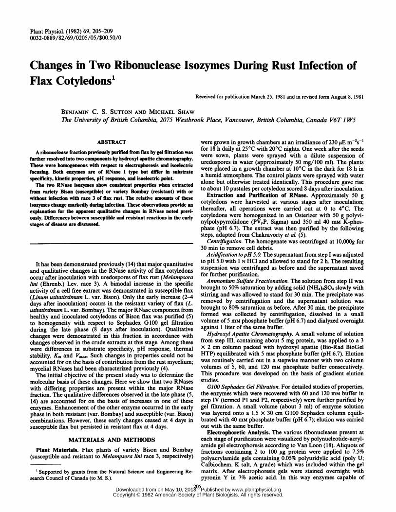
Table I) in younger plants was investigated by Sephadex G100gel filtration of cell free hotnogenates. The patterns from 1- and2-week-old flax (var. Bison) are presented in Figure 1. Theseresults show that an RNase component which elutes with the voidvolume (fractions 16 to 24) is more prevalent in the youngerplants. Gel filtration of the pH 5 supernatant demonstrates thatmost of this component is lost during step II. Unpublished datahave revealed that when pH 5 precipitated material is subjectedto G100 Sephadex gel fitration at pH 6.7 the RNase activity nowelutes as a major peak within the included volume (fractions 30 to44). Thus, it is indicated that some RNase activity becomes weaklybound to large molecules or particulate matter during pH 5precipitation; this phenomenon is enhanced in extracts fromyounger cotyledons.
Hydroxyl Apatite Chromatography. Gradient elution studiesusing gradients from 5 to 200 mM phosphate buffer (pH 6.7)revealed that a small amount of RNase activity was eluted at 5mm; two major components were eluted at 35 and 100 mm (termedP1 and P2, respectively). The amount of activity eluted at 5 mMwas highly inconsistent and this component appeared to be very
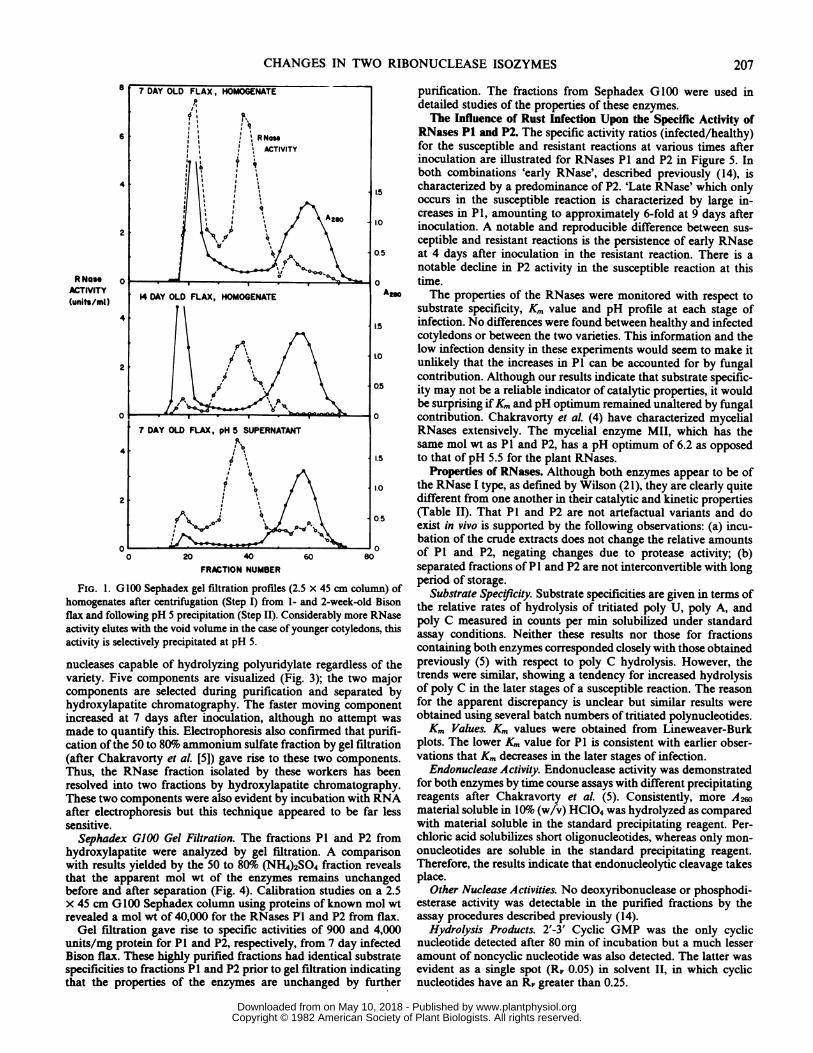
unstable. The relative amounts of activity eluted at 60 (P1) and120 (P2) mm in routine stepwise fractionations of partially purifiedextracts from susceptible (var. Bison) and resistant (var. Bombay)combinations are illustrated in Figure 2. These results show thefollowing: (a) healthy plants contain the same relative amounts ofP1 and P2 regardless of the variety or age of the plants during theexperimental period; (b) the early increases in RNase are charac-terized by an increase in the proportion of P2 and this changepersists at 4 days after inoculation in resistant but not in susceptibleplants; (c) the late increase in RNase, found only in the susceptiblecombination, is characterized by an increased proportion of P1.The A at 280 nm indicates not only the amount of protein
present but also the amount of contaminating material, the Amaximum being at about 270 nm. It is possible that such impuritiesmay interfere with protein estimation by the Lowry method.However, the amount of contaminating material is not influencedby infection and thus A at 280 nm is used in Figure 2 to providea standard by which to compare eluted RNase activity fromhealthy and infected plants.
Electrophoretic Analysis. Electrophoresis shows that the crudehomogenate from healthy flax and infected flax contains the same
Table I. Purification ofRNasefrom Healthy and Rust-Infected Flax (var. Bison) Cotyledons
RNase Protein Specific Activity YieldStep Fraction
H I H I H I H I
units mg units/mg %3 Days after inoculation
I Homogenate 2,650 3,830 1,300 1,500 2.0 2.5 100 100II pH 5 Supernatant 1,590 2,220 530 554 3.0 4.0 60 58
III 50 to 80%o(NH4)2SO4 1,060 1,600 53 57 20 28 40 42
IV Hydroxylapatite Pi 183 214 18 18 10 12 30 35P2 605 1,111 6.5 7.3 92 152
8 Days after inoculationI Homogenate 3,140 6,900 950 945 3.3 7.3 100 100
II pH 5 Supernatant 2,420 5,730 480 440 5 13 77 83III 50 to 80%
(NH4)2SO4 1,950 4,480 65 58 30 78 62 65IV Hydroxylapatite P1 425 2,077 13 14 33 139 41 43
P2 849 890 3.3 3.5 255 263a H, healthy plants; I, infected plants.
206 SUTTON AND SHAW
www.plantphysiol.orgon May 10, 2018 - Published by Downloaded from Copyright © 1982 American Society of Plant Biologists. All rights reserved.
CHANGES IN TWO RIBONUCLEASE ISOZYMES
7 DAY OLD FLAX, HOMOGENATE purification. The fractions from Sephadex G 100 were used indetailed studies of the properties of these enzymes.The Influence of Rust Infection Upon the Specific Activity of
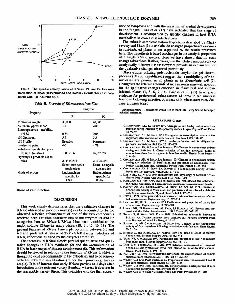
6 ? i i | RN.e RNases P1 and P2. The specific activity ratios (infected/healthy)li|ACTIVITY for the susceptible and resistant reactions at various times after
inoculation are illustrated for RNases P1 and P2 in Figure 5. Inz It t both combinations 'early RNase', described previously (14), is
4 @| |i i characterized by a predominance of P2. 'Late RNase' which onlyoccurs in the susceptible reaction is characterized by large in-
eI A* | fX ^20 g creases in P1, amounting to approximately 6-fold at 9 days after2 Kli 5.\,/ \ 10 inoculation. A notable and reproducible difference between sus-'1' q \ ceptible and resistant reactions is the persistence of early RNase
0.5 at 4 days after inoculation in the resistant reaction. There is aB'l,'°oO' V notable decline in P2 activity in the susceptible reaction at this
RNose 0 time.
AuTIVITY 14 DAY OLD FLAX, HOMOGENATE Am The properties of the RNases were monitored with respect to(units/ml) g t I l substrate specificity, Km value and pH profile at each stage of4 | \ .5 infection. No differences were found between healthy and infected
At\p <cotyledons or between the two varieties. This information and the
1.t\0 low infection density in these experiments would seem to make it
2 O unlikely that the increases in P1 can be accounted for by fungalcontribution. Although our results indicate that substrate specific-
05 ity may not be a reliable indicator of catalytic properties, it would1101. ' 'u^besurpnsing ifKm and pH optimum remained unaltered by fungal
0_--0 contribution. Chakravorty et aL (4) have characterized mycelial7 DAY OLD FLAX, pH 5 SUPERNATANT RNases extensively. The mycelial enzyme MII, which has the4a,Rssame mol wt as P1 and P2, has a pH optimum of 6.2 as opposed
15 to that of pH 5.5 for the plant RNases.Properties of RNases. Although both enzymes appear to be of
°J0 the RNase I type, as defined by Wilson (21), they are clearly quite2 1 different from one another in their catalytic and kinetic properties050|\(Table II). That P1 and P2 are not artefactual variants and do:%°tO<pwP>,V exist in vivo is supported by the following observations: (a) incu-,,= bation of the crude extracts does not change the relative amounts
0 20 40 60 0 of P1 and P2, negating changes due to protease activity; (b)FRACTION NUMBER separated fractions of P1 and P2 are not interconvertible with long
FiG. 1. G100 Sephadex gel filtration profiles (2.5 x 45 cm column) of period of storage.FIG. afterclO0Sephadexugelfation(epfrom 1- and 2-week-old Bison Substrate Specificity. Substrate specificities are given in terms ofhomogenates after centrifugation (Step I) frm1 n -ekodBsn the relative rates of hydrolysis of tritiated poly U, poly A, andflax and following pH 5 precipitation (Step II). Considerably more RNase the reasures of countsperitiatedpoly standactivity elutes with the void volume in the case of younger cotyledons, this poly C measured in counts per min solubilized under standardactivitisselctivelpreciptated a pH5.assay conditions. Neither these results nor those for fractionsactivity is selectively precipitated at pH 5. containing both enzymes corresponded closely with those obtained
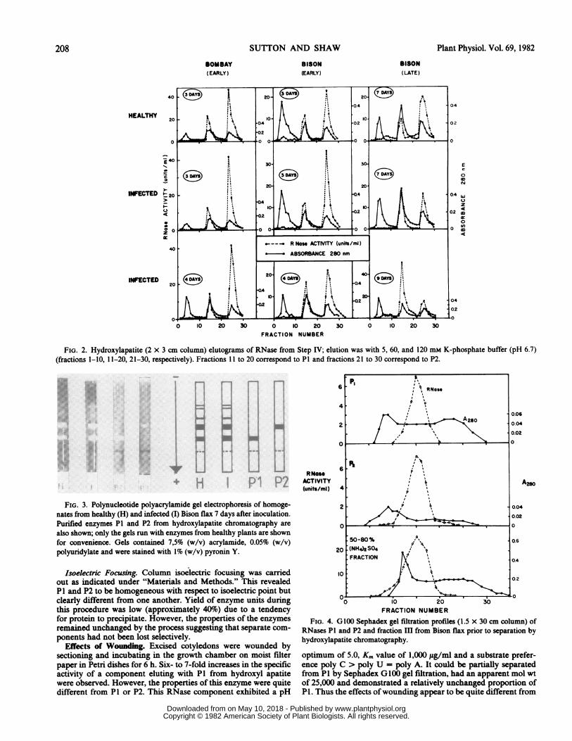
nucleases capable of hydrolyzing polyuridylate regardless of the previously (5) with respect to poly C hydrolysis. However, thevariety. Five components are visualized (Fig. 3); the two major trends were similar, showing a tendency for increased hydrolysiscomponents are selected during purification and separated by of poly C in the later stages of a susceptible reaction. The reasonhydroxylapatite chromatography. The faster moving component for the apparent discrepancy is unclear but similar results wereincreased at 7 days after inoculation, although no attempt was obtained using several batch numbers of tritiated polynucleotides.made to quantify this. Electrophoresis also confirmed that purifi- Km Values. Km values were obtained from Lineweaver-Burkcation of the 50 to 80%o ammonium sulfate fraction by gel filtration plots. The lower Km value for P1 is consistent with earlier obser-(after Chakravorty et aL [5]) gave rise to these two components. vations that Km decreases in the later stages of infection.Thus, the RNase fraction isolated by these workers has been Endonuclease Activity. Endonuclease activity was demonstratedresolved into two fractions by hydroxylapatite chromatography. for both enzymes by time course assays with different precipitatingThese two components were also evident by incubation with RNA reagents after Chakravorty et al. (5). Consistently, more A2Wafter electrophoresis but this technique appeared to be far less material soluble in 10%o (w/v) HC104 was hydrolyzed as comparedsensitive. with material soluble in the standard precipitating reagent. Per-
Sephadex G100 Gel Filtration. The fractions P1 and P2 from chloric acid solubilizes short oligonucleotides, whereas only mon-hydroxylapatite were analyzed by gel filtration. A comparison onucleotides are soluble in the standard precipitating reagent.with results yielded by the 50 to 80% (NH4)2S04 fraction reveals Therefore, the results indicate that endonucleolytic cleavage takesthat the apparent mol wt of the enzymes remains unchanged place.before and after separation (Fig. 4). Calibration studies on a 2.5 Other Nuclease Activities. No deoxyribonuclease or phosphodi-x 45 cm G100 Sephadex column using proteins of known mol wt esterase activity was detectable in the purified fractions by therevealed a mol wt of 40,000 for the RNases PI and P2 from flax. assay procedures described previously (14).Gel fitration gave rise to specific activities of 900 and 4,000 Hydrolysis Products. 2'-3' Cyclic GMP was the only cyclic
units/mg protein for P1 and P2, respectively, from 7 day infected nucleotide detected after 80 min of incubation but a much lesserBison flax. These highly purified fractions had identical substrate amount of noncyclic nucleotide was also detected. The latter wasspecificities to fractionsP1 and P2 prior to gel filtration indicating evident as a single spot (RF 0.05) in solvent II, in which cyclicthat the properties of the enzymes are unchanged by further nucleotides have an RF greater than 0.25.
207
www.plantphysiol.orgon May 10, 2018 - Published by Downloaded from Copyright © 1982 American Society of Plant Biologists. All rights reserved.
SUTTON AND SHAW
BISON(EARLY)
Plant Physiol. Vol. 69, 1982
BISON(LATE)
HEALTHY
0
INFECTED t 20
I-
u
.4
0 o
o40
INFECTED
E
0
N
z4
in
OD
0
4
0 10 20FRACTION NUMBER
FIG. 2. Hydroxylapatite (2 x 3 cm column) elutograms of RNase from Step IV; elution was with 5, 60, and 120 mm K-phosphate buffer (pH 6.7)(fractions 1-10, 11-20, 21-30, respectively). Fractions 11 to 20 correspond to P1 and fractions 21 to 30 correspond to P2.
FIG. 3. Polynucleotide polyacrylamide gel electrophoresis of homoge-
nates from healthy (H) and infected (I) Bison flax 7 days after inoculation.
Purified enzymes P1 and P2 from hydroxylapatite chromatography are
also shown; only the gels run with enzymes from healthy plants are shown
for convenience. Gels contained 7,5% (w/v) acrylamide, 0.05% (w/v)
polyuridylate and were stained with 1% (w/v) pyronin Y.
Isoelectric Focusing. Column isoelectric focusing was carried
out as indicated under "Materials and Methods." This revealed
P1 and P2 to be homogeneous with respect to isoelectric point but
clearly different from one another. Yield of enzyme units duringthis procedure was low (approximately 40%1) due to a tendencyfor protein to precipitate. However, the properties of the enzymes
remained unchanged by the process suggesting that separate com-
ponents had not been lost selectively.Effects of Wounding. Excised cotyledons were wounded by
sectioning and incubating in the growth chamber on moist filter
paper in Petri dishes for 6 h. Six- to 7-fold increases in the specificactivity of a component eluting with P1 from hydroxyl apatitewere observed. However, the properties of this enzyme were quitedifferent from P1 or P2. This RNase component exhibited a pH
FRACTION NUMBER
FIG. 4. G100 Sephadex gel filtration profiles (1.5 x 30 cm column) ofRNases P1 and P2 and fraction III from Bison flax prior to separation byhydroxylapatite chromatography.
optimum of 5.0, Km value of 1,000 ,ug/ml and a substrate prefer-ence poly C > poly U = poly A. It could be partially separatedfrom P1 by Sephadex GO00 gel filtration, had an apparent mol wtof 25,000 and demonstrated a relatively unchanged proportion ofP1. Thus the effects of wounding appear to be quite different from
208
BOMBAY(EARLY)
+b 4
www.plantphysiol.orgon May 10, 2018 - Published by Downloaded from Copyright © 1982 American Society of Plant Biologists. All rights reserved.
CHANGES IN TWO RIBONUCLEASE ISOZYMES
3
RATIO 2
SPECIFIC ACTIVITY(INFECTED/HEALTHY)
0
0 2 4 6 10
DAYS AFTER INOCULATION
FIG. 5. The specific activity ratios of RNases P1 and P2 following
inoculation of Bison (susceptible-S) and Bombay (resistant-R) flax coty-
ledons with flax rust race no. 3.
Table II. Properties of Ribonucleasesfrom Flax
EnzymeProperty
P1 P2
Molecular weight 40,000 40,000
Km value,,ug/ml RNA 105 200
Electrophoretic mobility,pH 8.3 0.84 0.66
pH Optimum 5.5 5.5
pH Profile Broader Narrower
Isoelectric point 4.0 4.75
Substrate specificity, polyU; A; C (relative) 100, 62, 60 86, 62, 20
Hydrolysis products (at 80min) 2'-3' cGMP 2'-3' cGMP
Some noncyclic Some noncyclicnucleotides nucleotides
Mode of action Endonuclease Endonuclease
specific for specific forRNA RNA
those of rust infection.
DISCUSSION
This work clearly demonstrates that the qualitative changes in
RNase observed in previous work (5) can be accounted for by the
observed selective enhancement of one of the two components
resolved here. Detailed characteristics of the enzymes P1 and P2
categorize them as RNaseI. RNase I has been identified as the
major soluble RNase in many plants (10, 11, 15, 16, 19). The
general features of RNase I are a pH optimum between 5.0 and
6.0 and preferential release of 2'-3' cGMP during hydrolysis of
RNA, conditions fulfilled by the enzymes from flax.
The increases in RNase closely parallel quantitative and quali-
tative changes in RNA synthesis (2) and the accumulation of
RNA in later stages of disease development (8). This informationshould be viewed in relation to the function of RNaseI, which is
thought to exist predominantly in the cytoplasm and to be respon-sible for substrate re-utilization (rather than processing, for ex-
ample).It is of interest that early RNase persists at 4 days after
inoculation in the resistant variety Bombay, whereas it does not in
the susceptible variety Bison. This coincides with the first appear-
ance of symptoms and with the initiation of uredial developmentin the fungus. Tani et al. (17) have indicated that this stage ofdevelopment is accompanied by specific changes in host RNAmetabolism in crown rust infected oats.The subunit complementation hypothesis described by Chak-
ravorty and Shaw (3) to explain the changed properties of enzymesin rust-infected plants is not supported by the results presentedhere. The hypothesis is based on changes in the catalytic propertiesof a single RNase species. Here we have shown that no suchchange takes place. Rather, changes in the relative amounts of twocatalytically different RNase isozymes provide an explanation forthe qualitative changes observed previously.
Observations utilizing polynucleotide acrylamide gel electro-phoresis (18 and unpublished) suggest that a multiplicity of ribo-nucleases are present in all plants as in Escherichia coli (7).Changes in the relative amounts of such enzymes may well accountfor the qualitative changes observed in many rust and mildewinfected plants (1, 5, 6, 9, 14). Sachse et aL (13) have givenevidence for preferential enhancement of three to ten nucleasefractions following infection of wheat with wheat stem rust, Puc-cinia graminis tritici.
Acknowledgment-The authors would like to thank Mr. Leroy Scrubb for experttechnical assistance.
LITERATURE CITED
1. CHAKRAVORTY AK, KJ Scorr 1979 Changes in two barley leaf ribonucleasefractions during infection by the powdery mildew fungus. Physiol Plant Pathol14: 95-97
2. CHAKRAVORTY AK, M SHAw 1971 Changes in the transcription pattern of flaxcotyledons after inoculation with flax rust. Biochem J 123: 551-557
3. CHAKRAVORTY AK, M SHAw 1977 A possible molecular basis for obligate hostpathogen interactions. Biol Rev 52: 147-179
4. CHAKRAVORTY AK, M SHAw, LA SCRUBB 1974 Changes in ribonuclease activityduring rust infection. I. Characterization of multiple molecular forms ofribonuclease from flax rust grown in host-free media. Physiol Plant Pathol 4:313-334
5. CHAKRAVORTY AK, M SHAw, LA SCRUBB 1974 Changes in ribonuclease activityduring rust infection. II. Purification and properties of ribonuclease fromhealthy and infected flax cotyledons. Physiol Plant Pathol 4: 335-358
6. CHAKRAVORTY AK, M SHAw, LA SCRUBB 1974 Ribonuclease activity of wheatleaves and rust infection. Nature 247: 577-580
7. DATTA AD, SK NiyoGi 1974 Biochemistry and physiology of bacterial ribonu-cleases. Prog Nucleic Acid Res Mol Biol 17: 271-308
8. HAMILTON WE 1969 RNA levels in healthy and rust-infected flax cotyledons.MA thesis. University of Saskatchewan, Saskatoon, Saskatchewan, Canada
9. HARVEy AE, AK CHAKRAVORTY, M SHAw, LA SCRUBB 1974 Changes inribonuclease activity in Ribes leaves and pine tissue culture infected with blisterrust, Cronartium ribicola Physiol Plant Pathol 4: 359-371
10. JERVIs L 1974 Partial purification and characterization of two Nicotiana tabacumleaf ribonucleases. Phytochemistry 13: 709-714
11. LANTERO OJ, HJ KLOSTERMAN 1973 Purification and properties of barley leafribonuclease. Phytochemistry 12: 775-784
12. LowRY OH, NJ ROSEBROUGH, AL FARR,RJ RANDAL 1951 Protein measure-ment with the Folin phenol reagent. J Biol Chem 193: 265-275
13. SACHSE B, G WOLF, WH FUCHS 1971 Nubleinsaure abbauenda Enzyme inBlattern von Triticum aestivum nach Infektion mit Puccinia graminis tritici.Acta Phytopathol Acad Sci Hung 6: 39-49
14. SCRUBB LA, AK CHADRAVORTY, M SHAW 1972 Changes in the ribonucleaseactivity of flax cotyledons following inoculation with flax rust. Plant Physiol50: 73-79
15. SHUSTER L, HG KHORANA, LAHEPPEL 1959 The mode of action of ryegrassribonuclease. Biochim Biophys Acta 33: 452-461
16. TANG WJ, A MARETrzi 1970 Purification and properties of leaf ribonucleasefrom sugar cane. Biochim Biophys Acta 212: 300-307
17. TANI T, M YOSHIKAWA, M NAITO 1975 Selective enhancement of ribosomalribonucleic acid synthesis of crown rust-infected oat leaves by stem excision.Physiol Plant Pathol 5:193-199
18. VAN LOON LS 1975 Polynucleotide-acrylamide gel electrophoresis of solublenucleases from tobacco leaves. FEBS Lett 51: 266-269
19. WILSON CM 1968 Plant nucleases. II. Properties of corn ribonucleases I and IIand corn nucleaseI. Plant Physiol 43: 1339-1346
20. WILSON CM 1971 Plant nucleases. III. Polyacrylamide electrophoresis of cornribonuclease isoenzymes. Plant Physiol 48: 64-68
21. WILSON CM 1975 Plant Nucleases. Annu Rev Plant Physiol 26: 187-208
Pi (S)
P2(R)
P2(S)
^O 2 4 6 8 10
209
www.plantphysiol.orgon May 10, 2018 - Published by Downloaded from Copyright © 1982 American Society of Plant Biologists. All rights reserved.


![CALCULATION OF ISOELECTRIC POINTS. · 808 Calculation of Isoelectric Points Except in the special case where the isoelectric point is at the “neutral” point of water [H+] does](https://img.pdfslide.net/doc/110x75/5f0a52187e708231d42b1422/calculation-of-isoelectric-808-calculation-of-isoelectric-points-except-in-the.jpg)