Embed Size (px)
Citation preview
Chap 47
Animal Development
Fertilization
• The Acrosomal Reaction.– Acrosomal reaction: when exposed to the jelly
coat the sperm’s acrosome discharges it contents by exocytosis.• Hydrolytic enzymes enable the acrosomal process to
penetrate the egg’s jelly coat.
• The tip of the acrosomal process adheres to the vitelline layer just external to the egg’s plasma membrane.
Copyright © 2002 Pearson Education, Inc., publishing as Benjamin Cummings
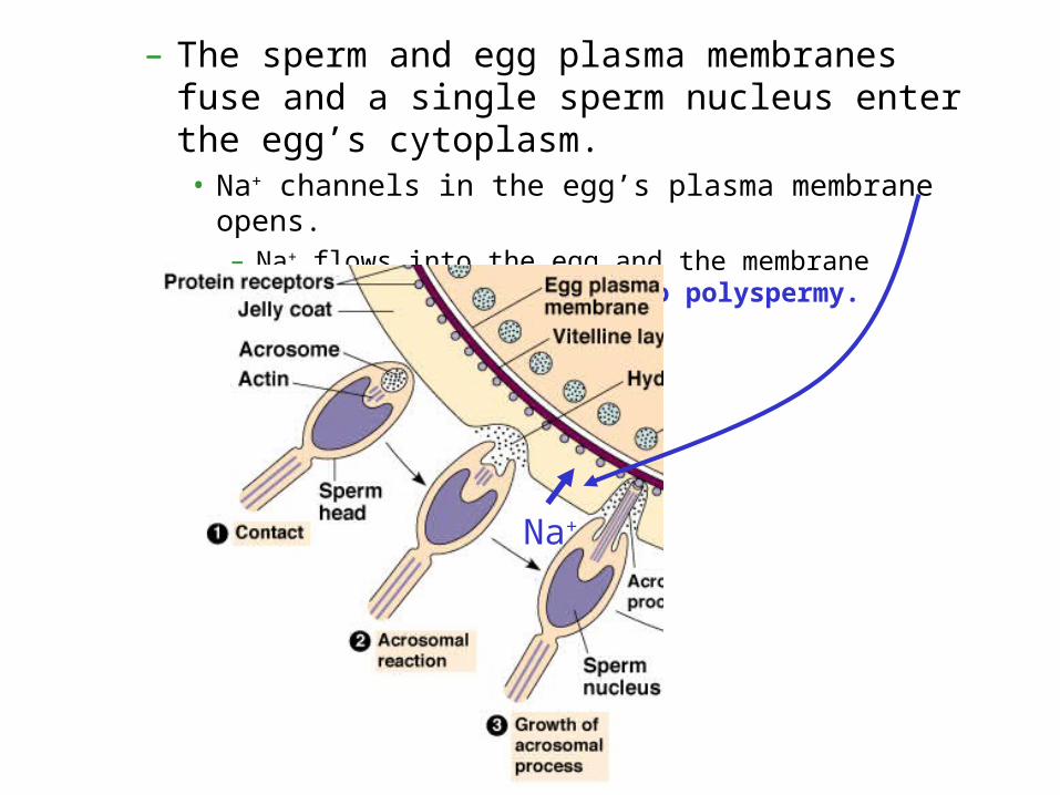
– The sperm and egg plasma membranes fuse and a single sperm nucleus enter the egg’s cytoplasm.• Na+ channels in the egg’s plasma membrane opens.
– Na+ flows into the egg and the membrane depolarizes: fast block to polyspermy.
Na+
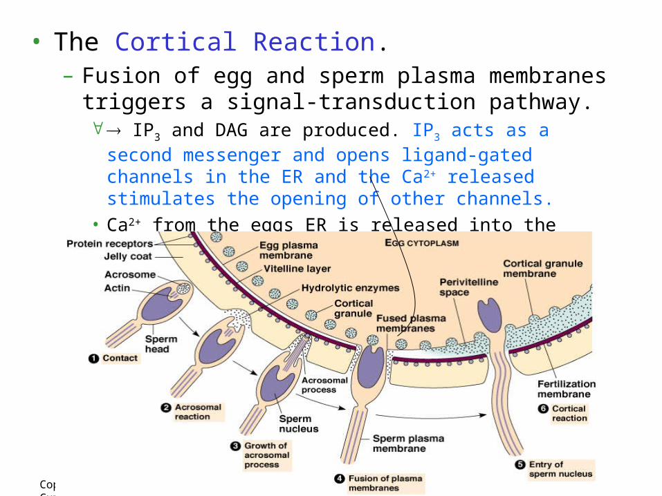
• The Cortical Reaction.– Fusion of egg and sperm plasma membranes triggers a
signal-transduction pathway. IP3 and DAG are produced. IP3 acts as a second messenger and
opens ligand-gated channels in the ER and the Ca2+ released stimulates the opening of other channels.
• Ca2+ from the eggs ER is released into the cytosol and propagates as a wave across the fertilized egg – cortical granules release contents
Copyright © 2002 Pearson Education, Inc., publishing as Benjamin Cummings
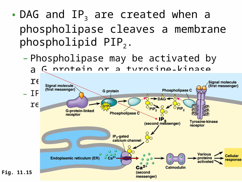
• DAG and IP3 are created when a phospholipase cleaves a membrane phospholipid PIP2.
– Phospholipase may be activated by a G protein or a tyrosine-kinase receptor.
– IP3 activates a gated-calcium channel, releasing Ca2+.
Fig. 11.15
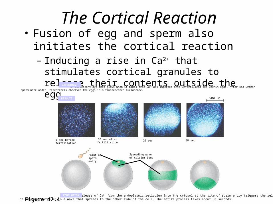
The Cortical Reaction• Fusion of egg and sperm also initiates the
cortical reaction– Inducing a rise in Ca2+ that stimulates cortical
granules to release their contents outside the egg
Figure 47.4
A fluorescent dye that glows when it binds free Ca2+ was injected into unfertilized sea urchin eggs. After sea urchin sperm were added, researchers observed the eggs in a fluorescence microscope.
EXPERIMENT
RESULTS
The release of Ca2+ from the endoplasmic reticulum into the cytosol at the site of sperm entry triggers the release of more and more Ca2+ in a wave that spreads to the other side of the cell. The entire process takes about 30 seconds.CONCLUSION
30 sec20 sec10 sec afterfertilization
1 sec beforefertilization
Point ofspermentry
Spreading waveof calcium ions
500 m
• The acrosomal reaction
Spermnucleus
Sperm plasmamembrane
Hydrolytic enzymes
Corticalgranule
Cortical granulemembrane
EGG CYTOPLASM
Basal body(centriole)
Spermhead
Acrosomalprocess
Actin
Acrosome
Jelly coatEgg plasmamembrane
Vitelline layer
Fused plasmamembranes
Perivitellinespace
Fertilizationenvelope
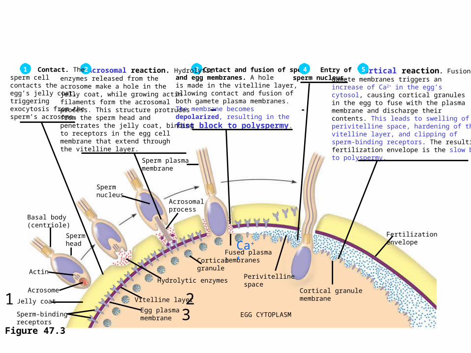
Cortical reaction. Fusion of the gamete membranes triggers an increase of Ca2+ in the egg’s cytosol, causing cortical granules in the egg to fuse with the plasma membrane and discharge their contents. This leads to swelling of the perivitelline space, hardening of thevitelline layer, and clipping of sperm-binding receptors. The resulting fertilization envelope is the slow block to polyspermy.
5 Contact and fusion of sperm and egg membranes. A hole is made in the vitelline layer, allowing contact and fusion of both gamete plasma membranes. The membrane becomes depolarized, resulting in the
fast block to polyspermy.
3 Acrosomal reaction. Hydrolytic enzymes released from the acrosome make a hole in the jelly coat, while growing actin filaments form the acrosomal process. This structure protrudes from the sperm head and penetrates the jelly coat, bindingto receptors in the egg cell membrane that extend through the vitelline layer.
2 Contact. The sperm cell contacts the egg’s jelly coat, triggering exocytosis from the sperm’s acrosome.
1
Sperm-bindingreceptors
Entry of sperm nucleus.4
Figure 47.3
1 23
Ca++

• Fertilization in Mammals.• Capacitation, a function of the female reproductive
system, enhances sperm function.– A capacitated
sperm migratesthrough a layerof follicle cellsbefore it reachesthe zona pellucida.
– Binding ofthe sperm cellinduces anacrosomalreaction similarto that seen in thesea urchin.
Copyright © 2002 Pearson Education, Inc., publishing as Benjamin Cummings Fig. 47.5
Cortical reaction forms slow block
Cortical reaction forms slow block
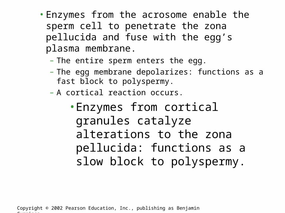
• Enzymes from the acrosome enable the sperm cell to penetrate the zona pellucida and fuse with the egg’s plasma membrane.– The entire sperm enters the egg.
– The egg membrane depolarizes: functions as a fast block to polyspermy.
– A cortical reaction occurs.
• Enzymes from cortical granules catalyze alterations to the zona pellucida: functions as a slow block to polyspermy.
Copyright © 2002 Pearson Education, Inc., publishing as Benjamin Cummings

– The envelopes of both the egg and sperm nuclei disperse.• The chromosomes from the two gametes share a common
spindle apparatus during the first mitotic division of the zygote.
Copyright © 2002 Pearson Education, Inc., publishing as Benjamin Cummings
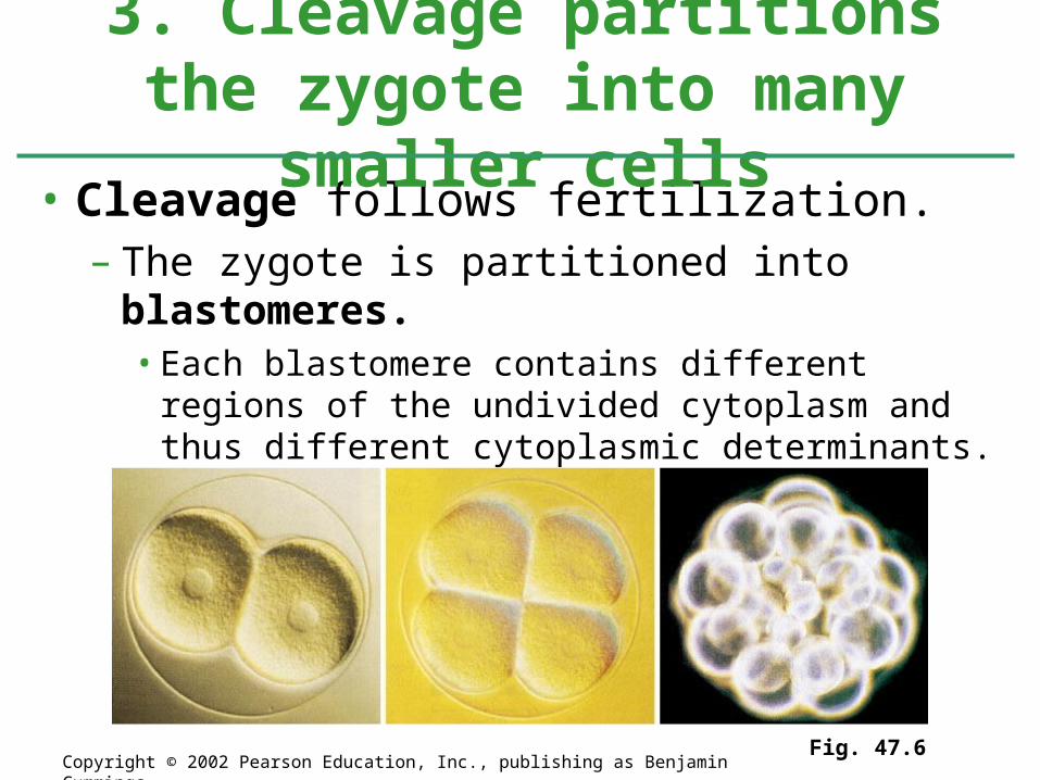
• Cleavage follows fertilization.– The zygote is partitioned into blastomeres.
• Each blastomere contains different regions of the undivided cytoplasm and thus different cytoplasmic determinants.
3. Cleavage partitions the zygote into many smaller cells
Fig. 47.6Copyright © 2002 Pearson Education, Inc., publishing as Benjamin Cummings

• Cleavage partitions the cytoplasm of one large cell– Into many smaller cells called blastomeres
Figure 47.7a–d
Fertilized egg. Shown here is thezygote shortly before the first cleavage division, surrounded by the fertilization envelope. The nucleus is visible in the center.
(a) Four-cell stage. Remnants of the mitotic spindle can be seen between the two cells that have just completed the second cleavage division.
(b) Morula. After further cleavage divisions, the embryo is a multicellular ball that is stillsurrounded by the fertilization envelope. The blastocoel cavityhas begun to form.
(c) Blastula. A single layer of cells surrounds a large blastocoel cavity. Although not visible here, the fertilization envelope is still present; the embryo will soon hatch from it and begin swimming.
(d)

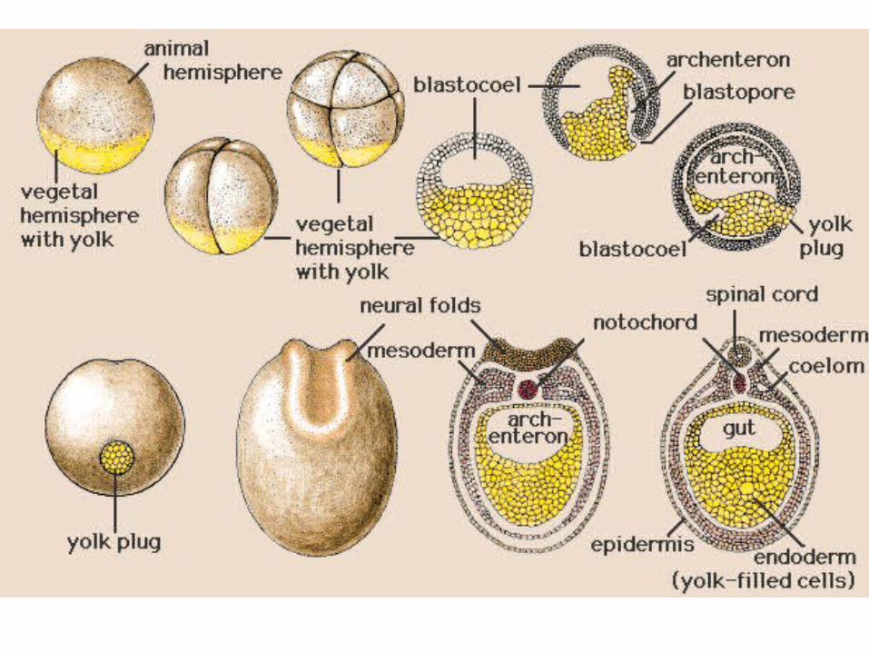
– Except for mammals, most animals have both eggs and zygotes with a definite polarity.• Thus, the planes of division follow a specific pattern
relative to the poles of the zygote.
• Polarity is defined by the heterogeneous distribution of substances such as mRNA, proteins, and yolk.– Yolk is most concentrated at the vegetal pole and least
concentrated at the animal pole.
• In some animals, the animal pole defines the anterior end of the animal.
Copyright © 2002 Pearson Education, Inc., publishing as Benjamin Cummings
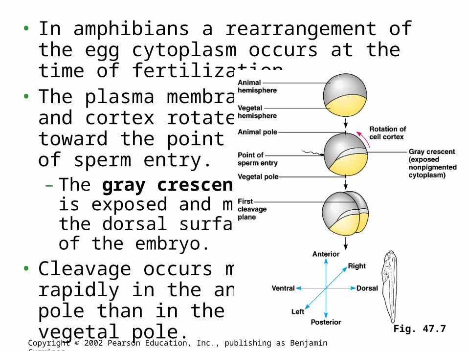
• In amphibians a rearrangement of the egg cytoplasm occurs at the time of fertilization.
• The plasma membraneand cortex rotatetoward the pointof sperm entry.– The gray crescent
is exposed and marksthe dorsal surfaceof the embryo.
• Cleavage occurs morerapidly in the animalpole than in thevegetal pole.
Copyright © 2002 Pearson Education, Inc., publishing as Benjamin Cummings
Fig. 47.7
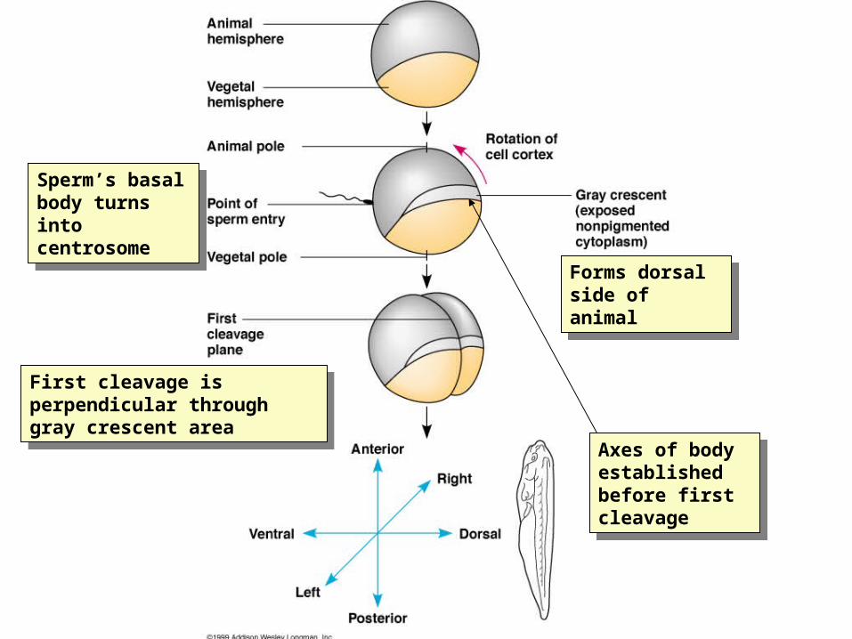
Forms dorsal side of animal
Forms dorsal side of animal
Sperm’s basal body turns into centrosome
Sperm’s basal body turns into centrosome
First cleavage is perpendicular through gray crescent area
First cleavage is perpendicular through gray crescent area
Axes of body established before first cleavage
Axes of body established before first cleavage
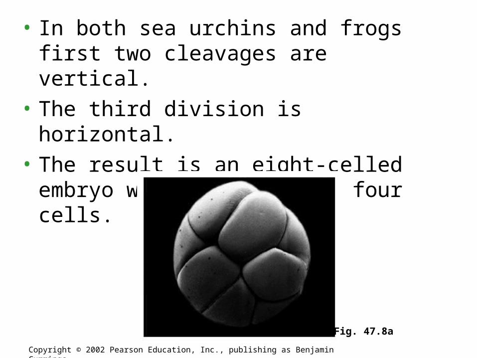
• In both sea urchins and frogs first two cleavages are vertical.
• The third division is horizontal.
• The result is an eight-celled embryo with two tiers of four cells.
Copyright © 2002 Pearson Education, Inc., publishing as Benjamin Cummings
Fig. 47.8a
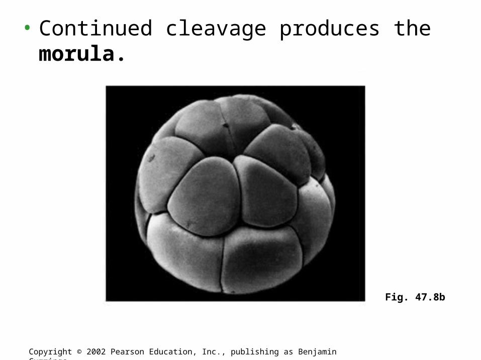
• Continued cleavage produces the morula.
Copyright © 2002 Pearson Education, Inc., publishing as Benjamin Cummings
Fig. 47.8b
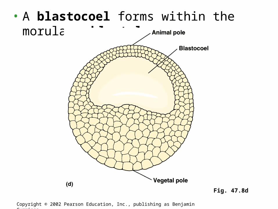
• A blastocoel forms within the morula blastula
Copyright © 2002 Pearson Education, Inc., publishing as Benjamin Cummings
Fig. 47.8d
• In animals with less yolk there is complete division of the egg: holoblastic cleavage.
Copyright © 2002 Pearson Education, Inc., publishing as Benjamin Cummings
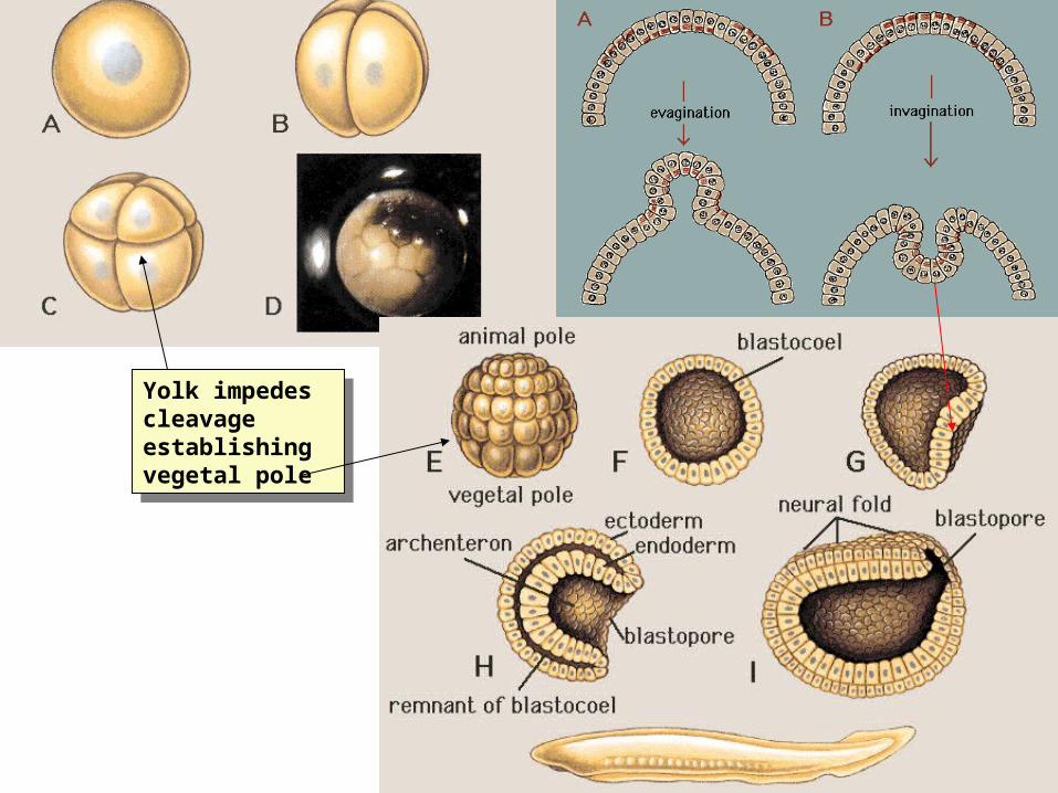
Yolk impedes cleavage establishing vegetal pole
Yolk impedes cleavage establishing vegetal pole

• Gastrulation in a sea urchin– Produces an
embryo with a primitive gut and three germ layers
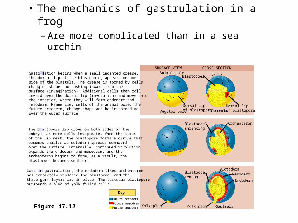
• The mechanics of gastrulation in a frog – Are more complicated than in a sea urchin
Figure 47.12
SURFACE VIEW CROSS SECTIONAnimal pole
Blastocoel
Dorsal lipof blastopore
Dorsal lipof blastoporeVegetal pole Blastula
Blastocoelshrinking
Archenteron
Blastocoelremnant
EctodermMesoderm
Endoderm
GastrulaYolk plugYolk plug
Key
Future ectoderm
Future mesoderm
Future endoderm
Gastrulation begins when a small indented crease, the dorsal lip of the blastopore, appears on one side of the blastula. The crease is formed by cellschanging shape and pushing inward from the surface (invagination). Additional cells then rollinward over the dorsal lip (involution) and move intothe interior, where they will form endoderm andmesoderm. Meanwhile, cells of the animal pole, the future ectoderm, change shape and begin spreading over the outer surface.
The blastopore lip grows on both sides of the embryo, as more cells invaginate. When the sides of the lip meet, the blastopore forms a circle thatbecomes smaller as ectoderm spreads downward over the surface. Internally, continued involutionexpands the endoderm and mesoderm, and the archenteron begins to form; as a result, the blastocoel becomes smaller.
1
2
3 Late in gastrulation, the endoderm-lined archenteron has completely replaced the blastocoel and the three germ layers are in place. The circular blastopore surrounds a plug of yolk-filled cells.
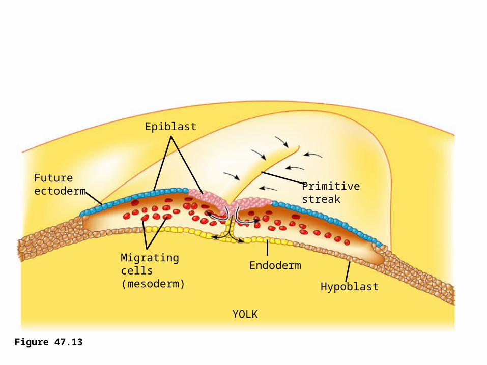
• Gastrulation in the chick– Is affected by the large amounts of yolk in the egg
Figure 47.13
Epiblast
Futureectoderm
Migratingcells(mesoderm)
Endoderm
Hypoblast
YOLK
Primitivestreak

Organogenesis
• Various regions of the three embryonic germ layers– Develop into the rudiments of organs during
the process of organogenesis

• Early in vertebrate organogenesis– The notochord forms from mesoderm and the
neural plate forms from ectoderm
Figure 47.14a
Neural plate formation. By the timeshown here, the notochord has developed from dorsal mesoderm, and the dorsal ectoderm hasthickened, forming the neural plate, in response to signals from thenotochord. The neural folds arethe two ridges that form the lateral edges of the neural plate. These are visible in the light micrographof a whole embryo.
Neural folds
1 mm
Neuralfold
Neuralplate
Notochord
Ectoderm
Mesoderm
Endoderm
Archenteron
(a)
LM

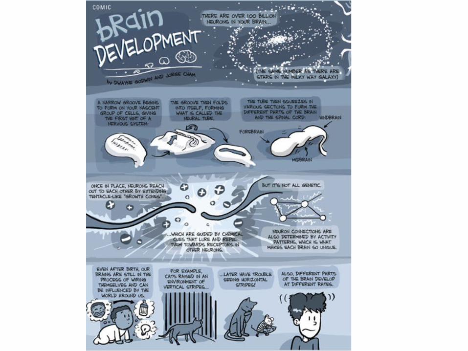
• The neural plate soon curves inward– Forming the neural tube
Figure 47.14b
Formation of the neural tube. Infolding and pinching off of the neural plate generates the neural tube. Note the neural crest cells, which will migrate and give rise to numerousstructures.
Neuralfold
Neural plate
Neural crest
Outer layer of ectoderm
Neural crest
Neural tube
(b)
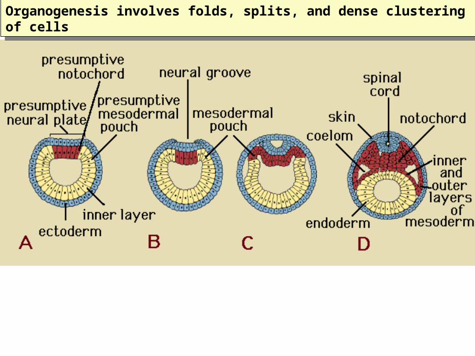
Organogenesis involves folds, splits, and dense clustering of cellsOrganogenesis involves folds, splits, and dense clustering of cells
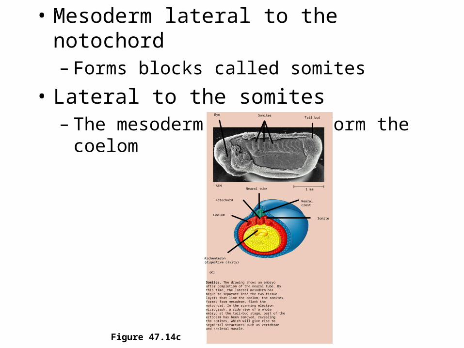
• Mesoderm lateral to the notochord– Forms blocks called somites
• Lateral to the somites– The mesoderm splits to form the coelom
Figure 47.14c
Somites. The drawing shows an embryoafter completion of the neural tube. By this time, the lateral mesoderm hasbegun to separate into the two tissuelayers that line the coelom; the somites, formed from mesoderm, flank thenotochord. In the scanning electron micrograph, a side view of a whole embryo at the tail-bud stage, part of the ectoderm has been removed, revealingthe somites, which will give rise to segmental structures such as vertebrae and skeletal muscle.
Eye SomitesTail bud
1 mmNeural tube
Notochord Neuralcrest
Somite
Archenteron(digestive cavity)
Coelom
(c)
SEM

• Organogenesis in the chick– Is quite similar to that in the frog
Figure 47.15a, b
Neural tube
Notochord
Archenteron
Lateral fold
Form extraembryonicmembranes
YOLKYolk stalk
Somite
Coelom
Endoderm
Mesoderm
Ectoderm
Yolk sac
Eye
Forebrain
Heart
Blood vessels
Somites
Neural tube
Early organogenesis. The archenteron forms when lateral folds pinch the embryo away from the yolk. The embryo remains opento the yolk, attached by the yolk stalk, about midway along its length,as shown in this cross section. The notochord, neural tube, and somites subsequently form much as they do in the frog.
(a) Late organogenesis. Rudiments of most major organs have already formed in this chick embryo, which is about 56 hours old and about 2–3 mm long (LM).
(b)

Meroblastic cleavage due to yolk
Meroblastic cleavage due to yolk
In birds the yolk is so plentiful that it restricts cleavage to the animal pole: meroblastic cleavage.
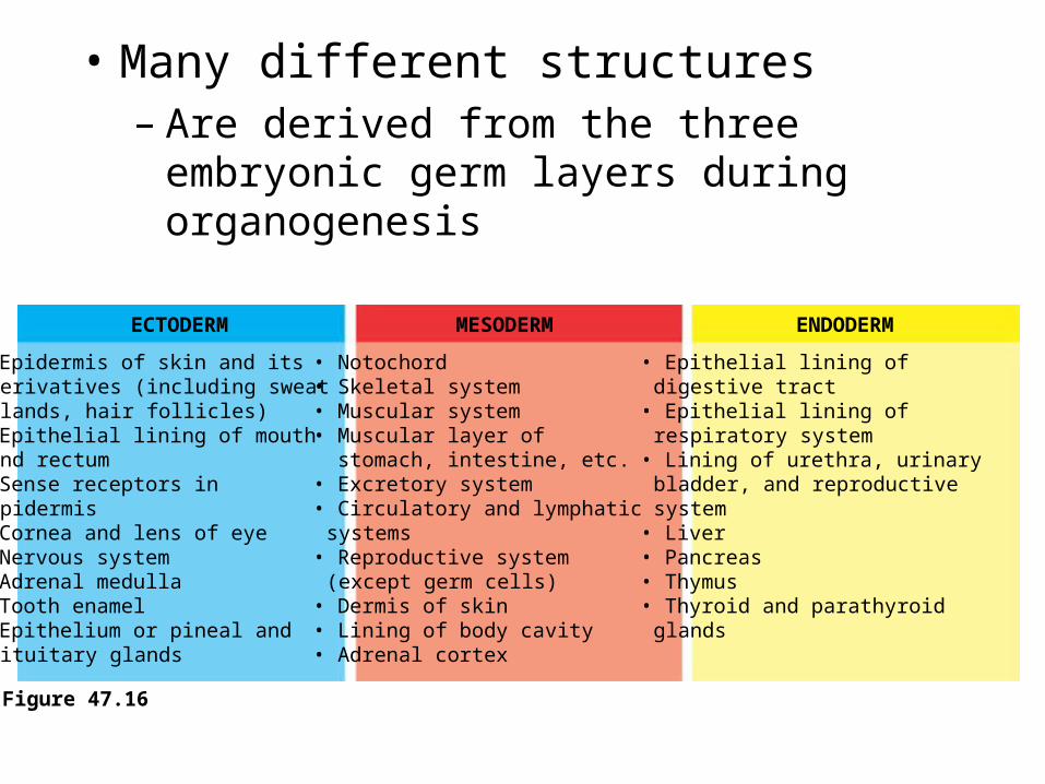
• Many different structures– Are derived from the three embryonic germ
layers during organogenesis
Figure 47.16
ECTODERM MESODERM ENDODERM
• Epidermis of skin and itsderivatives (including sweatglands, hair follicles)
• Epithelial lining of mouthand rectum
• Sense receptors inepidermis
• Cornea and lens of eye• Nervous system• Adrenal medulla• Tooth enamel• Epithelium or pineal and
pituitary glands
• Notochord• Skeletal system• Muscular system• Muscular layer of stomach, intestine, etc.• Excretory system• Circulatory and lymphatic
systems• Reproductive system
(except germ cells)• Dermis of skin• Lining of body cavity• Adrenal cortex
• Epithelial lining ofdigestive tract
• Epithelial lining ofrespiratory system
• Lining of urethra, urinarybladder, and reproductivesystem
• Liver• Pancreas• Thymus• Thyroid and parathyroid
glands

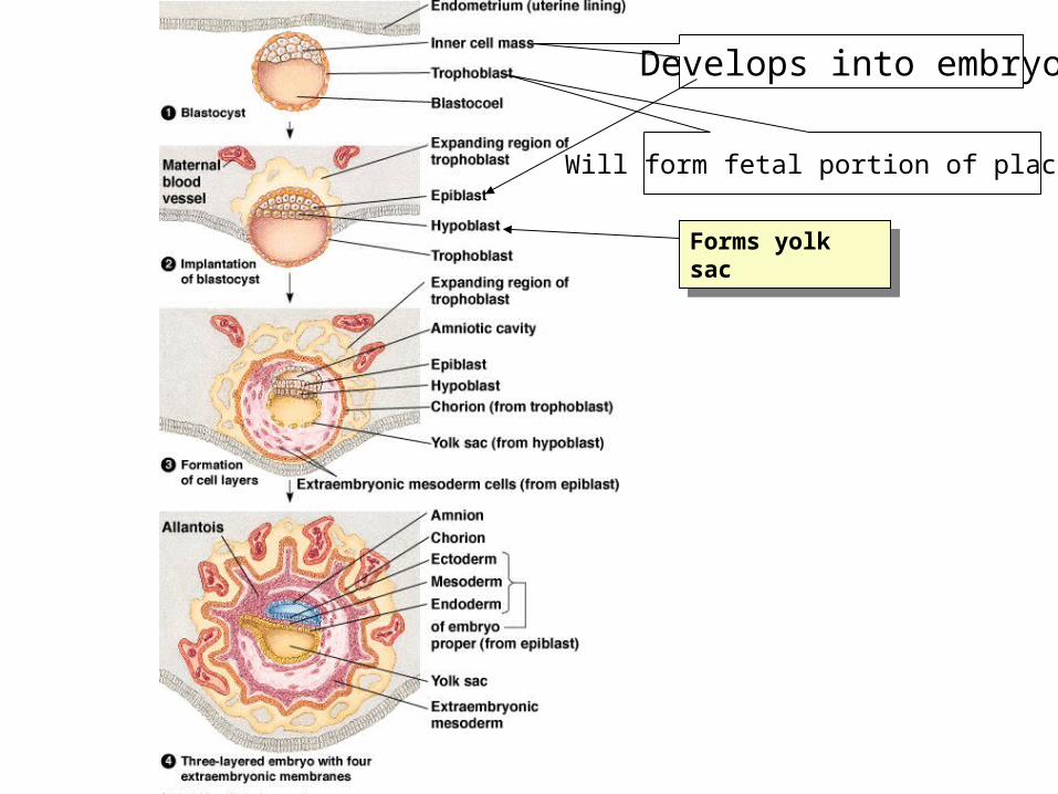
Develops into embryo
Will form fetal portion of placenta
Forms yolk sacForms yolk sac
The Cytoskeleton, Cell Motility, and Convergent Extension
• Changes in the shape of a cell– Usually involve reorganization of the
cytoskeleton
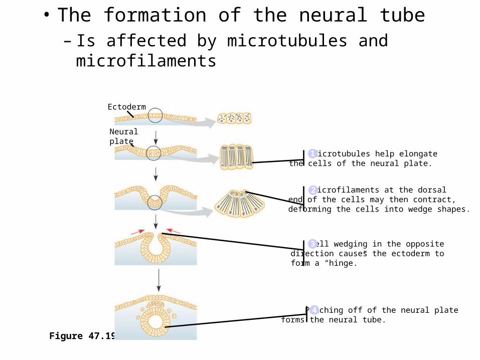
• The formation of the neural tube– Is affected by microtubules and microfilaments
Figure 47.19
Microtubules help elongatethe cells of the neural plate.1
Pinching off of the neural plate forms the neural tube.4
Ectoderm
Neuralplate
Microfilaments at the dorsal end of the cells may then contract,deforming the cells into wedge shapes.
Cell wedging in the opposite direction causes the ectoderm to form a “hinge.”
2
3
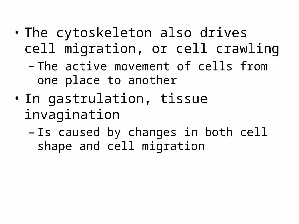
• The cytoskeleton also drives cell migration, or cell crawling– The active movement of cells from one place to
another
• In gastrulation, tissue invagination – Is caused by changes in both cell shape and cell
migration
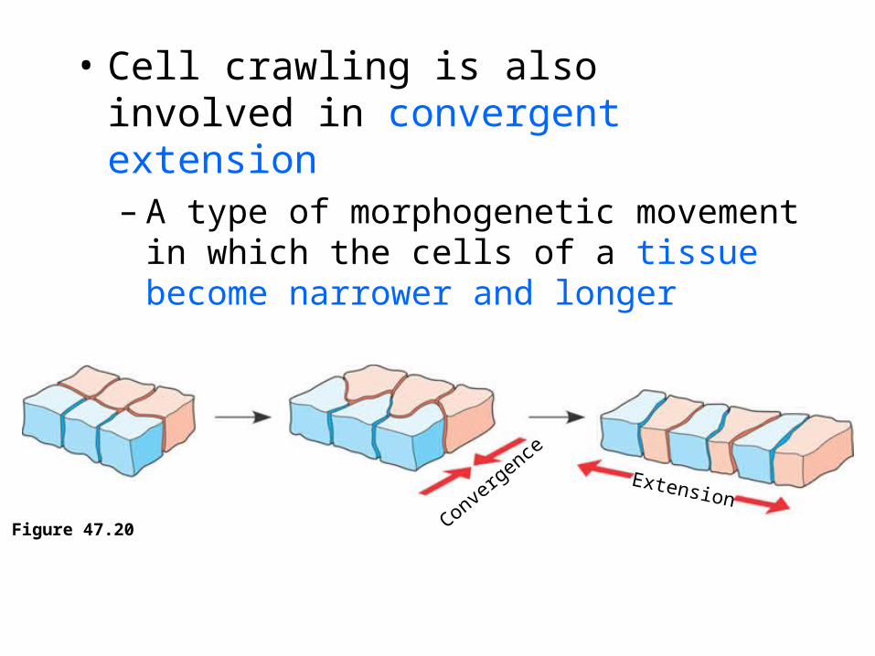
• Cell crawling is also involved in convergent extension– A type of morphogenetic movement in which
the cells of a tissue become narrower and longer
Figure 47.20Conve
rgence
Extension

Convergent Extension
cell crawling results in morphogenic movement
may involve the ECM (extracellular matrix) which is the mixture of glycoproteins outside the plasma membrane
ECM fibers may acts as tracks that act as routes for migrating cells in morphogenic movement or they inhibit migration in certain directions
Example
fibronectin lines the the roof of the blastocoel and acts as a guide to a sheet of migrating mesoderm cells.
Example
fibronectin lines the the roof of the blastocoel and acts as a guide to a sheet of migrating mesoderm cells.
Roles of the Extracellular Matrix and Cell Adhesion Molecules
• Fibers of the extracellular matrix– May function as tracks, directing migrating
cells along particular routes
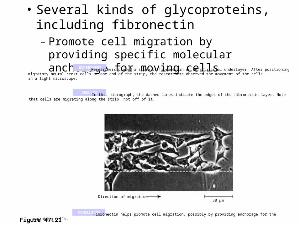
• Several kinds of glycoproteins, including fibronectin– Promote cell migration by providing specific
molecular anchorage for moving cells
Figure 47.21
EXPERIMENT Researchers placed a strip of fibronectin on an artificial underlayer. After positioning migratory neural crest cells at one end of the strip, the researchers observed the movement of the cells in a light microscope.
CONCLUSION
RESULTS In this micrograph, the dashed lines indicate the edges of the fibronectin layer. Note that cells are migrating along the strip, not off of it.
Fibronectin helps promote cell migration, possibly by providing anchorage for the migrating cells.
Direction of migration50 µm

• Cell adhesion molecules – Also contribute to cell migration and stable
tissue structure
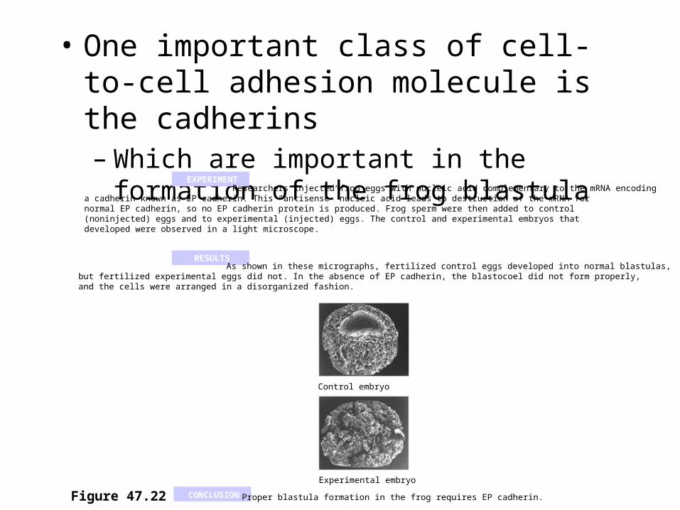
• One important class of cell-to-cell adhesion molecule is the cadherins– Which are important in the formation of the
frog blastula
Figure 47.22 CONCLUSION
EXPERIMENT Researchers injected frog eggs with nucleic acid complementary to the mRNA encoding a cadherin known as EP cadherin. This “antisense” nucleic acid leads to destruction of the mRNA for normal EP cadherin, so no EP cadherin protein is produced. Frog sperm were then added to control (noninjected) eggs and to experimental (injected) eggs. The control and experimental embryos that developed were observed in a light microscope.
RESULTS As shown in these micrographs, fertilized control eggs developed into normal blastulas, but fertilized experimental eggs did not. In the absence of EP cadherin, the blastocoel did not form properly, and the cells were arranged in a disorganized fashion.
Control embryo
Experimental embryo
Proper blastula formation in the frog requires EP cadherin.
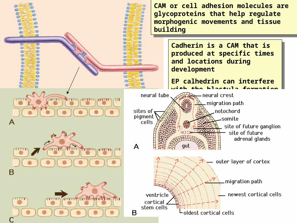
CAM or cell adhesion molecules are glycoproteins that help regulate morphogenic movements and tissue building
CAM or cell adhesion molecules are glycoproteins that help regulate morphogenic movements and tissue building
Cadherin is a CAM that is produced at specific times and locations during development
EP calhedrin can interfere with the blastula formation in frogs
Cadherin is a CAM that is produced at specific times and locations during development
EP calhedrin can interfere with the blastula formation in frogs
• Concept 47.3: The developmental fate of cells depends on their history and on inductive signals
• Coupled with morphogenetic changes– Development also requires the timely differentiation
of many kinds of cells at specific locations
Two general principlesUnderlie differentiation during embryonic
development
• First, during early cleavage divisions– Embryonic cells must somehow become different
from one another
• Second, once initial cell asymmetries are set up– Subsequent interactions among the embryonic cells
influence their fate, usually by causing changes in gene expression

Somites will give rise to segmental structures such as vertebrae and serially arranged skeletal muscles
Somites will give rise to segmental structures such as vertebrae and serially arranged skeletal muscles
Name the 3 different germ layers
Establishing Cellular Asymmetries
• To understand at the molecular level how embryonic cells acquire their fates– It is helpful to think first about how the basic
axes of the embryo are established
The Axes of the Basic Body Plan
• In nonamniotic vertebrates– Basic instructions for establishing the body
axes are set down early, during oogenesis or fertilization
• In amniotes, local environmental differences– Play the major role in establishing initial
differences between cells and, later, the body axes

Restriction of Cellular Potency
• In many species that have cytoplasmic determinants– Only the zygote is totipotent, capable of
developing into all the cell types found in the adult
• Unevenly distributed cytoplasmic determinants in the egg cell– Are important in establishing the body axes – Set up differences in blastomeres resulting from
cleavage

Blastomeres that receive half or all of the gray crescent develop into normal embryos, but a blastomere that receives none of the gray crescent gives rise to an abnormal embryo without dorsal structures. Spemann called it a “belly piece.”
EXPERIMENT
RESULTS
CONCLUSION The totipotency of the two blastomeres normally formed during the first cleavage division depends on cytoplasmic determinants localized in the gray crescent.
Left (control):Fertilizedsalamander eggswere allowed todivide normally,resulting in thegray crescent beingevenly dividedbetween the twoblastomeres.
Right (experimental):Fertilized eggs wereconstricted by athread so that thefirst cleavage planerestricted the graycrescent to oneblastomere.
Graycrescent
The two blastomeres werethen separated andallowed to develop.
Graycrescent
Normal
Bellypiece Normal
1
2
Figure 47.24

• As embryonic development proceeds– The potency of cells becomes progressively
more limited in all species

Cell Fate Determination and Pattern Formation by Inductive Signals
• Once embryonic cell division creates cells that differ from each other– The cells begin to influence each other’s fates
by induction

The “Organizer” of Spemann and Mangold
• Based on the results of their most famous experiment– Spemann and Mangold concluded that the
dorsal lip of the blastopore functions as an organizer of the embryo

• The organizer initiates a chain of inductions– That results in the formation of the notochord,
the neural tube, and other organs
Figure 47.25
EXPERIMENT
RESULTS
CONCLUSION
Spemann and Mangold transplanted a piece of the dorsal lip of a pigmented newt gastrula to the ventral side of the early gastrula of a nonpigmented newt.
During subsequent development, the recipient embryo formed a second notochord and neural tube in the region of the transplant, and eventually most of a second embryo. Examination of the interior of the double embryorevealed that the secondary structures were formed in part from host tissue.
The transplanted dorsal lip was able to induce cells in a different region of the recipient to form structures different from their normal fate. In effect, the dorsal lip “organized” the later development of an entire embryo.
Pigmented gastrula(donor embryo)
Dorsal lip ofblastopore
Nonpigmented gastrula(recipient embryo)
Primary embryo
Secondary (induced) embryoPrimarystructures:
Neural tube
Notochord
Secondarystructures:
Notochord (pigmented cells)
Neural tube (mostly nonpigmented cells)

In molluscs and annelids, the first cleavage is determinate, separating vital cytoplasmic constituents
In molluscs and annelids, the first cleavage is determinate, separating vital cytoplasmic constituents
Indeterminant cleavage

Formation of the Vertebrate Limb
• Inductive signals play a major role in pattern formation– The development of an animal’s spatial
organization
• The molecular cues that control pattern formation, called positional information – Tell a cell where it is with respect to the
animal’s body axes– Determine how the cell and its descendents
respond to future molecular signals
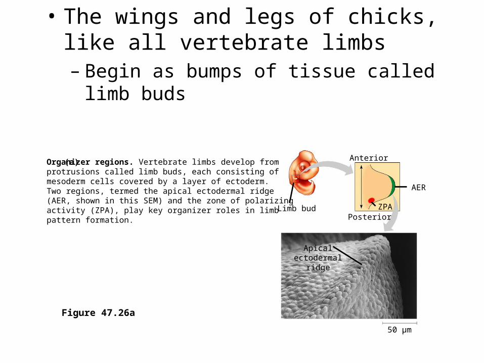
• The wings and legs of chicks, like all vertebrate limbs– Begin as bumps of tissue called limb buds
Figure 47.26a
Limb bud
Anterior
AER
ZPAPosterior
Organizer regions. Vertebrate limbs develop fromprotrusions called limb buds, each consisting of mesoderm cells covered by a layer of ectoderm. Two regions, termed the apical ectodermal ridge (AER, shown in this SEM) and the zone of polarizing activity (ZPA), play key organizer roles in limb pattern formation.
(a)
Apicalectodermal
ridge
50 µm

• The embryonic cells within a limb bud – Respond to positional information indicating
location along three axes
Figure 47.26b
Digits
Anterior
Ventral
DistalProximal
DorsalPosterior
Wing of chick embryo. As the bud develops into alimb, a specific pattern of tissues emerges. In the chick wing, for example, the three digits are always present in the arrangement shown here. Pattern formation requires each embryonic cell to receive some kind of positional information indicating location along the three axes of the limb. The AERand ZPA secrete molecules that help provide thisinformation.
(b)

Inductive signals causes pattern formation
Inductive signals causes pattern formation
Positional information in the chick limb bud responds to molecular cues from organizier regions
Positional information in the chick limb bud responds to molecular cues from organizier regions
AER apical
ZPA

• One limb-bud organizer region is the apical ectodermal ridge (AER)– A thickened area of ectoderm at the tip of the bud
• The second major limb-bud organizer region is the zone of polarizing activity (ZPA)– A block of mesodermal tissue located underneath the
ectoderm where the posterior side of the bud is attached to the body
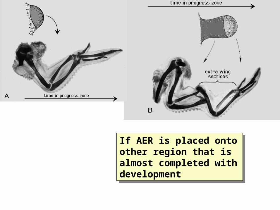
If AER is placed onto other region that is almost completed with development
If AER is placed onto other region that is almost completed with development
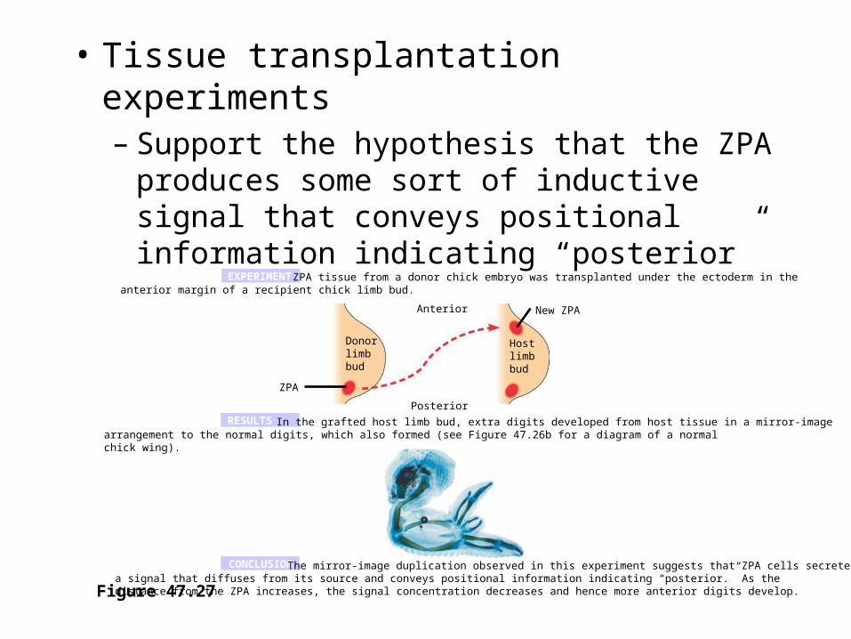
• Tissue transplantation experiments– Support the hypothesis that the ZPA produces
some sort of inductive signal that conveys positional information indicating “posterior”
Figure 47.27
EXPERIMENT
RESULTS
CONCLUSION
ZPA tissue from a donor chick embryo was transplanted under the ectoderm in the anterior margin of a recipient chick limb bud.
Anterior
Donorlimbbud
Hostlimbbud
Posterior
ZPA
The mirror-image duplication observed in this experiment suggests that ZPA cells secrete a signal that diffuses from its source and conveys positional information indicating “posterior.” As the distance from the ZPA increases, the signal concentration decreases and hence more anterior digits develop.
New ZPA
In the grafted host limb bud, extra digits developed from host tissue in a mirror-image arrangement to the normal digits, which also formed (see Figure 47.26b for a diagram of a normal chick wing).
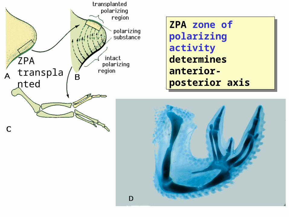
ZPA zone of polarizing activity determines anterior-posterior axis
ZPA zone of polarizing activity determines anterior-posterior axisZPA
transplanted
• Signal molecules produced by inducing cells– Influence gene expression in the cells that
receive them– Lead to differentiation and the development of
particular structures
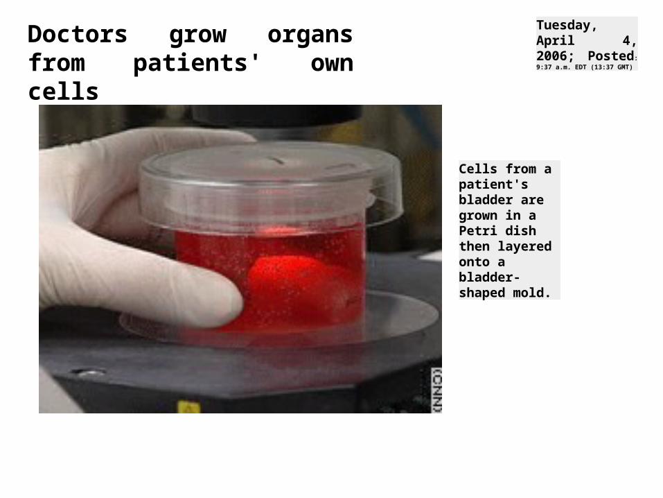
Cells from a patient's bladder are grown in a Petri dish then layered onto a bladder-shaped mold.
Tuesday, April 4, 2006; Posted:
9:37 a.m. EDT (13:37 GMT) Doctors grow organs from patients' own cells

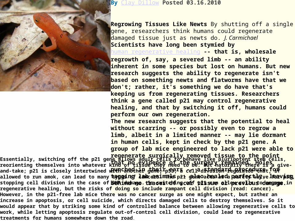
Humans Could Regenerate Tissue Like Newts Do By Switching Off a Single Gene By Clay Dillow Posted 03.16.2010 Regrowing Tissues Like Newts By shutting off a single gene, researchers think humans could regenerate damaged tissue just as newts do. J. Carmichael Scientists have long been stymied by human regenerative healing -- that is, wholesale regrowth of, say, a severed limb -- an ability inherent in some species but lost on humans. But new research suggests the ability to regenerate isn't based on something newts and flatworms have that we don't; rather, it's something we do have that's keeping us from regenerating tissues. Researchers think a gene called p21 may control regenerative healing, and that by switching it off, humans could perform our own regeneration.The new research suggests that the potential to heal without scarring -- or possibly even to regrow a limb, albeit in a limited manner -- may lie dormant in human cells, kept in check by the p21 gene. A group of lab mice engineered to lack p21 were able to regenerate surgically removed tissue to the point that no evidence of the surgery remained. Holes punched in their ears -- a standard procedure for tagging lab animals -- also healed perfectly, leaving behind no traces of scar tissue or previous damage.
Essentially, switching off the p21 gene allows adult cells to behave like pluripotent stem cells, reorienting themselves into whatever kind of tissue they need to be. But naturally there is a give-and-take; p21 is closely intertwined with another gene, p53, a cell-division regulator that, if allowed to run amok, can lead to many types of cancers. The p21 gene acts as a safety valve for p53, stopping cell division in the case of DNA damage. So switching off p21 can allow cells to engage in regenerative healing, but the risks of doing so include rampant cell division (read: cancer). However, in the p21-free lab mice there was no cancer surge as one might expect, but rather an increase in apoptosis, or cell suicide, which directs damaged cells to destroy themselves. So it would appear that by striking some kind of controlled balance between allowing regenerative cells to work, while letting apoptosis regulate out-of-control cell division, could lead to regenerative treatments for humans somewhere down the road.


![Reaction rates for mesoscopic reaction-diffusion … rates for mesoscopic reaction-diffusion kinetics ... function reaction dynamics (GFRD) algorithm [10–12]. ... REACTION RATES](https://img.pdfslide.net/doc/110x75/5b33d2bc7f8b9ae1108d85b3/reaction-rates-for-mesoscopic-reaction-diffusion-rates-for-mesoscopic-reaction-diffusion.jpg)