Embed Size (px)
Citation preview

1Copyright © McGraw-Hill Education. Permission required for reproduction or display.
Chapter 01
Lecture Outline
See separate PowerPoint slides for all figures and tables pre-
inserted into PowerPoint without notes.

• Anatomy studies the form and
structure of the body
• Physiology examines how the body
functions
• Form and function are interrelated
1.1 Anatomy and Physiology Compared
2

1.1a Anatomy: Details of Structure and Form
• Subdivisions of microscopic anatomy
Cytology—study of body cells and their internal
structure
Histology—study of tissues
3

1.1a Anatomy: Details of Structure and Form
• Subdivisions of gross anatomy
Systemic anatomy studies anatomy of each functional
body system
Regional anatomy examines all of the structures in a
particular region of the body
Surface anatomy focuses on superficial anatomic
markings and internal body structures
Comparative anatomy examines anatomical similarities
and differences in different species
Embryology studies developmental changes from
conception to birth
4
1.1b Physiology: Details of Function
• Physiologists examine the following organ systems,
focusing on the molecular and cellular level
Cardiovascular, functioning of the heart, blood vessels, and
blood
Neurophysiology, functioning of nerves and nervous system
organs
Respiratory physiology, functioning of respiratory organs
Reproductive physiology, functioning of reproductive
hormones and the reproductive cycle
Pathophysiology, relationship between the function of an
organ system and disease or injury to the system
5

Copyright © 2016 McGraw-Hill Education. All rights reserved. No reproduction or distribution without the prior written consent of McGraw-Hill EducationCopyright © 2016 McGraw-Hill Education. All rights reserved. No reproduction or distribution without the prior written consent of McGraw-Hill Education
What did you learn?
• What subdiscipline of
anatomy may explore how the
lower limb differs between
humans and chimpanzees?
• What subdiscipline of
anatomy focuses on cells?
• What subdiscipline of
physiology focuses on
disease?
6
1.3a Characteristics That Describe Living Things
Properties common to all organisms
• Organization
All organisms exhibit a complex structure and order
• Metabolism—the sum of all chemical reactions that
occur within the body
Anabolism—small molecules joined to form larger ones
Catabolism—large molecules broken down into smaller ones
• Growth and development
Organisms assimilate materials from environment; grow and
develop
7

1.3a Characteristics that Describe Living Things
Properties common to all organisms (continued )
• Responsiveness—ability to sense and react to stimuli
• Regulation
Ability to adjust internal bodily function to accommodate
environment changes
Homeostasis—ability to maintain body structure and function
• Reproduction
Produce new cells for growth, maintenance, and repair
With sex cells (gametes), can develop into new organisms
8
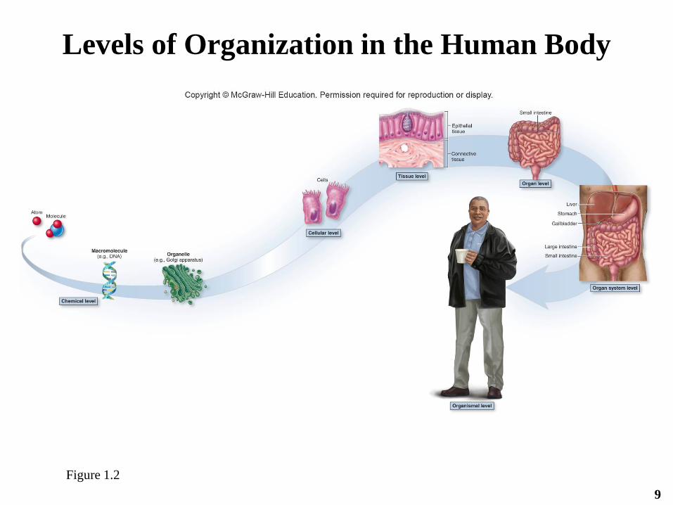
Figure 1.2
9
Levels of Organization in the Human Body

1.3b The View from Simplest to Most Complex
• Chemical level
Atoms—smallest units of matter
Molecules—one or more combined atoms
oE.g., sugar, water, vitamin
Macromolecules—more complex molecules
oE.g., proteins and deoxyribonucleic acid (DNA)
oOrganelles—microscopic structures within cells
10

1.3b The View from Simplest to Most Complex
• Cellular level
Cells—the smallest living structures
oBasic units of structure and function in
organisms
oFormed from molecules from the chemical level
oVary widely in structure, reflecting specialized
functions
11

1.3b The View from Simplest to Most Complex
• Tissue level
– Groups of similar cells performing common
functions
– Four main categories of tissues
oEpithelial tissue covers exposed surfaces and lines body
cavities
oConnective tissue protects, supports, and binds structures
and organs
oMuscle tissue produces movement
oNervous tissue conducts nerve impulses
12

1.3b The View from Simplest to Most Complex
• Organ level
– Two or more tissue types performing specific functions
o E.g., the small intestine composed of all four tissue types, working
to process and absorb digested nutrients
• Organ system level
– Related organs working together to achieve a common
function
o E.g., organs of the digestive system working together to digest
food, absorb nutrients, and expel waste products
• Organismal level
– Highest level of structural organization
– All body systems function interdependently
13

1.3b The View from Simplest to Most Complex
• Levels of organization from simplest to most
complex
Chemical level
____________
____________
____________
____________
____________
14

1.3b The View from Simplest to Most Complex
• Levels of organization from simplest to most
complex
Chemical level
Cellular level
Tissue level
Organ level
Organ system level
Organismal level
15

1.3c Introduction to Organ Systems
11 organ systems of the human body
1. Integumentary system
2. Skeletal system
3. Muscular system
4. Nervous system
5. Endocrine system
6. Cardiovascular system
16

1.3c Introduction to Organ Systems
11 organ systems of the human body (continued )
7. Lymphatic system
8. Respiratory system
9. Urinary system
10. Digestive system
11. Male and female reproductive systems
17

Figure 1.318
Organ
Systems

Figure 1.3 (continued)
19
Organ
Systems

Figure 1.3 (continued)
20
Organ
Systems

Copyright © 2016 McGraw-Hill Education. All rights reserved. No reproduction or distribution without the prior written consent of McGraw-Hill EducationCopyright © 2016 McGraw-Hill Education. All rights reserved. No reproduction or distribution without the prior written consent of McGraw-Hill Education
What did you learn?
• What characteristics are common to all life?
• List the levels of organization within the body, starting with the most complex?
• Which organ system is responsible for filtering waste products from the blood and excreting them in urine? For movement and generation of heat? Contains the site for blood cell formation?
21
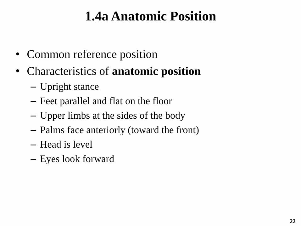
1.4a Anatomic Position
• Common reference position
• Characteristics of anatomic position
– Upright stance
– Feet parallel and flat on the floor
– Upper limbs at the sides of the body
– Palms face anteriorly (toward the front)
– Head is level
– Eyes look forward
22

1.4b Sections and Planes
• “Slices” of body called sections or planes
Section—actual cut or slice that exposes internal
anatomy
Plane—imaginary flat surface passing through body; 3
types
o Coronal (or frontal ) plane
˗ Vertical plane dividing the body into anterior (front) and
posterior (back) parts
o Transverse (or cross-sectional ) plane
˗ Horizontal plane dividing the body into superior (top) and
inferior (bottom) parts
23

1.4b Sections and Planes
Plane (continued )
o Midsagittal (or median) plane
˗ Vertical plane dividing the body into equal left and right
halves
o Sagittal plane
˗ Parallel to midsagittal, but left or right of midsagittal;
divides structure into unequal portions
o Oblique plane
˗ Passes through structure at an angle
24

Figure 1.425

Figure 1.6
26
Directional Terms in Anatomy

27

28

Copyright © 2016 McGraw-Hill Education. All rights reserved. No reproduction or distribution without the prior written consent of McGraw-Hill EducationCopyright © 2016 McGraw-Hill Education. All rights reserved. No reproduction or distribution without the prior written consent of McGraw-Hill Education
What did you learn?
• What plane divides the body into anterior/posterior halves?
• The elbow is ________ to the wrist?
• The toes are ________ to the knee?
• The mouth is ________ to the ear?
• The hairs on my head are ________ to the skin?
29

1.4d Regional Anatomy
• Human body is partitioned into two main
regions
Axial region
o Head, neck, and trunk
o Forms the main vertical axis of the body
Appendicular region
o Upper and lower limbs
• Several more regions within these two main
ones
30

Figure 1.7a
31

Figure 1.7b
32

Copyright © 2016 McGraw-Hill Education. All rights reserved. No reproduction or distribution without the prior written consent of McGraw-Hill EducationCopyright © 2016 McGraw-Hill Education. All rights reserved. No reproduction or distribution without the prior written consent of McGraw-Hill Education
What did you learn?
• The term antebrachial refers to which body region?
• The term crural refers to which body region?
• The term axillary refers to which body region?
• Where can I find the deltoid muscle?
• If a patient is complaining of lower back pain, what region could be involved?
• What region is involved with tarsal tunnel syndrome?
• A Baker’s cyst is usually found in the popliteal fossa which is the region of?
33

Figure 1.834
Body Cavities

1.4e Body Cavities and Membranes
• Significant difference between posterior aspect and
ventral cavity—subdivisions of ventral cavity are lined
with serous membranes
• Two layers of serous membranes
Parietal layer lines internal surface of body wall
Visceral layer covers external surface of organs (viscera)
o Serous cavity—space between membranes
• Serous fluid
– Liquid secreted by cells in serous membrane
– Acts as lubricant
– Reduces friction caused by movement of organs against body
wall35

1.4e Body Cavities and Membranes
Thoracic cavity• Mediastinum—median space in the thoracic cavity
– Contains heart, thymus, esophagus, trachea, and major blood vessels that connect to the heart
• Pericardial cavityo Space between parietal and visceral layers containing serous
fluid
– Parietal pericardium
o Outer layer, which forms the sac around the heart
– Visceral pericardium
o Forms the heart’s external surface
36

1.4e Body Cavities and Membranes
– Pleural cavity
o Space between parietal and visceral layers containing
serous fluid
– Parietal pleura
o Outer layer lines internal surface of thoracic wall
– Visceral pleura
o Inner layer covers external surface of lungs
37

Figure 1.9 b-c
38
Serous
Membranes in the
Thoracic and
Abdominopelvic
Body Cavities

1.4e Body Cavities and Membranes
Abdominopelvic cavity
• Abdominal cavity
– Superior area
– Contains most of the digestive system organs, kidneys, and
most of the ureters
• Pelvic cavity
– Inferior area, between hip bones
– Contains distal part of large intestine, remainder of ureters
and urinary bladder, and internal reproductive organs
39

1.4e Body Cavities and Membranes
Abdominopelvic cavity (continued )
• Peritoneum—two-layered serous membrane lining the
abdominopelvic cavity
– Parietal peritoneum
o Outer layer, which lines the internal walls of the abdominopelvic
cavity
– Visceral peritoneum
o Inner layer, which covers the external surface of most abdominal and
pelvic organs
40

Figure 1.9d
41
Serous
Membranes in the
Thoracic and
Abdominopelvic
Body Cavities

1.4f Abdominopelvic Regions and Quadrants
Abdominopelvic cavity
is partitioned into nine
compartments
• Umbilical region
– Middle region, named
for the umbilicus
(navel) that lies in its
center
• Epigastric region
– Superior to umbilical
• Hypogastric region
– Inferior to umbilicalFigure 1.10a 42
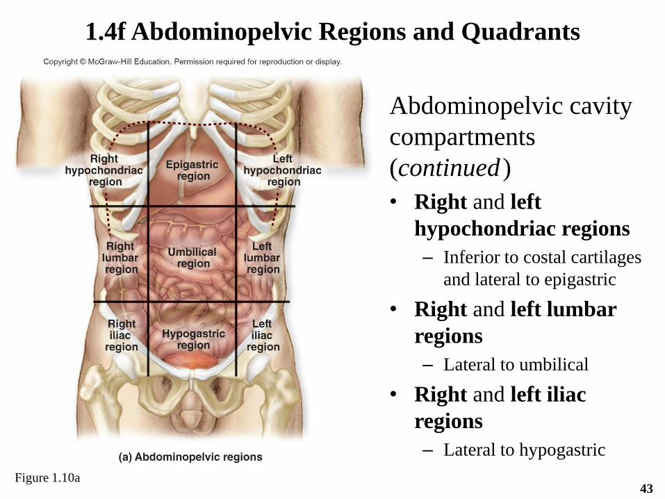
Abdominopelvic cavity
compartments
(continued )
• Right and left
hypochondriac regions
– Inferior to costal cartilages
and lateral to epigastric
• Right and left lumbar
regions
– Lateral to umbilical
• Right and left iliac
regions
– Lateral to hypogastric
1.4f Abdominopelvic Regions and Quadrants
Figure 1.10a43

• Abdominopelvic
cavity can also be
divided into four
compartments with
transverse and
midsagittal planes
through the umbilicus
– Right and left upper
quadrant
– Right and left lower
quadrant
1.4f Abdominopelvic Regions and Quadrants
Figure 1.10b44

Copyright © 2016 McGraw-Hill Education. All rights reserved. No reproduction or distribution without the prior written consent of McGraw-Hill EducationCopyright © 2016 McGraw-Hill Education. All rights reserved. No reproduction or distribution without the prior written consent of McGraw-Hill Education
What did you learn?
• Which body cavity is associated
with the lungs?
• Which body cavity is associated
with the heart?
• What serous membrane lines the
abdominopelvic cavity?
• Patient with pain in the RUQ may
cause you to suspect involvement
of which organ(s)?
45

1.5 Homeostasis: Keeping
Internal Conditions Stable
• Hundreds of anatomic structures and
physiologic processes are continuously
monitored and adjusted
• Homeostasis—the ability of an organism to
maintain consistent internal environment, or
“steady state,” in response to changing internal
or external conditions
46

1.5a Components of Homeostatic Systems
Three components of homeostatic systems
• Receptor detects changes in a variable
Stimulus (e.g., change in temperature sensed by skin)
• Control center interprets input from receptor and
initiates changes through effector
Nervous system can provide a quicker response
o E.g., regulation of blood pressure upon rising
Endocrine response is more sustained
o E.g., parathyroid hormone regulating calcium levels
• Effector is the structure that brings about changes to
alter the stimulus
47

Figure 1.1148
Components of a Homeostatic Control Mechanism

1.5b Homeostatic Systems Regulated
by Negative Feedback
• Negative feedback
Controls most processes in the body
Variable fluctuates within a normal range around a set
point
If a stimulus increases a variable, receptor informs
homeostatic control system, which activates effector to
decrease variable back into normal range
If a stimulus decreases a variable, receptor informs
homeostatic control system, which activates effector to
increase variable back into normal range
So, in negative feedback, homeostatic control responds to
move variable in opposite direction to bring it into
normal range49

Example: Temperature regulation
1. Body temperature drops
2. Sensory receptors detect this and signal hypothalamus in the brain
3. Hypothalamus sends nerve impulses to smooth muscle in blood
vessels in skin, causing them to contract, called vasoconstriction,
which decreases internal passageway, or lumen, of vessels
4. This decreases blood flow to the surface of the body
5. Thus, less heat is released through skin
6. Also, nerve impulses are sent to skeletal muscles, causing
shivering to warm body
7. Also, nerve impulses are sent to piloerector muscles attached to
hair follicles in skin, causing them to contract (goosebumps) to
warm the body
1.5b Homeostatic Systems Regulated
by Negative Feedback
50

Example: Temperature regulation (continued )
1. Body temperature rises
2. Sensory receptors detect rise and signal hypothalamus
3. Hypothalamus sends nerve impulses to smooth muscle in
blood vessels in the skin, causing relaxation or
vasodilation, which increases size of lumen
4. Increases blood flow to body surface
5. So, more heat is released through skin
1.5b Homeostatic Systems Regulated
by Negative Feedback
51

Figure 1.13a52

Figure 1.13b53

• Other examples of homeostatic regulation
– Withdrawal reflex in response to injury
– Regulating heart rate and blood pressure during exercise
– Changing breathing rate in response to increased carbon
dioxide
– Parathyroid hormone release in response to decreased
calcium
– Release of insulin by the pancreas in response to increased
blood glucose
1.5b Homeostatic Systems Regulated
by Negative Feedback
54

1.5c Homeostatic Systems Regulated
by Positive Feedback
• Positive feedback
Occurs much less frequently than negative
feedback
Stimulus reinforced to continue moving variable
in same direction until a climactic event occurs,
then body returns to homeostasis
55

• Positive feedback during breastfeeding
1. Sensory detectors detect baby suckling
2. Message is transmitted to the hypothalamus
3. Hypothalamus signals posterior pituitary to release the
hormone oxytocin
4. Oxytocin stimulates the mammary gland to eject breast milk
5. Cycle repeats as long as the baby suckles
• Other examples of positive feedback
– Blood clotting cascade
– Uterine contractions of labor
1.5c Homeostatic Systems Regulated
by Positive Feedback
56

Figure 1.1557
Positive Feedback

• Normal ranges for homeostatic variables
– Body temperature 98.6ºF
– Blood glucose 80–110 mg/dL
– Blood pressure 90–120/60-80 mm Hg
– Determined by sampling healthy individuals in a
population
– Normal range is value for 95% of individuals sampled
– 5% of healthy population have values outside normal range
58
Clinical View: Establishing Normal Ranges
for Clinical Practice

• Diabetes is an example of homeostatic imbalance
– Occurs when homeostatic mechanisms for regulating blood
glucose are not functioning normally
• Type 1 and Type 2 Diabetes
– Blood glucose fluctuations and high glucose readings
• Viral or Bacterial infections and fever
– Alters the set point of normal body temperature
– If not controlled, cell death can occur from inability to
maintain homeostasis
1.6 Homeostasis, Health, and Disease
59

• Treating patients involves finding a diagnosis, a
specific cause of the homeostatic imbalance
• Developing a diagnosis
– Examine patient and gather data
o E.g., patient health history, complaints, current vital signs such as
weight and blood pressure
– Initial hypothetical diagnosis
– Order tests
– Confirm, modify, or reject initial diagnosis
– Definitive diagnosis
60
Clinical View: Clinicians’ Use
of Scientific Method

Drugs may affect normal homeostatic control mechanisms (e.g.,
SSRIs)
• Patients with depression may have lower levels of serotonin in
their brains
• SSRI drugs block reuptake of serotonin into nerve cells in
brain, thus prolonging its effects; SSRIs help elevate mood of
patients with depression
• Side effects of SSRIs due to
– Serotonin also used in nerve cells of digestive system
– Digestive system becomes more excitable due to SSRI
– Symptoms include nausea and upset stomach
1.6 Homeostasis, Health, and Disease
61

62
Clinical View: Medical Imaging
Radiography
Positron Emission
Tomography (PET)
Magnetic
Resonance
Imaging (MRI)
Digital Subtraction
Angiography (DSA)
Computed Tomography (CT)
Sonography

63
Abdominal CAT Scan
MRI of the Lumbar Spine