Embed Size (px)
Citation preview

1
Chapter 04
Lecture and
Animation Outline
Copyright © McGraw-Hill Education. Permission required for reproduction or display.
To run the animations you must be in Slideshow View. Use the
buttons on the animation to play, pause, and turn audio/text on or
off.
Please Note: Once you have used any of the animation functions
(such as Play or Pause), you must first click on the slide’s
background before you can advance to the next slide.
See separate PowerPoint slides for all figures and tables pre-
inserted into PowerPoint without notes and animations.

Introduction
• Necessary to have some familiarity with DNA and genes in order to study genetic
disorders that effect hereditary traits – Color blindness, cystic fibrosis, diabetes mellitus,
hemophilia
• Mendelian genetics helps us discern and predict patterns of inheritance within a family line
4-2

4-3
DNA Structure and Function
• Deoxyribonucleic acid (DNA)—long, thread-like molecule with uniform diameter, but varied length – 46 DNA molecules
(chromosomes) in nucleus of most human cells
– Nucleotide consists of a sugar, phosphate group, and nitrogenous base

4-4
DNA Structure and Function
• Nitrogenous bases in nucleic acids
are purines and pyrimidines
• Purines—double ring
– Adenine (A)
– Guanine (G)
• Pyrimidines—single ring
– Cytosine (C)
– Thymine (T)
– Uracil (U)
Figure 4.1b
Copyright © The McGraw-Hill Companies, Inc. Permission required for reproduction or display.
C
NH2 N
N H
C C H
C H
N
N C
Adenine (A)
Purines
C
O N
N H
C C H
C N
H N C
NH2
Guanine (G)
H C
NH2
C
N H
C
H C N
O
Cytosine (C)
Uracil (U)
C
C
O
C
O
C H
H N C H
N H
N H
C
C
H C
CH 3
N H
O
O
Thymine (T)
Pyrimidines
(b)

4-5
DNA Structure and Function
• Nitrogenous bases united by hydrogen bonds – A purine on one strand always bound
to a pyrimidine on the other
– A–T two hydrogen bonds
– C–G three hydrogen bonds
• DNA base pairing – A–T
– C–G
• Law of complementary base pairing – One strand determines base
sequence of other Figure 4.2b,c
(b)
(c)
G C
Sugar–phosphate
backbone
Sugar–phosphate
backbone
G
A
C
T
T T
G C
Hydrogen
bond
Copyright © The McGraw-Hill Companies, Inc. Permission required for reproduction or display.

4-6
Chromatin and Chromosomes
• Chromatin—fine filamentous
DNA material complexed with
proteins
– Occurs as 46 chromosomes in
most cells
– 6 feet long thread packed in cell
nucleus of 5 μm diameter
– In nondividing cells, chromatin
is so slender it cannot be seen
with light microscope
– Granular appearance under
electron microscope
Figure 4.4b

4-7
RNA Structure and Function
• Ribonucleic acids (RNAs)—smaller molecules that
resemble DNA
– Can have less than 100 or just over 10,000 bases per
molecule
• Three important RNAs for protein synthesis
– Messenger RNA (mRNA)
– Ribosomal RNA (rRNA)
– Transfer RNA (tRNA)
• One nucleotide chain (not a double helix)
• Ribose replaces deoxyribose as the sugar
• Uracil replaces thymine as a nitrogenous base
• Functions mainly in cytoplasm
4-8
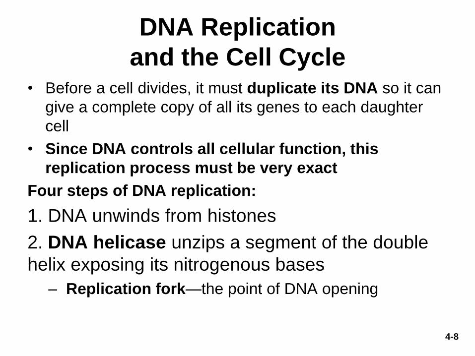
DNA Replication
and the Cell Cycle • Before a cell divides, it must duplicate its DNA so it can
give a complete copy of all its genes to each daughter
cell
• Since DNA controls all cellular function, this
replication process must be very exact
Four steps of DNA replication:
1. DNA unwinds from histones
2. DNA helicase unzips a segment of the double
helix exposing its nitrogenous bases
– Replication fork—the point of DNA opening

DNA Replication
3. DNA polymerase builds new DNA strands
– Polymerase reads exposed bases and matches
complementary free nucleotides
– Separate polymerase molecules work on each strand
proceeding in opposite directions
– The polymerase moving toward the replication fork
makes a long, continuous, new strand of DNA
– The polymerase moving away from the replication fork
makes short segments of DNA; DNA ligase joins them
together
– Ultimately, two daughter DNA molecules are made from
the original parental DNA
• Semiconservative replication—each daughter DNA consists of
one old and one new helix
4-9
4-10
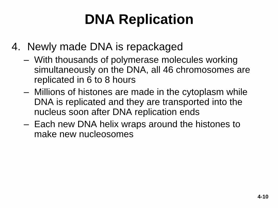
DNA Replication
4. Newly made DNA is repackaged – With thousands of polymerase molecules working
simultaneously on the DNA, all 46 chromosomes are replicated in 6 to 8 hours
– Millions of histones are made in the cytoplasm while DNA is replicated and they are transported into the nucleus soon after DNA replication ends
– Each new DNA helix wraps around the histones to make new nucleosomes

4-11
DNA Replication
Figure 4.14

4-12
Errors and Mutations
• DNA polymerase does make mistakes
– Multiple modes for correction of replication errors
– Double checks the new base pair and tends to replace
incorrect, biochemically unstable pairs with more stable,
correct pairs
– Result is only one error per 1 billion bases replicated
• Mutations—changes in DNA structure due to
replication errors or environmental factors
(radiation, viruses, chemicals)
– Some mutations cause no ill effects, others kill the cell,
turn it cancerous, or cause genetic defects in future
generations

4-13
The Cell Cycle
• Cell cycle—cell’s life from one division to the next – Includes interphase and
mitotic phase
– Interphase includes three subphases
• G1, S, G2
– Mitotic phase includes multiple subphases
• Prophase, Metaphase, Anaphase, Telophase
Figure 4.15

The Cell Cycle
• G1 phase—the first gap phase – Interval between cell birth (from division) and DNA replication
– Cell carries out normal tasks and accumulates materials for next phase
• S phase—synthesis phase – Cell replicates all nuclear DNA and duplicates centrioles
• G2 phase—second gap phase – Interval between DNA replication and cell division
– Cell repairs DNA replication errors, grows and synthesizes enzymes that control cell division
• M phase—mitotic phase – Cell replicates its nucleus
– Pinches in two to form new daughter cells
• G0 (G zero) phase—describes cells that have left the cycle and cease dividing for a long time (or permanently)
• Cell cycle duration varies between cell types
4-14

4-15
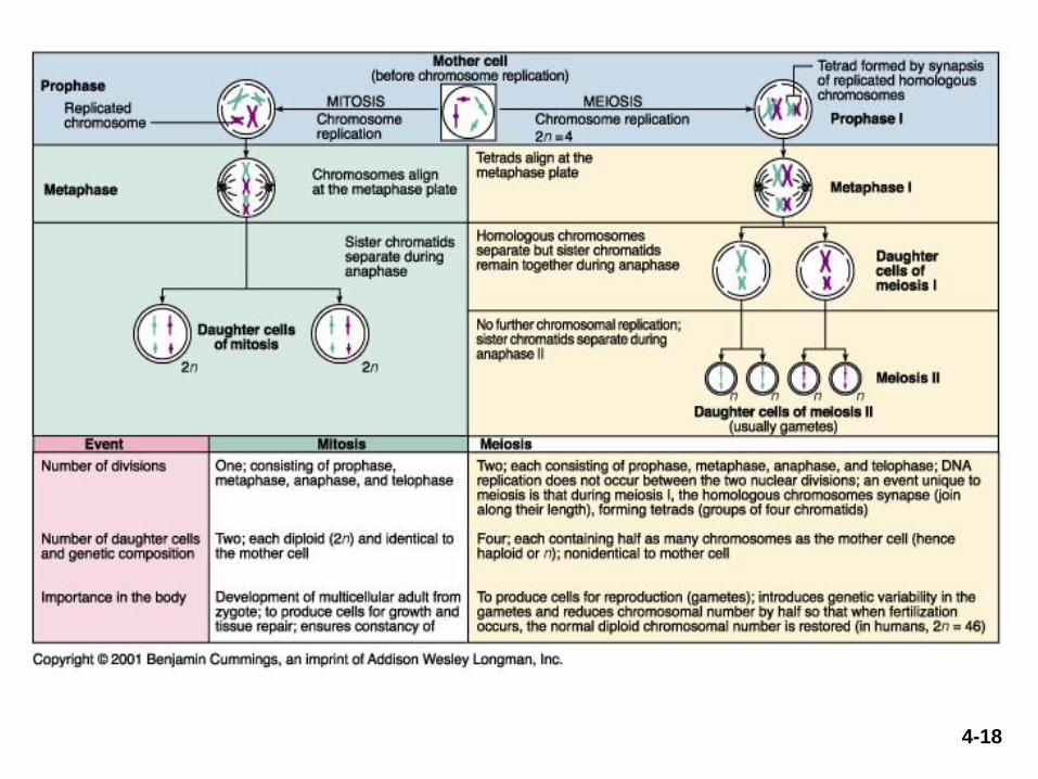
Mitosis
Figure 4.16 parts 1 & 2

4-16
Mitosis
Figure 4.16 parts 3 & 4

4-17
Mitosis
• Cytokinesis—division of cytoplasm into two
cells – Telophase is the end of nuclear division but
overlaps cytokinesis
• Achieved by myosin protein pulling on actin in
the terminal web of cytoskeleton
• Creates cleavage furrow around the equator
of cell
• Cell eventually pinches in two
4-18

4-19
The Timing of Cell Division
Cells divide when: • They have enough cytoplasm for two daughter cells
• They have replicated their DNA
• They have adequate supply of nutrients
• They are stimulated by growth factors (chemical signals)
• Neighboring cells die, opening up space
Cells stop dividing when: • They snugly contact neighboring cells
• Nutrients or growth factors are withdrawn
• They undergo contact inhibition—the cessation of cell division in response to contact with other cells

4-20
What Is a Gene?
• Gene—an information-containing segment of DNA that codes for the production of a molecule of RNA that plays a role in synthesizing one or more proteins
– Amino acid sequence of a protein is determined by the nucleotide sequence in the DNA
• Genome—all the DNA in one 23-chromosome set
• 46 human chromosomes come in two sets of 23 chromosomes – One set of 23 chromosomes came from each parent
• Human Genome Project (1990–2003) identified the base sequences of 99% of the human genome – Genomics—study of the whole genome and how its genes and
noncoding DNA interact to affect structure and function of the whole organism

4-21
The Genome
• Findings of Human Genome Project: – Homo sapiens has fewer than 100,000 genes
– A single gene can code for many different proteins
– A gene is on average 3,000 bases long (can be up to 2.4 million
bases long)
– All humans are at least 99.99% genetically identical
• Still, two individuals can differ by more than 3 million base pairs
• Combinations of single-nucleotide polymorphisms account for
all human genetic variation
– Currently we know locations of over 1,400 disease-producing
mutations
• Opens possibilities of genomic medicine
• Gene-substitution therapy

The Genetic Code
• Base triplet—a sequence of three DNA
nucleotides that stands for one amino acid
– Codon—the 3-base sequence in mRNA
– 64 possible codons available to represent the 20
amino acids
• 61 code for amino acids; 3 are stop codons
• Stop codons—UAG, UGA, and UAA: signal “end of
message,” like a period at the end of a sentence
• Start codon—AUG codes for methionine, and begins the
amino acid sequence of the protein
4-22

Protein Synthesis
• All body cells, except sex cells and some immune cells,
contain identical genes
• Different genes are activated in different cells
• When a gene is activated, messenger RNA (mRNA) is made
– mRNA—complementary to gene • Migrates from the nucleus to cytoplasm where it codes for amino
acids
• Process of protein synthesis
– DNA mRNA protein
– In transcription, DNA codes for mRNA
• Occurs within nucleus
– In translation, mRNA codes for protein
• Usually occurs in cytoplasm
4-23

4-24
Alternative Splicing of mRNA
• One gene can code for more than one protein
• Exons can be spliced together into a variety of
different mRNAs
Figure 4.6
Gene (DNA)
Pre-mRN A Intron Exon
mRN A 2 mRN A 1 mRN A 3
Protein 2 Protein 1 Protein 3
T ranscription 1
T ranslation 3
Splicing 2
A B C D E F
A C D A E F B D E
Copyright © The McGraw-Hill Companies, Inc. Permission required for reproduction or display.
Translation
• Three steps to translation: Initiation, Elongation, Termination
• Initiation – Leader sequence in mRNA binds to small ribosomal subunit
– Initiator tRNA (bearing methionine) pairs with start codon
– Large ribosomal subunit joins the complex and the now fully formed ribosome begins reading bases
4-25

Translation
• Elongation – Next tRNA (with its amino acid) binds to ribosome while its
anticodon pairs with next codon of mRNA
– Peptide bond forms between methionine and second amino acid
– Ribosome slides to read next codon and releases initiator tRNA (empty)
– Next tRNA with appropriate anticodon brings its amino acid to ribosome
– Another peptide bond forms (between 2nd and 3rd amino acids)
– Process continually repeats, extending peptide to a protein
• Termination
– When ribosome reaches stop codon a release factor binds to it
– Finished protein breaks away from ribosome
– Ribosome dissociates into two subunits
4-26
4-27
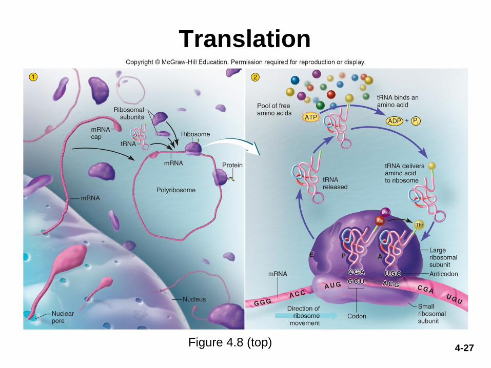
Translation
Figure 4.8 (top)
4-28
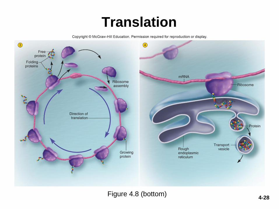
Translation
Figure 4.8 (bottom)

4-29
Protein Processing and Secretion
Figure 4.11
Nucleus
Rough ER
Golgi
complex
Lysosome
Ribosomes
Protein packaged into transport
vesicle, which buds from ER.
T ransport vesicles fuse into clusters that
unload protein into Golgi complex.
Golgi complex modifies
protein structure.
Golgi vesicle containing
finished protein is formed.
Secretory vesicles
release protein by
exocytosis.
1
2
3
4
5
6
Clathrin-coated
transport vesicle
Protein formed by
ribosomes on rough ER.
Copyright © The McGraw-Hill Companies, Inc. Permission required for reproduction or display.

Gene Regulation
• Genes can be turned on and off
– Cells can turn some genes permanently off
• Example: liver cells turn off hemoglobin genes
– Cells can turn genes on only when needed
• The level of gene expression can vary from day to day
or hour to hour
• This can be controlled by chemical messengers such
as hormones
• Example: mammary gland cells turn on gene for casein
protein only when breast milk is produced
4-30

4-31
Gene Regulation
Figure 4.12
mRNA
for casein
Casein
Exocytosis
Regulatory
protein
(transcription
activator)
Casein
gene
RNA
polymerase
Rough
endoplasmic
reticulum
Golgi
complex
Secretory
vesicles
Prolactin
Prolactin
receptor
2
1
3
4
5
6
7
ATP
Pi
AD P
+
Copyright © The McGraw-Hill Companies, Inc. Permission required for reproduction or display.

4-32
Synthesizing Compounds Other
Than Proteins
• Cells synthesize glycogen, fat, steroids, phospholipids, pigments, and other compounds – No genes for these products, but their synthesis is under indirect
genetic control
– They are produced by enzymatic reactions, and enzymes are proteins encoded by genes
• Example: production of testosterone (a steroid) – A cell of the testes takes in cholesterol
– Enzymatically converts it to testosterone
– Only occurs when genes for enzyme are active
Review Questions
• What is the difference between DNA and RNA?
• What are the complementary base pairings in
DNA? RNA?
• Sister chromatids separate to opposite poles
during which phase of mitosis?
• What is transcription and where does it occur?
Translation?
• What is the function of DNA polymerase vs.
RNA polymerase?
4-33

4-34
Chromosomes and Heredity
• Heredity—transmission of genetic characteristics from parent to offspring
• Karyotype—chart of 46 chromosomes laid out in order by size
• 23 pairs—the two members of each pair are called homologous chromosomes
– 1 chromosome from each pair inherited from each parent
• 22 pairs called autosomes
– Look alike and carry the same genes
• 1 pair of sex chromosomes (X and Y)
– Female has homologous pair of X chromosomes
– Male has one X and one much smaller Y chromosome

4-35
Karyotype
Figure 4.17

4-36
Genes and Alleles
• Locus—the location of a particular gene on a chromosome
• Alleles—different forms of gene at same locus on two homologous chromosomes
• Dominant allele (represented by capital letter) - If present, corresponding trait is usually seen in the
individual
- Masks effect of recessive allele
- Often produces protein responsible for visible trait
• Recessive allele (represented by lowercase letter) - Corresponding trait only seen when recessive allele
present on both homologous chromosomes
- Often codes for a nonfunctional variant of the protein

4-37
Genes and Alleles
• Genotype—alleles an individual possesses for a
particular trait
– Homozygous individuals—two identical alleles for the trait
– Heterozygous individuals—different alleles for that gene
• Phenotype—an observable trait
– An allele is expressed if it shows in the phenotype of an
individual
• Punnet square—diagram showing possible
genotype and phenotype outcomes from parents of
known genotype
– Example: Shows how two parents with dominant trait (cleft
chin) can produce child with recessive trait (uncleft chin)

4-38
Genetics of Cleft Chin
Figure 4.18 b
• Allele for cleft chin is
dominant
• Parents are heterozygous
• Each parent has cleft chin
phenotype
• One out of four offspring
will have uncleft chin

4-39
Genes and Alleles
• Parents can be healthy, heterozygous
carriers of hereditary diseases
• Genetic counselors—perform genetic testing
and advise couples on the probability of
transmitting genetic diseases

4-40
Multiple Alleles, Codominance, and
Incomplete Dominance
• Gene pool—genetic makeup of whole population
• Multiple alleles—more than two allelic forms of gene
– Example: three alleles for ABO blood types
• IA, IB, i alleles for ABO blood types
• Codominance—both alleles equally dominant
– Both are phenotypically expressed
– Example: IAIB = type AB blood
• Incomplete dominance
– Heterozygous individual shows phenotype intermediate
between traits each allele would have produced alone
– Example: familial hypercholesterolemia

4-41
Polygenic Inheritance and Pleiotropy
• Polygenic inheritance—genes at two or more loci contribute to a single phenotypic trait – Examples: eye color, skin color, some forms of cancer
Figure 4.19

4-42
Polygenic Inheritance and Pleiotropy
• Pleiotropy—one gene produces multiple phenotypic effects
– Example: alkaptonuria—disorder resulting from mutation on
chromosome 3 that blocks the breakdown of tyrosine
Figure 4.20

4-43
Sex Linkage
• Sex-linked traits—carried on X or Y chromosome, and therefore tend to be inherited by one sex more than the other
• Recessive color blindness allele on X, no gene locus for trait on Y, so color blindness more common in men (mother is carrier in illustrated example)
Figure 4.21
X X X Y
C c c
Female (XX)
Genotype Cc
Normal vision
Male (XY)
Genotype c–
Color blindness
Copyright © The McGraw-Hill Companies, Inc. Permission required for reproduction or display.

4-44
Epigenetics
• Epigenetics—field examining nongenetic
changes that alter gene expression and can be
passed to offspring
– Gene expression is changed without genetic mutation
to base sequence
– DNA methylation—mechanism of epigenetic change in
which methyl groups are added to DNA
• Often silences the gene
• Inappropriate DNA methylation implicated in some forms of
cancer

4-45
Cancer
• Oncology—medical specialty dealing with tumors
• Tumor angiogenesis—growth of blood vessels by energy-hungry tumors
• Cancers are named for tissue of origin
– Carcinomas: in epithelial tissue
– Lymphomas: in lymph nodes
– Melanomas: in pigment cells of epidermis (melanocytes)
– Leukemias: in blood-forming tissues
– Sarcomas: in bone, other connective tissue, or muscle

4-46
Cancer
• Malignant tumors cause cancer – Fast growing, tend to
metastasize—to give off cells that spread and seed the growth of tumors elsewhere
– Distinguished from slow growing, encapsulated benign tumors
Figure 4.23

4-47
Cancer
• Carcinogens—environmental cancer-causing
agents
– Radiation—ultraviolet rays, X-rays
– Chemical—cigarette tar, food preservatives, industrial
chemicals
– Viruses—human papillomavirus, hepatitis C, and type 2
herpes simplex
• Only 5% to 10% of cancers are hereditary
• Carcinogens trigger gene mutations

4-48
Cancer
• Oncogenes – Cause cell division to accelerate out of control
• Excessive production of growth factors or growth-factor receptors that stimulate mitosis
• Tumor-suppressor (TS) genes – Healthy tumor suppressor genes inhibit development of
cancer • Some code for DNA-repair enzymes
– Mutated TS genes or silenced TS genes leave oncogene action unopposed


![Chapt09 Holes Lecture Animation[1]](https://img.pdfslide.net/doc/110x75/55493f66b4c9050a4d8b4f6d/chapt09-holes-lecture-animation1.jpg)
![Chapt10 Holes Lecture Animation[1]](https://img.pdfslide.net/doc/110x75/55572092d8b42a067f8b4aad/chapt10-holes-lecture-animation1.jpg)
![Chapt07 Holes Lecture Animation[1]](https://img.pdfslide.net/doc/110x75/555a9195d8b42a3e268b469f/chapt07-holes-lecture-animation1.jpg)


![Chapt06 Holes Lecture Animation[1]](https://img.pdfslide.net/doc/110x75/554b4455b4c9054b5e8b4c10/chapt06-holes-lecture-animation1.jpg)


![Chapt03 Holes Lecture Animation[1]](https://img.pdfslide.net/doc/110x75/55503bd3b4c905b2788b45df/chapt03-holes-lecture-animation1.jpg)