Embed Size (px)
Citation preview

1
Chapter 05
Cardiovascular
System

2
Cardiovascular System:
Heart and Blood Vessels

3
Points to ponder
• What are the functions of the cardiovascular system?
• What is the anatomy of the heart? Of blood vessels, such as veins and arteries?
• How is the heart beat regulated?
• What is blood pressure?
• What are common cardiovascular diseases and how might you prevent them?

4
5.1 Overview of the Cardiovascular System
Figure 5.1 The cardiovascular system
and homeostasis.
What is the cardiovascular system?Copyright © The McGraw-Hill Companies, Inc. Permission required for reproduction or display.
O2
tissue cellsRespiratory System
kidneys
food
liver
Urinary SystemDigestive System
metabolic wastes
(urine)
indigestible
food residues (feces)
CO2
Cardiovascular System
• Includes Heart & blood vessels
• Functions:
• Organs that refresh blood:
• What’s involved?
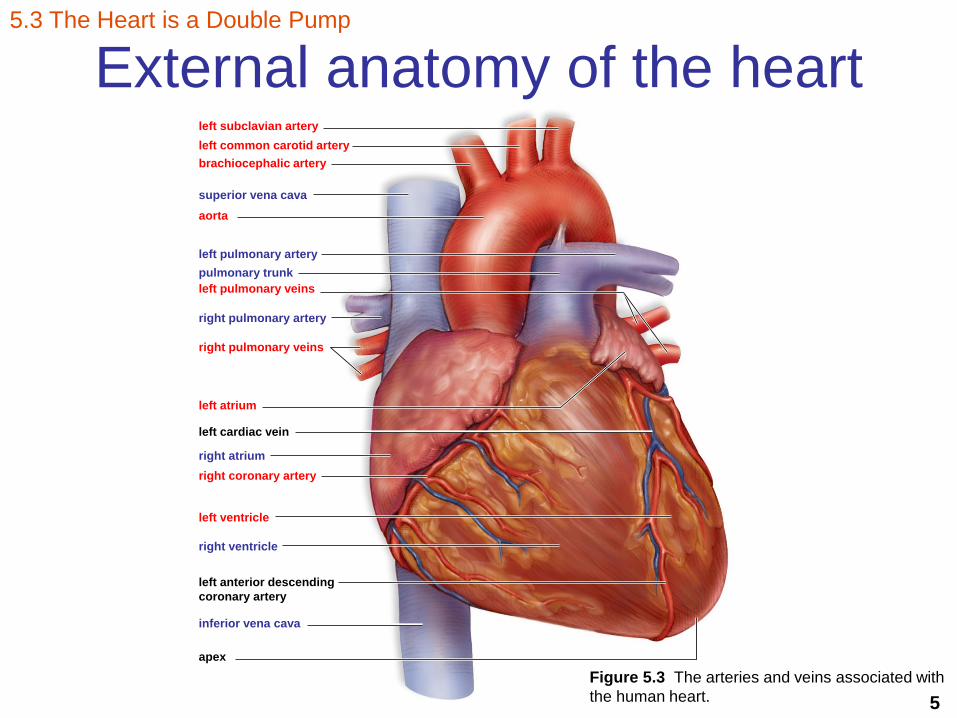
5
External anatomy of the heartleft subclavian artery
left common carotid artery
brachiocephalic artery
superior vena cava
aorta
left pulmonary artery
pulmonary trunk
left pulmonary veins
right pulmonary artery
right pulmonary veins
left atrium
left cardiac vein
right atrium
right coronary artery
left ventricle
right ventricle
left anterior descending
coronary artery
inferior vena cava
apex
Figure 5.3 The arteries and veins associated with
the human heart.
5.3 The Heart is a Double Pump

6
Internal anatomy of the heartCopyright © The McGraw-Hill Companies, Inc. Permission required for reproduction or display.
left subclavian artery
left common carotid artery
brachiocephalic artery
superior vena cava
aorta
left pulmonary artery
pulmonary trunk
left pulmonary veins
right pulmonary artery
right pulmonary veins
semilunar valve
left atrium
right atrium
atrioventricular
(bicuspid) valve
atrioventricular
(tricuspid) valve
chordae tendineae
papillary muscles
right ventricle
septum
left ventricle
inferior vena cava
a.Figure 5.4a The heart is a double pump.
5.3 The Heart is a Double Pump

7
Figure 42.7
Pulmonary artery
Right
atrium
Pulmonary
Semilunar
valve
Right
ventricle
Left
ventricle
Atrioventricular Valve
• Bicuspid/Mitral
Aortic Semilunar
valve
Left
atrium
Pulmonary
artery
Aorta
Atrioventricular Valve
• Tricuspid

8
Visualizing blood flow through the heart
Figure 5.4 The heart is a double pump.
5.3 The Heart is a Double Pump
Intercalated disc with gap junctions

9
Figure 42.10Artery
Red blood cells
Endothelium
Artery
Smoothmuscle
Connectivetissue
Capillary
Valve
Vein
Vein
Basal lamina
Endothelium
Smoothmuscle
Connectivetissue
100 m
LM
Venule
15
mL
M
Arteriole
Red blood cell
Capillary
Main pathway of
blood in the
body?

10
What are the 2 cardiovascular pathways
in the body?
• Pulmonary circuit
• Systemic circuit
5.5 Two Cardiovascular Pathways
O2
O2CO2
head and armscarotid artery
(also subclavian
artery to arms)
jugular vein
(also subclavian
vein from arms)
CO2
CO2
O2
O2
lungs
pulmonary
artery
superior
vena cava
inferior
vena cava
hepatic
vein
hepatic
portal
vein
renal
vein
iliac vein
CO2
trunk and legs
iliac
artery
renal
artery
intestinal
artery
aorta
pulmonary
vein
liver digestive
tract
kidneys
heart
Copyright © The McGraw-Hill Companies, Inc. Permission required for reproduction or display.
Figure 5.10 Overview of the cardiovascular system.
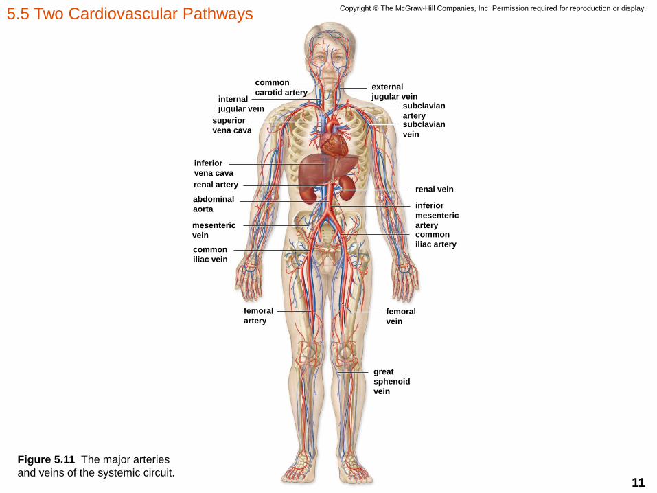
11
Copyright © The McGraw-Hill Companies, Inc. Permission required for reproduction or display.
common
carotid arteryinternal
jugular vein
superior
vena cava
inferior
vena cava
renal artery
abdominal
aorta
mesenteric
vein
common
iliac vein
femoral
artery
great
sphenoid
vein
femoral
vein
common
iliac artery
inferior
mesenteric
artery
renal vein
subclavian
vein
subclavian
artery
external
jugular vein
Figure 5.11 The major arteries
and veins of the systemic circuit.
5.5 Two Cardiovascular Pathways
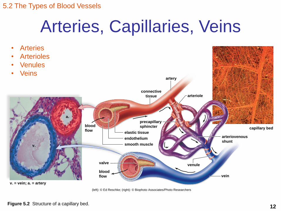
12Figure 5.2 Structure of a capillary bed.
5.2 The Types of Blood Vessels
Arteries, Capillaries, Veins
a.
v.
v. = vein; a. = artery
valve
blood
flow
blood
flow
connective
tissue
artery
arteriole
capillary bed
arteriovenous
shunt
vein
venule
smooth muscle
endothelium
elastic tissue
precapillary
sphincter
(left): © Ed Reschke; (right): © Biophoto Associates/Photo Researchers
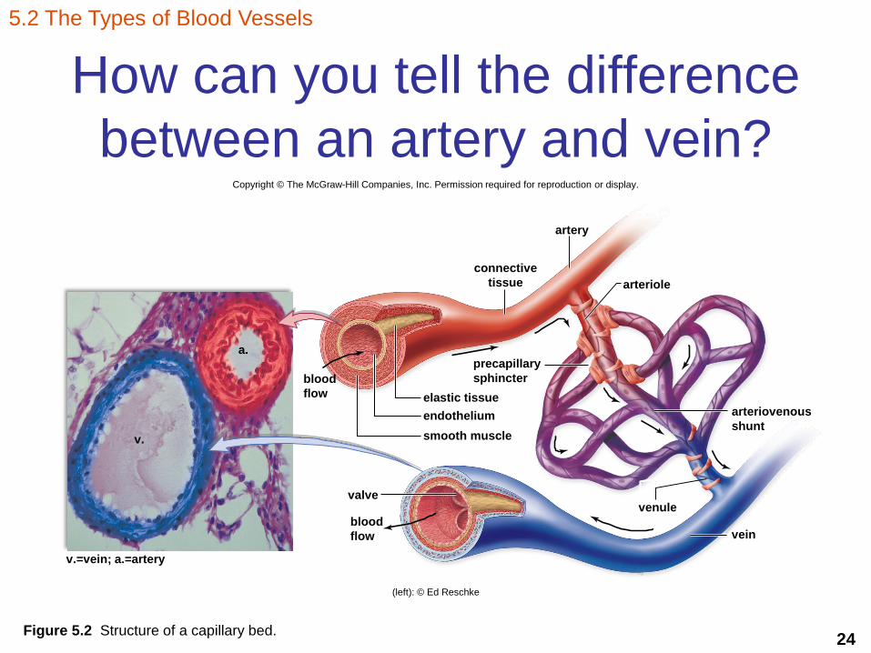
• Arteries
• Arterioles
• Venules
• Veins

13
How can you tell the difference
between an artery and vein?Copyright © The McGraw-Hill Companies, Inc. Permission required for reproduction or display.
arteriovenous
shunt
vein
venule
arteriole
artery
connective
tissue
v.=vein; a.=artery
blood
flow
valve
blood
flow
smooth muscle
endothelium
elastic tissue
precapillary
sphincter
v.
a.
(left): © Ed Reschke
Figure 5.2 Structure of a capillary bed.
5.2 The Types of Blood Vessels
14
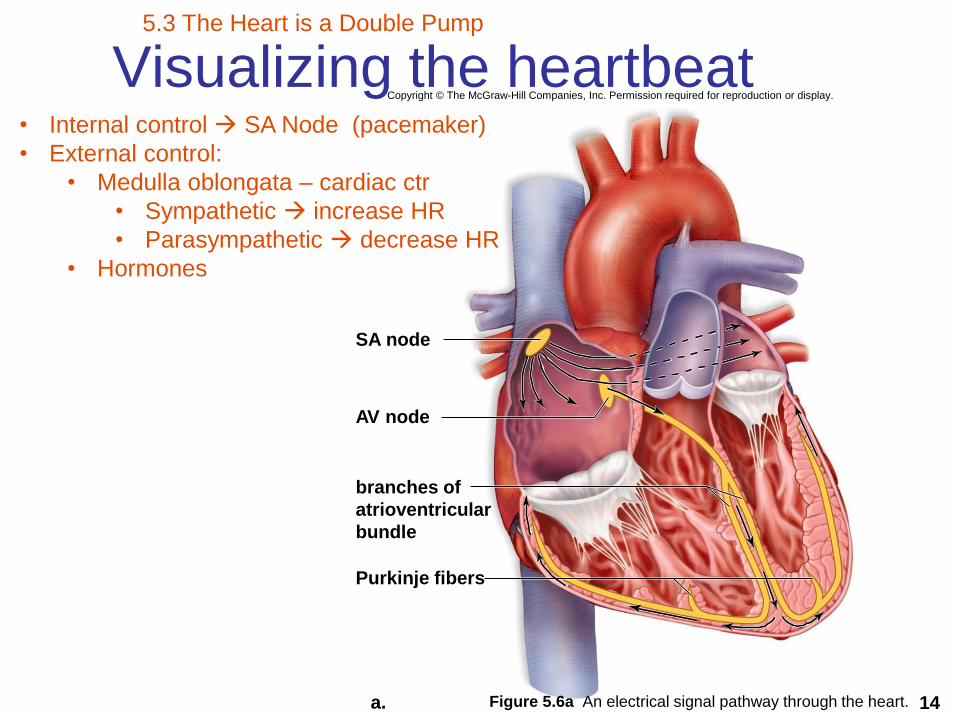
Visualizing the heartbeat
SA node
AV node
branches of
atrioventricular
bundle
Purkinje fibers
a.
Copyright © The McGraw-Hill Companies, Inc. Permission required for reproduction or display.
Figure 5.6a An electrical signal pathway through the heart.
5.3 The Heart is a Double Pump
• Internal control SA Node (pacemaker)
• External control:
• Medulla oblongata – cardiac ctr
• Sympathetic increase HR
• Parasympathetic decrease HR
• Hormones
15
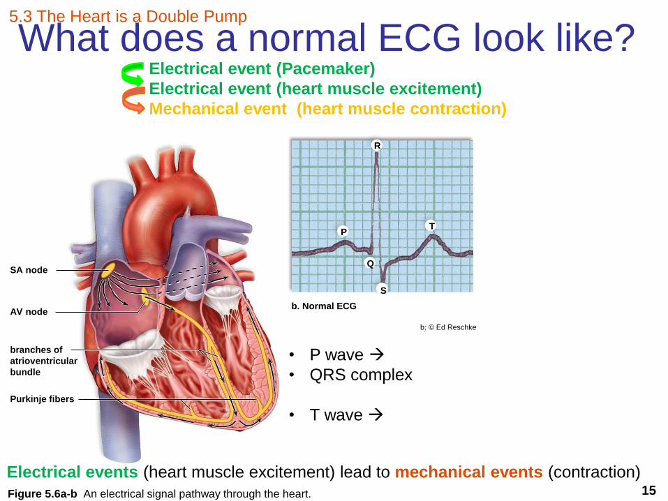
What does a normal ECG look like?
SA node
AV node
branches of
atrioventricular
bundle
Purkinje fibers
b. Normal ECG
P
Q
T
S
R
b: © Ed Reschke
Figure 5.6a-b An electrical signal pathway through the heart.
5.3 The Heart is a Double Pump
• P wave
• QRS complex
• T wave
Electrical events (heart muscle excitement) lead to mechanical events (contraction)
Electrical event (Pacemaker)
Electrical event (heart muscle excitement)
Mechanical event (heart muscle contraction)
16
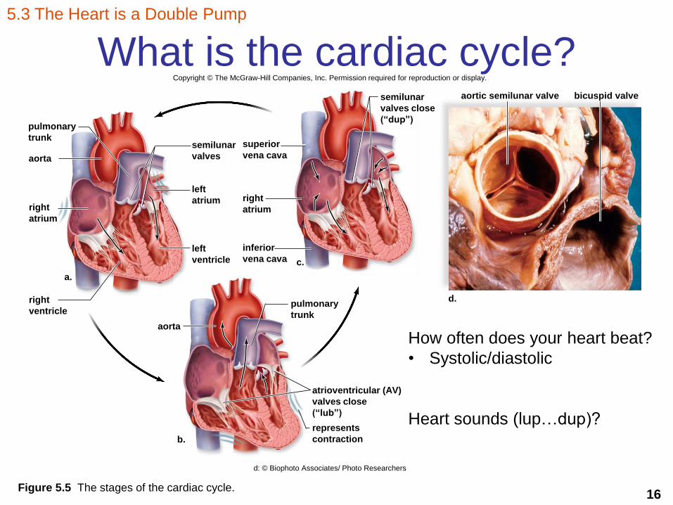
What is the cardiac cycle?
Figure 5.5 The stages of the cardiac cycle.
5.3 The Heart is a Double Pump
bicuspid valveaortic semilunar valvesemilunar
valves close
(“dup”)
superior
vena cavasemilunar
valves
pulmonary
trunk
aorta
right
atrium
right
ventricle
a.
aorta
b.
atrioventricular (AV)
valves close
(“lub”)
d.pulmonary
trunk
c.
right
atrium
inferior
vena cava
left
atrium
left
ventricle
represents
contraction
Copyright © The McGraw-Hill Companies, Inc. Permission required for reproduction or display.
d: © Biophoto Associates/ Photo Researchers
How often does your heart beat?
• Systolic/diastolic
Heart sounds (lup…dup)?
17
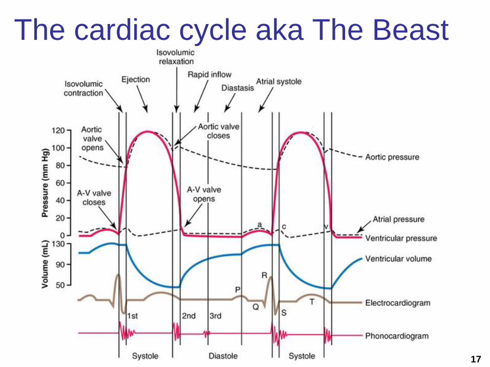
The cardiac cycle aka The Beast

18
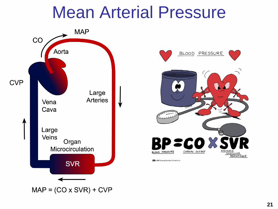
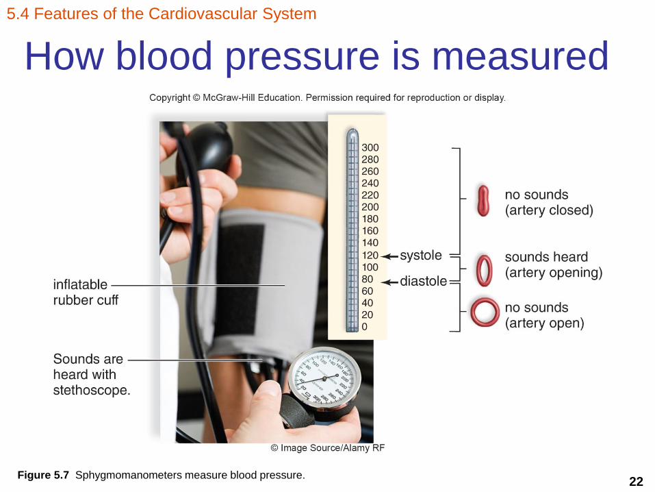
• Systole
• Diastole
• Average: 120/80 mmHg (systolic/diastolic)
• Mean arterial pressure (MAP)
DP + 1/3 Pulse pressure; (PP = SP – DP)
5.3 The Heart is a Double Pump
What is blood pressure?

19
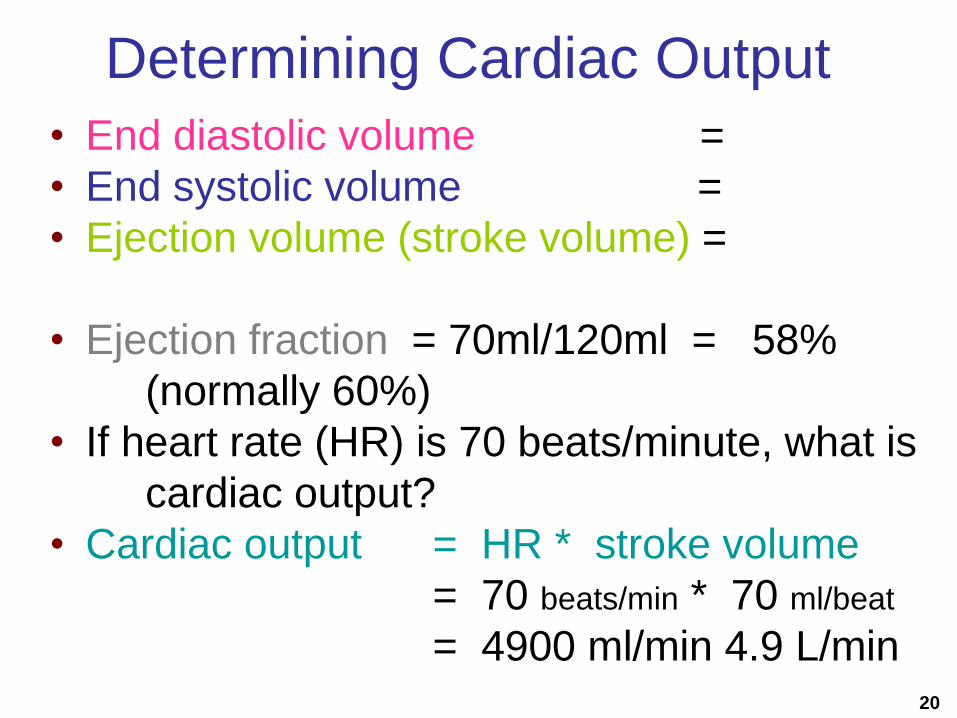
Cardiac Output
• Amount ejected by a ventricle in 1 minute
• CO = Heart Rate x Stroke Volume• HR = how many times your heart beats/min
• SV = volume of blood ejected/beat (mL/beat)
• Resting values, usually about 4 to 6L/min
• Vigorous exercise
– CO to 21 L/min for fit person
– up to 35 L/min for world class athlete
20
Determining Cardiac Output
• End diastolic volume =
• End systolic volume =
• Ejection volume (stroke volume) =
• Ejection fraction = 70ml/120ml = 58%
(normally 60%)
• If heart rate (HR) is 70 beats/minute, what is
cardiac output?
• Cardiac output = HR * stroke volume
= 70 beats/min * 70 ml/beat
= 4900 ml/min 4.9 L/min
21
Mean Arterial Pressure
22
5.4 Features of the Cardiovascular System
Figure 5.7 Sphygmomanometers measure blood pressure.
How blood pressure is measured

23
How is blood pressure categorized?
5.4 Features of the Cardiovascular System
24
How can you tell the difference
between an artery and vein?Copyright © The McGraw-Hill Companies, Inc. Permission required for reproduction or display.
arteriovenous
shunt
vein
venule
arteriole
artery
connective
tissue
v.=vein; a.=artery
blood
flow
valve
blood
flow
smooth muscle
endothelium
elastic tissue
precapillary
sphincter
v.
a.
(left): © Ed Reschke
Figure 5.2 Structure of a capillary bed.
5.2 The Types of Blood Vessels

25Figure 5.8 Blood velocity and pressure in the blood vessels.
5.4 Features of the Cardiovascular System
What is important about blood flow?Copyright © The McGraw-Hill Companies, Inc. Permission required for reproduction or display.
Rela
tive
ma
gn
itu
de
total
cross-sectional
area of
vessels
velocity
blood
pressure
arteries arterioles capillaries venules veins
Blood flow (starting from heart)
Pressure greatest
Blood flow slowest
Why???
Pressure minimal,
Flow increased

26
If blood pressure is so low in the veins,
why does the blood flow increase?
• They have help.
to heartto heart
a. Contracted skeletal
muscle pushes blood
past open valve.
b. Closed valve prevents
backward flow of blood.
Copyright © The McGraw-Hill Companies, Inc. Permission required for reproduction or display.
Figure 5.9 The skeletal muscle
pump.
5.4 Features of the Cardiovascular System
27
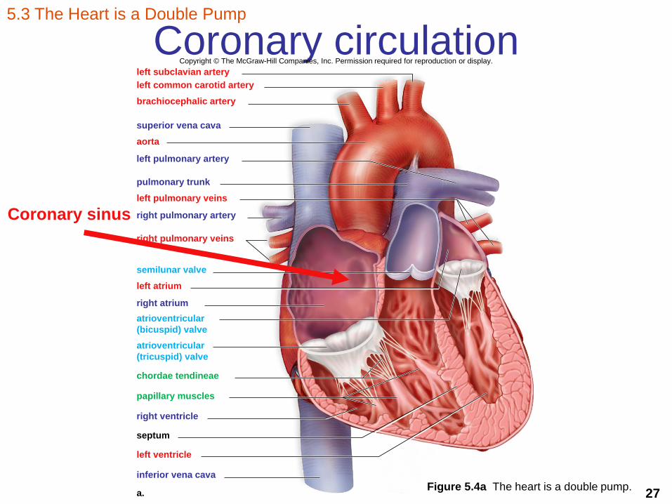
Coronary circulationCopyright © The McGraw-Hill Companies, Inc. Permission required for reproduction or display.
left subclavian artery
left common carotid artery
brachiocephalic artery
superior vena cava
aorta
left pulmonary artery
pulmonary trunk
left pulmonary veins
right pulmonary artery
right pulmonary veins
semilunar valve
left atrium
right atrium
atrioventricular
(bicuspid) valve
atrioventricular
(tricuspid) valve
chordae tendineae
papillary muscles
right ventricle
septum
left ventricle
inferior vena cava
a.Figure 5.4a The heart is a double pump.
5.3 The Heart is a Double Pump
Coronary sinus

28
Coronary circulationleft subclavian artery
left common carotid artery
brachiocephalic artery
superior vena cava
aorta
left pulmonary artery
pulmonary trunk
left pulmonary veins
right pulmonary artery
right pulmonary veins
left atrium
left cardiac vein
right atrium
right coronary artery
left ventricle
right ventricle
left anterior descending
coronary artery
inferior vena cava
apex
Figure 5.3 The arteries and veins associated with
the human heart.
5.3 The Heart is a Double Pump
29
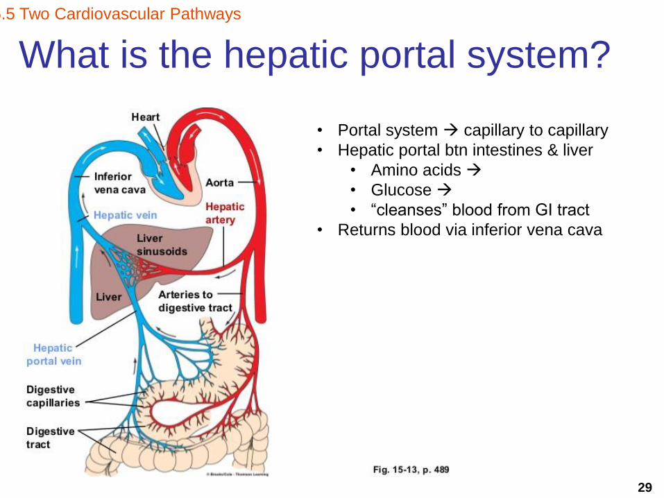
What is the hepatic portal system?
5.5 Two Cardiovascular Pathways
• Portal system capillary to capillary
• Hepatic portal btn intestines & liver
• Amino acids
• Glucose
• “cleanses” blood from GI tract
• Returns blood via inferior vena cava
30
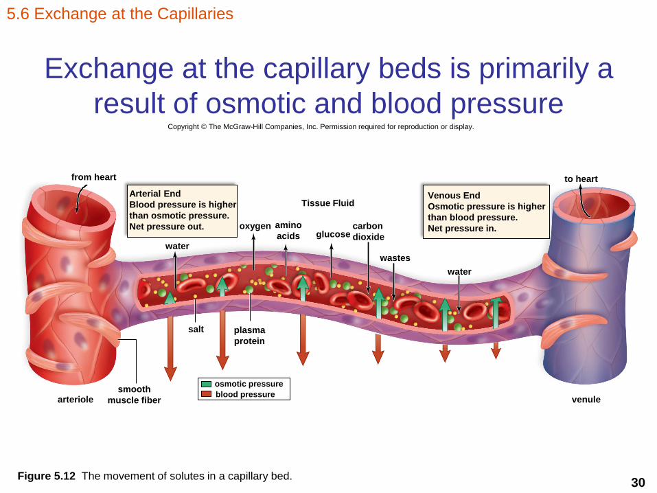
Exchange at the capillary beds is primarily a
result of osmotic and blood pressure
5.6 Exchange at the Capillaries
Copyright © The McGraw-Hill Companies, Inc. Permission required for reproduction or display.
from heart
Arterial End
Blood pressure is higher
than osmotic pressure.
Net pressure out.
water
oxygen amino
acids
Tissue Fluid
glucosecarbon
dioxide
wastes
arteriolesmooth
muscle fiberblood pressure
osmotic pressure
plasma
protein
Venous End
Osmotic pressure is higher
than blood pressure.
Net pressure in.
venule
to heart
salt
water
Figure 5.12 The movement of solutes in a capillary bed.

31
Exchange at the capillaries
Figure 5.13 Interaction of lymphatic and capillary beds.
5.6 Exchange at the Capillaries
Copyright © The McGraw-Hill Companies, Inc. Permission required for reproduction or display.
venule
Lymphatic duct with lymph subclavian veins
blood
capillary
lymphatic
capillary
tissue
cells
arteriole
Precapillary sphincter
32
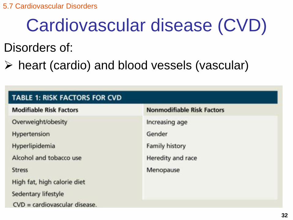
Cardiovascular disease (CVD)Disorders of:
heart (cardio) and blood vessels (vascular)
5.7 Cardiovascular Disorders

33
Cardiovascular disease (CVD)
Risk Factors

34
Disorders of the blood vessels
• Hypertension/high blood pressure
• Atherosclerosis
• Stroke
• Heart attack
• Aneurysm
5.7 Cardiovascular Disorders
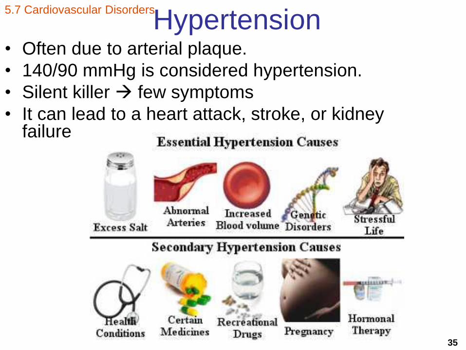
35
Hypertension• Often due to arterial plaque.
• 140/90 mmHg is considered hypertension.
• Silent killer few symptoms
• It can lead to a heart attack, stroke, or kidney failure
5.7 Cardiovascular Disorders
36
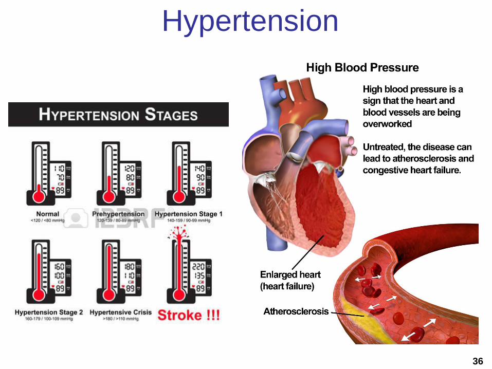
Hypertension
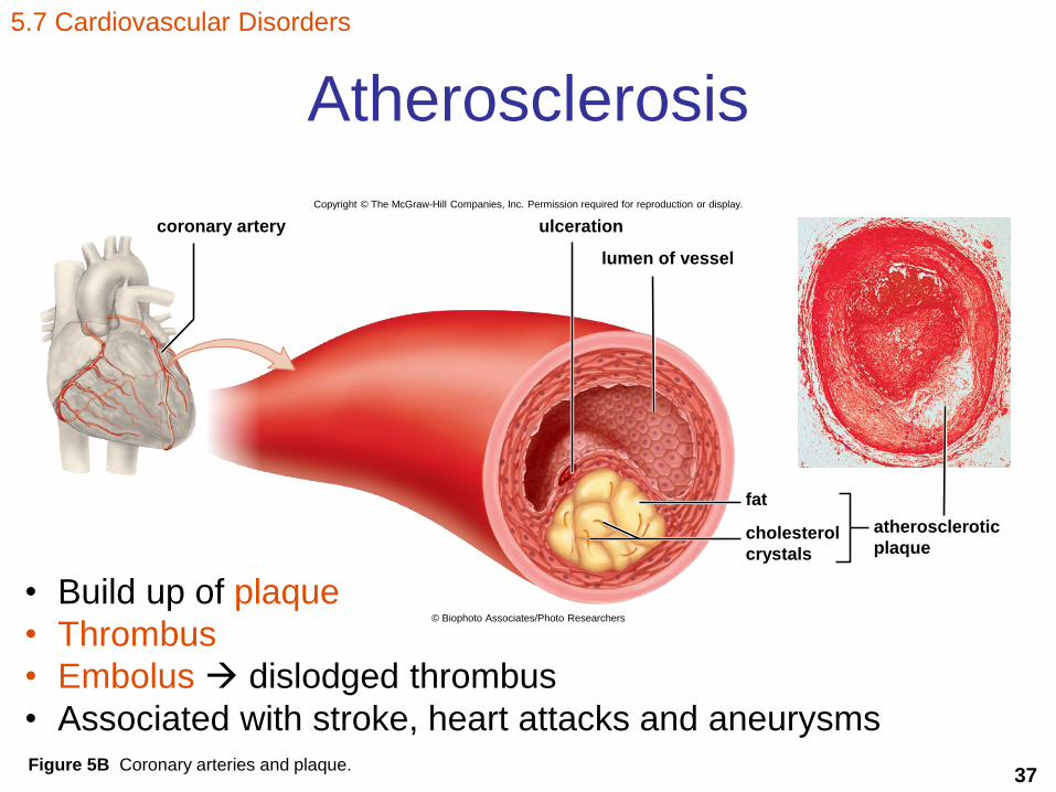
37Figure 5B Coronary arteries and plaque.
5.7 Cardiovascular Disorders
Atherosclerosis
coronary artery ulceration
lumen of vessel
atherosclerotic
plaquecholesterol
crystals
fat
Copyright © The McGraw-Hill Companies, Inc. Permission required for reproduction or display.
© Biophoto Associates/Photo Researchers
• Build up of plaque
• Thrombus
• Embolus dislodged thrombus
• Associated with stroke, heart attacks and aneurysms
38
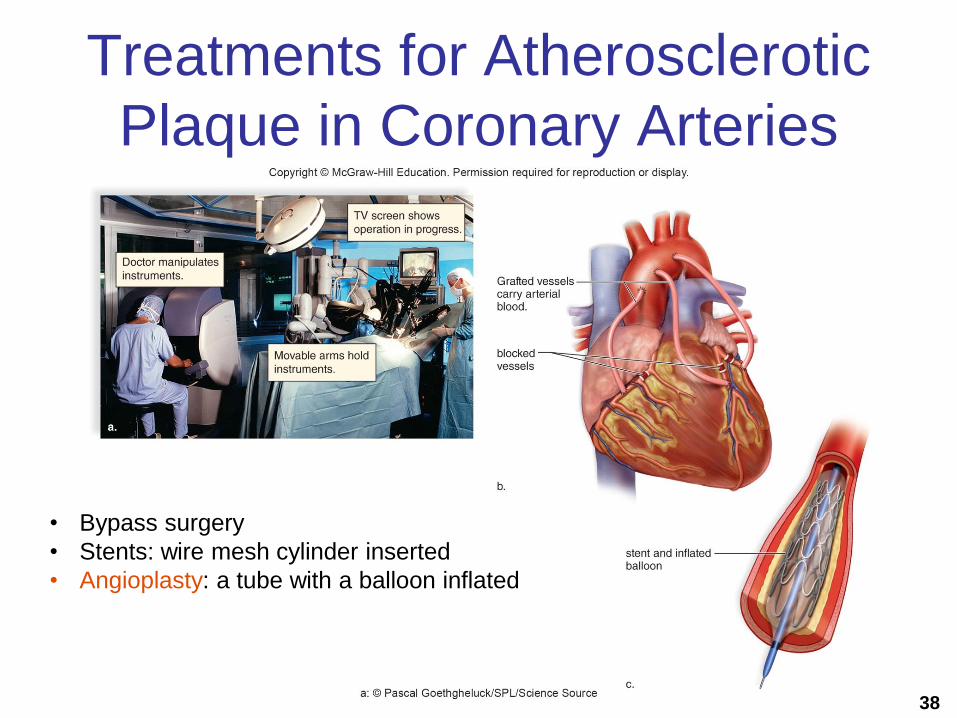
Treatments for Atherosclerotic
Plaque in Coronary Arteries
• Bypass surgery
• Stents: wire mesh cylinder inserted
• Angioplasty: a tube with a balloon inflated

39
Stroke• Stroke aka cerebrovascular accident (CVA)
• Cranial artery is blocked or bursts
• Part of the brain dies
• Symptoms may include:• numbness of hands or face
• difficulty speaking
• inability to see in one eye
5.7 Cardiovascular Disorders
Sudden burst

40
Aneurysm• Blood vessel balloons (weaken walls)
• Atherosclerosis and hypertension
• Most commonly affected
– abdominal artery or the arteries leading to the brain.
5.7 Cardiovascular Disorders

41
Heart attack• Heart attack or myocardial infarction (MI)
• Angina pectoris:
5.7 Cardiovascular Disorders

42
How are disorders of the blood
vessels treated?
• Dissolving blood clotst-PA (tissue plasmogen activator)
drug that dissolves clots
5.7 Cardiovascular Disorders

43
Disorders of the heart and its treatment
• Disorders
Congestive heart failure
• Treatments
Left ventricular assist device (LVAD)
Heart transplant either natural or artificial
5.7 Cardiovascular Disorders
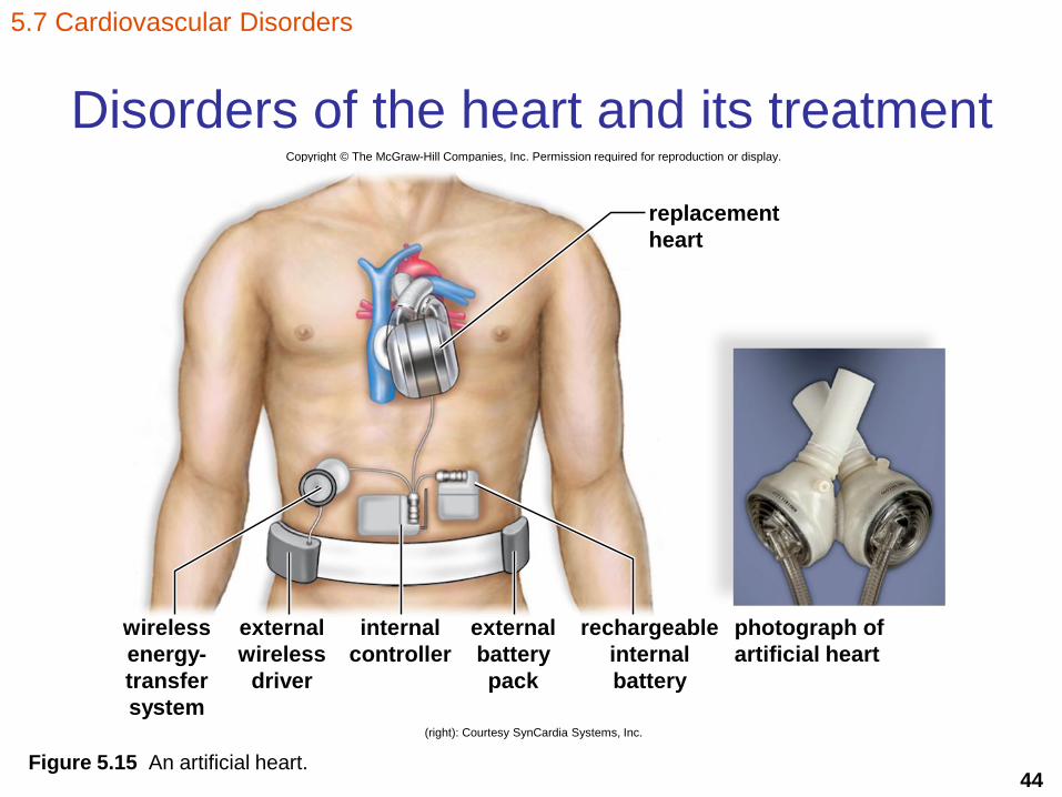
44Figure 5.15 An artificial heart.
5.7 Cardiovascular Disorders
Disorders of the heart and its treatmentCopyright © The McGraw-Hill Companies, Inc. Permission required for reproduction or display.
replacement
heart
photograph of
artificial heart
wireless
energy-
transfer
system
rechargeable
internal
battery
external
battery
pack
internal
controller
external
wireless
driver
(right): Courtesy SynCardia Systems, Inc.

45
Cardiovascular Disease Prevention
5.7 Cardiovascular Disorders