Embed Size (px)
Citation preview
Cardiovascular System
Cardiovascular System
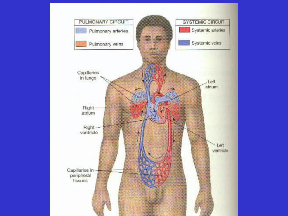
• Two divisions
Cardiovascular System
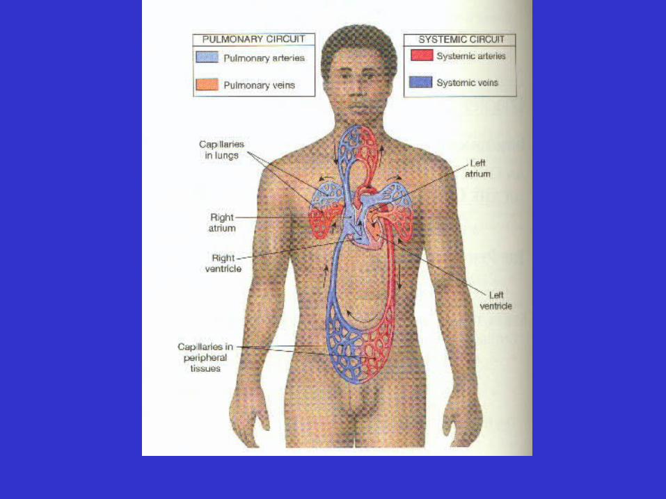
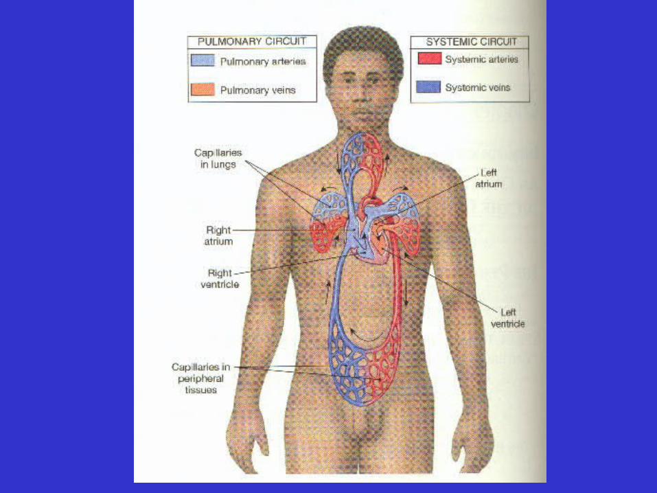
• Two divisions: pulmonary
Cardiovascular System
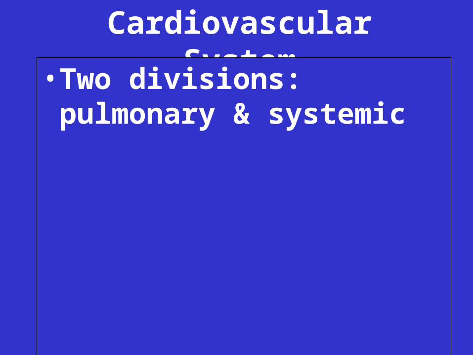
• Two divisions: pulmonary & systemic
Cardiovascular System
• Pulmonary Division
–Blood flows from heart
Cardiovascular System
• Pulmonary Division
–Blood flows from heart to alveolar capillaries
Cardiovascular System
• Pulmonary Division
–Blood flows from heart to alveolar capillaries and back to heart
Cardiovascular System
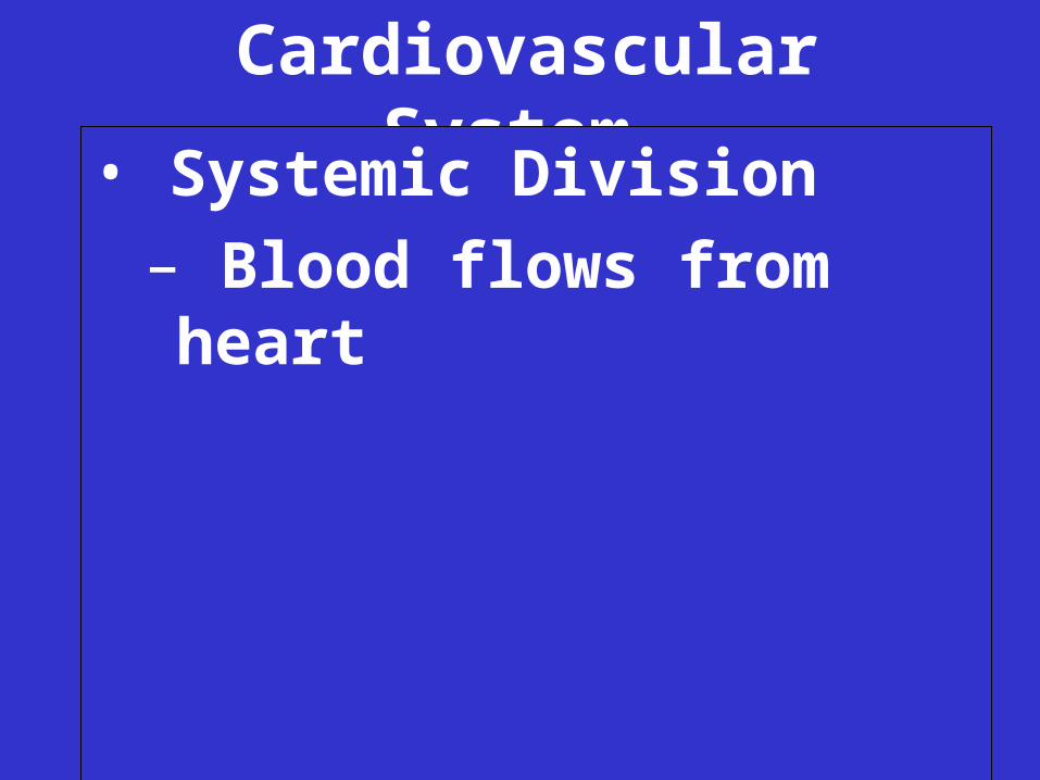
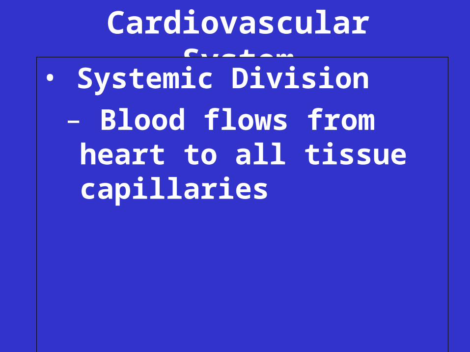
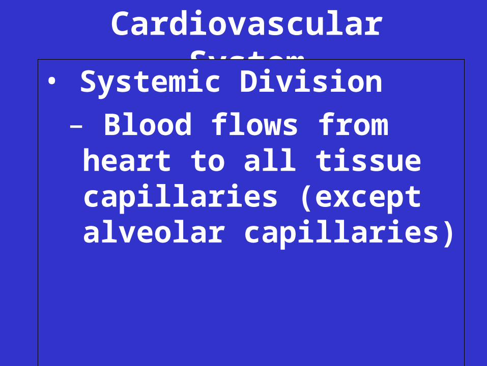
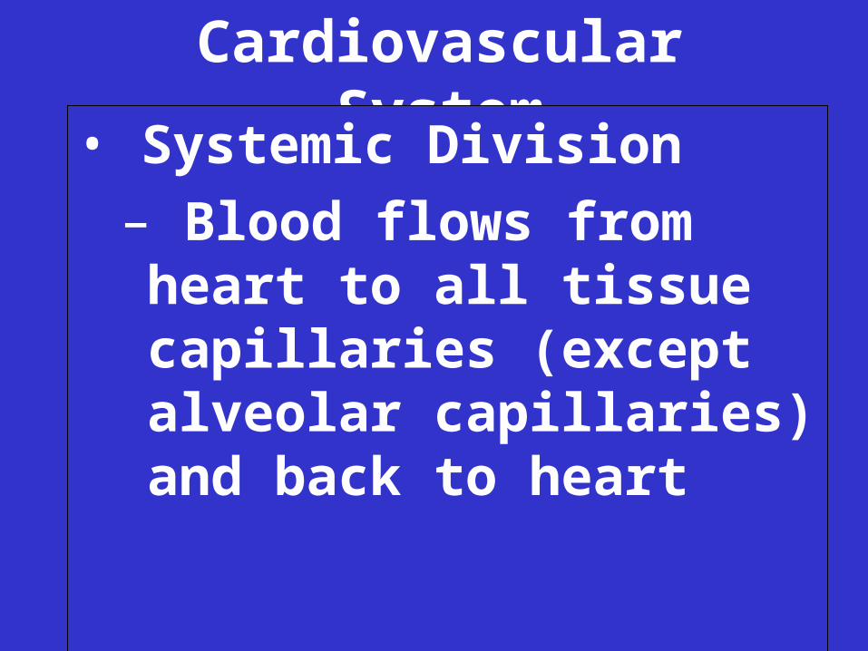
• Systemic Division
Cardiovascular System
• Systemic Division
– Blood flows from heart
Cardiovascular System
• Systemic Division
– Blood flows from heart to all tissue capillaries
Cardiovascular System
• Systemic Division
– Blood flows from heart to all tissue capillaries (except alveolar capillaries)
Cardiovascular System
• Systemic Division
– Blood flows from heart to all tissue capillaries (except alveolar capillaries) and back to heart
• Pulmonary arteries carry blood:
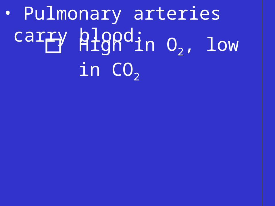
• Pulmonary arteries carry blood:
High in O2, low in CO2
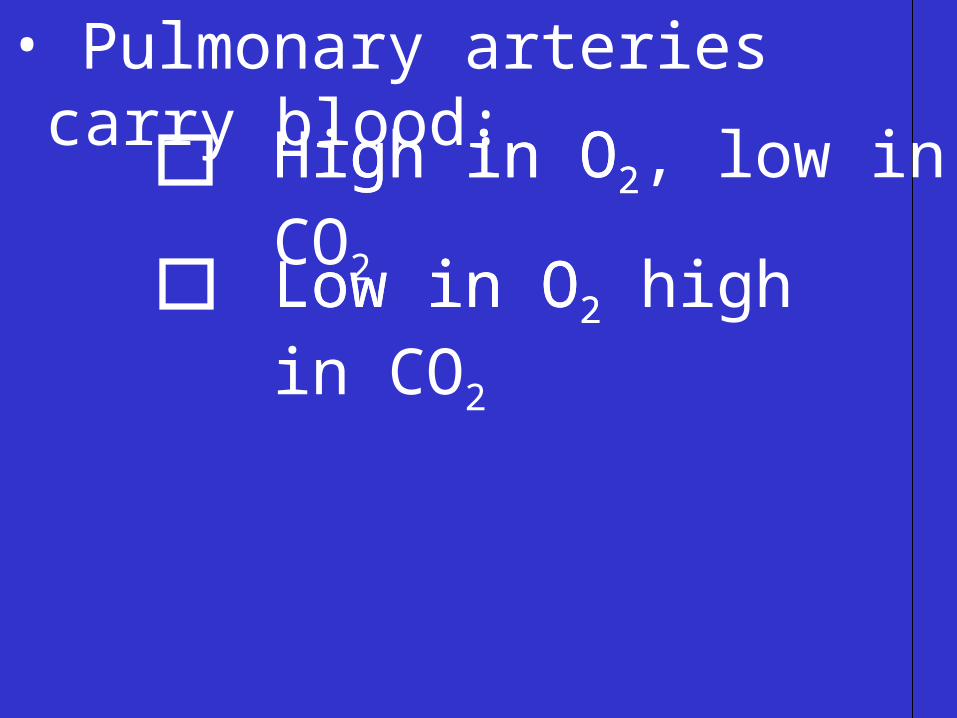
• Pulmonary arteries carry blood:
High in O2
Low in O2 Low in O2 high in CO2
High in O2, low in CO2

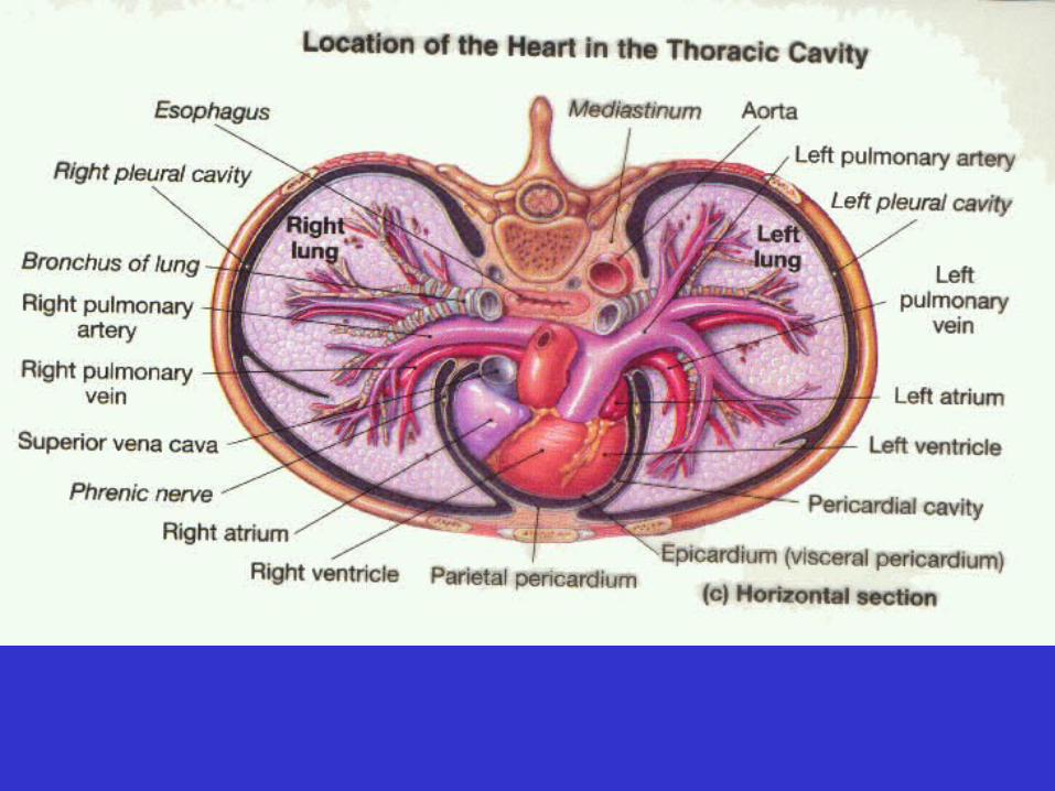
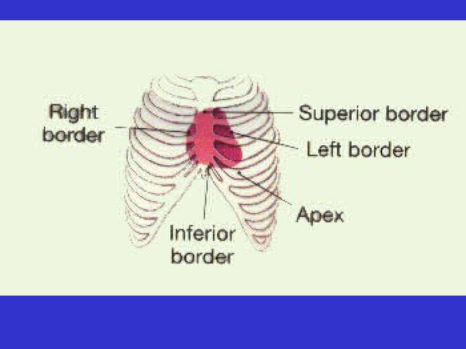
• Heart

• Heart
– Location

• Heart
– Location: mediastinum
• Heart
– Location: mediastinum
• Heart
– Location: mediastinum
– Size?
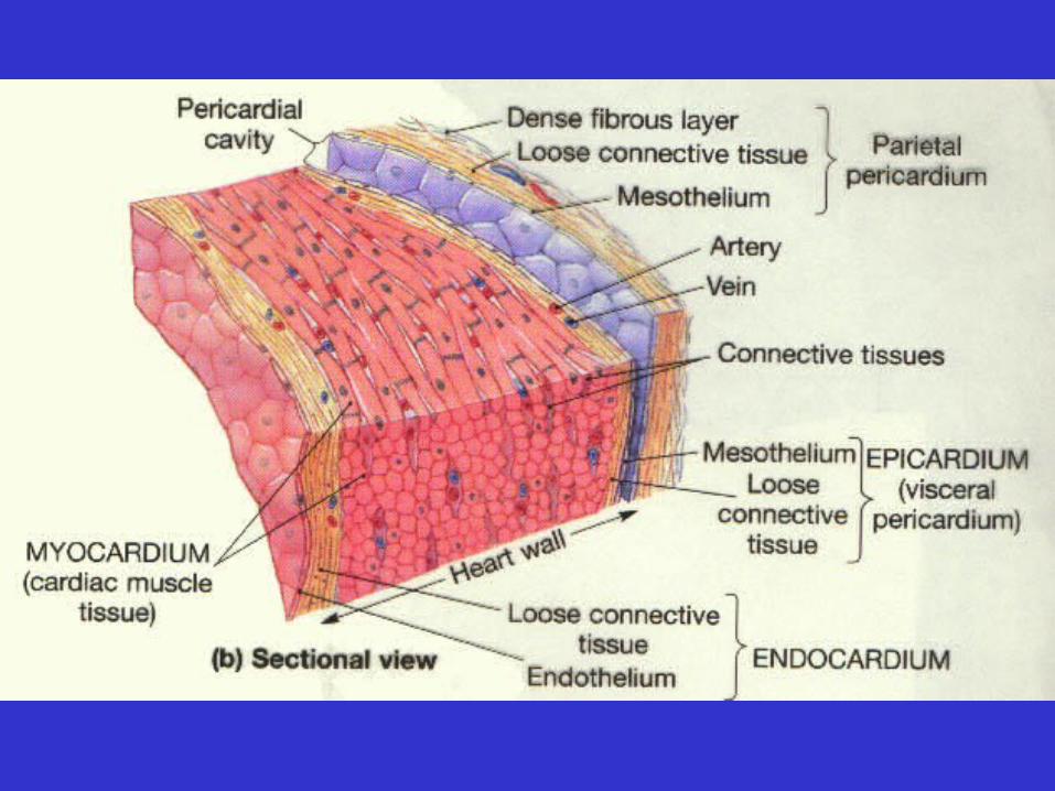
• Pericardium
• Pericardium: protective sac surrounding the heart
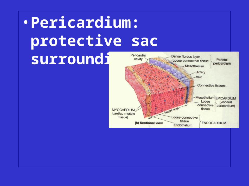
• Pericardium: protective sac surrounding the heart
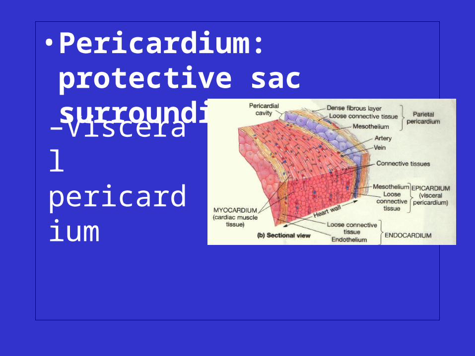
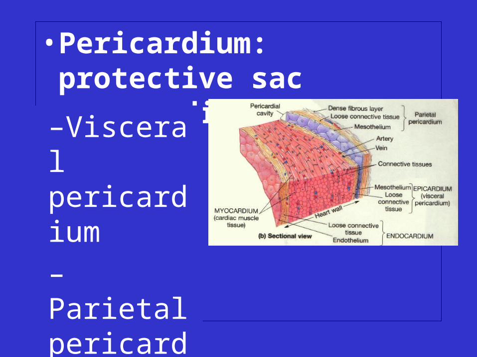
• Pericardium: protective sac surrounding the heart
–Visceral pericardium

• Pericardium: protective sac surrounding the heart
–Visceral pericardium
– Parietal pericardium
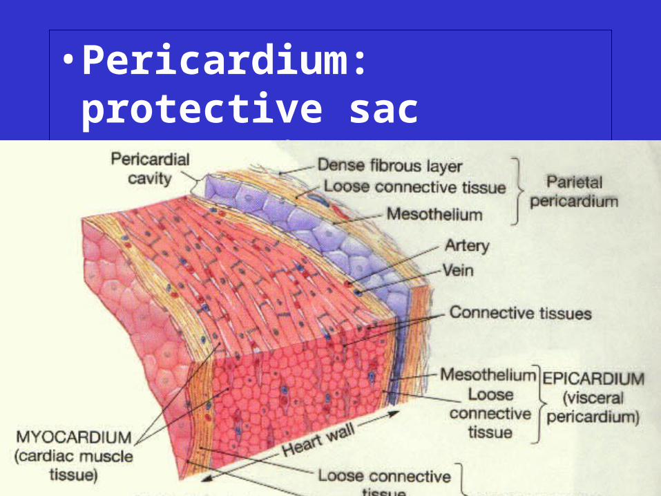
• Pericardium: protective sac surrounding the heart
–Visceral pericardium
– Parietal pericardium
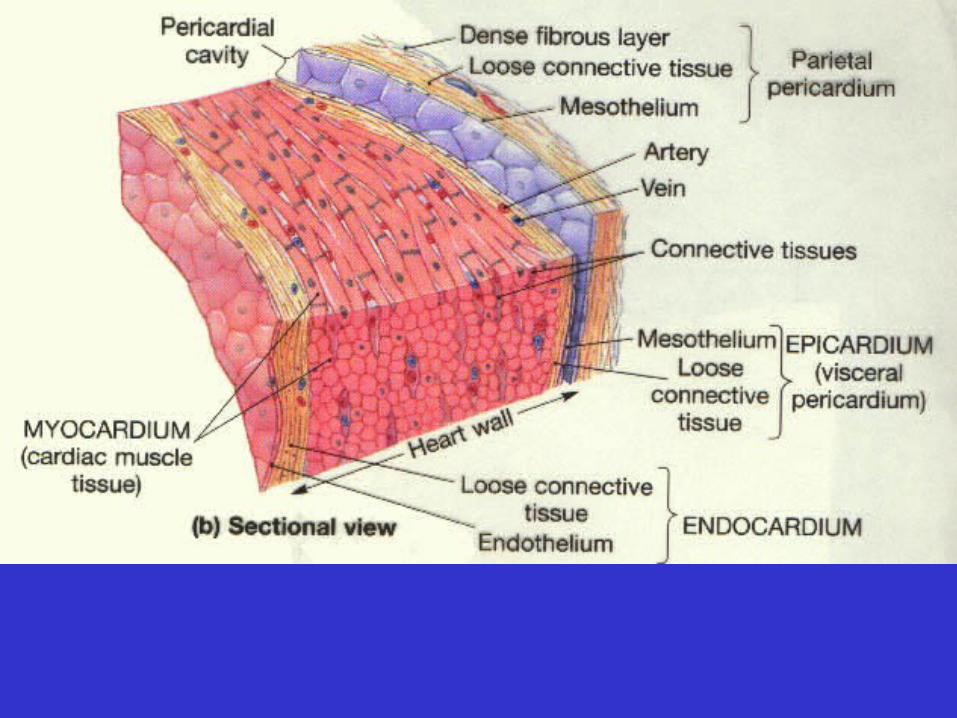

• Layers of the heart wall
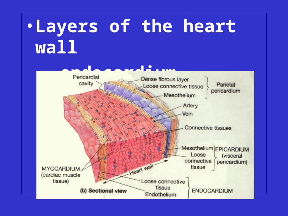
• Layers of the heart wall
– endocardium
• Layers of the heart wall
– endocardium
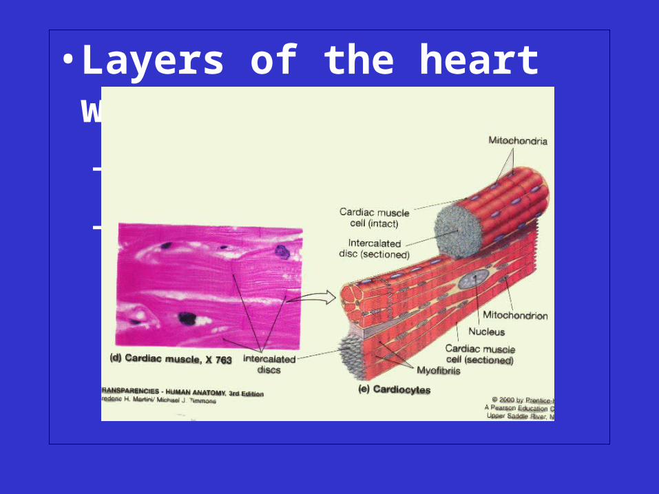
– myocardium
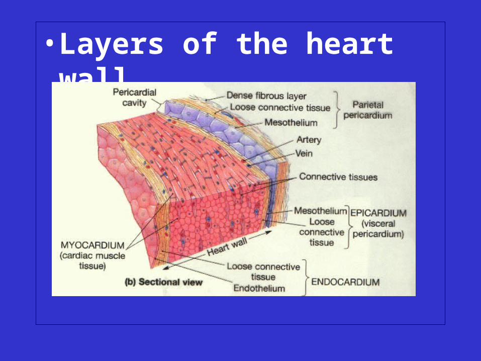
• Layers of the heart wall
– endocardium
– myocardium
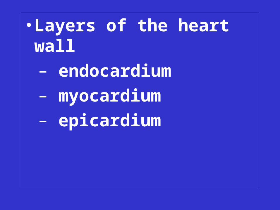
• Layers of the heart wall
– endocardium
– myocardium
– epicardium
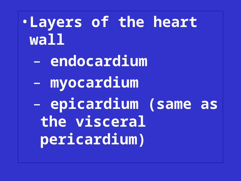
• Layers of the heart wall
– endocardium
– myocardium
– epicardium (same as the visceral pericardium)

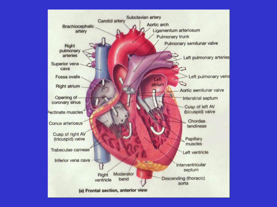
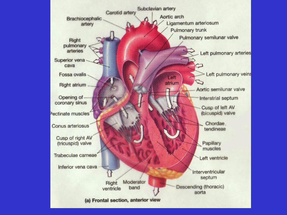
• Chambers of the heart

• Chambers of the heart
– Receiving chambers:
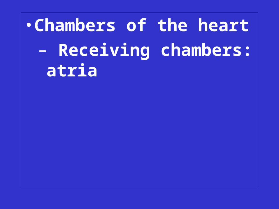
• Chambers of the heart
– Receiving chambers: atria
• Chambers of the heart
– Receiving chambers: atria (singular:
• Chambers of the heart
– Receiving chambers: atria (singular: atrium)

• Chambers of the heart
– Receiving chambers: atria (singular: atrium)
• Chambers of the heart
– Receiving chambers: atria (singular: atrium)
–Pumping chambers: ventricles
• Right ventricle
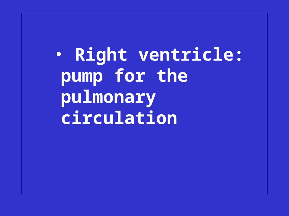
• Right ventricle: pump for the pulmonary circulation
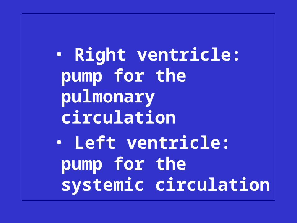
• Right ventricle: pump for the pulmonary circulation
• Left ventricle
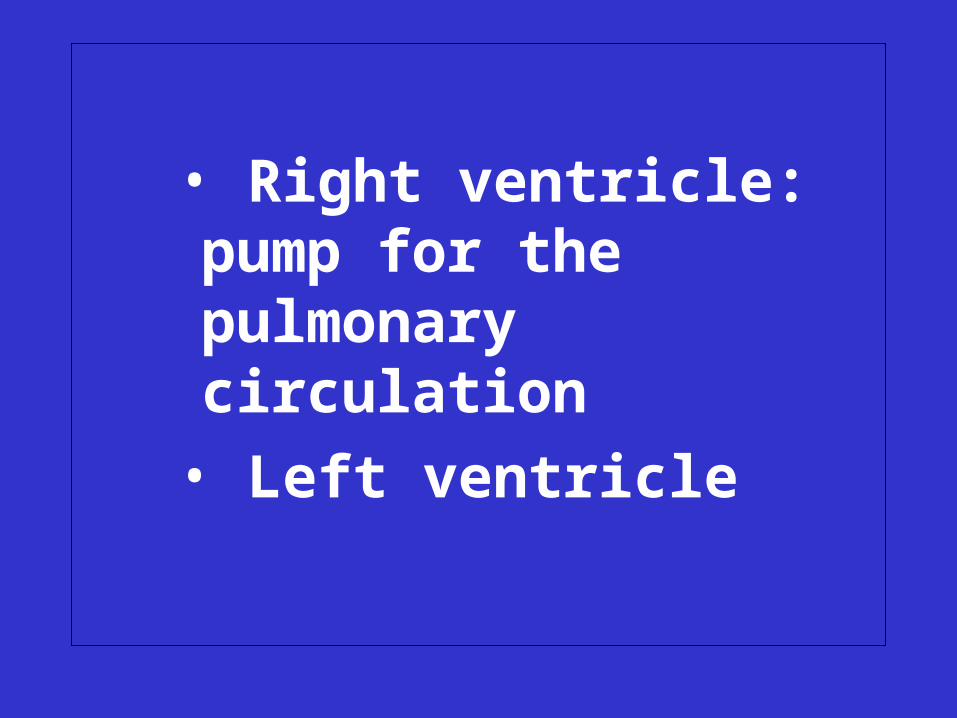
• Right ventricle: pump for the pulmonary circulation
• Left ventricle: pump for the systemic circulation
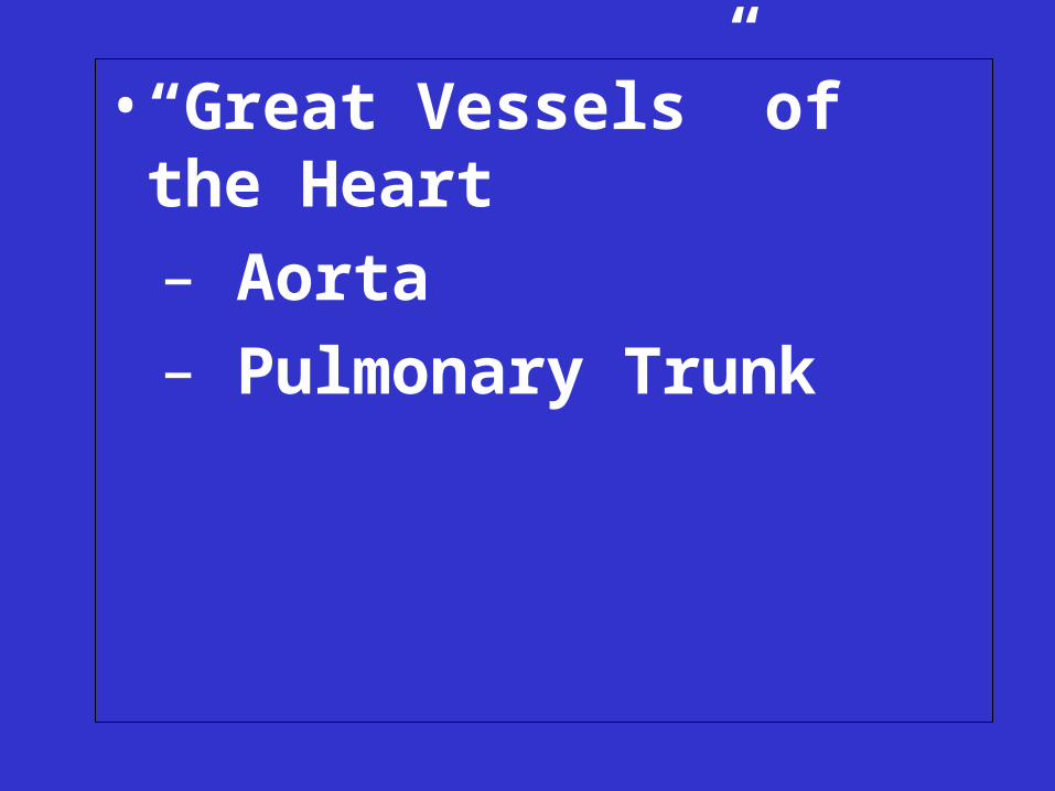
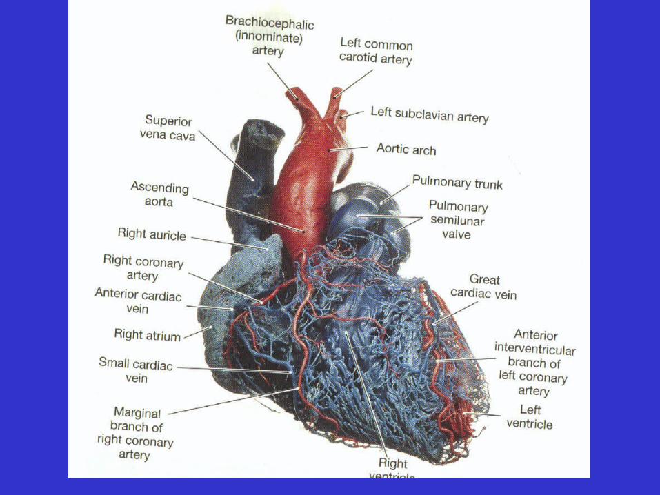
• “Great Vessels” of the Heart
– Aorta
– Pulmonary Trunk
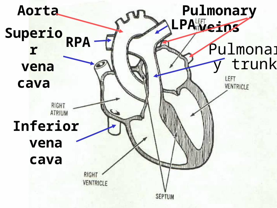
Inferior vena cava
Superior vena cava Pulmonary
trunk
Pulmonary veinsAorta
RPALPA
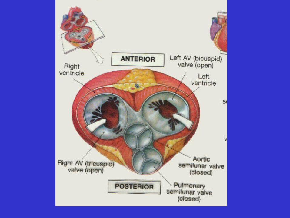
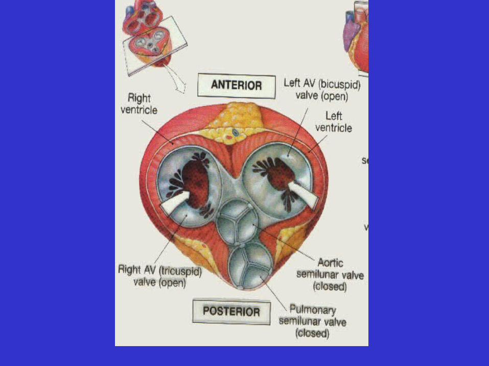
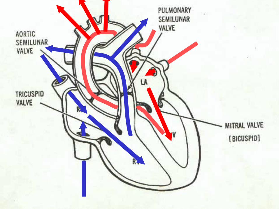
• Heart valves
• Heart valves
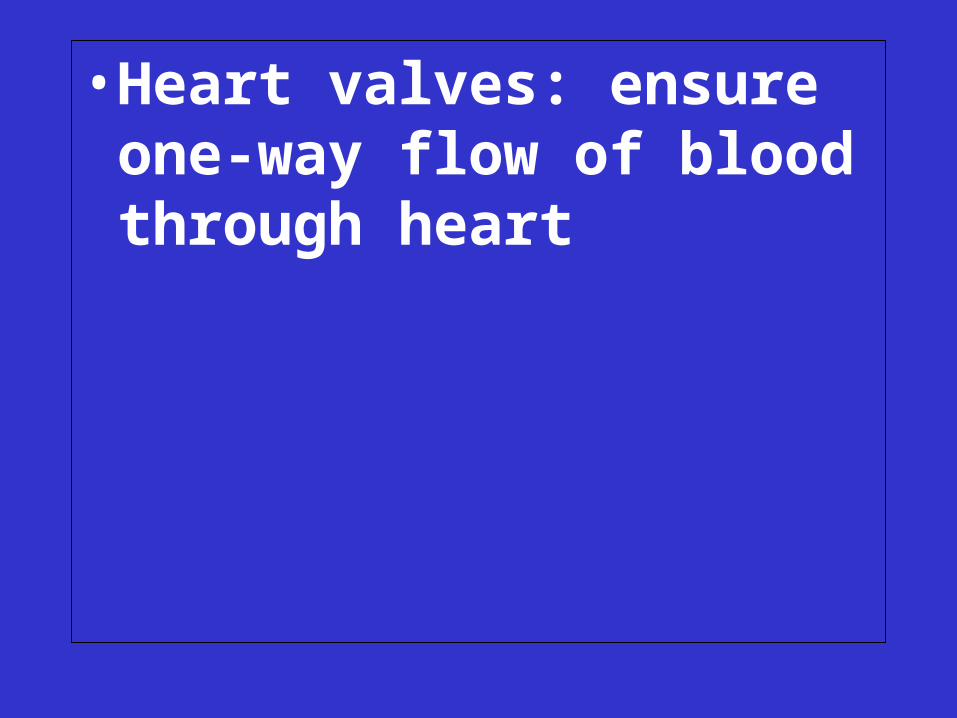
• Heart valves: ensure one-way flow of blood through heart
– Semilunar valves

– Semilunar valves
• pulmonary semilunar
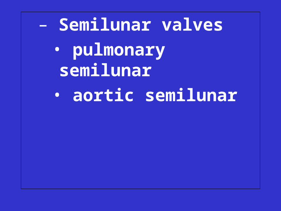
– Semilunar valves
• pulmonary semilunar
• aortic semilunar
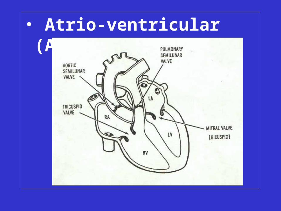
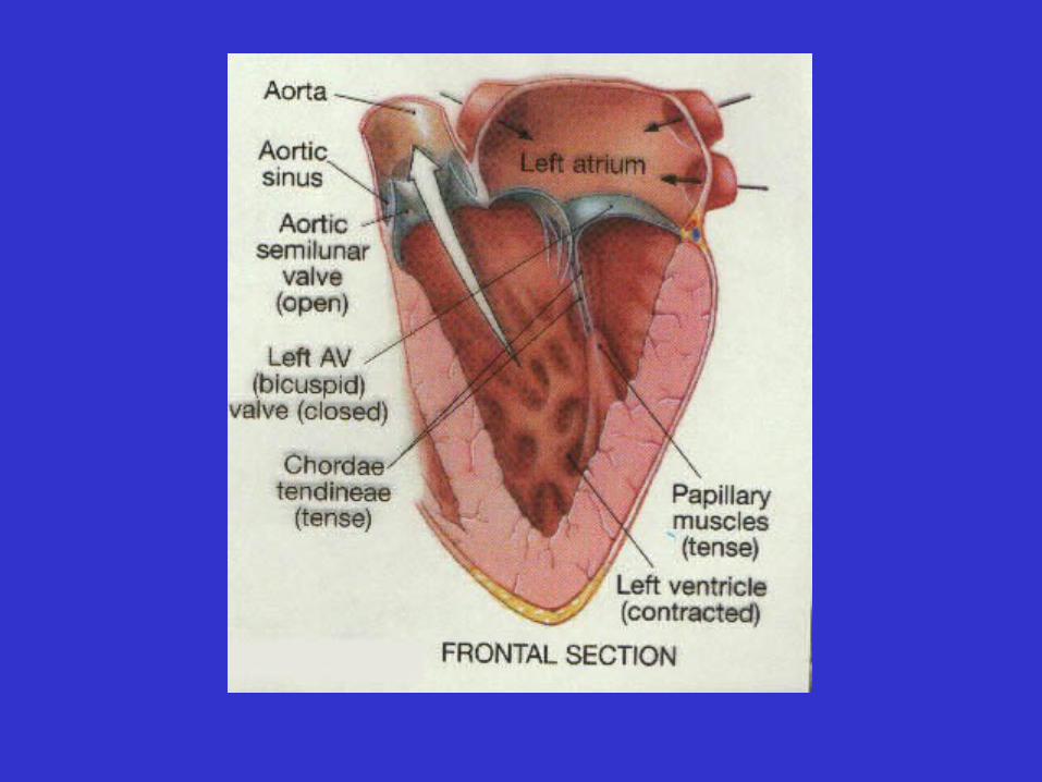
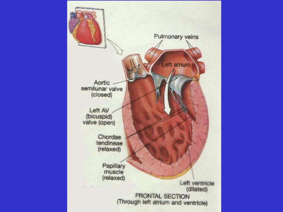
• Atrio-ventricular (AV) valves
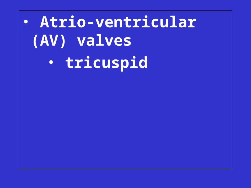
• Atrio-ventricular (AV) valves
• tricuspid
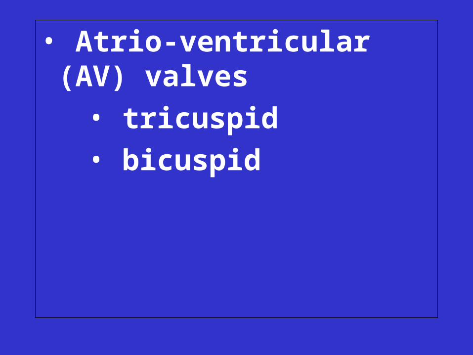
• Atrio-ventricular (AV) valves
• tricuspid
• bicuspid
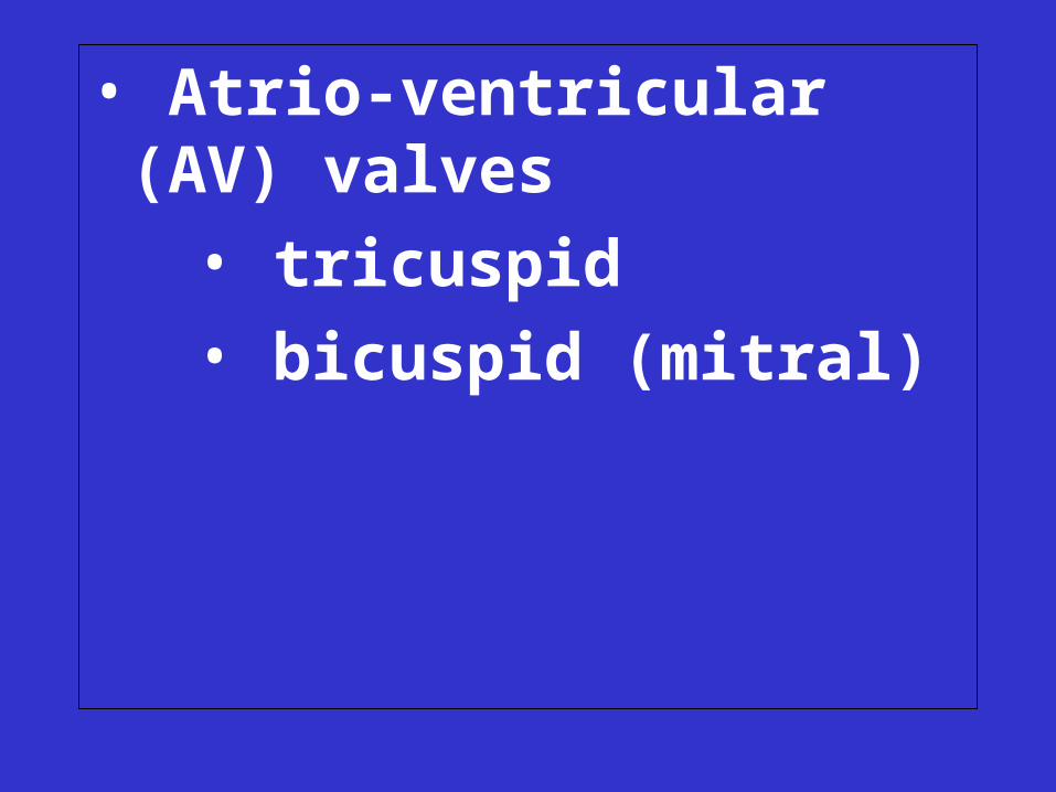
• Atrio-ventricular (AV) valves
• tricuspid
• bicuspid (mitral)
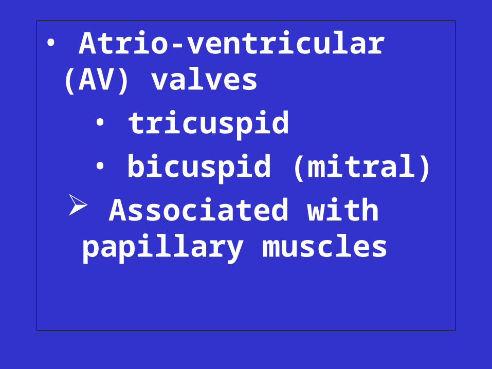
• Atrio-ventricular (AV) valves
• tricuspid
• bicuspid (mitral) Associated with papillary
muscles
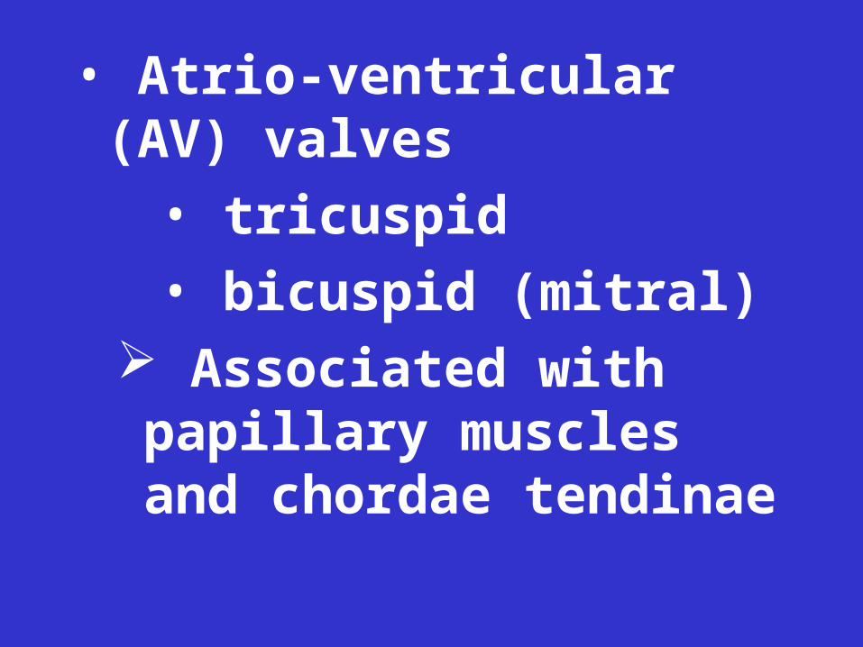
• Atrio-ventricular (AV) valves
• tricuspid
• bicuspid (mitral) Associated with papillary
muscles and chordae tendinae
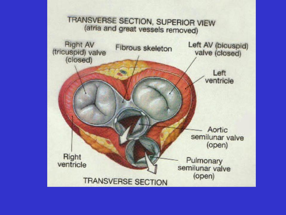
• Skeleton of the heart
– set of 4 fibrous rings near base of heart
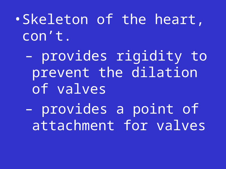
• Skeleton of the heart, con’t.
– provides rigidity to prevent the dilation of valves
– provides a point of attachment for valves
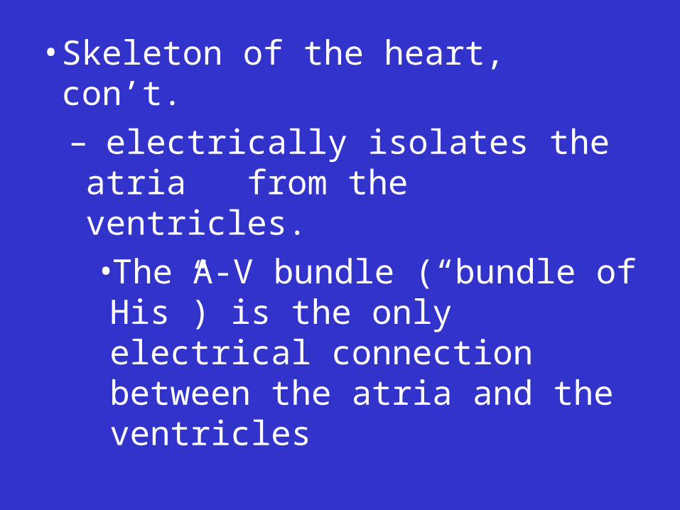
• Skeleton of the heart, con’t.
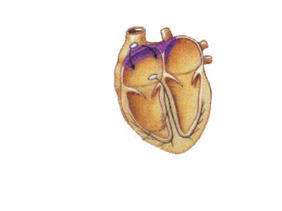
– electrically isolates the atria from the ventricles.
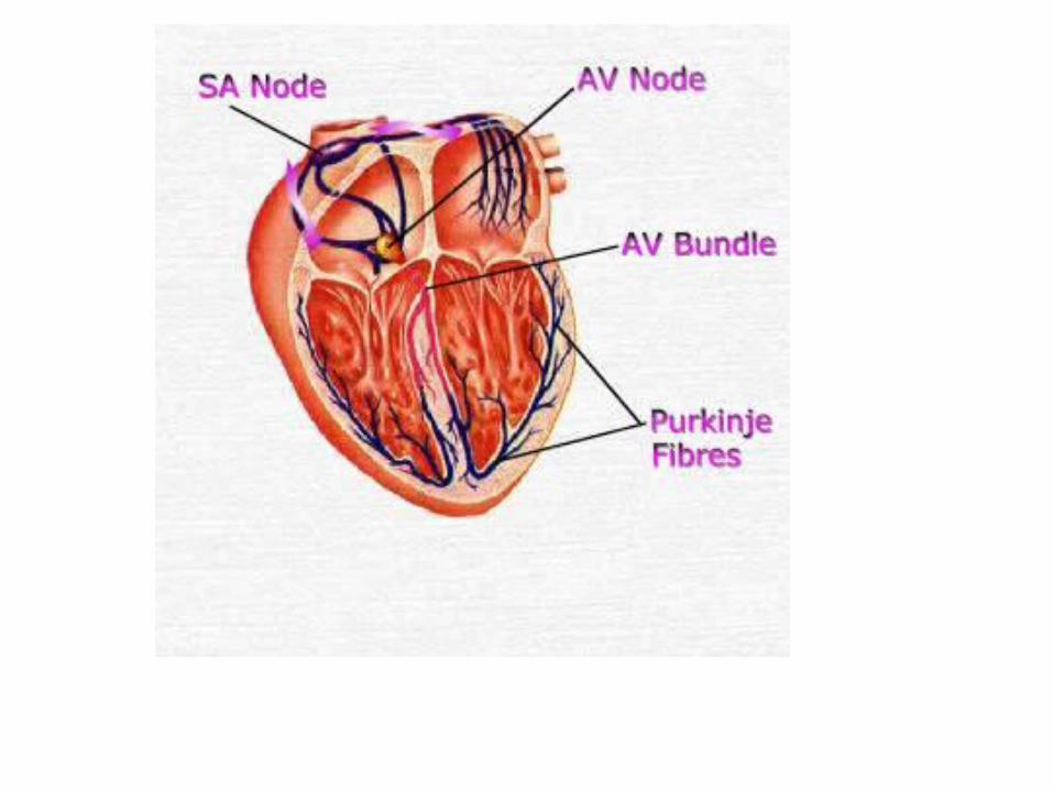
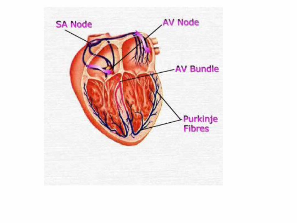
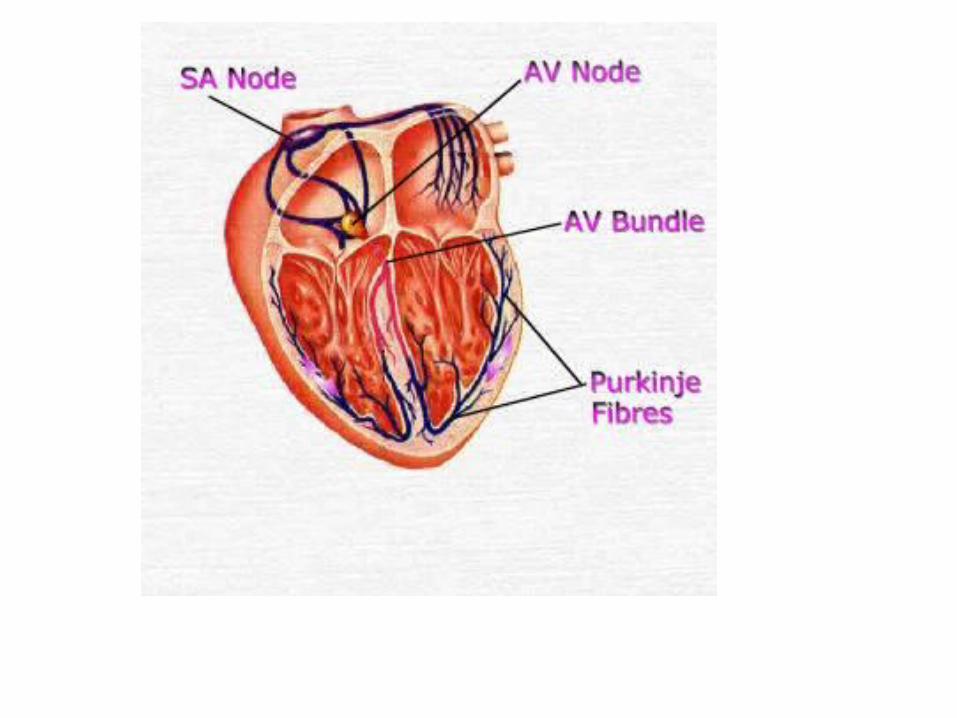
•The A-V bundle (“bundle of His”) is the only electrical connection between the atria and the ventricles
• Path of Blood Flow Through the Heart

http://www.innerbody.com/anim/heart.html
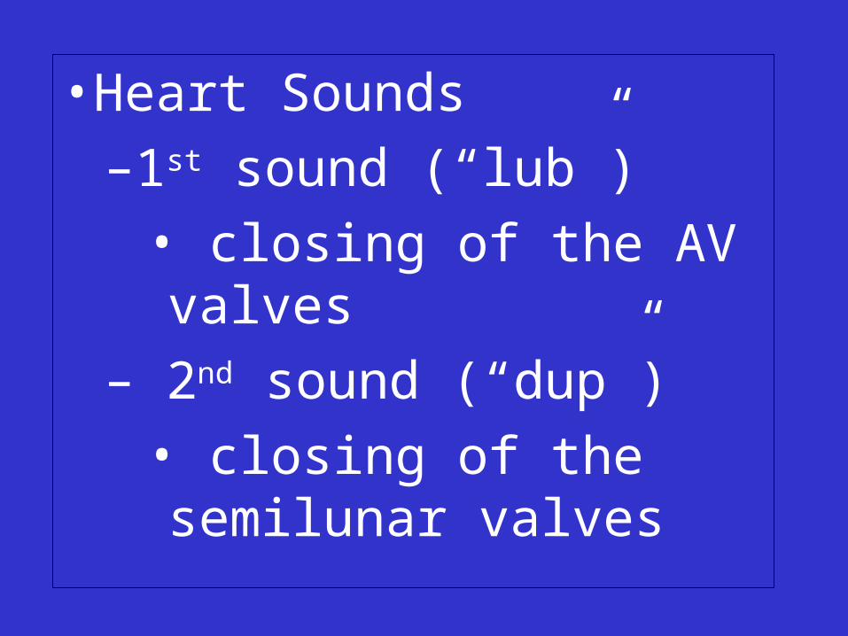
• Heart Sounds
–1st sound (“lub”)
• closing of the AV valves
– 2nd sound (“dup”)
• closing of the semilunar valves
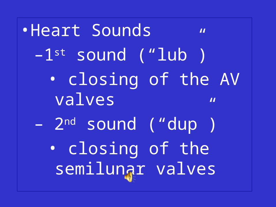
• Heart Sounds
–1st sound (“lub”)
• closing of the AV valves
– 2nd sound (“dup”)
• closing of the semilunar valves
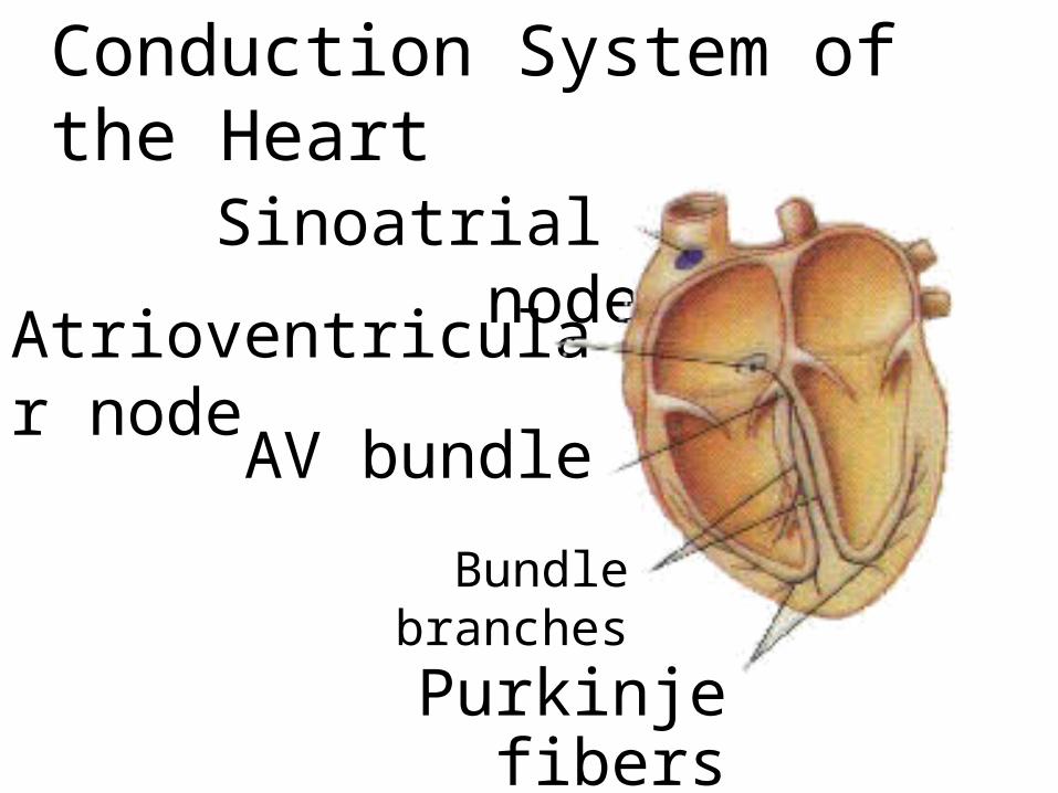
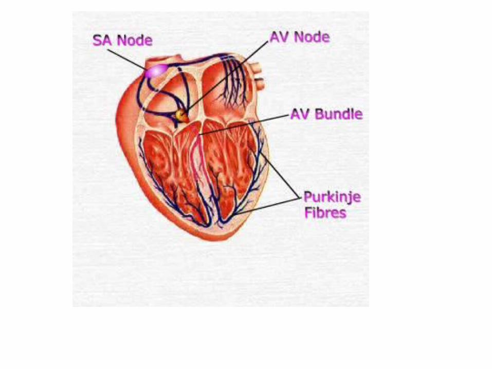
Conduction System of the Heart
Sinoatrial node
Atrioventricular node
AV bundle
Purkinje fibers
Bundle branches


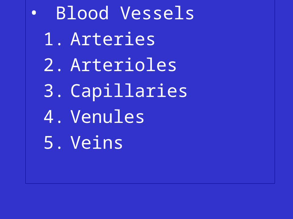
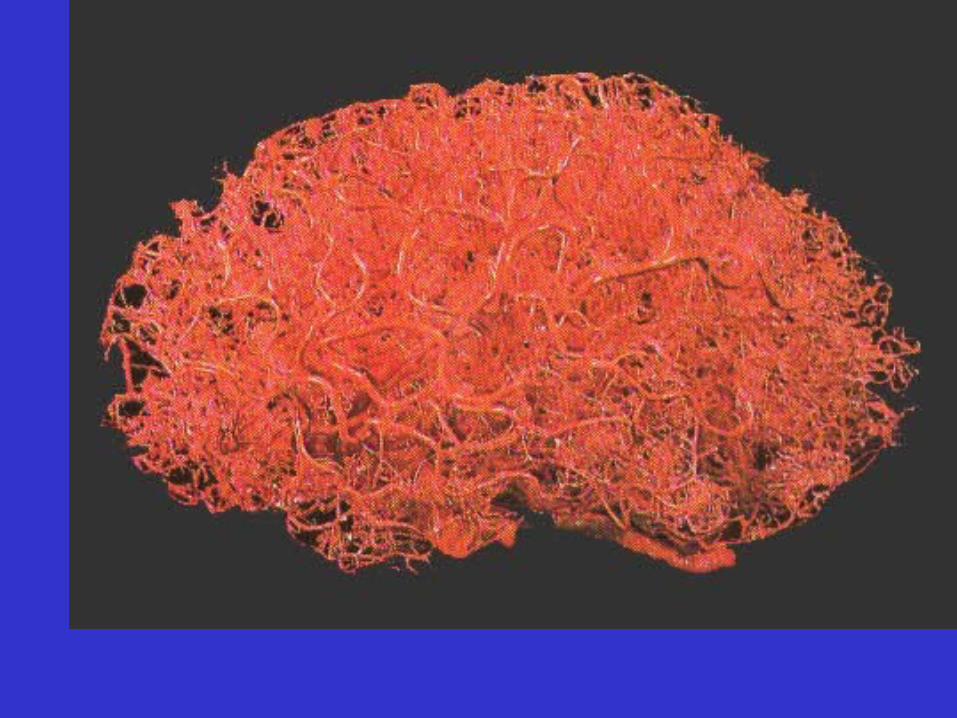
• Blood Vessels
1. Arteries
2. Arterioles
3. Capillaries
4. Venules
5. Veins
• All blood vessels are lined with endothelium
1. Arteries
• Carry blood away from the heart
• Subject to the highest blood pressure


1. Arteries
• Carry blood away from the heart
• Subject to the highest blood pressure
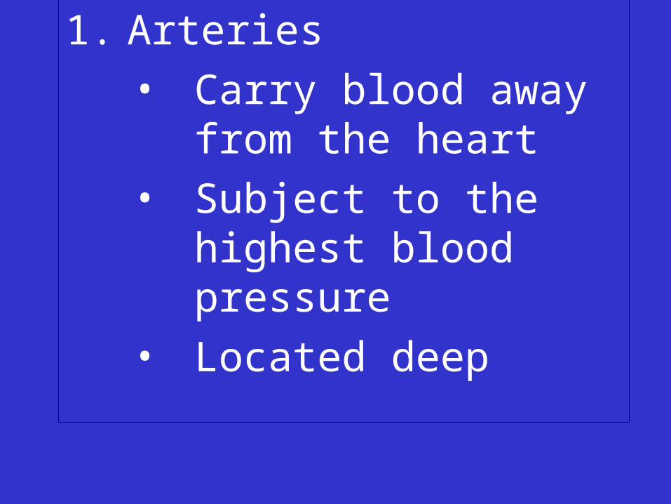
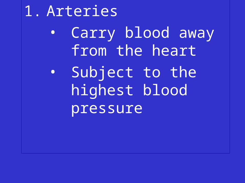
1. Arteries
• Carry blood away from the heart
• Subject to the highest blood pressure
• Located deep
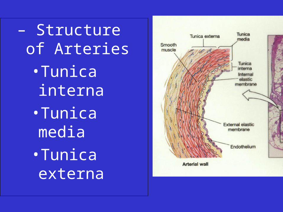
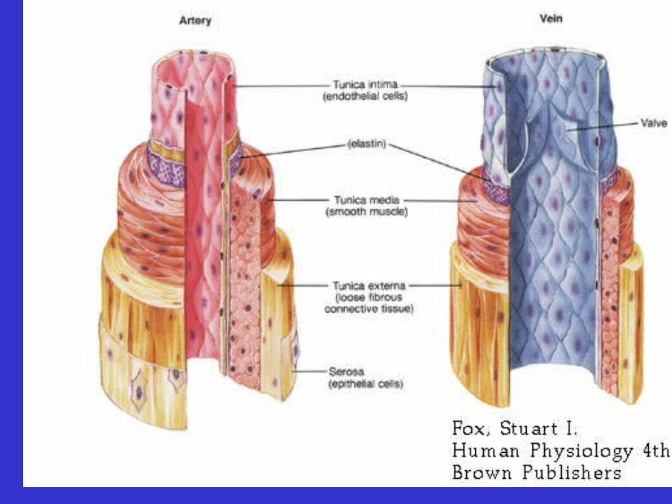
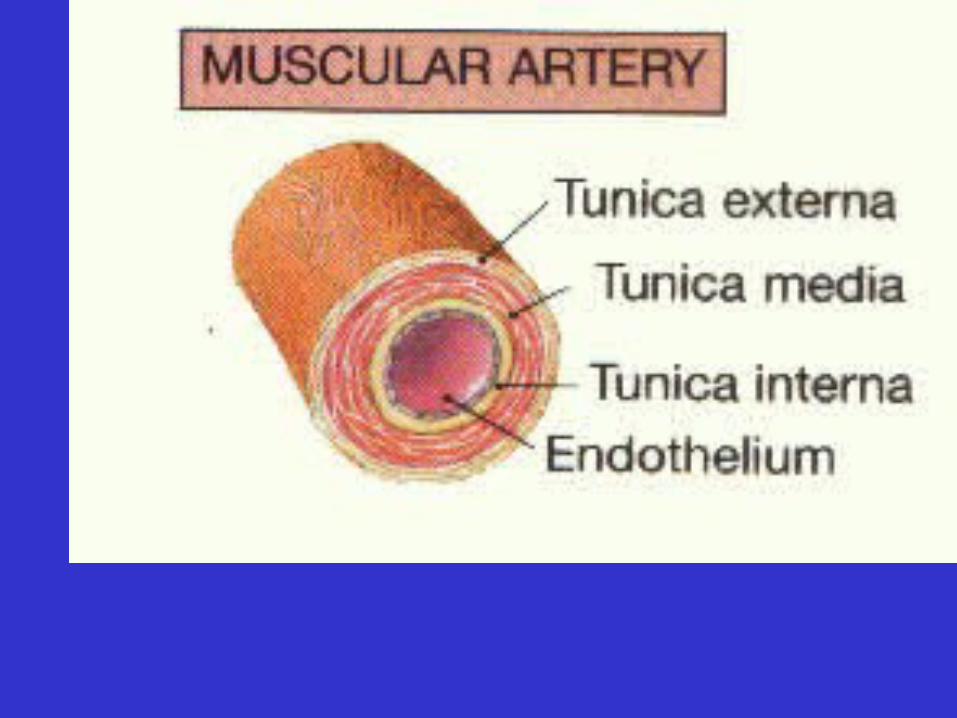
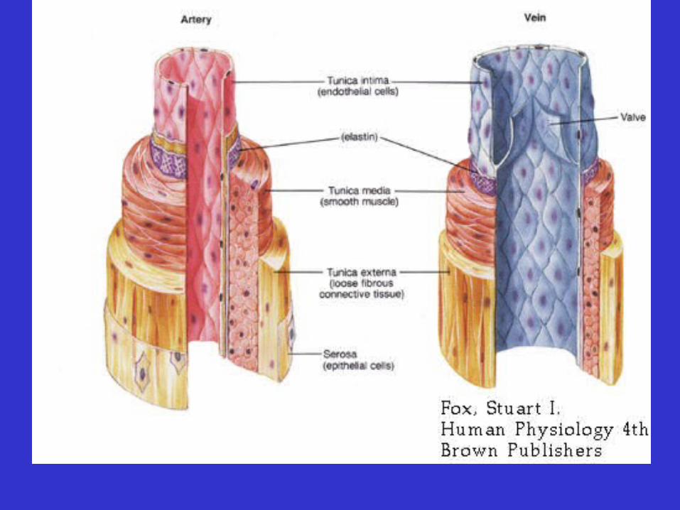
– Structure of Arteries
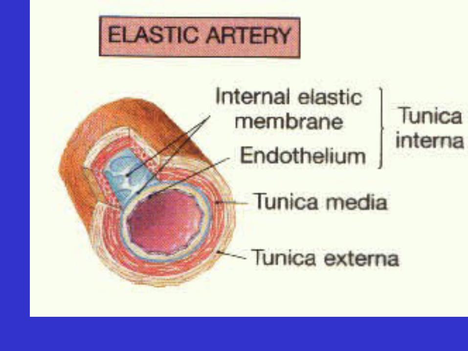
•Tunica interna
•Tunica media
•Tunica externa
• Types of arteries
– Elastic arteries
• Contain elastic fibers in the tunica media and interna
•Largest arteries
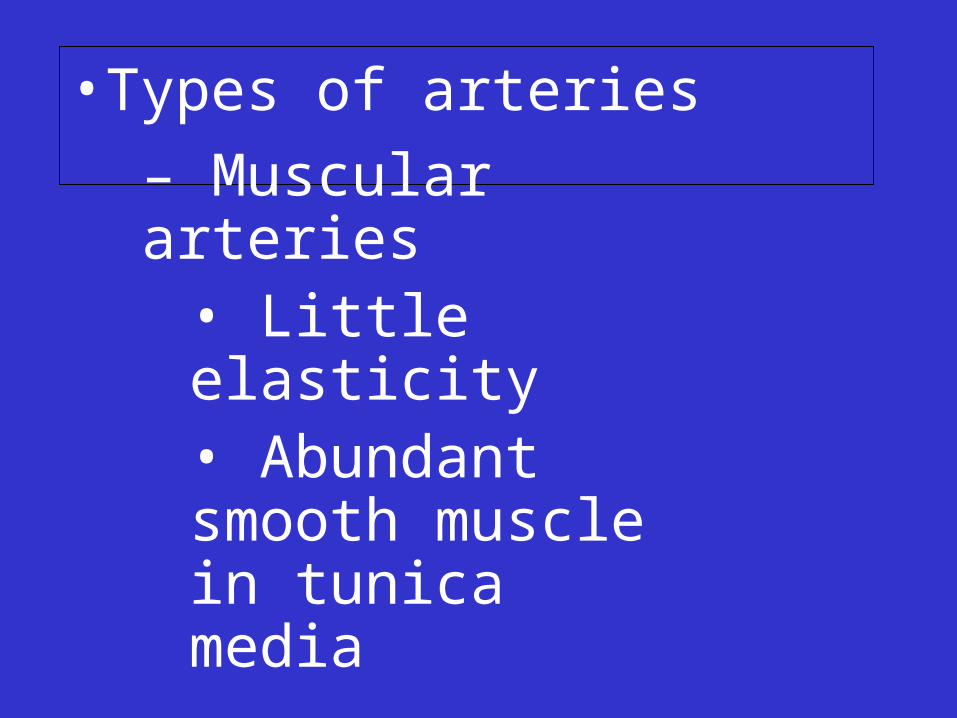
• Types of arteries
– Muscular arteries• Little elasticity• Abundant smooth muscle in tunica media
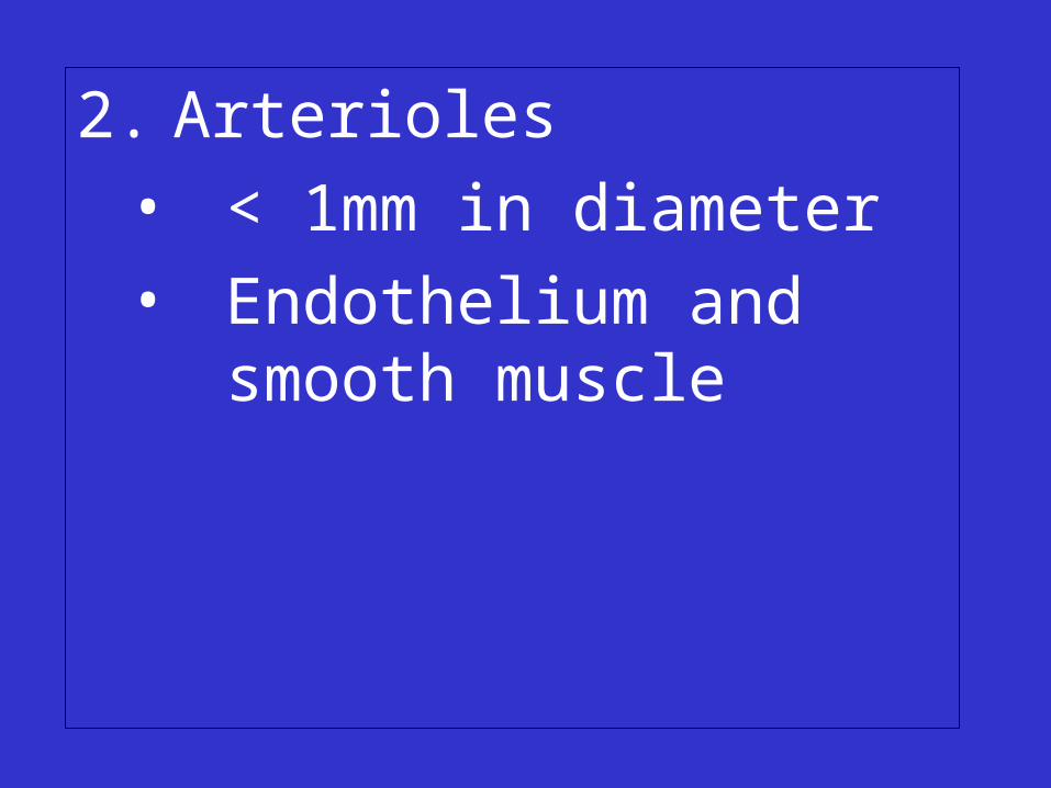
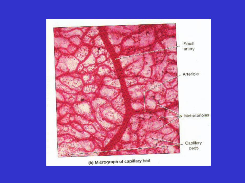
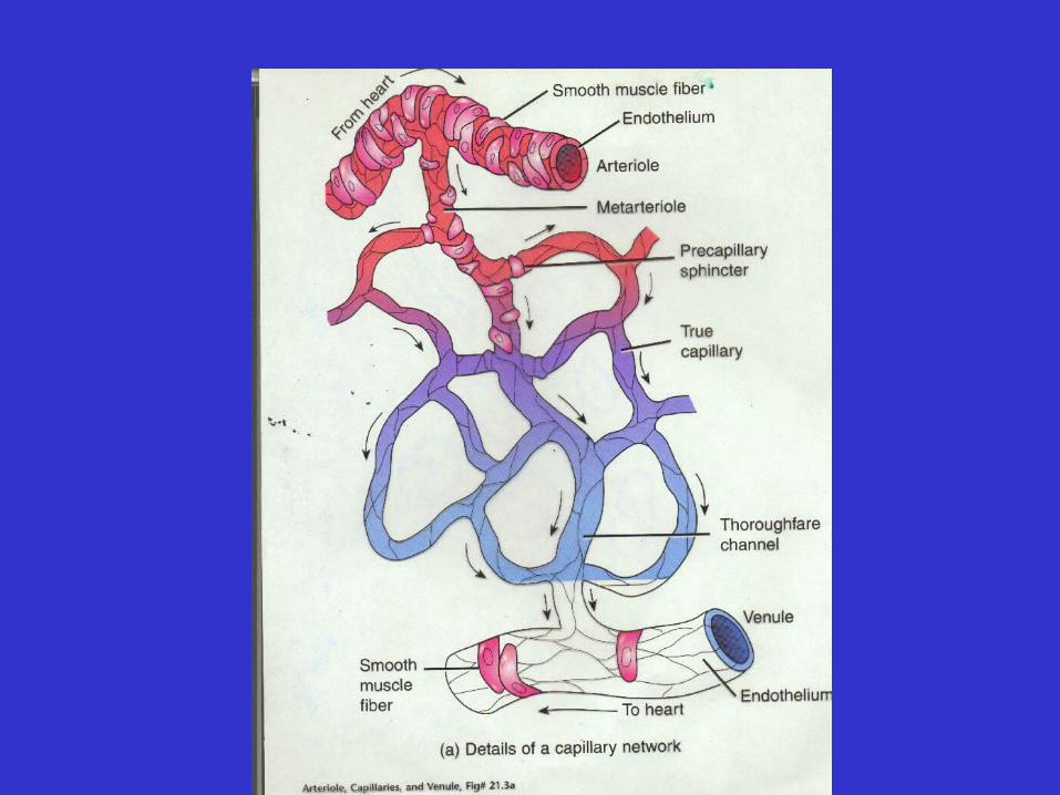
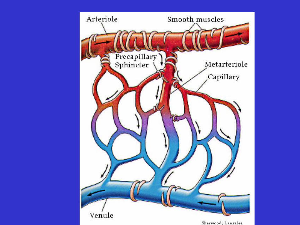
2. Arterioles
• < 1mm in diameter
• Endothelium and smooth muscle
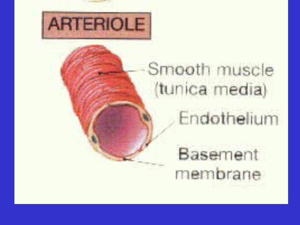

2. Arterioles
• < 1mm in diameter
• Endothelium and smooth muscle
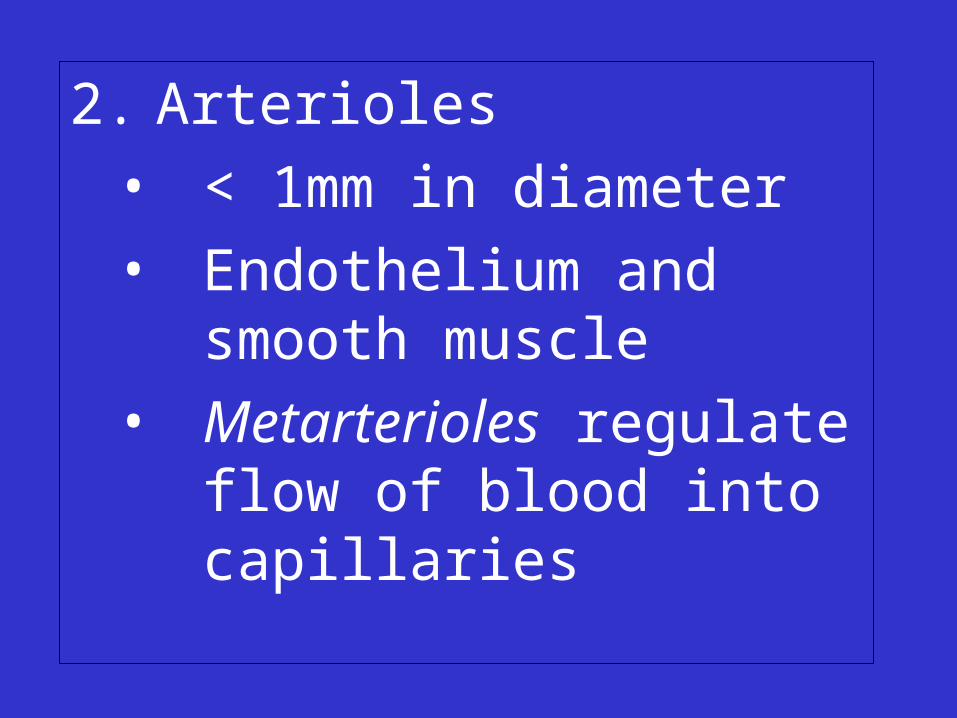
2. Arterioles
• < 1mm in diameter
• Endothelium and smooth muscle
• Metarterioles regulate flow of blood into capillaries
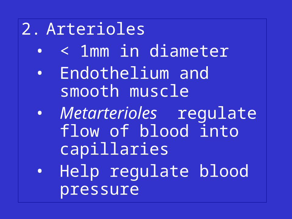
2. Arterioles• < 1mm in diameter• Endothelium and smooth
muscle • Metarterioles regulate flow
of blood into capillaries • Help regulate blood
pressure
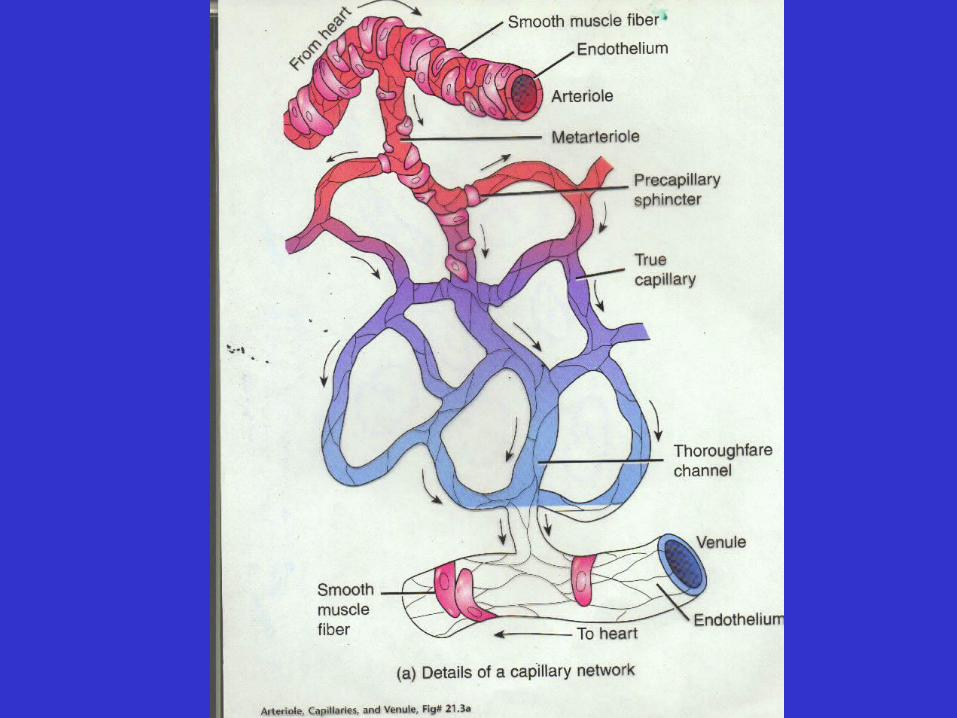
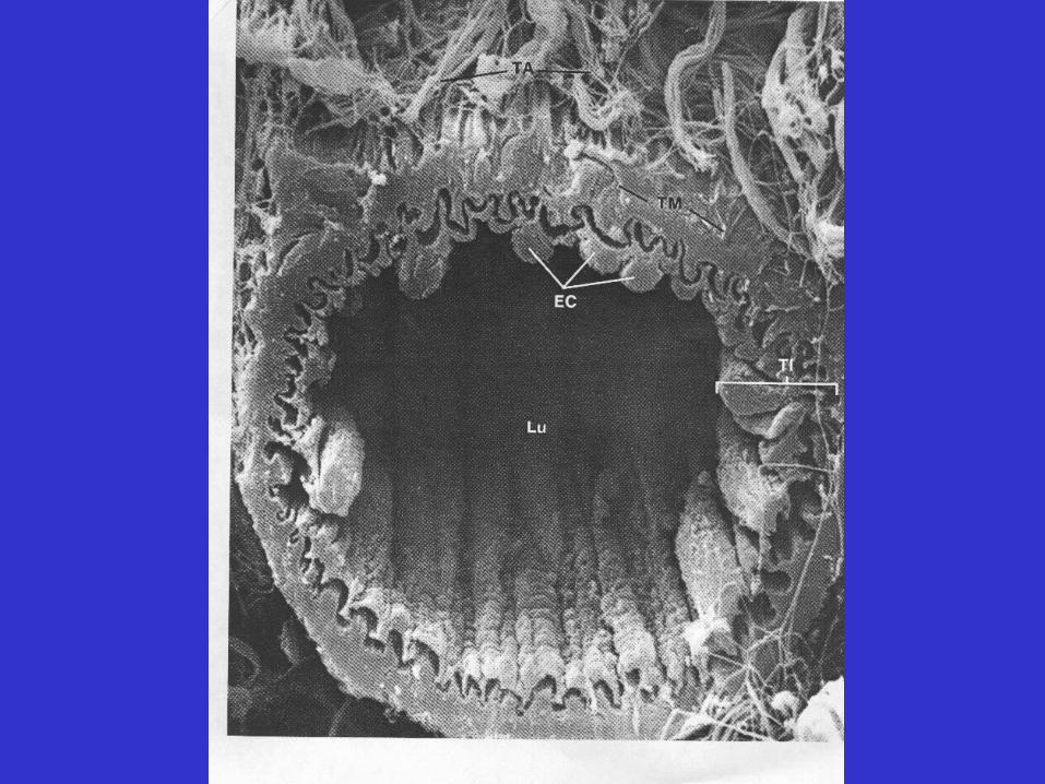
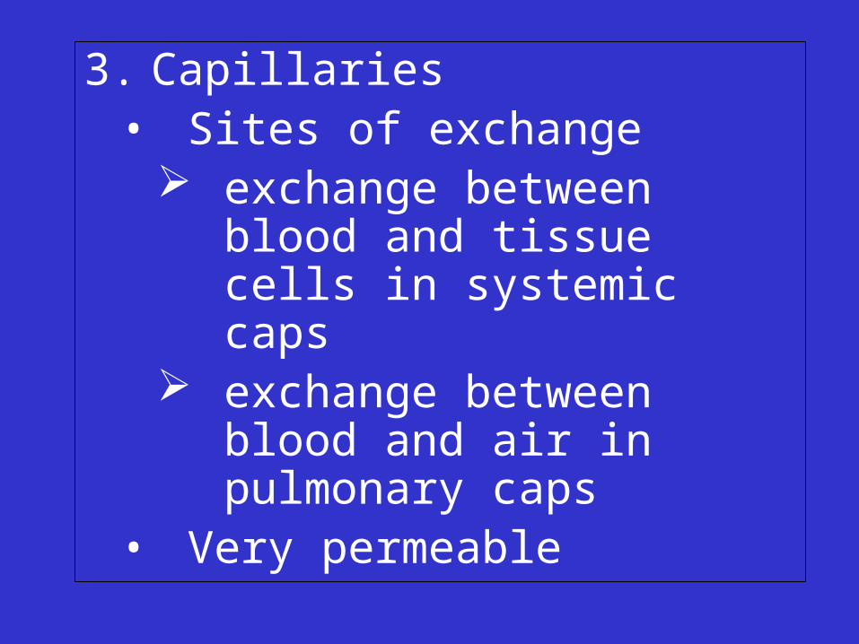
3. Capillaries• Sites of exchange
exchange between blood and tissue cells in systemic caps
exchange between blood and air in pulmonary caps
• Very permeable

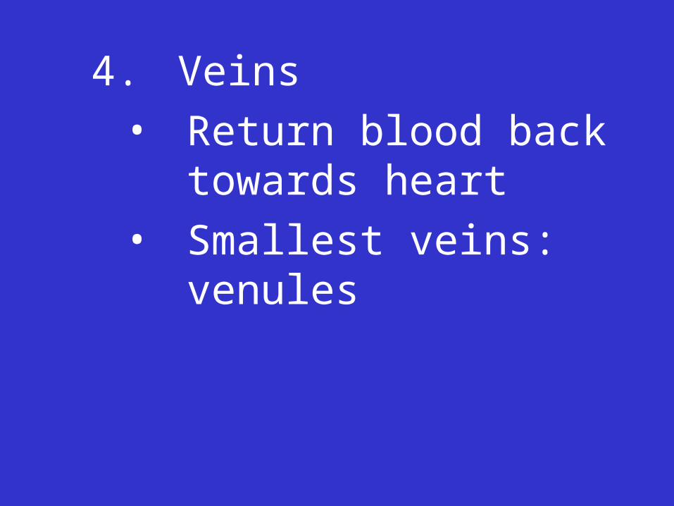
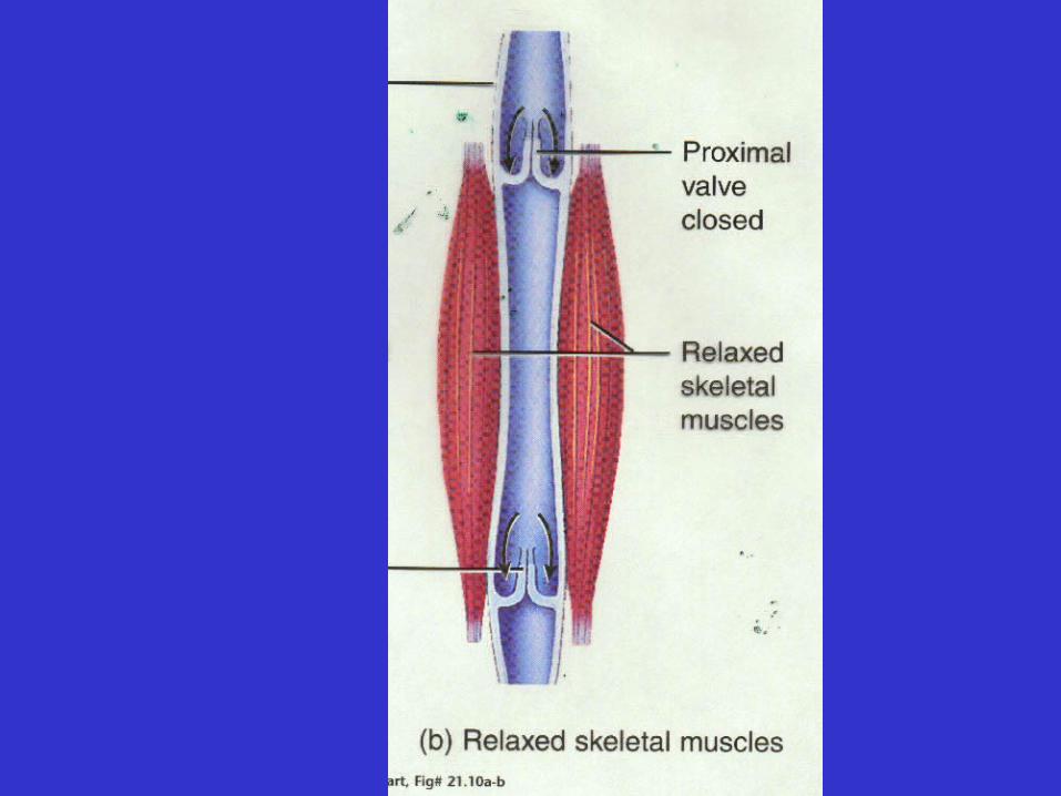
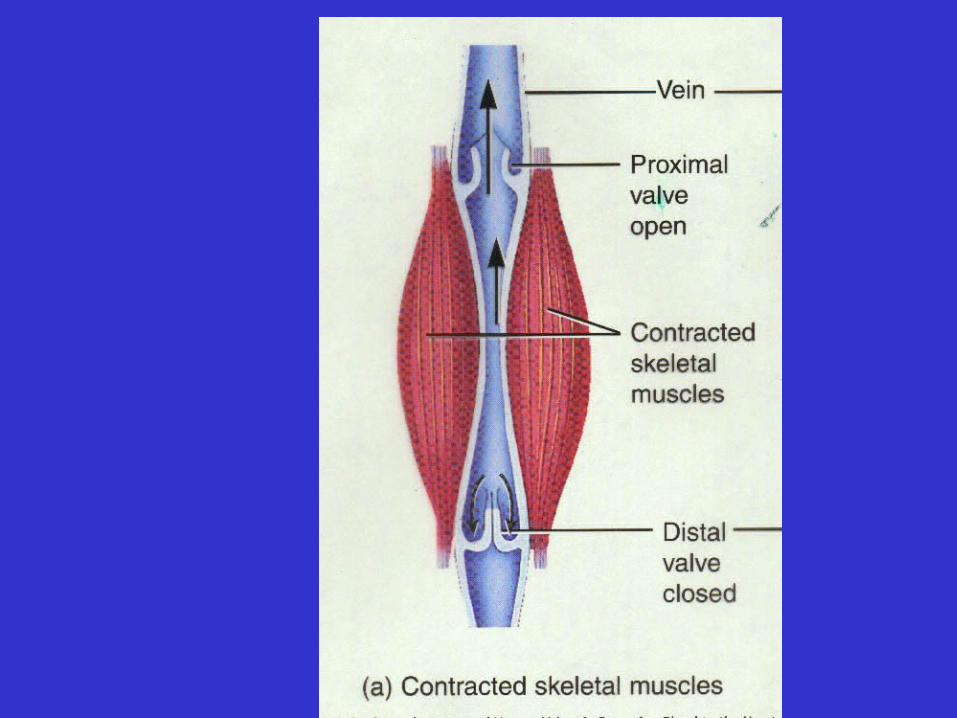
4. Veins
• Return blood back towards heart
• Smallest veins: venules
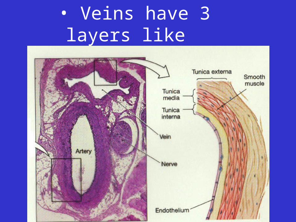
• Veins have 3 layers like arteries
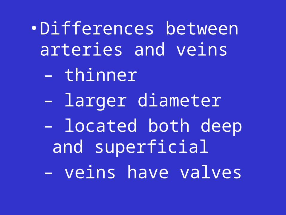
• Differences between arteries and veins
– thinner
– larger diameter
– located both deep and superficial
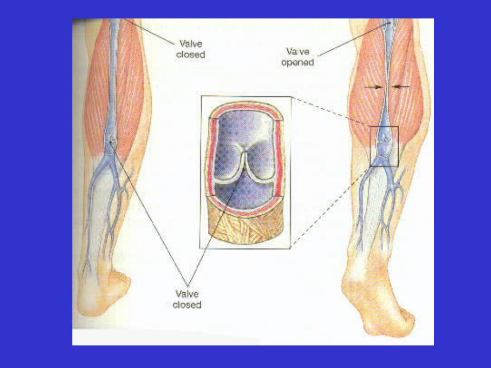
– veins have valves
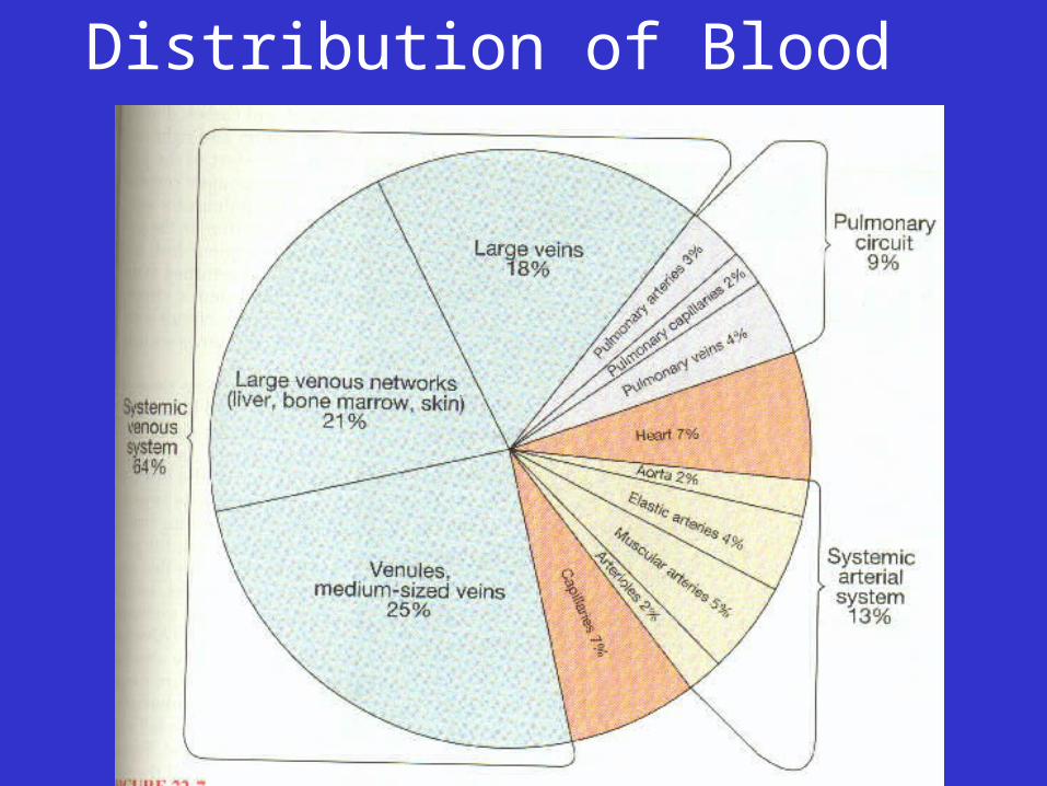
Distribution of Blood
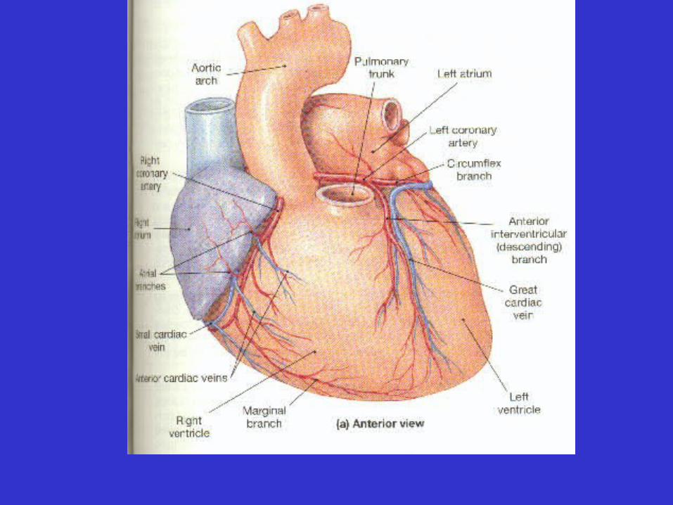
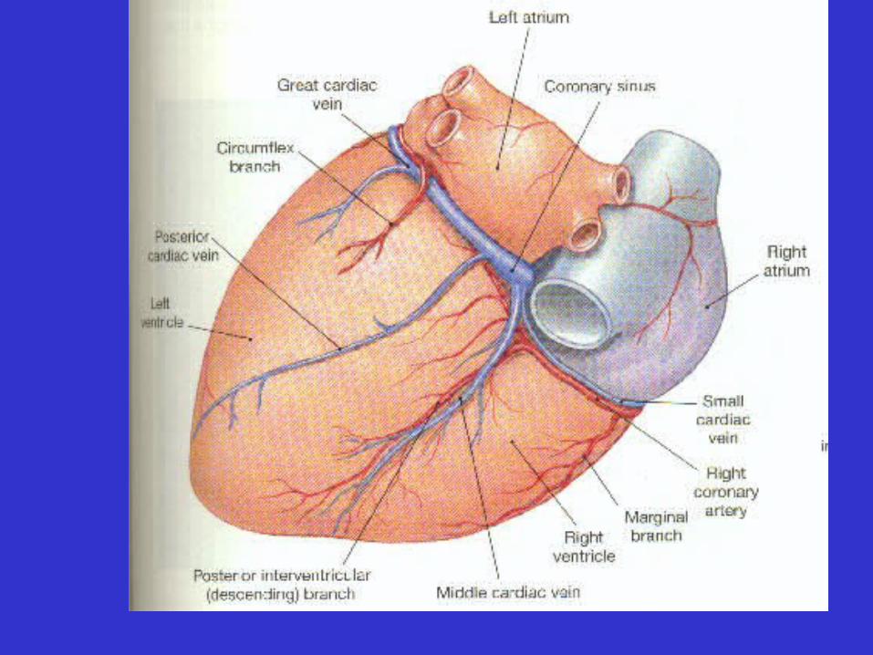
• Coronary Circulation
– Blood flow to and from the myocardial capillaries
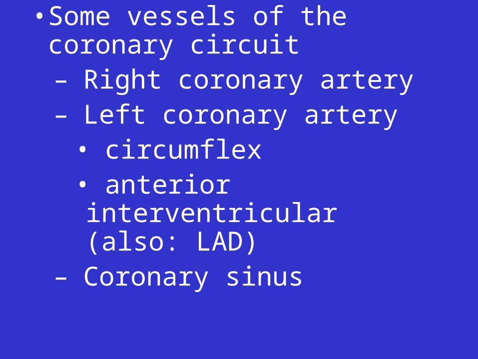
• Some vessels of the coronary circuit– Right coronary artery– Left coronary artery
• circumflex• anterior interventricular (also: LAD)
– Coronary sinus
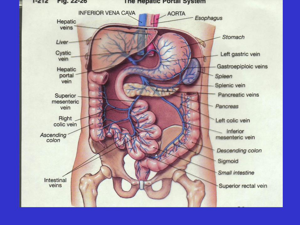
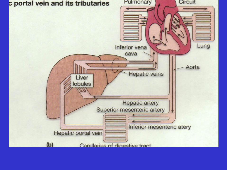
• Hepatic Portal Circulation
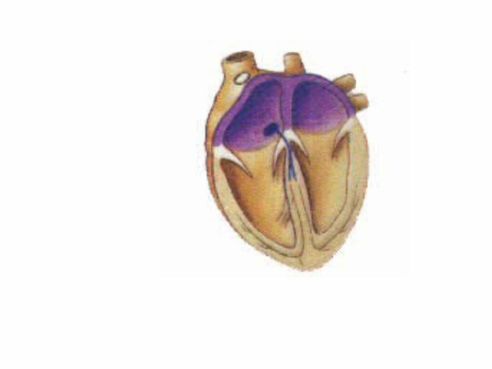
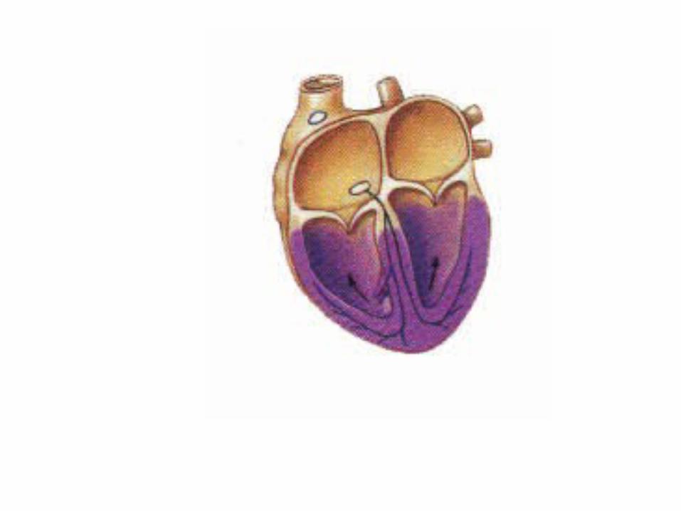
• Features of fetal heart
– Foramen ovale
– Ductus arteriosus
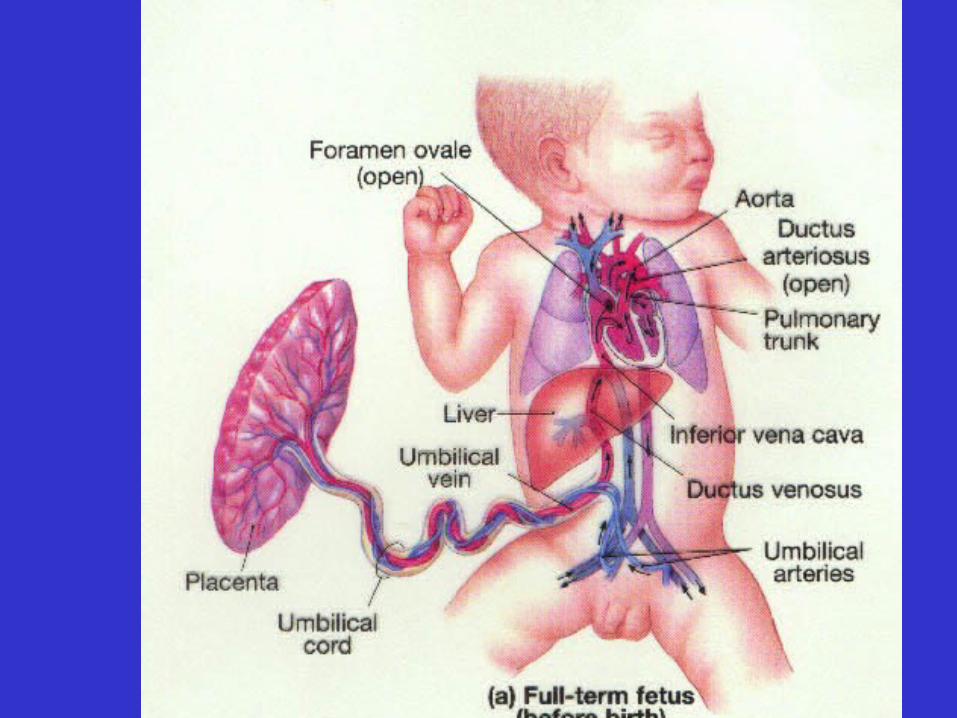
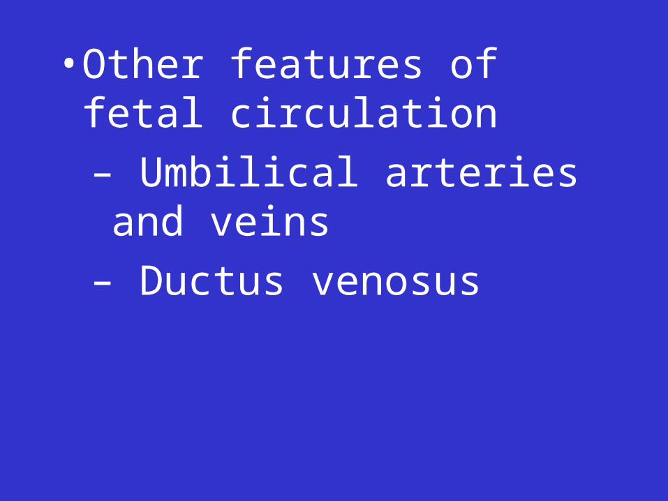
• Other features of fetal circulation
– Umbilical arteries and veins
– Ductus venosus
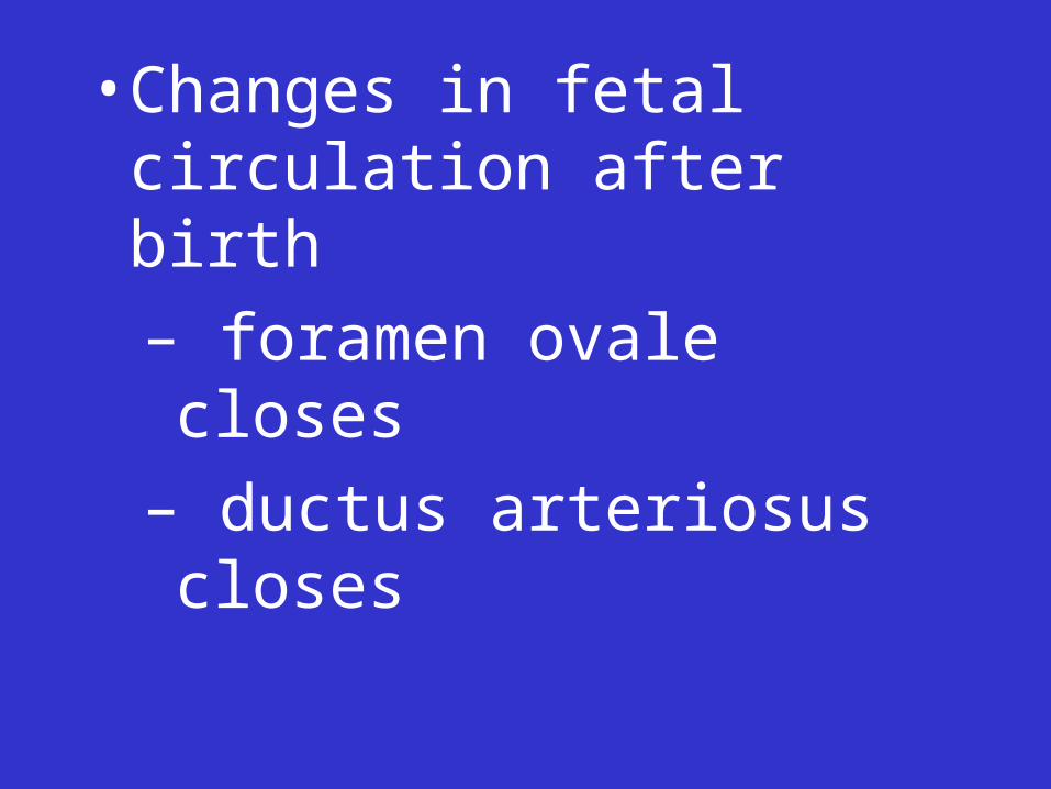
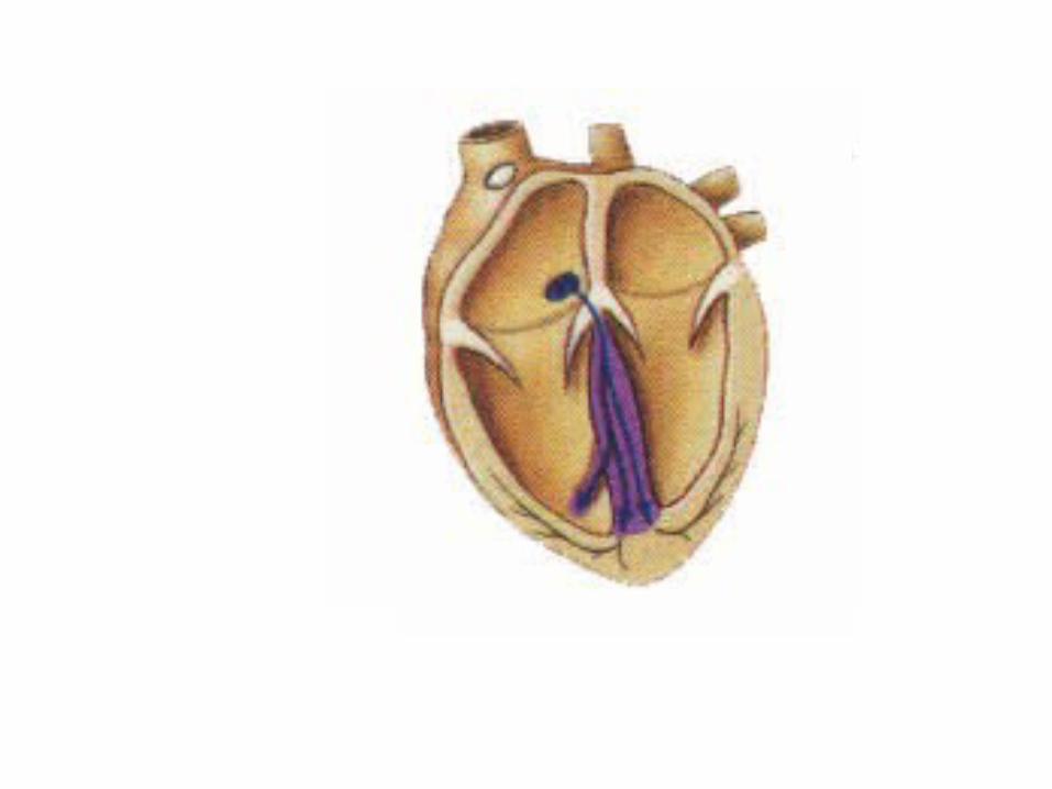
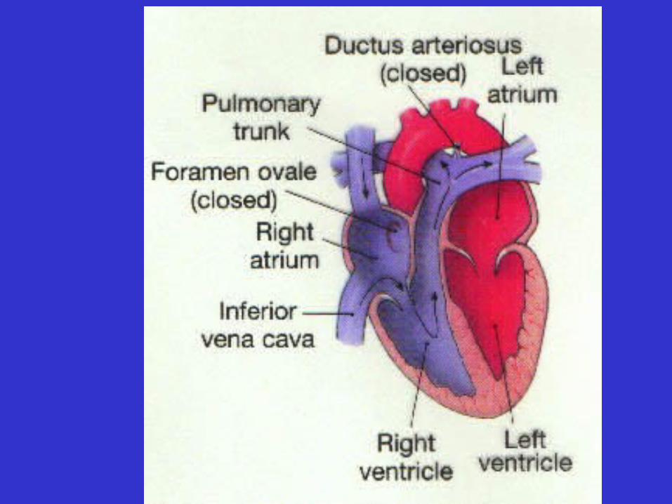
• Changes in fetal circulation after birth
– foramen ovale closes
– ductus arteriosus closes
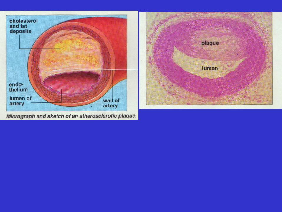
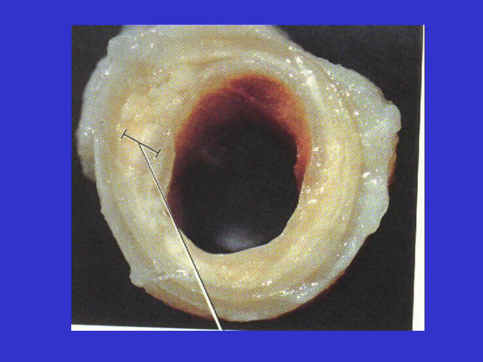
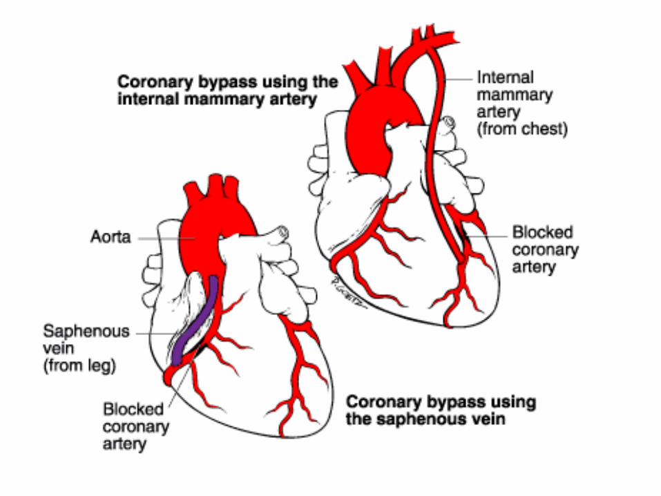
Coronary Artery Disease
• Degenerative changes in coronary arteries
• Often associated with arterial plaques

•


![12079 CSR Umschlag en RZ - The Linde Group · the linDe woRlD [3] The Group comprises three divisions: Gases and Engineering (the two core divisions) and Gist (logistics services)](https://img.pdfslide.net/doc/110x75/5b4334387f8b9a26268bcf2b/12079-csr-umschlag-en-rz-the-linde-the-linde-world-3-the-group-comprises.jpg)