Embed Size (px)
Citation preview
Chapter 10
Chapter 10 – The Respiratory System
Applied Learning Outcomes
Use the terminology associated with the respiratory system
Learn about the following:• Respiratory system components• Development and histology of the
respiratory system• Respiratory system function• Breathing processUnderstand the aging and pathology
of the respiratory system
Overview
Chapter 10 – The Respiratory System
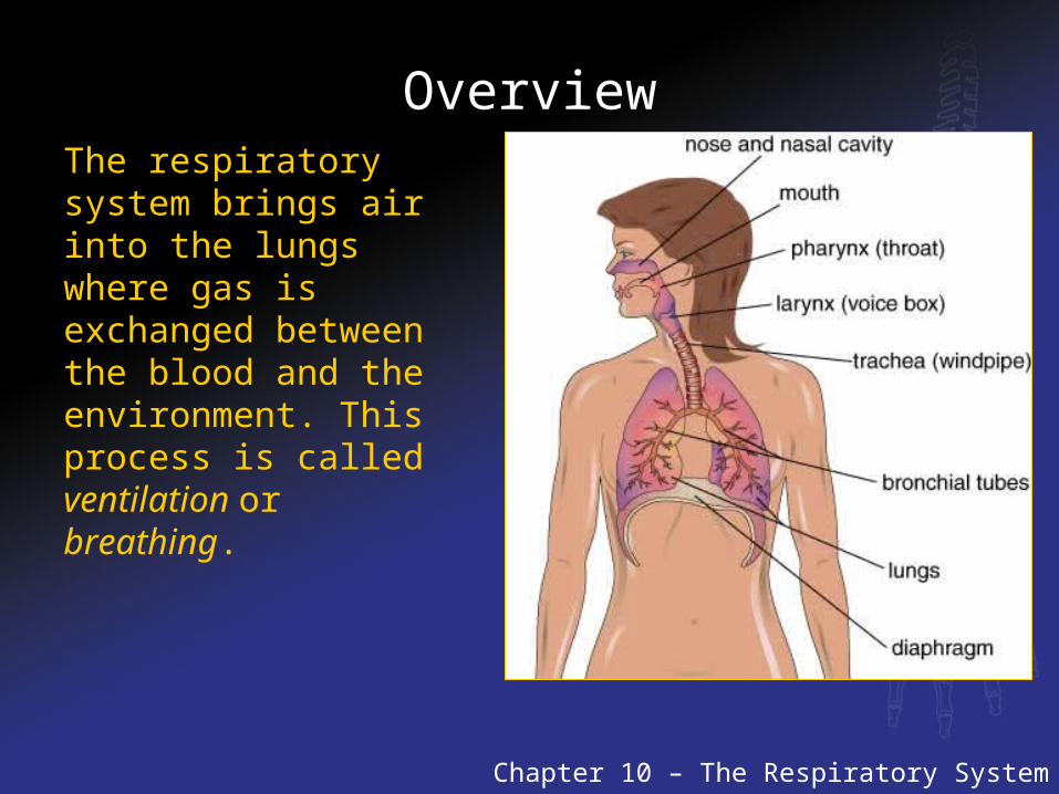
The respiratory system brings air into the lungs where gas is exchanged between the blood and the environment. This process is called ventilation or breathing.
Overview
Respiratory system •Able to exchange certain materials by facilitating the passive diffusion of carbon dioxide and oxygen.•Accomplished by bringing air to the lungs
Conditions needed for the diffusion of materials into and out of the body.
•Moist surface for diffusion of gases•Thin layer of cells
Drawback of having an internal diffusion surface•Air must be moved to come in contact with the lung surface•Ventilation or breathing brings the air into the lungs
Components of the Respiratory System
Chapter 10 – The Respiratory System
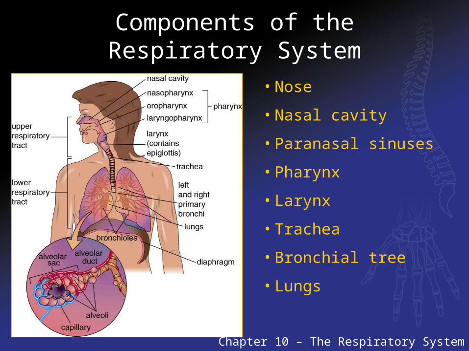
• Nose
• Nasal cavity
• Paranasal sinuses
• Pharynx
• Larynx
• Trachea
• Bronchial tree
• Lungs
Component of the Respiratory System
Nose—The entrance to the respiratory tract
Nasal cavity—The body cavity behind the nose
Paranasal sinuses—Air cavities within the facial bones
Pharynx—The throat; the cavity behind the mouth
Larynx—The area of the throat that houses the vocal cords
Trachea—The windpipe; a passage for the admission of air into the lungs
Bronchial tree—A network of passages that supplies the lungs with air
Lungs—Two large organs in which gas is exchanged between the blood and the environment
Respiratory System Divided
Upper Respiratory System:Composed of:The nose, nasal cavity, paranasal sinuses, larynx, mouth, and the eustachian tubes of the middle ear.
Lower Respiratory System:Composed of:The trachea, bronchial tree, and lungs.
Upper respiratory infections are usually less serious than lower respiratory infections.
Upper Respiratory System
Nostrils or nares:•major point of entry for air entering the respiratory system. hairs in nostrils protect respiratory system by restricting passage of large particles•mucous covering the hairs reduces number of microorganisms that can cause diseases.
Nasal cavity:•Large air filled space•Lined with mucous membrane with a rich blood supply•Mucous assists with cleaning and moistening the air•Blood supply warms the air•Nasal septum separates the nasal cavity into right and left airways
Paranasal sinuses:•Connected to nasal cavity through small openings in the bones•Four pairs of sinuses
•Maxillary sinuses-under the eyes in maxillary bone•Frontal sinuses-just above the eyes•Ethmoid sinuses-between the nose and the eyes•Sphenoid sinuses-at base of skull behind the nasal cavity
Upper Respiratory System
Function: help warm and moisten the air and provide resonance for speech
Pharynx:•Nasopharynx- openings to the Eustachian tubes and the adenoids are found here
•Adenoids are part of the immune system•Oropharynx- part of digestive system, it carries air and food in.
•Tonsils are located in this area. •Laryngopharynx- distal portion of the pharynx
Upper Respiratory System
Larynx:•Voicebox•Lies below the pharynx•Passageway for air to enter into and out of the trachea•Houses the vocal cords which are in a region called the glottis•It is composed of the hyoid bone, muscles, and nine pieces of irregularly shaped cartilage.The epiglottis projects over the larynx, covering the opening of the trachea and assists in swallowing.
•It is a flap of cartilage covered by mucous membrane.
Three major types of cartilage form the larynx:
•Thyroid -the anterior ridge of this cartilage forms the Adam’s apple
a difference in shape accounts for the more prominent
Adam’s apple in males. Male hormones cause this change.•Cricoid - are the ring shaped cartilage in the larynx•Arytenoid- are where the vocal cords are attached
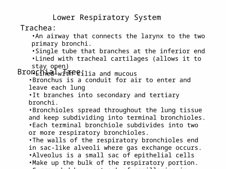
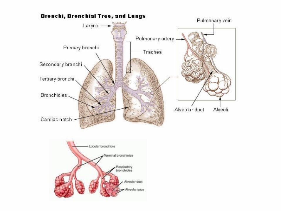
Lower Respiratory SystemTrachea:
•An airway that connects the larynx to the two primary bronchi.•Single tube that branches at the inferior end•Lined with tracheal cartilages (allows it to stay open)•Lined with cilia and mucousBronchial Tree:•Bronchus is a conduit for air to enter and leave each lung•It branches into secondary and tertiary bronchi.•Bronchioles spread throughout the lung tissue and keep subdividing into terminal bronchioles.•Each terminal bronchiole subdivides into two or more respiratory bronchioles.•The walls of the respiratory bronchioles end in sac-like alveoli where gas exchange occurs.•Alveolus is a small sac of epithelial cells•Make up the bulk of the respiratory portion.•Surrounded by a network of capillaries in which gases pass back and forth between the lungs and blood.
Lower Respiratory System
Smooth muscle lines the terminal bronchioles.This is what controls the flow of air through the lungs by constricting and dilating.
•Constriction of the terminal bronchioles is called bronchoconstriction.•Expansion of the terminal bronchioles is called bronchodilation.
The parasympathetic nervous system stimulates bronchoconstriction.The sympathetic nervous system stimulates bronchodilation.
Lungs:•Are paired organs that exchange atmospheric gases with the blood•They lie within the thoracic cavity•The thoracic cavity is surrounded by two layers of serous membranes
•The parietal pleura -the outer layer•The visceral pleura- the inner layer that covers each lung.•Right lung composed of three lobes.•Left lung composed of two lobes.
These membranes secrete fluid that reduce abrasion as lungs rub against the thoracic cavity.
•Clara cells which are special cells of the terminal bronchioles remove toxins from the lung surface.
•Type I aveolar cells are responsible for gas exchange.•Type II aveolar cells are responsible for secretion of surfactant•Surfactant reduces evaporation of water from the lung’s wet surface.•It also prevents the respiratory portion of lung from collapsing during ventilation.
Lower Respiratory System
Breathing
Chapter 10 – The Respiratory System
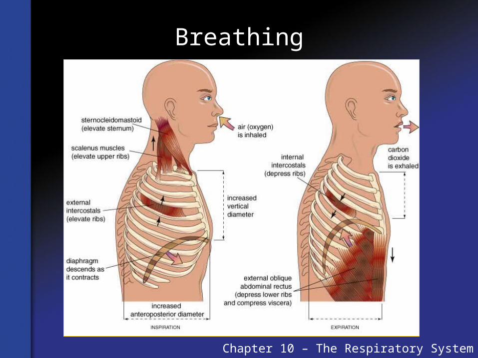
Breathing is the process of inhalation – the entering of air into the lungs – and exhalation – air leaving the lungs.
Inhalation (Inspiration): The entering of air into the lungs; caused by contraction of the diaphragm and expansion of the rib cage, increasing the volume of the thoracic cavity. The lungs stretch out and increase in volume accordingly.
Exhalation (Expiration): Air leaving the lungs; caused by relaxation of the diaphragm and external contraction of intercostal muscles. This returns the rib cage to its original, smaller volume. The volume of the lungs decreases and air is forced out of the lungs and into the airways.
Mechanics of Breathing
Inspiration:•Carried out by diaphragm and intercostal muscles•Neural signals from the respiratory center of the brain stem have involuntary control over breathing•During inspiration
•the diaphragm contracts and moves downward.•The intercostal muscles contract
This action expands and lifts up the rib cage increasing the volume ofthe thoracic cavity.Air flows in because the air pressure inside the lungs drop as they expand.Air flows from a region of high pressure to a region of low pressure
•Inspiration is effective only if the lungs and alveoli are expanded•Alveoli are kept inflated by expansion of the lungs•A layer of surfactant keeps them from completely from deflating•Surfactant makes the walls of the alveoli rigid which supports their shape.
Mechanics of Breathing
Expiration forces air out of the lungs.•Initiated by relaxation of the diaphragm and external intercostal muscles.•Causes diaphragm to return to resting curved position.•Relaxation of intercostal muscles causes rib cage to return to its original smaller volume.•Both of these actions cause the volume of the lungs to decrease•Air flows out because the air pressure inside the lungs rises as they
contract.•Air flows from a region of high pressure to a region of low pressure
Intrapleural pressure and surfactants prevent the collapse of the lungsand alveoli during expiration.
•Damage to the pleural membranes can cause one or both lungs to collapse after expiration.•Puncture wounds to the chest can produce this condition
Gas exchange
Diffusion has to occur in the lungs for gas exchange to occur.
It can only occur if the concentration of a gas in the atmosphere differsfrom the body
Wellness and Illness over the Life Span
Chapter 10 – The Respiratory System
Diseases of the respiratory system are developmental or infectious. Developmental diseases are due to genetic conditions or life style factors. Infectious diseases are produced by microorganisms.
Aging of the respiratory system reduces the ability to carry out the process of breathing and reduces the diffusion of gases across the alveoli. Some aging is due to wear and tear on the body; life style also plays a significant role.
Pathology of the Respiratory System
Developmental diseases•results from a condition that produces pathology of one or more respiratory system components.•Cardiovascular system diseases commonly cause developmental respiratory system disorders
Infectious diseases•Produced by microorganisms•Lower respiratory system - microorganisms survive best in a moist environment of the bronchial tree and lungs
Bronchitis can be developmental or infectious in nature.•Due to smoking or living in areas of air pollution.•Can lead to a condition called emphysema•Alveoli will deteriorate over time if they don’t get enough oxygen.•Eventually, they will collapse and capillaries will cut off the blood supply to the alveoli.
Developmental Diseases
Acute Respiratory Distress Syndrome (ARDS)•The rapid development of respiratory system failure.•Widespread inflammation of the lungs and capillaries of the alveoli•Can be caused by breathing irritating fumes•Respiratory failure usually occurs within 24 hours to 3 days •Even with medical intervention kills approximately 30-40% •Death usually results from failure of other organ systems which results from lung failure•Treatment- anti-inflammatory drugs and artificial ventilation
Atelectasis•Complete or partial collapse of a lung•Results from absence of gas in one or more parts of the lungs•May be due to excessive pressure on lung tissue or obstruction of airways•Causes are air in the pleural cavity, fluid build up in the pleura, foreign object in the airways, obesity, and bronchial tumors •This condition causes breathing difficulties, chest pain, and coughing•Treatment involves expansion of affected lung, only if a large amount of lung tissue is affected.
Developmental Diseases
Pneumothorax•Condition in which air enters the pleura membranes•Can cause Atelectasis•Can be due to lung damage produced by severe lung infection or thorax injury
Bronchiectasis•Abnormal stretching and dilation of the bronchi or bronchioles•Caused by long term mucous blockage of airways weakens the walls of these structures, leading to excessive dilation.•Disease is characterized by abnormal breathing sounds, coughing, fatigue, shortness of breath, weakness, and weight loss. •Treatment- bronchodilators
Chronic Obstructive Pulmonary Disease (COPD)•Caused by long term lung disorders due to genetic conditions or certain lifestyles.•Usually due to long term chronic bronchitis or emphysema•Leads to permanent narrowing of small airways•Shortness of breath•Treatment- bronchodilators and antispasmodics •Patients advised not to smoke and avoid any irritating chemicals
Developmental Diseases
Restrictive lung disease•Caused by a decrease in the amount of air the lungs can hold.•This is due to a stiffness of the tissue, which a variety of lung diseases can cause.•Inhalation difficult because lungs resist expansion.Sleep apnea
• “Without breath during sleep”•Stop breathing repeatedly while sleeping•Most common type due to blockage of the bronchial tree•Another type problems with neural pathways from brain that control breathing•Untreated can cause loss of sleep, headaches, high blood pressure, and cardiovascular diseases.
Lung Cancer•Most common in ages 55 to 60•Prevalent in people who smoke and/or exposed to irritating chemicals•Usually originates in the lining of the bronchi and can spread throughout body
Infectious Diseases
Flu or influenza•Contagious•Caused by a variety of influenza viruses•Spread through contact with respiratory system fluids•Symptoms are dry cough, diarrhea, headache, high fever, muscle aches, nausea, sore throat, runny nose, fatigue, and vomiting.•Possible to spread from various animals to humans•No cure•Do have some vaccines against specific types of flu•Vaccines recommended for babies and adults over 65 years of age
Hantavirus pulmonary syndrome•Very little know about it•Contract virus through contact with rodent feces, saliva, and urine.•Recently also contracted from prairie dogs and exotic pet rodents.•High fatality rate•No well established treatment or vaccine
Other viruses cause common colds, headaches, and sinus infections.
Infectious Diseases
Pneumonia•Inflammation of the lungs •Caused by bacteria, fungi, protista, and viruses•Usually develops in people with weakened immune systems
Types of Pneumonia•Lobar Pneumonia-occurs in one lobe of the lung•Bronchopneumonia- is scattered throughout the lungs.
Viruses cause about 50% of pneumonia in North AmericaTend to mild forms of the diseaseBacterial pneumonia are more severe
can be treated with antibioticcharacterized by breathing difficulties, chills, chest pains, and coughing
Infectious DiseasesTuberculosis (TB)
•Caused by a bacterium•Use of vaccinations, antibiotics, improved health and sanitation have limited the spread of TB in the US.•Spread by coughing, sneezing and spitting•Most people can fight off the bacterium but it can lie dormant in the body for years.•It can become active later as the person’s immune system weakens with age or other illnesses.•It is fatal if not treated with antibiotics.•The bacteria irritates the lung, causing damage to
the alveoli. Hydatid lung disease
•Caused by a worm that forms an inactive cyst.•It does not usually cause problems•Severe cases produce cough, fever, and chest pain.•Chest x-rays reveal the cysts and inflammation of the lungs.Fungal diseases•Very serious•More common in people with weakened immune systems•Commonly found in soil and may be spread by bird droppings
Aging of the respiratory system
Some typical signs of respiratory system aging:
•Decrease in elasticity of the lung and rib cage, which makes inspiration difficult•Loss of skeletal muscle mass, which weakens inspiration muscles•Decrease in smooth muscle control, which makes it more difficult to regulate air flow to the alveoli•Decrease in alveolar gas exchange surface, which reduces the rate of diffusion•Decrease in nervous system control, which makes it more difficult to coordinate the rate of breathing with body activity•Decrease in laryngeal function, which diminishes the cough and gagging response.•Decrease in mucus production, which makes lungs more susceptible to infection•Decrease in blood flow through alveoli, which reduces gas exchange•Decrease in diameter of the bronchi, which reduces the flow of air into and out of the lungs.
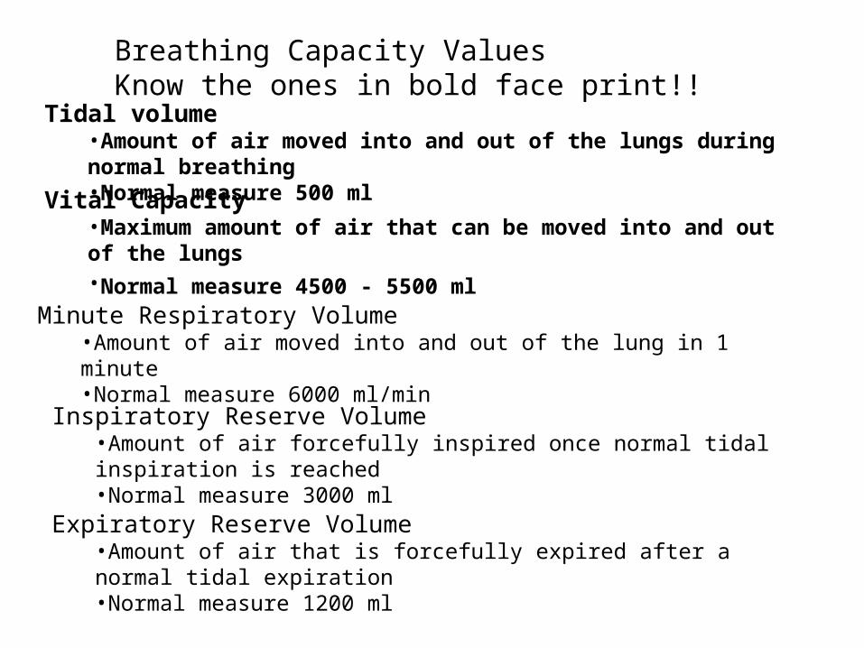
Breathing Capacity ValuesKnow the ones in bold face print!!
Tidal volume•Amount of air moved into and out of the lungs during normal breathing•Normal measure 500 mlVital Capacity•Maximum amount of air that can be moved into and out of the lungs•Normal measure 4500 - 5500 ml
Minute Respiratory Volume•Amount of air moved into and out of the lung in 1 minute•Normal measure 6000 ml/min
Inspiratory Reserve Volume•Amount of air forcefully inspired once normal tidal inspiration is reached•Normal measure 3000 ml
Expiratory Reserve Volume•Amount of air that is forcefully expired after a normal tidal expiration•Normal measure 1200 ml
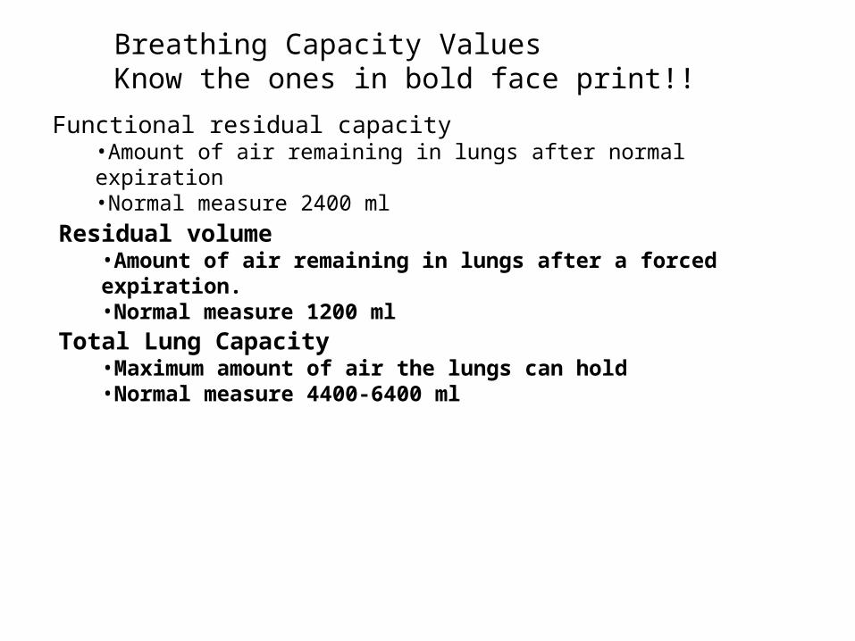
Breathing Capacity ValuesKnow the ones in bold face print!!
Functional residual capacity•Amount of air remaining in lungs after normal expiration•Normal measure 2400 ml
Residual volume•Amount of air remaining in lungs after a forced expiration.•Normal measure 1200 ml
Total Lung Capacity•Maximum amount of air the lungs can hold•Normal measure 4400-6400 ml
Summary
The respiratory system is responsible for exchanging atmospheric gases with those in the body’s internal environment. Ventilation is carried out to ensure that atmospheric air comes in contact with the alveoli of the lungs – the site of gas diffusion.
Lung diseases either affect the flow of air through the airways to the lungs, or they are caused by deterioration of the alveoli.
Aging mostly affects lung elasticity and contributes to diseases that impair the airways.
Chapter 10 – The Respiratory System