Embed Size (px)
Citation preview

Chapter 13
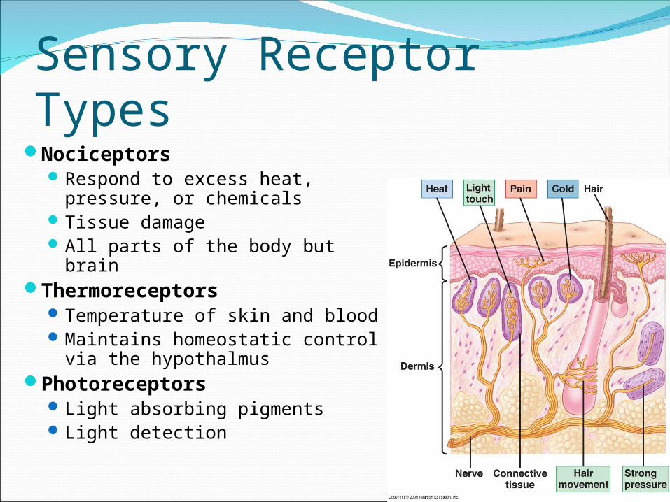
Sensory Receptor TypesNociceptors
Respond to excess heat, pressure, or chemicals
Tissue damageAll parts of the body but brain
ThermoreceptorsTemperature of skin and bloodMaintains homeostatic control
via the hypothalmusPhotoreceptors
Light absorbing pigmentsLight detection

Sensory Receptor TypesMechanoreceptors ChemoreceptorsTouch, pressure, &
vibrationsBend or stretch PM of
receptor cell = changing permeabilityStretch receptors –
position of body partsHair cells - sound waves
and H2O movements
Chemicals in the internal & external environment
O2 in arteriolesOsmoreceptors -
changes in [blood solute]Pheromone detection

Sensory Receptor LocationsExteroceptors
Stimuli outside the bodySkin and special sense organs
InteroceptorsStimuli within the bodyChemical messengers, tissue stretch, and
temperature Proprioceptors
Internal stimuliMonitor position and stretch of joints, tendons,
and muscles

Sensory Receptor StructuresUnencapsulated Encapsulated
Free nerve endingsMost body tissuesTemperature and painful
stimuliCapsaicin and itch
Merkel discsDeeper epidermal layersLight touch
Hair follicle receptorsShaft of hair follicleLight touch and hair
bending
Meissner’s corpuscles Dermal papillae of sensitive and
hairless skin Discriminative touch
Pacinian corpuscles Deep in the dermis Deep pressure initially, vibration
Ruffini endings Deep dermis and hypodermis Deep continuous pressure
Muscle spindles Perimysium of skeletal muscle Detect muscle stretch & initiate
a reflex Golgi tendon
Insertion tendons Activation inhibits contracting
muscle

Sensory InputAll senses trigger the same TYPE of signal
Distinction occurs in activated brain areaTypically graded response, but AP’s possible
Sensory receptors detect sensations and carry to the brainAwareness of environmental change
Brain constructs perceptions by integrating sensations with other informationNeuronal communication involving multiple
brain areas

The Working Brain

Sensory AdaptationSensory receptors
become less responsiveFewer action
potentials
Limits reactions to normal background stimuliShower or hot tub
temperatureOdors over time

Decoding NervesPNS organ consisting of parallel
bundles of axons enclosed by connective tissue layers
EpineuriumPerineuriumEndoneuriumDirection of transmission
Afferents: only to CNS Dorsal root ganglia
Efferents: only from CNS Sympathetic & parasympathetic ganglia
Mixed: carry both; mostClassified as cranial or spinal

Mature neurons don’t divide*Cell body damage = deathCut/compressed axons regenerate
Separated ends seal off and swellDistal end of injury disintegrates
Lack of nutrients Neurilemma maintained in
endoneuriumSchwann cells proliferate &
encourage axon growth Guide ‘sprouting’ axons to original
contactsGreater distance decreases chancesRegrowth never exact = retrainingExtremely rare in CNS
Nerve Fiber Regeneration

Cranial NervesVentral portion of the brain
1st 2 pairs attach to forebrain
Remainders originate on brainstem

Numbers Cranial Nerve FunctionI OLFACTORY Smell (sensory)
II OPTIC Vision (sensory)
III OCULOMOTOR Eye movement (motor)(medial, inferior, superior rectus muscle & inferior oblique muscle)
IV TROCHLEAR Eye movements (motor)(superior oblique muscle)
V TRIGEMINAL Temperature, pain, crude touch of face (sensory) & mastication (motor)
VI ABDUCENS Eye movement (motor)(lateral rectus muscle)
VII FACIAL Taste (2/3 of anterior tongue) (sensory)Facial expressions (motor)
VIII VESTIBULOCOCHLEAR Hearing & Equilibrium (sensory)
IX GLOSSOPHRAYNGEAL Taste (1/3 of posterior tongue) (sensory)Pharynx (swallowing & gag reflex) (motor)
X VAGUS Senses blood pressure (sensory)Stimulate heart rate and digestive organs (motor)
XI ACCESSORY Head and neck movement (motor)e.g. trapezius, levator scapula
XII HYPOGLOSSAL Tongue movement (motor)

Testing Cranial Nerves for Disorders Olfactory
Smell substances Anosima
Optic Eye chart Anopsias
Oculomotor Follow object; pupil reflex Strabismus, double vision, ptosis
Trochlear See oculomotor
Trigeminal Close/move jaws; touch face with
objects Abducens
See oculomotor
Facial Make various faces; tasting
substances Bell’s palsy, loss of taste, can’t
close eye Vestibulocochlear
Tuning fork; distance of sound Deafness, vertigo, tinnitus
Glossopharyngeal Swallowing & gag reflex; say ‘ah’
Vagus See glossopharyngeal Horseness, swallowing problems,
death (Spinal) accessory
Move head/shoulders against resistance
Hypoglossal Stick out, retract, & move tongue to
sides

Roots: medial & afferent OR efferentDorsal root: peripheral
receptors to spinal cordVentral root: ventral horn to
skeletal musclesBranches: laterally pass
through intervertebral foramenDorsal ramus: dorsal trunkVentral ramus: limbs & rest of
trunkMeningeal branch: meninges
and blood vesselsPlexus
Criss cross joining of ventral rami
Excludes T2 – T12
Spinal Nerve Anatomy

31 Pairs of Spinal Nerves8 cervical
Cervical plexusBrachial plexus
12 thoracicIntercostal nerves &
enlargements5 lumbar
Lumbar plexus5 sacral
Sacral plexus1 coccygeal
Tailbone & perineum

Cervical Nerves Cervical plexus
Phrenic nerve: diaphragm Irritation causes hiccups
Brachial plexus C5 – C8
Median nerve: flexor muscles of the anterior forearm and small hand muscles Carpal tunnel syndrome and suicide attempts
Radial nerve: extensor muscle of posterior forearm and triceps brachii ‘Saturday night paralysis’
Ulnar nerve: similar to median nerve ‘Funny bone’ and paralysis/distortion of medial fingers

Lumbosacral PlexusInnervates lower limbs, buttocks, and pelvic
musclesLumbar plexus L1 – L4
Femoral nerve: quadriceps and sartorius Branches to saphenous
Obturator nerve: adductor musclesSacral plexus L4 – S4
Sciatic nerve: entire lower leg (except anteriomedial thigh) Tibial: hamstrings Common fibular nerve: anterior tibialis

Reflex ArcReceptor
Senses stimulusSensory neuron (afferents)
Message to the CNSIntegration center
Synapses in CNS Monosynaptic (single motor or sensory neuron) Polysynaptic (multiple interneurons)
Motor neuron (efferents)Message to effectors
EffectorMuscle fibers or glands
Reflexes are rapid, predictable motor responses to a stimulus
http://a248.e.akamai.net/7/248/430/20080327144023/www.mercksource.com/ppdocs/us/common/dorlands/dorland/images/arc_reflex%20a.(1).jpg

Classifying ReflexesSomatic: activate skeletal muscle
Spinal: integration center is spinal cord Stretch: ensures muscle length maintained (knee-jerk reflex) Crossed extensor: withdrawl from painful stimuli (pin prick) Superficial: cutaneous stimulation (plantar reflex)
Cranial nerve: integration center is brain stem Corneal: stimulation causes blinking
Autonomic (visceral): activate smooth or cardiac musclePupillary light: controls diameter of pupil
(inside/outside)Ciliospinal: ipsilateral pupil dilation from pain