Embed Size (px)
Citation preview

e376
CHAPTER
140Thyroid DisordersJennifer A. SipoS and WilliAm G. CAnCe
InTroDucTIon
The purpose of this chapter is to present thyroid disease as it may be seen in an intensive care unit (ICU). Interpretation of thyroid function tests in a critically ill patient requires knowl-edge of the perturbations of hormone synthesis that may occur with illness and certain medications. This text will also discuss the diagnosis and treatment of thyroid emergencies: myxedema coma, thyroid storm, postthyroidectomy hypocal-cemia, and airway obstruction by goiter.
ThyroID FuncTIon TesTs
Numerous tests of thyroid function exist; interpretation of the results and their individual roles in elucidating the status of the hypothalamic–pituitary–thyroid axis can confound even the most astute clinician. This section will provide an overview of the frequently ordered labs. A later section will delineate how to interpret these values in the critically ill patient.
Measurement of serum thyroid-stimulating hormone (TSH) is sufficient to diagnose the majority of thyroid disorders (1). It is recommended to use the ultrasensitive TSH (2). In patients for whom there is a concern of pituitary dysfunction, it is essential to measure serum T4 as well. Some clinicians elect to measure serum T4 concomitantly with the TSH regardless of the pituitary status.
Total T4 (TT4) measurement includes both bound and free thyroxine. Therefore, conditions or medications that affect serum levels of thyroid-binding globulin (TBG) will also affect the total T4 value; the use of FT4 can eliminate this shortcom-ing. If, however, FT4 is not available, this level can be esti-mated by looking at the FT4 index (FTI). The FTI is calculated by multiplying the total T4 by the T3 resin uptake (T3RU) (see below for explanation). Though free T4 is now more read-ily available in standard laboratories, some facilities may still substitute this calculated value when reporting an FT4.
Similar to thyroxine, serum triiodothyronine (T3) may be measured in the free and bound fractions. The total T3 is gen-erally used, however, as only a minute fraction of T3 is free, and the serum half-life of free T3 is very short (3). It is important
to measure T3 levels in those suspected of having hyperthy-roidism, as some patients may have excess secretion of only T3 early in the course of thyrotoxicosis. The resultant pattern of thyroid function tests is one of the suppressed TSH, nor-mal free T4, and an elevated total T3 (T3 toxicosis). On the contrary, measurement of T3 is less helpful in hypothyroidism as the elevated TSH stimulates preferential formation of T3, typically maintaining these levels in the normal range (4,5). Consequently, even in the face of significant hypothyroidism, many patients will have a normal serum T3.
T3 resin uptake is an indirect, inverse test to estimate the num-ber of unoccupied serum protein-binding sites. Radiolabeled T3 is added to the patient’s serum and distributes between unoccu-pied T4-binding sites on TBG in the serum and an adsorbent that has been added to the solution. Binding of 125I–T3 to the adsor-bent is increased (high T3RU) if the number of unoccupied bind-ing sites is decreased. This may be due to either low TBG levels, as seen in nephrotic syndrome and chronic liver disease, or due to increased thyroid hormone levels, as seen in hyperthyroidism (6). In contrast, the T3RU value is low if the number of unoccu-pied binding sites is increased, a situation which would be seen with low thyroid hormone levels (hypothyroidism) or high TBG concentrations (with estrogen therapy or pregnancy) (7).
Reverse T3 differs from T3 in that the iodine is missing from the inner ring instead of the outer ring of T4. It is largely bound to proteins in the serum. Its half-life in the serum is quite short (8) and, frankly, the biologic function of rT3 is not completely understood in humans. The clinical utility of measuring these levels is chiefly in the setting of nonthyroidal illness and will be discussed in greater detail in the section dealing with the interpretation of thyroid function in critically ill patients. Table 140.1 details the interpretation of thyroid function tests.
Drugs anD ThyroID FuncTIon
A number of drugs commonly used in the ICU will interfere with thyroid homeostasis. While many of these actions are viewed as detrimental, some effects may also be utilized for a therapeutic benefit, particularly in thyrotoxicosis. The majority of the effects
Table 140.1 Interpretation of Thyroid Function Tests
TSH Free T4 Total T3 T3U rT3
Hypothyroidism
primary High low normal low low / normal
Secondary low / normal low normal low low / normal
Hyperthyroidism low High / normal High High low / normal
Sick euthyroid low / normal low / normal low High High
T3U = T3 resin uptakerT3 = reverse T3
LWBK1580-CH140E_p376-388.indd 376 01/08/17 9:00 PM

CHAPTER 140 Thyroid Disorders e377
Since atrial fibrillation may be the only indication of thyro-toxicosis, it is important to screen such patients with a TSH and FT4. Congestive heart failure typically occurs only in patients with underlying heart disease, but may also manifest as a result of chronic tachycardia-induced cardiomyopathy (11). Physical examination findings in a thyrotoxic patient include widened pulse pressure, hyperdynamic precordium, tachycardia, and systolic ejection murmur. The pathogenesis of cardiovascular diseases from exposure to excessive thyroid hormone is not entirely understood. Thyroid hormone has both indirect and direct effects on vascular smooth muscle tone and increases cardiac output. Thyroxine also regulates expression of myocardial genes involved in the handling of calcium (12).
The typical electrocardiographic changes seen in thyro-toxicosis are sinus tachycardia and atrial fibrillation (11). Patients may also present with complete heart block, and cases have been reported which showed reversal of the car-diac dysfunction with treatment of the underlying thyroid disorder (13–16).
of pharmacologic agents on thyroid hormone homeostasis may be divided into four categories. First, they may alter the synthe-sis or secretion of thyroid hormones. Second, they may alter hormone concentration by changing serum levels of binding proteins or by competing for binding sites. Third, the pharmaco-logic agents may modify the cellular uptake and metabolism of thyroid hormones. Fourth, drugs may interfere with thyroid hor-mone action at the tissue level. Typically, these effects on thyroid hormone metabolism are transient, but they may complicate the interpretation of the thyroid function tests (9). The more com-monly used compounds that interfere with thyroid function and their mechanisms of action are listed in Table 140.2.
ThyroToxIcosIs
Clinical Presentation
In the ICU, patients with thyrotoxicosis may have an atypical presentation, with tachyarrhythmias or central nervous sys-tem disturbance as the primary sign. It is important for the intensivist to consider thyroid dysfunction in the differential diagnosis of such patients, since treatment with beta blockers and antithyroid medications can rapidly improve the clinical course (10). The diagnosis may need to be a clinical one, how-ever, as the laboratory testing can take days to return. Table 140.3 lists the symptoms and signs that may be seen in a patient with thyrotoxicosis.
Cardiovascular Manifestations
Sinus tachycardia and atrial fibrillation are the most com-monly seen cardiovascular disorders in hyperthyroidism.
Table 140.2 Commonly Used Drugs that affect Thyroid Function
Drug Mode of action Thyroid Function abnormality
Amiodarone inhibits cellular hormone uptakeAlters hormone secretionAlters intracellular metabolism
HyperthyroidismHypothyroidism (10)
lithium reduced hormone secretion (acute)Autoantibody immune response (chronic)
Hypothyroidism (acute)Hyperthyroidism (chronic) (10)
Dopamine reduced hormone secretion reduced TSH, T4 (11)
Cholestyramineferrous sulfateCharcoalSucralfate
reduced absorption from gut elevated TSH, reduced T4 (12)
propranolol Altered hormone synthesis reduced T4 to T3 conversion (13)
phenobarbital Alters intracellular metabolism reduced total T4 (13)
Sodium ipodateiopanoic acid
reduced hormone secretionAltered hormone synthesis
reduced T4 to T3 conversion (14)
Glucocorticoids reduced TSH secretionAltered hormone synthesis
reduced total T4
reduced T4 to T3 conversion (15)
Heparin inhibits T4 binding to TBG Transient increase in fT4 (13)
lasix inhibits T4 binding to TBG reduced total T3, total T4 (16)
Salicylates inhibits T4 binding to TBG reduced total T3, total T4 (13)increased free T3, free T4
Carbamazepine Alters intracellular metabolism reduced total T4 (17)
radiographicContrast agents
Alters hormone secretion increased free T4
Decreased free T3 (10)
Cytokines Autoantibody immune response Transient hypo- or hyperthyroidism (18)
Tyrosine kinase inhibitors Unknown elevated TSH [140]
Table 140.3 Symptoms and Signs of Thyrotoxicosis
Symptoms Signs
Anxiety Goiter
Hyperdefecation lid lag / lid retraction
Sweating / heat intolerance proximal muscle weakness
palpitations Tremor
Weight loss Tachycardia / arrhythmias
Weakness Hyperreflexia
increased appetite Thyroid bruit
Scant / absent menses Dermopathy
LWBK1580-CH140E_p376-388.indd 377 01/08/17 9:00 PM
e378 SECTion 15 enDoCrine DiSeASe AnD DySfUnCTion
Pulmonary Manifestations
Dyspnea on exertion is a common presenting symptom of patients with hyperthyroidism. Respiratory muscle strength is significantly reduced in thyrotoxicosis, and improves with reduction of thyroid hormone levels (17). Several studies also have shown that thyrotoxicosis is a risk factor for the develop-ment of pulmonary hypertension (18-20). Pulmonary emboli may be seen in patients with atrial fibrillation not on antico-agulation therapy (21). Further, pulmonary edema has been described in uncontrolled thyrotoxic patients (22–24).
Laboratory Findings
The laboratory findings in hyperthyroidism are the combina-tion of a low TSH and a high FT4 (see Table 140.1) (25). If the FT4 is normal and TSH suppressed in a patient suspected of having thyrotoxicosis, it is important to check a T3 level, as this may be elevated in early Graves’ disease or in T3-secreting toxic adenomas. In the event that the patient presents with an elevated T4 or T3 and a detectable or “normal” TSH, the cli-nician should consider the effects of nonthyroidal illness (see below) or drugs (see Table 140.2) on thyroid function test-ing. Rarely, the patient may have a TSH-secreting pituitary tumor or thyroid hormone resistance (25). Consultation with an endocrinologist may be warranted if the pattern of thyroid function tests cannot be readily corroborated with the entire clinical picture.
Etiology
The two most common causes of thyrotoxicosis in the outpa-tient setting are Graves disease and toxic multinodular goiter. Elderly patients tend to have a solitary or multiple nodules which become autonomously functioning and lead to hyper-secretion of thyroid hormone. The goiter may not always be palpable if the offending nodule is small or if the patient has a substernal goiter. Tracheal deviation on chest x-ray may be the only finding to alert the physician of the underlying disease process. By way of contrast, young women are more likely to present with the classic stigmata of Graves disease—thyroid bruit, ophthalmopathy, and diffuse goiter. In the ICU, how-ever, it is important to consider other causes of hyperthyroid-ism. Factitious thyrotoxicosis is rare but should be considered in a patient with a history of taking herbal supplements or over-the-counter weight-loss medications (26). Surreptitious use of thyroid hormone may be diagnosed by measurement of serum thyroglobulin levels. If low, this would indicate that the patient is self-medicating (27). Typically, the thyro-globulin levels are high in patients with endogenous thyroid disorders. Two additional clues may be a low uptake on radio-iodine scanning (if done) and absence of a goiter on physical examination. Iodinated contrast media as used with com-puted tomography or cardiac catheterization may also cause hyperthyroidism because these agents contain free iodine (28). Generally, patients with normal thyroid function are not at risk of this complication. Patients with a history of Graves disease, multinodular goiter, or even subclinical hyperthyroid-ism, however, may develop frank thyrotoxicosis several days after the administration of contrast media (see Contrast Media subsection in Drug-Induced Alterations in Thyroid Function Section) (28,29).
Treatment
The ultimate treatment of thyrotoxicosis is based upon the underlying pathophysiology. Patients with Graves disease, toxic multinodular goiter, or toxic adenoma initially should be started on a thionamide, such as methimazole (MMI) or propylthiouracil (PTU). MMI is the preferred agent in nearly all patients with thyrotoxicosis because PTU can cause fulmi-nant hepatic necrosis that can be fatal. Routine monitoring of liver function tests has not been shown to prevent severe hepatotoxicity. The three indications for the use of PTU are during the first trimester of pregnancy, in thyroid storm, and in a patient who has minor reactions to MMI and refuses RAI or surgery (30). The dosage of antithyroid medication should be tailored to the degree of thyrotoxicosis; hence con-sultation with an endocrinologist may be advisable. Patients with peripheral manifestations of hyperthyroidism may also benefit from the addition of a beta-adrenergic antago-nist. This agent will assist with the symptoms of agitation, tremor, palpitations, and diarrhea. Propranolol is the drug most commonly used in the United States. Patients who have overdosed on thyroid hormone or are taking a supplement with thyroid hormone extract should be counseled regarding the complications of taking thyroid hormone supplements in excess and the offending agent should be discontinued. If the patient requires treatment, beta-adrenergic antagonists and bile acid sequestrants—for example, cholestyramine—may be used (31).
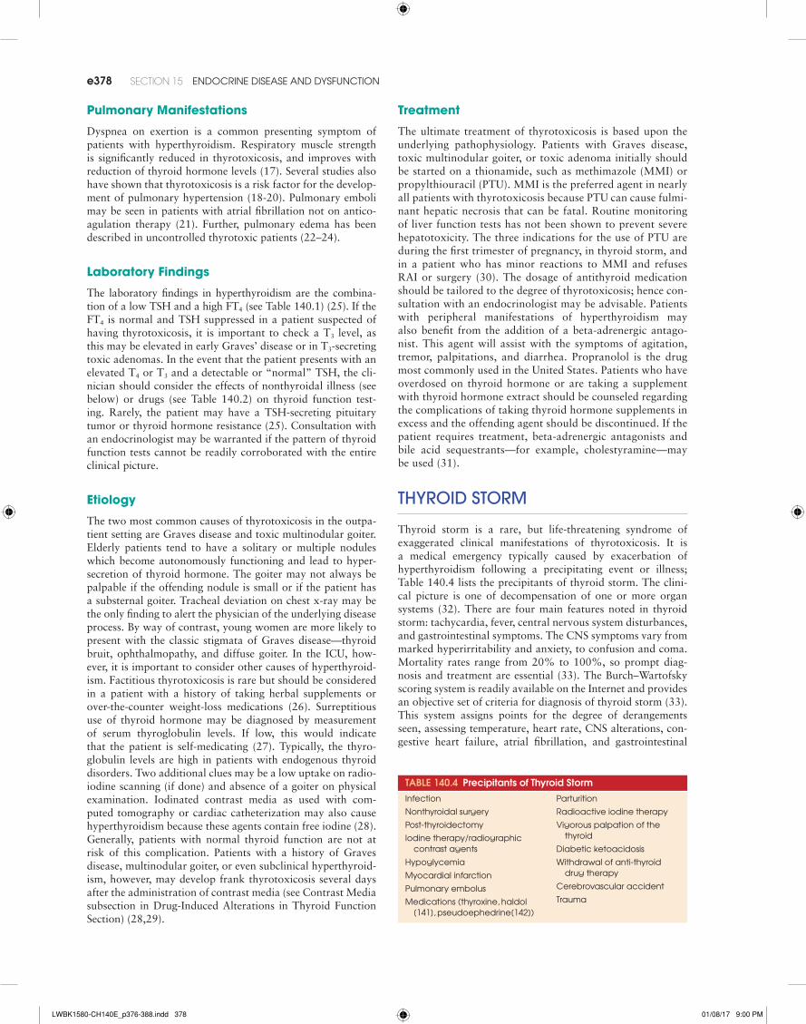
ThyroID sTorm
Thyroid storm is a rare, but life-threatening syndrome of exaggerated clinical manifestations of thyrotoxicosis. It is a medical emergency typically caused by exacerbation of hyperthyroidism following a precipitating event or illness; Table 140.4 lists the precipitants of thyroid storm. The clini-cal picture is one of decompensation of one or more organ systems (32). There are four main features noted in thyroid storm: tachycardia, fever, central nervous system disturbances, and gastrointestinal symptoms. The CNS symptoms vary from marked hyperirritability and anxiety, to confusion and coma. Mortality rates range from 20% to 100%, so prompt diag-nosis and treatment are essential (33). The Burch–Wartofsky scoring system is readily available on the Internet and provides an objective set of criteria for diagnosis of thyroid storm (33). This system assigns points for the degree of derangements seen, assessing temperature, heart rate, CNS alterations, con-gestive heart failure, atrial fibrillation, and gastrointestinal
Table 140.4 Precipitants of Thyroid Storm
infection parturition
nonthyroidal surgery radioactive iodine therapy
post-thyroidectomy Vigorous palpation of the thyroid
Diabetic ketoacidosisiodine therapy/radiographic
contrast agents
Hypoglycemia
myocardial infarction
Withdrawal of anti-thyroid drug therapy
Cerebrovascular accident
Traumapulmonary embolus
medications (thyroxine, haldol [141], pseudoephedrine[142])
LWBK1580-CH140E_p376-388.indd 378 01/08/17 9:00 PM

CHAPTER 140 Thyroid Disorders e379
symptoms. A score greater than 45 indicates thyroid storm, a score between 25 and 45 suggests thyroid storm, and a score less than 25 indicates that thyroid storm is unlikely (33).
Treatment
The treatment of thyroid storm takes a four-pronged approach (Table 140.5). First, an antithyroid drug must be given to reduce thyroid hormone production and peripheral conver-sion of T4 to T3. Second, supportive care must be administered against the systemic disturbances of fever, hypovolemia, and cardiovascular compromise. Third, the peripheral actions of thyroid hormone should be blocked. And finally, any precipi-tating factors should be addressed.
A thionamide is given to block synthesis of T3 and T4. PTU is the favored agent because it also inhibits peripheral conver-sion of T4 to T3. By reducing T3 concentrations in the serum, it is postulated that the manifestations of thyrotoxicosis are more rapidly improved with PTU than with MMI (33). Nei-ther of these drugs is available parenterally, so administration is typically by mouth or nasogastric (NG) tube. In patients with altered mental status, or in whom an NG cannot be placed, rectal administration of PTU has been reported in a few patients (34,35). It is conventional to use high doses of antithyroid drugs, such as 200 to 400 mg PTU every 4 hours or 20 mg MMI every 4 hours.
Thionamides do not inhibit release of preformed T3 and T4 from the thyroid. Inorganic iodide, however, can accomplish this goal. It may be administered orally as Lugol’s solution (10 drops every 8 hours) or as saturated solution of potassium iodide (SSKI, five drops every 6 hours). Oral radiographic con-trast agents, such as sodium ipodate or iopanoic acid, may be substituted for iodine. These drugs block the release of preformed thyroid hormone from the gland and inhibit the extrathyroidal conversion of T4 to T3 (36). It is critical to give the thionamide therapy at least an hour before the iodide or
contrast agent is administered, because the sudden influx of iodide into the thyroid can lead to increased thyroid hormone production and can thereby exacerbate the thyrotoxicosis (37). However, when the iodide or contrast agent is given after the antithyroid drug, serum T3 and T4 levels are substantially reduced in 2 to 3 days and may reach the normal range in 5 to 7 days (33,38).
If the patient has an allergy to iodide or cannot tolerate thionamides, lithium may be substituted to inhibit T3 and T4 synthesis (39). It may be given initially at a dose of 300 mg every 6 hours and is titrated to maintain serum lithium con-centrations around 1 mEq/L.
Supportive care should also be provided. Fever is preferen-tially treated with acetaminophen. Salicylates should not be used as they competitively inhibit T3 and T4 binding to serum proteins and thus increase serum-free T3 and T4 levels (40). The patient’s fluid losses should be appropriately replaced, bearing in mind the insensible losses from high fever and, if present, diarrhea. Hypercalcemia, if present, will usually be reversed by adequate hydration. High-dose glucocorticoids have been given historically for empiric treatment of relative adrenal insufficiency. Such treatment also has the added benefit of inhibition of peripheral conversion of T4 to T3. In patients with Graves disease, glucocorticoids also directly inhibit secre-tion of thyroid hormone. A loading dose of hydrocortisone 200 mg may be given initially followed by 100 mg every 8 hours; this therapy can be tapered rapidly after 2 to 3 days. Dexamethasone or methylprednisolone at equivalent doses may be substituted for hydrocortisone if preferred.
Therapy directed against the peripheral actions of thyroid hormone should be administered as well. Beta-adrenergic antagonist drugs can provide rapid amelioration of many of the symptoms of thyroid storm and should be dispensed immediately. Propranolol is the most commonly used agent, and may be given intravenously or orally depending upon the clinical setting. It is important to consider that in the thyro-
Table 140.5 Summary of Treatment for Thyroid Storm
Medication Dose & Route of administration action
propranolol 0.5 to 1 mg iv over ten minutes then 1 to 3 mg iv as needed
80 to 120 mg po q6°
Beta-adrenergic blockade and inhibition of T4 to T3 conversion
Thionamides
pTU 200 to 400 mg po / ng / pr q4° inhibit hormone synthesis and block con-version of T4 to T3
methimazole 20 mg po / ng q 4° inhibit hormone synthesis
iodine1
SSKi 5 drops po / ng q6° Blocks release of thyroid hormone
lugol's solution 10 drops po / ng q8°iodinated contrast agents1
Sodium ipodate or iopanoic acid 0.5 g po / ng q12° Blocks release of thyroid hormone and con-version of T4 to T3
Glucocorticoids
Hydrocortisone 200 mg iv load then 100 mg iv q8° Stress dose steroids and blocks conversion T4 to T3
Dexamethasone 2 mg po / ng / iv q6°lithium 300 mg po / ng q6° titrate to lithium level of
1meq / linhibits hormone synthesis
Cholestyramine 4 g po / ng q6° lowers serum T3 and T4
1either iodine or iodinated contrast agents should be used, but not both. Administration of iodine should be preceded by pTU by 2 to 3 hours to avoid enhancement of thy-roid hormone synthesis.
LWBK1580-CH140E_p376-388.indd 379 01/08/17 9:00 PM

e380 SECTion 15 enDoCrine DiSeASe AnD DySfUnCTion
toxic state, drug clearance is increased and higher than usual doses may be necessary to achieve the desired effect. If rapid beta-blockade is necessary to reduce the heart rate or if the patient’s mental status precludes oral drugs, intravenous administration is preferred. Propranolol attenuates the effects of catecholamines and weakly inhibits peripheral conversion of T4 to T3. This inhibition occurs over a period of a week, however, and thus does not solely account for the beneficial effects of propranolol in the thyrotoxic patient.
In extreme cases, it may be beneficial to utilize a method to remove T3 and T4 from the patient’s serum. The simplest approach is to administer oral cholestyramine. This drug binds the hormones in the GI tract, interrupting the enterohepatic cir-culation (31). Plasmapheresis (41–45) and charcoal hemoper-fusion (46,47) also have been used successfully to lower T3 and T4 levels in patients who were unable to tolerate thionamides.
Finally, it is important to search for and treat underlying illnesses, which may have precipitated the thyroid storm. This process can be difficult in obtunded patients, but a systematic approach is usually successful in uncovering the cause.
Patients treated with the above regimen may clinically improve within 12 to 24 hours of starting therapy if the syn-drome is recognized and treated in a timely fashion. As the patient’s condition stabilizes, it is important to wean the glu-cocorticoids, switch to oral rehydration, and taper the beta-blocking drugs. Long-term treatment of the hyperthyroidism is required if the patient has Graves disease or toxic multi-nodular goiter. In patients with Graves, it may be preferable to treat with thionamides, as there is a chance of remission of the autoimmune condition. Others advocate total thyroidectomy if a patient’s disease is severe enough to lead to thyroid storm; a highly skilled surgeon is required, however (30,48). Radio-iodine ablation is not an option for several months because the inorganic iodide used in the treatment of the thyroid storm saturates the gland and precludes further uptake of iodide.
Drug-InDuceD alTeraTIons In ThyroID FuncTIon
Amiodarone and Thyroid Function
Amiodarone is a lipophilic drug that contains 75 mg iodine per 200-mg tablet. The drug has a half-life of several months and during that time it releases approximately 6 mg of inorganic iodine per day (49). In euthyroid patients, chronic administra-tion of the drug results in increased serum TT4 (see the above section on thyroid function tests), FT4, and rT3 levels, lower T3 concentrations, and normal TSH (50). The reason for these
changes is the drug’s strong inhibition of 5′-deiodinase, the enzyme responsible for conversion of T4 to T3 and rT3 to T2. Most patients remain euthyroid while on amiodarone, despite the hormonal derangements that may be seen. However, about 14% to 18% of patients develop either hypothyroidism or hyperthyroidism while on amiodarone (51). Hypothyroidism is more commonly encountered in iodine-replete areas, such as the United States (52). Treatment is aimed at normalization of the TSH with levothyroxine replacement while the amioda-rone therapy is continued. Upon discontinuation of the amiod-arone, most patients return to euthyroidism though it may take several months because of the prolonged half-life of the drug.
In iodine-deficient regions, it is more common to see hyper-thyroidism as a result of amiodarone therapy (52). There are two mechanisms (Table 140.6) of amiodarone-induced thy-rotoxicosis (AIT) and distinction of these two disorders is relevant because their treatment differs. Type I AIT occurs in glands with an underlying anatomic abnormality. Areas of autonomous thyroid tissue such as a toxic nodule or autoim-mune disease in the thyroid produce increased the levels of hormone in response to the excess iodine released from the amiodarone (51). Type II AIT develops as a result of a direct cytotoxic effect of amiodarone on the thyrocyte (53). Treat-ment of type I AIT can be difficult because most patients do not respond to thionamides as these drugs have decreased efficacy in states of iodine excess (54). Use of potassium per-chlorate (KClO4) blocks further iodine entry into the thyro-cyte and may enhance the efficacy of thionamides (50). It is important to note that both thionamides and KClO4 may cause agranulocytosis, so serial monitoring of blood counts is advisable (30). The treatment of type II AIT is primarily with glucocorticoids (55). Whether it is advisable to stop the amiodarone with either form of AIT is a controversial issue particularly because the long half-life of the drug indicates that its effects may persist for many months even after discontinua-tion. Additionally, amiodarone may be the only effective treat-ment for the arrhythmia. Consequently, a careful discussion with the cardiologist is warranted to aid in the decision of whether to discontinue the medication. For patients in whom chronic therapy with amiodarone is essential, thyroidectomy is a potential treatment for AIT (50). Radioiodine ablation, however, is not an immediate option given the low iodine uptake as a result of the iodine excess from the amiodarone.
Contrast Media
Another potential source of excess iodine is radiographic contrast media. As noted in the section about the management of thyroid storm, the oral cholecystographic agents, iopanoic acid and
Table 140.6 Features of amiodarone-Induced Thyrotoxicosis
Iodine Induced Thyrotoxicosis (Type I) Destructive Thyrotoxicosis (Type II)
Underlying thyroid abnormality yes no
Goiter Diffuse or multinodular usually present occasionally small, firm goiter
RaIU low / normal / high low
Serum Il-6 concentrations Slightly elevated markedly elevated
Pathogenic mechanism excessive thyroid hormone synthesis excessive hormone release (destructive thyroiditis)
Treatment Thionamides & KClo4 Glucocorticoids
Subsequent hypothyroidism Unlikely possible
Color flow Doppler sonograpy normal or increased blood flow Decreased blood flow
LWBK1580-CH140E_p376-388.indd 380 01/08/17 9:00 PM

CHAPTER 140 Thyroid Disorders e381
sodium ipodate, may be used short-term in thyrotoxic patients for their side effect of decreasing peripheral conversion of T4 to T3 and blocking hormone secretion from the thyroid. Such agents used over a longer period of time, however, can exacer-bate the underlying hyperthyroidism (36). There are a multitude of other agents available which have variable effects on the thy-roid gland. Typically, patients with no underlying thyroid disease will not be affected by the use of these agents (56). Patients with Graves disease, multinodular goiter, or the elderly, are at risk to develop thyrotoxicosis after the use of these contrast agents (28). The lipid-soluble agents used for myelography, bronchog-raphy, and uterosalpingography are cleared slowly and release inorganic iodine for months to years. Newer, water-soluble prep-arations used in arteriography and computed tomography are cleared from the plasma more quickly but the iodine they release during these procedures can still have an effect on thyroid func-tion. The degree of thyroid dysfunction can range from mild, transient subclinical hyperthyroidism to thyroid storm (57–61). The majority of patients experience only transient thyrotoxicosis and the syndrome resolves when the excess iodine is cleared. If treatment is required, thionamides and beta-adrenergic blockade may be used until the thyrotoxicosis resolves (59).
ThyroToxIc PerIoDIc ParalysIs
Thyrotoxic periodic paralysis (TPP) is a complication of hyper-thyroidism characterized by localized or generalized attacks of weakness or flaccid paralysis and hypokalemia (62). Although it has been reported in Western countries and in women, it is more common in Asian men, where the incidence is 1.9% in thyrotoxic patients (63). The clinical presentation is identical to familial hypokalemic periodic paralysis but the pathophysi-ology is distinct. Though the mechanism of the syndrome is not clearly defined, hypokalemia alone is not enough to elicit the paralysis. Hypokalemia sufficient to create the paralysis in a hyperthyroid patient has no effect on the same patient when euthyroid (64). This finding points to the importance of thy-roid hormone excess in the pathophysiology of this process. It is most commonly associated with Graves disease but may be seen with any form of thyrotoxicosis (65).
The clinical presentation is one of symmetrical flaccid weak-ness with lower extremities generally affected more than upper extremities (64). Breathing may be impaired if the patient has a more generalized weakness. The onset of the attacks is usu-ally sudden and may be preceded by cramping. Ingestion of alcohol or carbohydrates and strenuous physical exercise com-monly precipitate the episodes of weakness (64). Patients have decreased or absent deep tendon reflexes. The symptoms may last from a few hours to several days (64).
Treatment of TPP is aimed at correction of the hyperthy-roidism. If hypokalemia is present, replacement should be given. Some patients are given a potassium-sparing diuretic in addition to the potassium supplementation until euthyroid-ism is achieved. Beta-adrenergic antagonists also decrease the frequency of attacks in these patients (63).
Preoperative Management of Hyperthyroidism
Adequate preparation for surgery in thyrotoxic patients is crit-ical to the successful outcome of the procedure. Surgery in a
hyperthyroid patient can precipitate thyroid storm, with high morbidity and mortality if preoperative care is inadequate. The type of treatment will depend upon the amount of time before the surgery. Elective procedures should be postponed until the T3 and T4 levels are normalized with thionamides and beta-adrenergic blockade (30). This can usually be achieved within approximately 2 weeks. It is important to note that a suppressed TSH may not normalize for months and this value should not be used as the criteria to assess the thyroid status. Urgent or emergent procedures may be safely done after initia-tion of a thionamide and a beta-adrenergic antagonist. Iodide, as either SSKI, Lugol’s solution, or an oral radiographic con-trast agent—sodium ipodate or iopanoic acid—should also be administered to block the release of thyroid hormone and decrease peripheral conversion of T4 to T3 (66). Finally, a glu-cocorticoid, such as hydrocortisone or dexamethasone, should also be used if the patient is suspected of having concomitant adrenal insufficiency or additional inhibition of extrathyroidal conversion of T4 to T3 is needed (67). Considerable lowering of T3 and T4 levels can be achieved within 1 to 3 days and normalization within 3 to 5 days (67,68).
hyPoThyroIDIsm
Hypothyroidism is a common clinical problem, affecting approximately 4.6% of the population in the United States (69). It is important to recognize the clinical features and potential complications of a patient with hypothyroidism. Nonthyroidal illness, surgery, or diagnostic testing can lead to metabolic decompensation in patients with undiagnosed or untreated hypothyroidism. Additionally, it is important to note that untreated hypothyroidism may slow the metabolism of certain drugs, thereby increasing the risk of problematic side effects. Hypothyroidism is most often caused by autoimmune thyroiditis, also known as Hashimoto’s thyroiditis. Other com-mon causes of hypothyroidism are noted in Table 140.7.
The clinical manifestations of hypothyroidism are mani-fold. A majority of the symptoms are nonspecific which can lead to a delay in the diagnosis. In elderly patients, the diag-nosis may be missed because the patient may be asymptomatic or the signs attributed to aging (70). Patients in the ICU may present with severe CNS disturbances, cardiovascular derange-ments, hyponatremia, or respiratory failure. Table 140.8 notes the signs and symptoms of hypothyroidism.
Table 140.7 Causes of HypothyroidismAutoimmune, Hashimoto's thyroiditis
post-thyroidectomy
post-radiation 131i treatment external beam radiation
iodine deficiency
Drugs lithium
iodine containing drugs (amiodarone, radiocontrast agents)
Secondary hypothyroidism pituitary tumor, irradiation, empty sella syndrome, infiltrative
disorders Hypothalamic disease
Transient disorders Silent, subacute thyroiditis
LWBK1580-CH140E_p376-388.indd 381 01/08/17 9:00 PM

e382 SECTion 15 enDoCrine DiSeASe AnD DySfUnCTion
Cardiovascular Manifestations
The symptoms of cardiovascular dysfunction are much less pronounced in patients with hypothyroidism compared to their thyrotoxic counterparts. These cardiovascular changes may manifest themselves in the hypothyroid patient who is undergoing the stress of anesthesia and surgery. Cardiovascu-lar hemodynamics are affected by hypothyroidism in several ways. In particular, patients have decreased cardiac output, mediated by reduced contractility and heart rate (71). This reduction in cardiac output contributes to the dyspnea on exertion seen in many hypothyroid patients. These patients also have increased systemic vascular resistance, predispos-ing them to hypertension (72). Diastolic filling and com-pliance are reduced, leading to diastolic dysfunction (73). Patients may have an elevation of diastolic pressure out of proportion to systolic pressure leading to a reduction in pulse pressure (74).
The presence of decreased cardiac output, diastolic hyper-tension, and increased systemic vascular resistance suggests that hypothyroidism can cause congestive heart failure. It is rare, however, for hypothyroidism to be the sole causative agent in the development of heart failure (75). Typically, patients have underlying cardiac disease that is exacerbated by the hypothyroidism. Pericardial effusion associated with hypothyroidism also may compromise cardiac function (76). Angina may worsen in patients with hypothyroidism. Patients without pre-existing cardiac dysfunction typically do not manifest symptoms and signs of congestive heart fail-ure or coronary artery syndrome, unless the hypothyroidism is profound (77).
Pulmonary Manifestations
Dyspnea on exertion is a common presenting complaint in patients with hypothyroidism, in part due to the impaired car-diac function and reduced pulmonary function. Respiratory muscle weakness also appears to play a role in this dyspnea (78–80). There is a reduction in central pulmonary drive in response to hypoxia and hypercapnia, leading to hypoven-tilation (22,81). Upper airway obstruction may occur as a result of goiter (see Acute Airway Obstruction and Goiter) (82). Sleep apnea may occur as a result of macroglossia (83). Patients may thus require continuous positive airway pressure in addition to replacement of the thyroid hormone. Finally, a restrictive pattern on pulmonary function testing may be seen as a result of an effusion or an intrinsic decrease in lung volumes (22).
Gastrointestinal Manifestations
Peristalsis is slowed in patients with hypothyroidism. Most patients have normal bowel motility, but a small proportion with hypothyroidism requires laxative use. Patients may report vague abdominal pain and distension. Rarely, severe cases may present with ileus (84–86). Severely hypothyroid patients may also have malabsorption. The mechanism of this abnormal absorption is not clearly defined, theories include myxedema-tous infiltration of the mucosa, associated autoimmunity (87), and decreased intestinal motility (88).
Metabolic Manifestations
Though classically taught that untreated hypothyroidism may lead to hyponatremia, the actual incidence is exceedingly rare, leading some to doubt the relationship (89). It has been described in myxedema coma, but in patients with less severe forms of hypothyroidism, a continued search for other etiolo-gies is advisable (89). Though the mechanism is not completely understood, the higher systemic vascular resistance of severe hypothyroidism may cause decreased glomerular filtration and renal perfusion which can result in reductions in free water clearance (89–92). Hyperlipidemia, on the other hand, is a more commonly seen metabolic derangement than hyponatre-mia. Lipid clearance is decreased in patients with hypothyroid-ism, resulting in elevated levels of free fatty acids, low-density lipoprotein (LDL), and total cholesterol (93). Treatment of the underlying hypothyroidism will also improve the lipid panel (94,95).
Treatment
Thyroid hormone is preferentially replaced with T4. Levothy-roxine is then converted to T3 in the peripheral tissues; there-fore, it is unnecessary to administer T3 in most situations (1). In adults, the starting dose of T4 is typically 1.7 µg/kg/d (based on ideal body weight). Elderly patients or those with coronary disease should be started at lower doses and cautiously titrated based upon TSH levels every 6 weeks (1).
Myxedema Coma
Myxedema coma is a rare, but life-threatening complication of hypothyroidism. It may occur after severe, long-standing hypothyroidism or after an acute precipitating event such as surgery or infection. It is more likely to occur in elderly women during the winter months (96). Any of the usual causes of hypothyroidism (see Table 140.6) may induce myxedema coma. Prompt recognition and treatment are essential, even before laboratory results are available. Mortality rates are improving due to early diagnosis and treatment, but mortality remains elevated at 30% to 50% (97). Patients with cardiac complications and the elderly are at greatest risk of mortality (98,99).
Clinical Presentation
The majority of patients with myxedema coma have had symp-toms of hypothyroidism for many months. There is a gradual onset of lethargy, progressing to stupor, which is precipitated by cold exposure, infection, or medications. Other precipitating
Table 140.8 Clinical Manifestations of Hypothyroidism
Symptoms Signs
fatigue & weakness Delayed relaxation of tendon reflexes
Cold intolerance Bradycardia
Weight gain Hypoventilation
Constipation Diastolic hypertension
Dyspnea on exertion reduced pulse pressure
Depression pericardial & pleural effusions
menorrhagia Generalized & periorbital edema
myalgia macroglossia
Dry skin & hair loss of eyebrows
LWBK1580-CH140E_p376-388.indd 382 01/08/17 9:00 PM

CHAPTER 140 Thyroid Disorders e383
events are stroke, congestive heart failure, trauma, or gastro-intestinal bleeding. The patient is typically an obese elderly woman with yellow discoloration of the skin. The principal features of myxedema are hypothermia, bradycardia, and decreased mental status or coma (32). Patients also character-istically have a decreased respiratory rate as a result of reduced hypoxic ventilatory drive (100). The resultant carbon dioxide retention can exacerbate the altered mental status. Similar to the patient with profound hypothyroidism, the respiratory muscle weakness and pleural effusions, if present, can make ventilating these patients difficult (79,100).
Hypothermia is present in nearly all patients with myx-edema coma (100). Temperature may be quite low (less than 80ºF, 20ºC); values below 90ºF (32ºC) predict a poorer prog-nosis (98). The hypothermia may go unrecognized if the proper thermometer is not utilized; many thermometers may not be able to measure below 93ºF (34ºC). The diagnosis of myx-edema coma should be considered in any unconscious patient with infection who does not have a fever. Warming should be gradual with ordinary blankets. Electric heating blankets should not be used as they may cause peripheral vasodilation and subsequent hypotension.
The cardiovascular abnormalities seen in myxedema coma are similar to those associated with severe hypothyroidism. Patients can present with bradycardia, reduced cardiac out-put, and decreased cardiac contractility, which may lead to hypotension. Signs of congestive heart failure may be found. In patients with diminished heart sounds, low-voltage ECG, or cardiomegaly on chest x-ray, an investigation for pericardial effusion should be performed (101).
This profound level of hypothyroidism may lead to impaired free water excretion. As a result, up to half of patients may have hyponatremia (96,102). Severely reduced sodium levels can also exacerbate the altered mental status. Impaired free water clearance may be manifested through gen-eralized nonpitting edema and periorbital swelling. Patients should be managed by free water restriction, but the condition will improve with thyroid hormone replacement.
Additional clinical features of myxedema include ileus or megacolon as a result of decreased intestinal motility (84). Bladder atony may occur so patients should be monitored for post-void residual volumes. Hypoglycemia is a common fea-ture, which is the result of the hypothyroidism or concomitant adrenal insufficiency.
Diagnosis
The diagnosis of myxedema coma is initially made based upon historical and clinical clues. Recently, a scoring system was developed to aid in making the diagnosis (97). Though the performance of this system has not been validated in other clinical settings, it is important to investigate other causes of altered mental status, such as cerebrovascular accident and infection. Serum should be obtained for measurement of TSH, free T4, and cortisol. If the clinical picture is consistent with myxedema coma, treatment should be initiated before labora-tory confirmation of the diagnosis.
Treatment
Because of the high mortality rate of this endocrine emergency, patients with myxedema coma should be treated aggressively
(99). Patients should be presumed to have adrenal insuffi-ciency and treated with stress–dose intravenous steroids until laboratory data excludes adrenal dysfunction. Administration of levothyroxine prior to glucocorticoids in such patients can provoke an adrenal crisis.
The optimal replacement strategy for levothyroxine is unknown because of the rarity of the condition. Clinical judg-ment must be used to weigh the risk of rapid administration of thyroid hormone, and its attendant possibility of precipitating a myocardial infarction, against the risk of not replacing the thyroid hormone fast enough in light of the high mortality of under-treated myxedema coma. Whether to administer T3, T4, or combination therapy and the dose is a subject of debate among endocrinologists. It is preferable to utilize intravenous adminis-tration of thyroid hormone in patients with myxedema because of the possibility of impaired gastrointestinal absorption.
Supportive care should be directed to the co-existing medi-cal conditions. Hyponatremia can usually be managed with free water restriction, but 3% saline may be given in extreme circumstances. Hypotension will usually improve with ini-tiation of levothyroxine. Refractory hypotension should be treated with vasopressor agents until the thyroid hormone has had time to act. Patients may require mechanical ventilation because of respiratory muscle weakness, depressed mental sta-tus, or decreased hypoxic ventilatory drive. Warming should be passive with ordinary blankets, as heating blankets can exacerbate hypotension because of peripheral vasodilation. Finally, it is important to address the underlying medical illness that precipitated the myxedema coma.
Preoperative Management of Hypothyroidism
Patients with mild-to-moderate hypothyroidism may proceed to surgery as no convincing evidence exists to show that there is an adverse effect on perioperative outcomes (103–106). If the procedure is elective, however, it is optimal to begin replacement with thyroid hormone and delay the surgery until the patient is biochemically euthyroid. The exception to this rule is a patient with coronary artery disease awaiting bypass or stenting. Such patients should have their coronary vascu-lature addressed first, then their thyroid hormone replaced postoperatively. Evidence suggests that replacement of thyroid hormone before restoring coronary blood flow could tax an already ischemic myocardium (107). A patient with severe hypothyroidism who requires urgent surgery should be given a loading dose of intravenous T4 and possibly T3. In addition, stress–dose glucocorticoids should be given if adrenal or pitu-itary function is uncertain as replacement of thyroxine in a patient with adrenal insufficiency can precipitate adrenal crisis. The patient may be given an initial dose of T4 at 200 to 300 µg IV, followed by 50 µg daily. Depending upon the patient’s age and cardiac risk factors, T3 may be given simultaneously at 10 µg every 8 to 12 hours (108).
ThyroID FuncTIon In nonThyroIDal Illness
Aberrations in thyroid function during illness occur along a continuum, with wider deviations from the normal range as the patient becomes more severely ill (109). Several names have
LWBK1580-CH140E_p376-388.indd 383 01/08/17 9:00 PM

e384 SECTion 15 enDoCrine DiSeASe AnD DySfUnCTion
been ascribed to the condition, including euthyroid sick syn-drome, low T3 syndrome, low T4 syndrome, and nonthyroidal illness. Considerable debate exists as to whether this syndrome represents a pathologic process marked by hypothyroidism or an adaptive response to systemic illness that allows the body to lower its tissue energy requirements and limit catabolism (109). In light of this controversy, it is understandable that no consensus exists on whether or how to treat this entity.
Interpretation of thyroid function tests in critically ill patients is complex. Accordingly, thyroid function should not be measured in this setting unless a thyroid disorder is strongly suspected. When it is deemed appropriate to evaluate the hypothalamic–pituitary–thyroid axis, the clinician should check a TSH, total T4, free T4, and total T3.
The most commonly seen change in thyroid hormone func-tion tests among acutely ill and fasting healthy patients is a low serum T3 concentration (110). The majority of T3 in the serum is produced by de-iodination of T4 to T3 in the peripheral tis-sues. The levels of the enzyme responsible for this conversion, 5′-monodeiodinase, are decreased with even mild illness (111). As described in the above section regarding drugs and thyroid function, many commonly used medications in the ICU may also decrease the peripheral conversion of T4 to T3, further lowering the circulating T3 levels. Glucocorticoids and beta-adrenergic antagonists are the most common offending agents. In addition, free fatty acids inhibit the activity of this de-iodinase (112). Cytokines have also been shown to have a role in the development of the sick euthyroid syndrome by their role in decreasing the conversion of T4 to T3 (113). Concomitant with the decline in T3 levels is a rise in reverse T3 (rT3) during nonthyroidal illness. Fasting may produce this clinical picture within 24 to 36 hours and is reversed as quickly with refeed-ing (114). This pattern of low T3/high rT3 is found in many patients with various acute and chronic illnesses, whether due to infection, surgery, cancer, cardiovascular diseases, pul-monary processes, burns, or trauma. The metabolic rate of formation of rT3 is unchanged in the setting of illness. The increase in this value is instead a reflection of attenuated rates of clearance of rT3 (115). The complicating factor with rou-tinely measuring the rT3 levels in patients suspected of having nonthyroidal illness is that it may take up to a week to process this test in the laboratory.
During the chronic phase of illness, thyroxine (T4) levels may also decline in up to 20% of hospitalized patients and 50% of critically ill patients (116). Low T4 levels are corre-lated with a higher mortality rate during acute illness (117). The reduction in T4 can, in part, be attributed to decreased concentrations of one of the three thyroid hormone–binding proteins: TBG, transthyretin, and albumin. Inhibition of T4 attachment to its binding proteins has also been identified as responsible for the lowering of T4 levels in the serum of criti-cally ill patients. Some data points to high levels of free fatty acids as a causative agent in this process (118). Serum TSH levels are typically normal in most patients with nonthyroi-dal illness (119). During the chronic phase of critical illness, the TSH may simultaneously fall with the decline in T4 levels, a clinical picture consistent with central hypothyroidism. A reduction in the secretion of thyrotropin-releasing hormone (TRH) from the hypothalamus occurs simultaneously and appears to be responsible for this clinical picture (119). Thy-roid hormone replacement has not been shown to improve outcomes in critically ill patients, however (119,120). Thy-
rotropin concentrations may rise during the recovery phase of critical illness, further supporting the notion of a clinical picture of central hypothyroidism (121).
Medications may also alter TSH levels (see Table 140.2). Dopamine infusions may be associated with a reduction in serum thyrotropin concentration (122,123). There is evidence to suggest that TSH has a reduced biologic activity during ill-ness, which may, in part, be due to changes in glycosylation of the hormone (109).
When measuring TSH levels in the ICU, it is important to utilize a high-sensitivity assay with a lower detection limit of at least 0.01 mU/L (109,124). Most hospitalized patients with low, but detectable, TSH by this assay have sick euthyroid syndrome. In contrast, patients with undetectable thyrotro-pin are more likely to be hyperthyroid (109). Finally, those patients with high TSH (<20 mU/L) are likely recovering from a nonthyroidal illness and should be reassessed 6 weeks after the hospitalization (109,124). A serum TSH of greater than 20 mU/L is more apt to be related to an underlying primary thy-roid disorder and replacement of thyroxine may be appropriate (109,124).
Acute Airway Obstruction and Goiter
Acute airway obstruction is a life-threatening complication of an enlarged thyroid gland. Typically, development of a goiter is a gradual process, but rapid growth of the gland may occur in certain circumstances. Fortunately quite rare, but of utmost importance to consider, is anaplastic thyroid cancer. Patients with this disease may present with considerable growth of the thyroid within a few weeks or even days (125). Primary thy-roid lymphoma can also show a rapid growth pattern, but will quickly respond to appropriate chemotherapy and/or radia-tion therapy. Riedel’s thyroiditis, also rare, with a prevalence of 0.06% to 0.3%, may present with a rapidly enlarging, hard neck mass that must be differentiated from thyroid cancer or lymphoma. The fibrous tissue may invade soft tissue and mus-cle and lead to tracheal compression (126). The more common scenario is a patient who presents with a nodule that rapidly increases in size over several minutes to hours. In these cases, the patient likely has underlying nodular disease, which has encroached upon a nearby blood vessel and bled into a cystic compartment of the nodule. Typically, patients have regression of such a nodule over the ensuing weeks.
The clinical presentation of a compressive goiter is varied. Patients may present with complaints of a pressure sensation in the anterior neck, particularly with the movement of the head. Difficulty swallowing and vocal cord paralysis also may be encountered. Pemberton’s sign, facial flushing and jugular venous distension upon raising the arms over the head, is an indication of impeded of venous outflow from the head as the goiter shifts into and obstructs the thoracic inlet (127). Many patients with a goiter have a mild degree of airway obstruction when screened with pulmonary function tests. While chest radiographs accurately indicate retrosternal extension of goi-ters, it cannot predict airway obstruction as reliably as flow-volume loops (82). It is important to recognize that a patient presenting with new-onset wheezing or stridor may have a substernal goiter (128).
The management of acute airway compromise is primarily surgical (129). If airway collapse is imminent, it is critical to protect the airway with intubation (130). The type of surgery
LWBK1580-CH140E_p376-388.indd 384 01/08/17 9:00 PM

CHAPTER 140 Thyroid Disorders e385
necessarily depends upon the size of the goiter and whether there is an associated malignancy. Radioiodine treatment can take months to years to shrink the goiter (131).
Postthyroidectomy Hypocalcemia
Hypoparathyroidism
The most common cause of hypoparathyroidism is surgery on the neck, with resultant removal of, or injury to, the parathy-roid glands. This complication is typically seen after cancer surgery, total thyroidectomy, or parathyroidectomy, though it is most often a transient condition with symptoms occurring 1 to 2 days postoperatively. Symptoms may vary from subtle perioral numbness and tingling to profound fatigue to tetany. Table 140.9 lists the symptoms and signs that may be seen in patients with hypocalcemia. Risk of hypocalcemia is depen-dent upon the extent of surgery, localization and preservation of the parathyroids, and skill of the surgeon. Incidence rates of postoperative, transient hypocalcemia range from 1.6% up to greater than 50%, but most of these patients will regain parathyroid function over the ensuing months. The risk of per-manent hypoparthyroidism is variable, between 0% and 10% (132). During nonparathyroid neck surgery, it is important that the surgeon recognize a compromised parathyroid gland and autotransplant it into the adjacent neck muscle in order for the gland to possibly regain function.
Treatment of hypoparathyroidism is aimed at the main-tenance of the serum calcium in the low end of the normal range (133). Some surgeons begin thrice-daily prophylactic oral calcium supplementation the night of the surgery (134). If the patient develops progressive symptoms, tetany or sei-zures, then the use of IV calcium gluconate is warranted. In life-threatening situations, 10 mL of calcium gluconate may be administered intravenously over a 5- to 10-minute period and repeated as necessary. In less acute situations, a continu-ous calcium infusion may be used by mixing 10 ampules of calcium gluconate in 500 mL of 5% dextrose in water. The infusion rate may vary between 0.3 and 2 mg/kg/hr, depend-ing upon the clinical setting. The goal of therapy is to reverse hypocalcemic symptoms and restore calcium levels to the low–normal range. Chronic management of permanent hypopara-thyroidism is beyond the scope of this chapter.
Hungry bone Syndrome
Hungry bone syndrome is a well-recognized complication of sur-gical correction of severe hyperparathyroidism (135). Patients with very high levels of PTH may develop significant hypocal-cemia after the surgical removal of the offending parathyroid adenoma(s) (135). The mechanism of this metabolic derange-ment is rapid skeletal mineralization. Less commonly seen is
HBS after the treatment of severe thyrotoxicosis (136–139). Patients with hyperthyroidism may develop secondary osteo-porosis and resultant hypercalcemia from the increased bone resorption. After correction of hyperthyroidism, patients have a reversal of the thyrotoxic osteodystrophy and instead have a net flux of calcium and phosphorous deposition into bone. In extreme thyrotoxicosis, the patient may develop hypocalcemia (136–139). This condition typically resolves within a few days to weeks with the treatment of the hypocalcemia (139).
Table 140.9 Symptoms and Signs of acute Hypocalcemia
Symptoms Signs
perioral numbness and tingling Chvostek's sign
Tingling paresthesias in distal extremities
Trousseau's sign
muscle cramps/twitching Arrhythmias
Seizures Bradycardia
Tetany prolonged QT interval
• Measurement of serum TSH is sufficient to diagnose the majority of thyroid disorders.
• A number of drugs commonly used in the ICU will interfere with thyroid homeostasis.
• In the ICU, patients with thyrotoxicosis may have an atypical presentation, with tachyarrhythmias or central nervous system disturbance as the primary sign.
• About 14% to 18% of patients develop either hypothy-roidism or hyperthyroidism while on amiodarone.
• Nonthyroidal illness, surgery, or diagnostic testing can lead to metabolic decompensation in patients with undiagnosed or untreated hypothyroidism. Addition-ally, it is important to note that untreated hypothyroid-ism may slow the metabolism of certain drugs, thereby increasing the risk of problematic side effects.
• Interpretation of thyroid function tests in critically ill patients is complex. Accordingly, thyroid function should not be measured in this setting unless a thyroid disorder is strongly suspected.
Key Points
References 1. Jonklaas J, Bianco AC, Bauer AJ, et al. Guidelines for the treatment of hypo-
thyroidism: prepared by the American Thyroid Association Task Force on Thyroid Hormone Replacement. Thyroid. 2014;24(12):1670–1751.
2. De los Santos ET, Mazzaferri EL. Sensitive thyroid-stimulating hormone assays: clinical applications and limitations. Compr Ther. 1988;14(9):26–33.
3. Stockigt JR. Free thyroid hormone measurement: a critical appraisal. Endocrinol Metab Clin North Am. 2001;30(2):265–289.
4. Adami HO, Rimsten A, Thoren L, et al. Thyroid disease and function in breast cancer patients and non-hospitalized controls evaluated by determination of TSH, T3, rT3 and T4 levels in serum. Acta Chir Scand. 1978;144(2):89–97.
5. Stepanas AV, Mashiter G, Maisey MN. Serum triiodothyronine: clinical experience with a new radioimmunoassay kit. Clin Endocrinol (Oxf). 1977;6(3):171–183.
6. Afrasiabi MA, Vaziri ND, Gwinup G, et al. Thyroid function studies in the nephrotic syndrome. Ann Intern Med. 1979;90(3):335–338.
7. Swanson MA, Custer TR, Suey CM. Free thyroxine and free thy-roxine index in women taking oral contraceptives. Clin Nucl Med. 1981;6(4):168–171.
8. Schimmel M, Utiger RD. Thyroidal and peripheral production of thyroid hormones: rview of recent findings and their clinical implications. Ann Intern Med. 1977;87(6):760–768.
9. Gittoes NJ, Franklyn JA. Drug-induced thyroid disorders. Drug Saf. 1995; 13(1):46–55.
10. Ringel MD. Management of hypothyroidism and hyperthyroidism in the intensive care unit. Crit Care Clin. 2001;17(1):59–74.
11. Roffi M, Cattaneo F, Brandle M. Thyrotoxicosis and the cardiovascular system. Minerva Endocrinol. 2005;30(2):47–58.
12. Moolman JA. Thyroid hormone and the heart. Cardiovasc J S Afr. 2002; 13(4):159–163.
13. Jalal S, Khan KA, Rauoof MA, et al. Thyrotoxicosis presenting with com-plete heart block. Saudi Med J. 2004;25(12):2057–2058.
LWBK1580-CH140E_p376-388.indd 385 01/08/17 9:00 PM

e386 SECTion 15 enDoCrine DiSeASe AnD DySfUnCTion
14. Kernoff LM, Rossouw JE, Kennelly BM. Complete heart block complicat-ing thyrotoxicosis. S Afr Med J. 1974;47(12):513–515.
15. Osman F, Ayuk J, Dale J, et al. Thyrotoxicosis with heart block. J R Soc Med. 2001;94(7):346–348.
16. Zargar AH, Bashir MI, Wani AI, et al. Reversible complete heart block in Grave’s disease. J Assoc Physicians India. 1999;47(11):1120–1121.
17. McElvaney GN, Wilcox PG, Fairbarn MS, et al. Respiratory muscle weak-ness and dyspnea in thyrotoxic patients. Am Rev Respir Dis. 1990;141 (5 Pt 1):1221–1227.
18. Armigliato M, Paolini R, Aggio S, et al. Hyperthyroidism as a cause of pulmonary arterial hypertension: a prospective study. Angiology. 2006; 57(5):600–606.
19. Ma RC, Chow CC. Thyrotoxicosis as a risk factor for pulmonary arterial hypertension. Ann Intern Med. 2006;144(3):222–223.
20. Ma RC, Cheng AY, So WY, et al. Thyrotoxicosis and pulmonary hyperten-sion. Am J Med. 2005;118(8):927–928.
21. Werner D, Misfeld M, Regenfus M, et al. Emergency coronary angiogra-phy with gadolinium in a patient with thyrotoxicosis, pulmonary embolism and persistent right atrial thrombi. Clin Res Cardiol. 2006;95(8):418–421.
22. Brussel T, Matthay MA, Chernow B. Pulmonary manifestations of endo-crine and metabolic disorders. Clin Chest Med. 1989;10(4):645–653.
23. Glikson M, Freimark D, Leor R, et al. Unstable anginal syndrome and pulmonary oedema due to thyrotoxicosis. Postgrad Med J. 1991;67(783): 81–83.
24. Wald DA, Silver A. Cardiovascular manifestations of thyroid storm: a case report. J Emerg Med. 2003;25(1):23–28.
25. Surks MI, Chopra IJ, Mariash CN, et al. American Thyroid Associa-tion guidelines for use of laboratory tests in thyroid disorders. JAMA. 1990;263(11):1529–1532.
26. Ohye H, Fukata S, Kanoh M, et al. Thyrotoxicosis caused by weight-reducing herbal medicines. Arch Intern Med. 2005;165(8):831–834.
27. Cohen JH, III, Ingbar SH, Braverman LE. Thyrotoxicosis due to ingestion of excess thyroid hormone. Endocr Rev. 1989;10(2):113–124.
28. van der Molen AJ, Thomsen HS, Morcos SK; Contrast Media Safety Committee, European Society of Urogenital Radiology (ESUR). Effect of iodinated contrast media on thyroid function in adults. Eur Radiol. 2004;14(5):902–907.
29. Chen CC, Huang WS, Huang SC, et al. Thyrotoxicosis aggravated by iodinated contrast medium: a case report. Zhonghua Yi Xue Za Zhi (Taipei). 1994;53(6):379–382.
30. Bahn RS, Burch HB, Cooper DS, et al. Hyperthyroidism and other causes of thyrotoxicosis: management guidelines of the American Thyroid Asso-ciation and American Association of Clinical Endocrinologists. Endocr Pract. 2011;17(3):456–520.
31. Solomon BL, Wartofsky L, Burman KD. Adjunctive cholestyramine ther-apy for thyrotoxicosis. Clin Endocrinol (Oxf). 1993;38(1):39–43.
32. Burger AG, Philippe J. Thyroid emergencies. Baillieres Clin Endocrinol Metab. 1992;6(1):77–93.
33. Burch HB, Wartofsky L. Life-threatening thyrotoxicosis: thyroid storm. Endocrinol Metab Clin North Am. 1993;22(2):263–277.
34. Cansler CL, Latham JA, Brown PM Jr, et al. Duodenal obstruction in thy-roid storm. South Med J. 1997;90(11):1143–1146.
35. Zweig SB, Schlosser JR, Thomas SA, et al. Rectal administration of propylthiouracil in suppository form in patients with thyrotoxicosis and critical illness: case report and review of literature. Endocr Pract. 2006;12(1):43–47.
36. Braga M, Cooper DS. Clinical review 129: oral cholecystographic agents and the thyroid. J Clin Endocrinol Metab. 2001;86(5):1853–1860.
37. Livadas DP, Koutras DA, Souvatzoglou A, et al. The toxic effect of small iodine supplements in patients with autonomous thyroid nodules. Clin Endocrinol (Oxf). 1977;7(2):121–127.
38. Bal C, Nair N. The therapeutic efficacy of oral cholecystographic agent (iopanoic acid) in the management of hyperthyroidism. J Nucl Med. 1990;31(7):1180–1182.
39. Ng YW, Tiu SC, Choi KL, et al. Use of lithium in the treatment of thyro-toxicosis. Hong Kong Med J. 2006;12(4):254–259.
40. Faber J, Waetjen I, Siersbaek-Nielsen K. Free thyroxine measured in undiluted serum by dialysis and ultrafiltration: effects of non-thyroi-dal illness, and an acute load of salicylate or heparin. Clin Chim Acta. 1993;223(1-2):159–167.
41. Carhill A, Gutierrez A, Lakhia R, Nalini R. Surviving the storm: two cases of thyroid storm successfully treated with plasmapheresis. BMJ Case Rep. 2012.
42. Jha S, Waghdhare S, Reddi R, Bhattacharya P. Thyroid storm due to inappro-priate administration of a compounded thyroid hormone preparation suc-cessfully treated with plasmapheresis. Thyroid. 2012;22(12):1283–1286.
43. Vyas AA, Vyas P, Fillipon NL, et al. Successful treatment of thyroid storm with plasmapheresis in a patient with methimazole-induced agranulocyto-sis. Endocr Pract. 2010;16(4):673–676.
44. Ashkar FS, Katims RB, Smoak WM III, et al. Thyroid storm treatment with blood exchange and plasmapheresis. JAMA. 1970;214(7):1275–1279.
45. Tshirch LS, Drews J, Liedtke R, Schemmel K. Treatment of thyroid storm with plasmapheresis (author’s transl) [in German]. Med Klin. 1975; 70(18):807–811.
46. Kreisner E, Lutzky M, Gross JL. Charcoal hemoperfusion in the treatment of levothyroxine intoxication. Thyroid. 2010;20(2):209–212.
47. Candrina R, Di Stefano O, Spandrio S, Giustina G. Treatment of thyro-toxic storm by charcoal plasmaperfusion. J Endocrinol Invest. 1989;12(2): 133–134.
48. De Groot LJ, Bartalena L. Thyroid storm. In: De Groot LJ, ed. Endotext: Thyroid Emergencies. South Dartmouth, MA: MDtext.com; 2000.
49. Wiersinga WM, Trip MD. Amiodarone and thyroid hormone metabolism. Postgrad Med J. 1986;62(732):909–914.
50. Newman CM, Price A, Davies DW, et al. Amiodarone and the thyroid: a practical guide to the management of thyroid dysfunction induced by amiodarone therapy. Heart. 1998;79(2):121–127.
51. Martino E, Bartalena L, Bogazzi F, et al. The effects of amiodarone on the thyroid. Endocr Rev. 2001;22(2):240–254.
52. Martino E, Safran M, Aghini-Lombardi F, et al. Environmental iodine intake and thyroid dysfunction during chronic amiodarone therapy. Ann Intern Med. 1984;101(1):28–34.
53. Bartalena L, Grasso L, Brogioni S, et al. Serum interleukin-6 in amiodarone-induced thyrotoxicosis. J Clin Endocrinol Metab. 1994;78(2):423–427.
54. Bogazzi F, Bartalena L, Gasperi M, et al. The various effects of amiodarone on thyroid function. Thyroid. 2001;11(5):511–519.
55. Loh KC. Amiodarone-induced thyroid disorders: a clinical review. Post-grad Med J. 2000;76(893):133–140.
56. Woeber KA. Iodine and thyroid disease. Med Clin North Am. 1991;75(1): 169–178.
57. Blum M, Kranjac T, Park CM, et al. Thyroid storm after cardiac angiogra-phy with iodinated contrast medium: occurrence in a patient with a previ-ously euthyroid autonomous nodule of the thyroid. JAMA. 1976;235(21): 2324–2325.
58. Lorberboym M, Mechanick JI. Accelerated thyrotoxicosis induced by iodinated contrast media in metastatic differentiated thyroid carcinoma. J Nucl Med. 1996;37(9):1532–1535.
59. Martin FI, Tress BW, Colman PG, et al. Iodine-induced hyperthyroidism due to nonionic contrast radiography in the elderly. Am J Med. 1993; 95(1):78–82.
60. Shimura H, Takazawa K, Endo T, et al. T4-thyroid storm after CT-scan with iodinated contrast medium. J Endocrinol Invest. 1990;13(1): 73–76.
61. Weber C, Scholz GH, Lamesch P, et al. Thyroidectomy in iodine induced thyrotoxic storm. Exp Clin Endocrinol Diabetes. 1999;107(7):468–472.
62. Kung AW. Clinical review: thyrotoxic periodic paralysis: a diagnostic chal-lenge. J Clin Endocrinol Metab. 2006;91(7):2490–2495.
63. Hsieh CH, Kuo SW, Pei D, et al. Thyrotoxic periodic paralysis: an over-view. Ann Saudi Med. 2004;24(6):418–422.
64. Ober KP. Thyrotoxic periodic paralysis in the United States: report of 7 cases and review of the literature. Medicine (Balt). 1992;71(3):109–120.
65. Lin SH. Thyrotoxic periodic paralysis. Mayo Clin Proc. 2005;80(1):99–105. 66. Langley RW, Burch HB. Perioperative management of the thyrotoxic
patient. Endocrinol Metab Clin North Am. 2003;32(2):519–534. 67. Baeza A, Aguayo J, Barria M, et al. Rapid preoperative preparation in
hyperthyroidism. Clin Endocrinol (Oxf). 1991;35(5):439–442. 68. Pandey CK, Raza M, Dhiraaj S, et al. Rapid preparation of severe uncon-
trolled thyrotoxicosis due to Graves’ disease with iopanoic acid: a case report. Can J Anaesth. 2004;51(1):38–40.
69. Hollowell JG, Staehling NW, Flanders WD, et al. Serum TSH, T(4), and thyroid antibodies in the United States population (1988 to 1994): National Health and Nutrition Examination Survey (NHANES III). J Clin Endocrinol Metab. 2002;87(2):489–499.
70. Wallace K, Hofmann MT. Thyroid dysfunction: how to manage overt and subclinical disease in older patients. Geriatrics. 1998;53(4):32–38.
71. Forfar JC, Muir AL, Toft AD. Left ventricular function in hypothyroid-ism. Responses to exercise and beta adrenoceptor blockade. Br Heart J. 1982;48(3):278–284.
72. Klein I. Thyroid hormone and the cardiovascular system. Am J Med. 1990; 88(6):631–637.
73. Wieshammer S, Keck FS, Waitzinger J, et al. Acute hypothyroidism slows the rate of left ventricular diastolic relaxation. Can J Physiol Pharmacol. 1989;67(9):1007–1010.
LWBK1580-CH140E_p376-388.indd 386 01/08/17 9:00 PM

CHAPTER 140 Thyroid Disorders e387
74. Danzi S, Klein I. Thyroid hormone and blood pressure regulation. Curr Hypertens Rep. 2003;5(6):513–520.
75. Ladenson PW. Recognition and management of cardiovascular disease related to thyroid dysfunction. Am J Med. 1990;88(6):638–641.
76. Karu AK, Khalife WI, Houser R, et al. Impending cardiac tamponade as a primary presentation of hypothyroidism: case report and review of litera-ture. Endocr Pract. 2005;11(4):265–271.
77. Ladenson PW, Sherman SI, Baughman KL, et al. Reversible alterations in myocardial gene expression in a young man with dilated cardiomy-opathy and hypothyroidism. Proc Natl Acad Sci U S A. 1992;89(12): 5251–5255.
78. Laroche CM, Cairns T, Moxham J, et al. Hypothyroidism presenting with respiratory muscle weakness. Am Rev Respir Dis. 1988;138(2):472–474.
79. Siafakas NM, Salesiotou V, Filaditaki V, et al. Respiratory muscle strength in hypothyroidism. Chest. 1992;102(1):189–194.
80. Weiner M, Chausow A, Szidon P. Reversible respiratory muscle weakness in hypothyroidism. Br J Dis Chest. 1986;80(4):391–395.
81. Duranti R, Gheri RG, Gorini M, et al. Control of breathing in patients with severe hypothyroidism. Am J Med. 1993;95(1):29–37.
82. Miller MR, Pincock AC, Oates GD, et al. Upper airway obstruction due to goitre: detection, prevalence and results of surgical management. Q J Med. 1990;74(274):177–188.
83. Skatrud J, Iber C, Ewart R, et al. Disordered breathing during sleep in hypothyroidism. Am Rev Respir Dis. 1981;124(3):325–329.
84. Abbasi AA, Douglass RC, Bissell GW, et al. Myxedema ileus: a form of intestinal pseudo-obstruction. JAMA. 1975;234(2):181–183.
85. Bassotti G, Pagliacci MC, Nicoletti I, et al. Intestinal pseudoobstruction secondary to hypothyroidism. Importance of small bowel manometry. J Clin Gastroenterol. 1992;14(1):56–58.
86. Thompson M, Fischer PM. Ileus with hypothyroidism. J Fam Pract. 1986;23(3):269–270.
87. Siurala M, Varis K, Lamberg BA. Intestinal absorption and autoimmunity in endocrine disorders. Acta Med Scand. 1968;184(1-2):53–64.
88. Misra GC, Bose SL, Samal AK. Malabsorption in thyroid dysfunctions. J Indian Med Assoc. 1991;89(7):195–197.
89. Verbalis JG, Goldsmith SR, Greenberg A, et al. Diagnosis, evaluation, and treatment of hyponatremia: expert panel recommendations. Am J Med. 2013;126(10 Suppl 1):S1–42.
90. Macaron C, Famuyiwa O. Hyponatremia of hypothyroidism. Appro-priate suppression of antidiuretic hormone levels. Arch Intern Med. 1978;138(5):820–822.
91. Nakano M, Higa M, Ishikawa R, et al. Hyponatremia with increased plasma antidiuretic hormone in a case of hypothyroidism. Intern Med. 2000;39(12):1075–1078.
92. Schmitz PH, de Meijer PH, Meinders AE. Hyponatremia due to hypothy-roidism: a pure renal mechanism. Neth J Med. 2001;58(3):143–149.
93. Diekman T, Lansberg PJ, Kastelein JJP, et al. Prevalence and correction of hypothyroidism in a large cohort of patients referred for dyslipidemia. Arch Intern Med. 1995;155:1490–1495.
94. O’Brien T, Dinneen SF, O’Brien PC, et al. Hyperlipidemia in patients with pri-mary and secondary hypothyroidism. Mayo Clin Proc. 1993;68(9):860–866.
95. Pucci E, Chiovato L, Pinchera A. Thyroid and lipid metabolism. Int J Obes Relat Metab Disord. 2000;24(Suppl 2):S109–S112.
96. Wartofsky L. Myxedema coma. Endocrinol Metab Clin North Am. 2006; 35(4):687–698.
97. Chiong YV, Bammerlin E, Mariash CN. Development of an objective tool for the diagnosis of myxedema coma. Transl Res. 2015;166(3):233–243.
98. Hylander B, Rosenqvist U. Treatment of myxoedema coma–factors associ-ated with fatal outcome. Acta Endocrinol (Copenh). 1985;108(1):65–71.
99. Yamamoto T, Fukuyama J, Fujiyoshi A. Factors associated with mortality of myxedema coma: report of eight cases and literature survey. Thyroid. 1999;9(12):1167–1174.
100. Behnia M, Clay AS, Farber MO. Management of myxedematous respira-tory failure: review of ventilation and weaning principles. Am J Med Sci. 2000;320(6):368–373.
101. Benvenga S, Squadrito S, Saporito F, et al. Myxedema coma of both pri-mary and secondary origin, with non-classic presentation and extremely elevated creatine kinase. Horm Metab Res. 2000;32(9):364–366.
102. Wall CR. Myxedema coma: diagnosis and treatment. Am Fam Physician. 2000;62(11):2485–2490.
103. Connery LE, Coursin DB. Assessment and therapy of selected endocrine disorders. Anesthesiol Clin North Am. 2004;22(1):93–123.
104. Jones TH, Hunter SM, Price A, et al. Should thyroid function be assessed before cardiopulmonary bypass operations? Ann Thorac Surg. 1994;58(2):434–436.
105. Ladenson PW, Levin AA, Ridgway EC, et al. Complications of surgery in hypothyroid patients. Am J Med. 1984;77(2):261–266.
106. Weinberg AD, Brennan MD, Gorman CA, et al. Outcome of anesthe-sia and surgery in hypothyroid patients. Arch Intern Med. 1983;143(5): 893–897.
107. Stathatos N, Wartofsky L. Perioperative management of patients with hypothyroidism. Endocrinol Metab Clin North Am. 2003;32(2):503–518.
108. Schiff RL, Welsh GA. Perioperative evaluation and management of the patient with endocrine dysfunction. Med Clin North Am. 2003;87(1):175–192.
109. De Groot LJ. Dangerous dogmas in medicine: the nonthyroidal illness syn-drome. J Clin Endocrinol Metab. 1999;84(1):151–164.
110. McIver B, Gorman CA. Euthyroid sick syndrome: an overview. Thyroid. 1997;7(1):125–132.
111. Davidson MB, Chopra IJ. Effect of carbohydrate and noncarbohydrate sources of calories on plasma 3,5,3'-triiodothyronine concentrations in man. J Clin Endocrinol Metab. 1979;48(4):577–581.
112. Chopra IJ, Huang TS, Beredo A, et al. Evidence for an inhibitor of extrathyroidal conversion of thyroxine to 3,5,3'-triiodothyronine in sera of patients with nonthyroidal illnesses. J Clin Endocrinol Metab. 1985;60(4):666–672.
113. van der PT, Romijn JA, Wiersinga WM, et al. Tumor necrosis factor: a putative mediator of the sick euthyroid syndrome in man. J Clin Endocri-nol Metab. 1990;71(6):1567–1572.
114. Wartofsky L, Burman KD. Alterations in thyroid function in patients with systemic illness: the “euthyroid sick syndrome.” Endocr Rev. 1982;3(2): 164–217.
115. Papanicolaou DA. Euthyroid sick syndrome and the role of cytokines. Rev Endocr Metab Disord. 2000;1(1–2):43–48.
116. Arem R, Deppe S. Fatal nonthyroidal illness may impair nocturnal thyro-tropin levels. Am J Med. 1990;88(3):258–262.
117. Slag MF, Morley JE, Elson MK, et al. Hypothyroxinemia in critically ill patients as a predictor of high mortality. JAMA. 1981;245(1):43–45.
118. Lim CF, Curtis AJ, Barlow JW, et al. Interactions between oleic acid and drug competitors influence specific binding of thyroxine in serum. J Clin Endocrinol Metab. 1991;73(5):1106–1110.
119. Van den Berghe G. Non-thyroidal illness in the ICU: a syndrome with dif-ferent faces. Thyroid. 2014;24(10):1456–1465.
120. Brent GA, Hershman JM. Thyroxine therapy in patients with severe non-thyroidal illnesses and low serum thyroxine concentration. J Clin Endocri-nol Metab. 1986;63(1):1–8.
121. Hamblin PS, Dyer SA, Mohr VS, et al. Relationship between thyrotropin and thyroxine changes during recovery from severe hypothyroxinemia of critical illness. J Clin Endocrinol Metab. 1986;62(4):717–722.
122. Besses GS, Burrow GN, Spaulding SW, Donabedian RK. Dopamine infu-sion acutely inhibits the TSH and prolactin response to TRH. J Clin Endo-crinol Metab. 1975;41(5):985–958.
123. Leebaw WF, Lee LA, Woolf PD. Dopamine affects basal and augmented pitu-itary hormone secretion. J Clin Endocrinol Metab. 1978;47(3):480–487.
124. Spencer C, Eigen A, Shen D, et al. Specificity of sensitive assays of thyrotro-pin (TSH) used to screen for thyroid disease in hospitalized patients. Clin Chem. 1987;33(8):1391–1396.
125. Nel CJ, van Heerden JA, Goellner JR, et al. Anaplastic carcinoma of the thy-roid: a clinicopathologic study of 82 cases. Mayo Clin Proc. 1985;60:51–58.
126. Few J, Thompson NW, Angelos P, et al. Riedel’s thyroiditis: treatment with tamoxifen. Surgery. 1996;120:993–998.
127. Anders H, Keller C. Pemberton’s maneuver: a clinical test for latent supe-rior vena cava syndrome caused by a substernal mass. Eur J Med Res. 1997;2(11):488–490.
128. Katlic MR, Grillo HC, Wang CA. Substernal goiter: analysis of 80 patients from Massachusetts General Hospital. Am J Surg. 1985;149(2):283–287.
129. Ben Nun A, Soudack M, Best LA. Retrosternal thyroid goiter: 15 years experience. Isr Med Assoc J. 2006;8(2):106–109.
130. Nielsen VM, Lovgreen NA, Elbrond O. Intrathoracic goiter: surgical treat-ment in an ENT department. J Laryngol Otol. 1983;97(11):1039–1045.
131. Huysmans D, Hermus A, Edelbroek M, et al. Radioiodine for nontoxic multinodular goiter. Thyroid. 1997;7(2):235–239.
132. Pattou F, Combemale F, Fabre S, et al. Hypocalcemia following thyroid surgery: incidence and prediction of outcome. World J Surg. 1998;22(7): 718–724.
133. Cusano NE, Rubin MR, Sliney J Jr, Bilezikian JP. Mini-review: new thera-peutic options in hypoparathyroidism. Endocrine. 2012;41(3):410–414.
134. Carty SE. Prevention and management of complications in parathyroid surgery. Otolaryngol Clin North Am. 2004;37(4):897–907.
135. Headley CM. Hungry bone syndrome following parathyroidectomy. ANNA J. 1998;25(3):283–239.
LWBK1580-CH140E_p376-388.indd 387 01/08/17 9:00 PM

e388 SECTion 15 enDoCrine DiSeASe AnD DySfUnCTion
136. Lazareva O, Panayiotopoulos A, Kazachkova I, Jacobson-Dickman E. A teenage boy with hypocalcemia after radioablation for Graves’ disease. J Pediatr Endocrinol Metab. 2014;27(3-4):379–382.
137. Grieff M. The hungry bone syndrome after medical treatment of thyro-toxicosis. Ann Intern Med. 2003;139(8):706–707.
138. Jones RM, Davidson CM. Thyrotoxicosis and the hungry bone syndrome: a cause of postoperative tetany. J R Coll Surg Edinb. 1987;32(1):24–88.
139. Dembinski TC, Yatscoff RW, Blandford DE. Thyrotoxicosis and hungry bone syndrome: a cause of posttreatment hypocalcemia. Clin Biochem. 1994;27(1):69–74.
140. Illouz F, Braun D, Briet C, et al. Endocrine side-effects of anti-cancer drugs: thyroid effects of tyrosine kinase inhibitors. Eur J Endocrinol. 2014;171(3): R91–9.
141. Hoffman WH, Chodoroff G, Piggott LR. Haloperidol and thyroid storm. Am J Psychiatry. 1978;135(4):484–486.
142. Wilson BE, Hobbs WN. Case report: pseudoephedrine-associated thy-roid storm: thyroid hormone-catecholamine interactions. Am J Med Sci. 1993;306(5):317–319.
LWBK1580-CH140E_p376-388.indd 388 01/08/17 9:00 PM


![[1] 1 2 5 6 15 a 15 a Q) ¥33, ¥33, 000. - 000. - 000. - 15 14 29 04 51 16 53 53 00 17 50 56 08 50 00 40 15 46 09 18 00 09 35 141 141 141 141 141 141 141 141 141 141 141 141 54 49](https://img.pdfslide.net/doc/110x75/5f09a6d27e708231d427dc4e/1-1-2-5-6-15-a-15-a-q-33-33-000-000-000-15-14-29-04-51-16-53-53.jpg)