Embed Size (px)
DESCRIPTION
Chapter 20 Tumor Immunology. Contents. Introduction PartⅠ Tumor antigens PartⅡ Immune response to tumors Part Ⅲ Mechanism of tumor escape from immune surveillance PartⅣ Immunotherapy of tumors. Introduction. - PowerPoint PPT Presentation
Citation preview
Chapter 20
Tumor Immunology
Introduction
Part Tumor antigensⅠPart Immune response to tumorsⅡPart Mechanism of tumor escape Ⅲ
from immune surveillance
Part Immunotherapy of tumorsⅣ
ContentsContentsContentsContents
Tumor immunology is mainly to study the immunogenicity of tumor and the mechanism of immune response to tumor, to demonstrate the relationship between the status of immune system and the generation, development of tumor, to explore the method of tumor diagnosis, therapy and prevention.
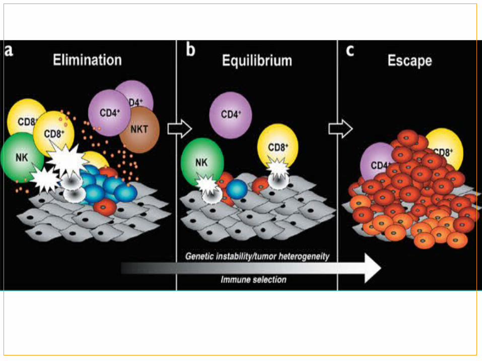
Immunosurveillance
IntroductionIntroduction
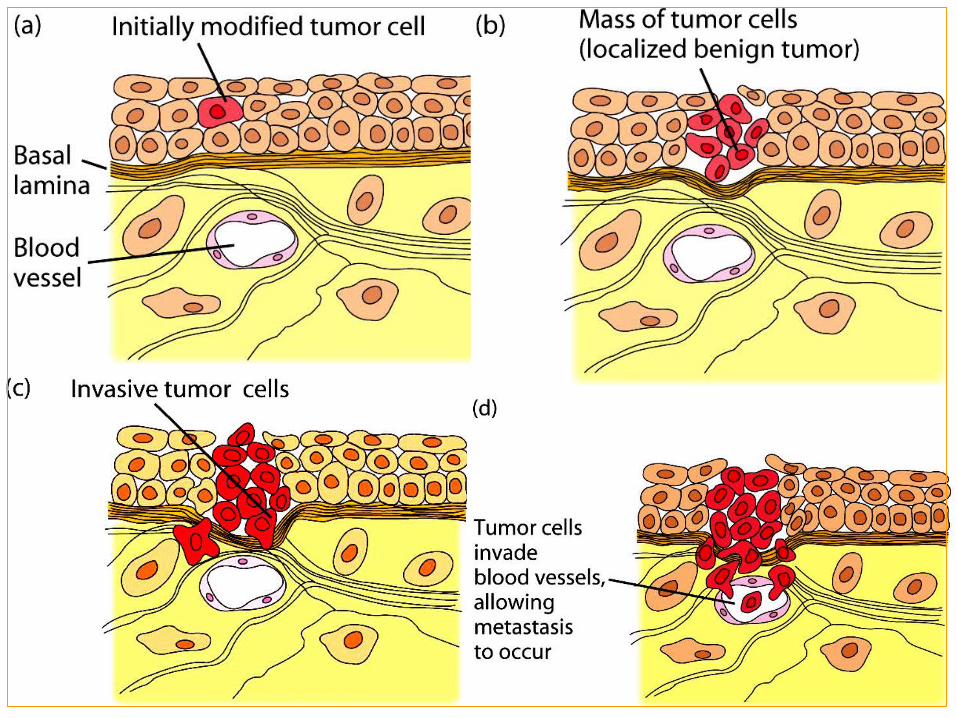
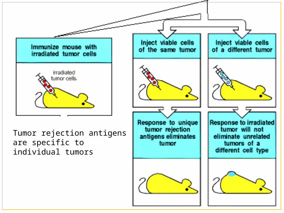
Tumor rejection antigens are specific to individual tumors
Tumor antigens: Refer to all newly expressed antigens or over expressed antigens during the generation and development of the tumor.
Part Tumor antigensⅠPart Tumor antigensⅠ
Base on their patterns of expression:
Tumor specific antigen (TSA) Tumor associated antigens (TAA)
Ⅰ.Classification of tumor antigensⅠ.Classification of tumor antigens
1.Tumor-specific antigens (TSA)1.Tumor-specific antigens (TSA)
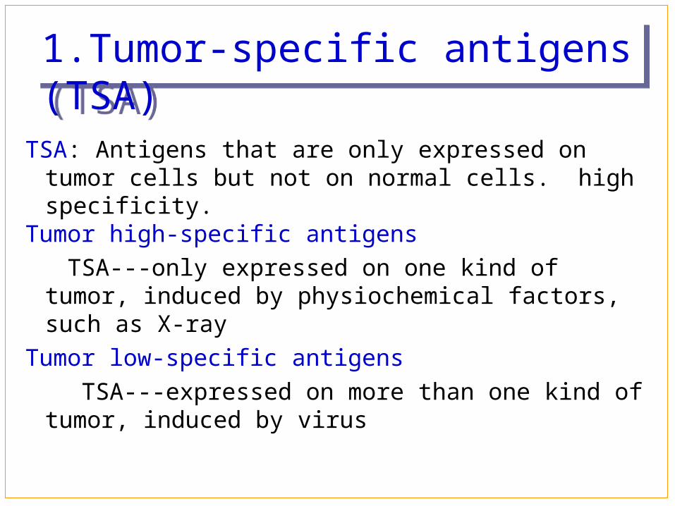
TSA: Antigens that are only expressed on tumor cells but not on normal cells. high specificity.
Tumor high-specific antigens
TSA---only expressed on one kind of tumor, induced by physiochemical factors, such as X-ray
Tumor low-specific antigens
TSA---expressed on more than one kind of tumor, induced by virus
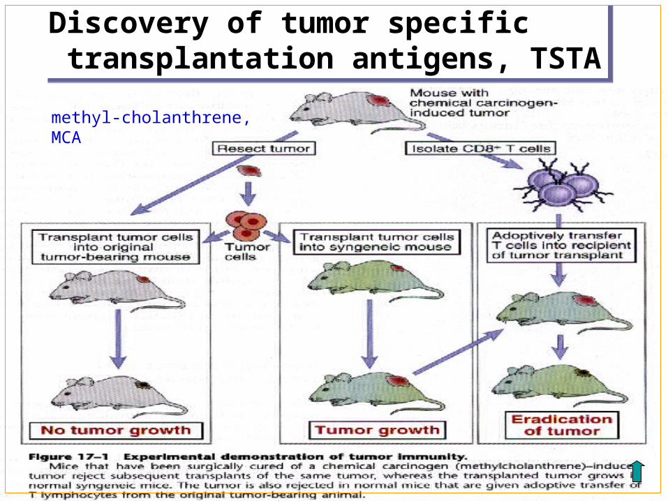
Discovery of tumor specific transplantation antigens, TSTADiscovery of tumor specific transplantation antigens, TSTA
methyl-cholanthrene,MCA
Tumors express antigens that are recognized as foreign by the immune system of the tumor-bearing host.
Immune response frequently fail to prevent the growth of tumors.
The immune system can be activated by external stimulator to effectively kill tumor cells and eradicate tumors.
Conclusion from this experimentConclusion from this experiment
2.Tumor-associated antigens,TAA2.Tumor-associated antigens,TAA
Antigens that are also expressed on normal cells, but high expressed on tumor cells. Without tumor specificity: CEA, AFP
Ⅱ.Common human tumor antigensⅡ.Common human tumor antigens
1. Embryonic antigens
2. Tumor antigens induced by viruses
3. proteins coded by Mutated oncogene or suppressor oncogene
4. TATAS expressed on human melanoma cells
embryonic antigens are proteins that are express at high levels on cancer cells and in normal developing fetal, but peter out or very low level in adult.
Their main function is that they provide markers that aid diagnosis of tumor.
Carcinoembryonic antigen (CEA)
alpha-fetoprotein (AFP)
1. embryonic antigens1. embryonic antigens
High CEA level is normally restricted to cells of the gut, pancreas, and liver in the course of 2-6 months of gestation, and low level is found in serum of normal adult(<5g/ml).
CEA level of serum is increased in many carcinomas ,such as the colon, pancreas, stomach, and breast.
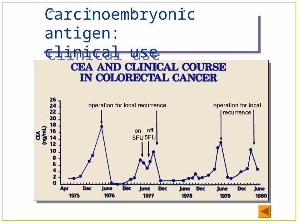
The level of serum CEA is used to monitor the persistence or recurrence of the tumors after treatment.
(1) (1) Carcinoembryonic antigen (CEA)(1) (1) Carcinoembryonic antigen (CEA)
CEA levels in normal individuals are below 2.5 ng/ml, but it increases significantly in certain malignancies, particularly colo-rectal cancers. It may also rise in some nonmalignant conditions (e.g., chronic cirrhosis, pulmonary emphysema, heavy smoking). Levels 4-5-fold of normal have been used to predict recurrence of colo-rectal tumors.
Carcinoembryonic antigen:clinical useCarcinoembryonic antigen:clinical use
Adjunct in diagnosis
Staging and prognosis
Monitoring response to therapy
Detection of tumor recurrence
Carcinoembryonic antigen:clinical useCarcinoembryonic antigen:clinical use
AFP is a circulating glycoprotein normally synthesized and secreted by the yolk sac and liver of fetal.
Serum levels of AFP is very low in serum of adult (≤20ng/ml), and the concentration of AFP is up to 500ng/ml in serum of patients with hepatocellular carcinoma.
higher rise in this protein is used for monitoring hepatomas and testicular cancers. AFP level may also be raised in some nonmalignant conditions, such as cirrhosis, hepatitis and other forms of liver damage.
(2) alpha-fetoprotein (AFP)(2) alpha-fetoprotein (AFP)
Alpha fetoprotein: concentrationsAlpha fetoprotein: concentrations
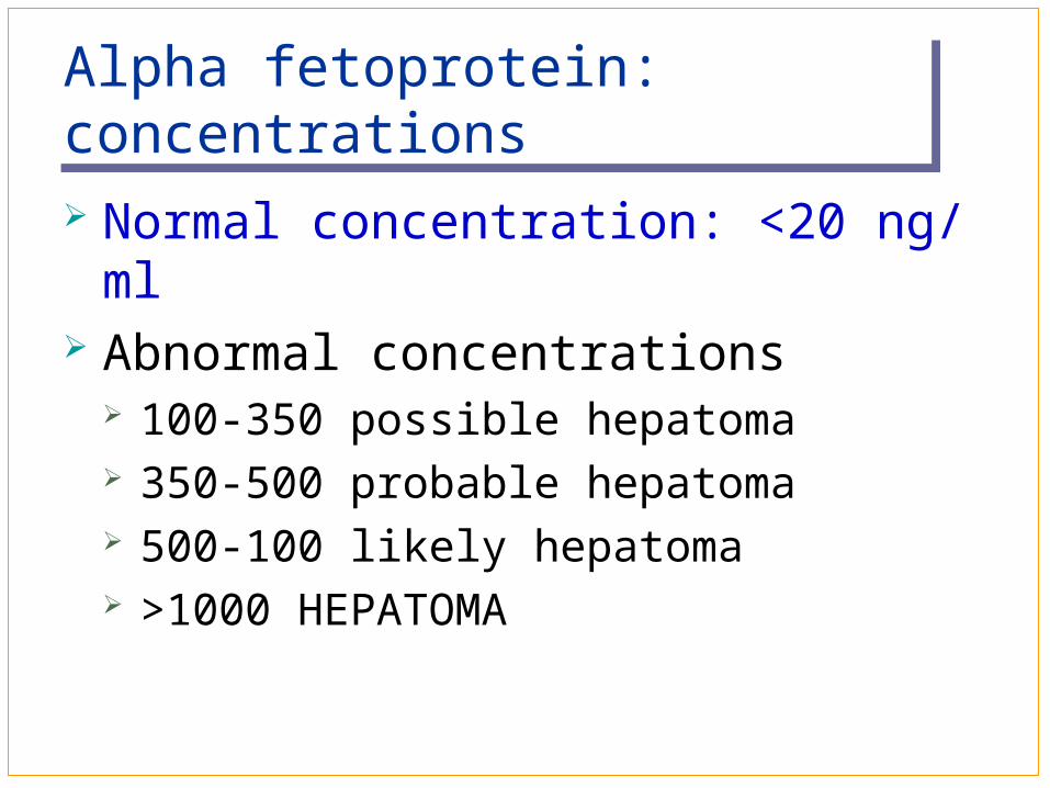
Normal concentration: <20 ng/ml Abnormal concentrations
100-350 possible hepatoma 350-500 probable hepatoma 500-100 likely hepatoma >1000 HEPATOMA
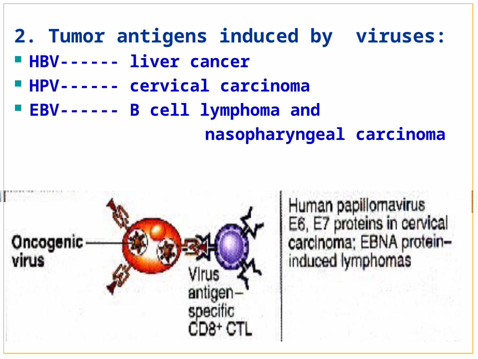
2. Tumor antigens induced by viruses: HBV------ liver cancer HPV------ cervical carcinoma EBV------ B cell lymphoma and
nasopharyngeal carcinoma
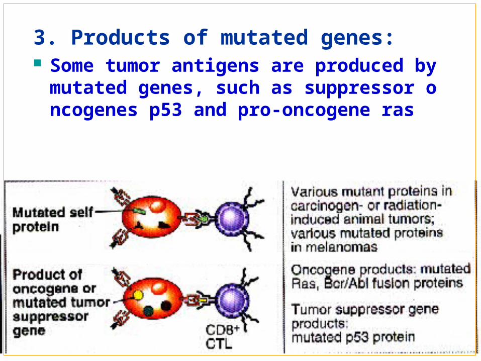
3. Products of mutated genes: Some tumor antigens are produced by mutate
d genes, such as suppressor oncogenes p53 and pro-oncogene ras
Some patients with cancer have circulating CD4+ and CD8+T cells that can respond to the products of mutated genes such as Ras and P53.
Furthermore, in animals, immunization with mutated Ras or P53 proteins induces CTLs and rejection responses against tumors expressing these mutants.
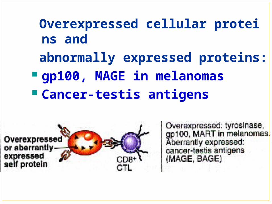
Overexpressed cellular proteins and
abnormally expressed proteins: gp100, MAGE in melanomas Cancer-testis antigens

Part ⅡMechanism of Immune ResponsePart ⅡMechanism of Immune Response
T cells: αβT, γδT
NK cells Cellular immunity
Macrophages
Dendritic cells Humoral immunity
Ⅰ.Cell-mediated Immune ResponseⅠ.Cell-mediated Immune Response
T cells NK cells Macrophages(MΦ) Dendritic cells (DCs)
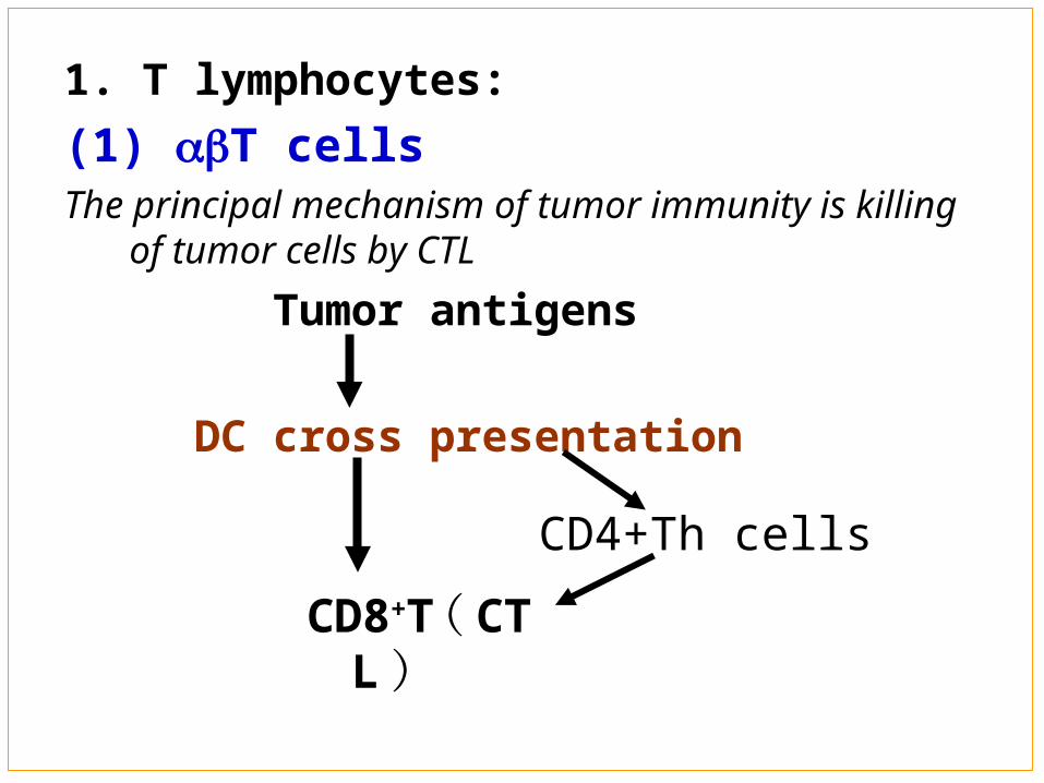
1. T lymphocytes:
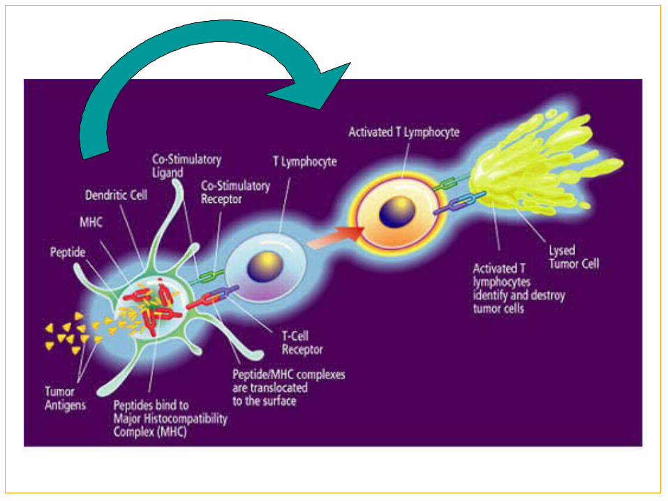
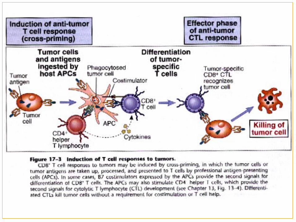
(1) T cells The principal mechanism of tumor immunity is killing of
tumor cells by CTL
Tumor antigens
DC cross presentation
CD8+T ( CTL )CD4+Th cells
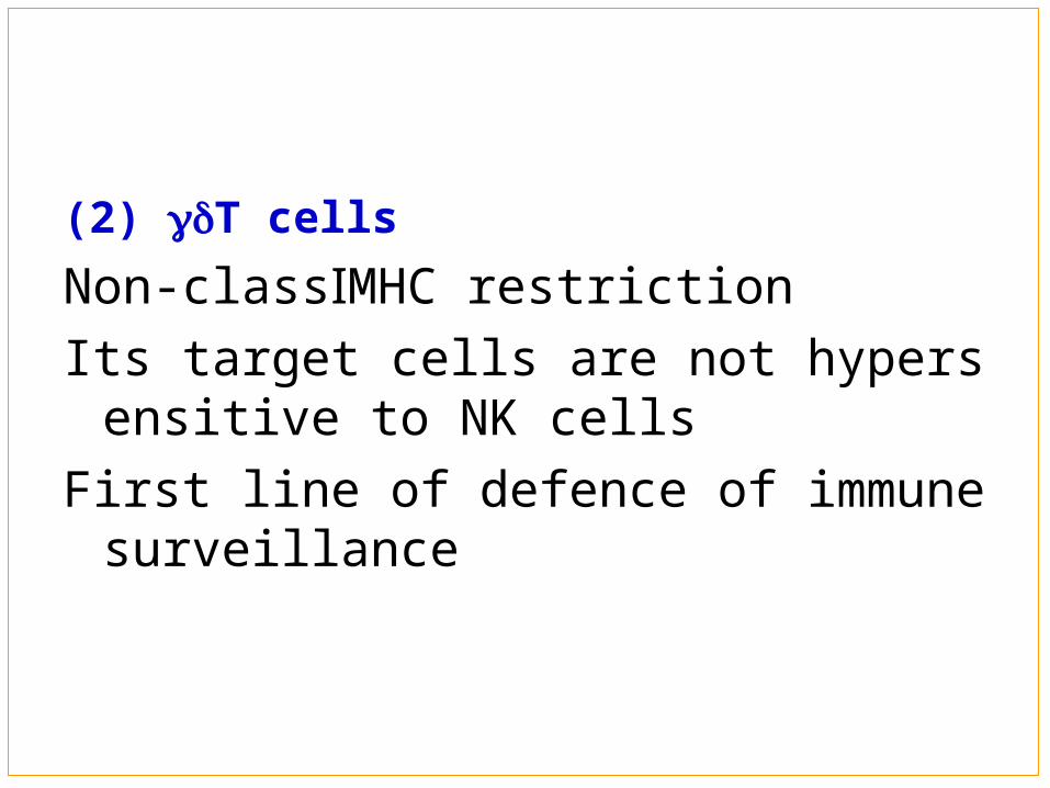
(2) T cells
Non-classⅠMHC restriction
Its target cells are not hypersensitive to NK cells
First line of defence of immune surveillance
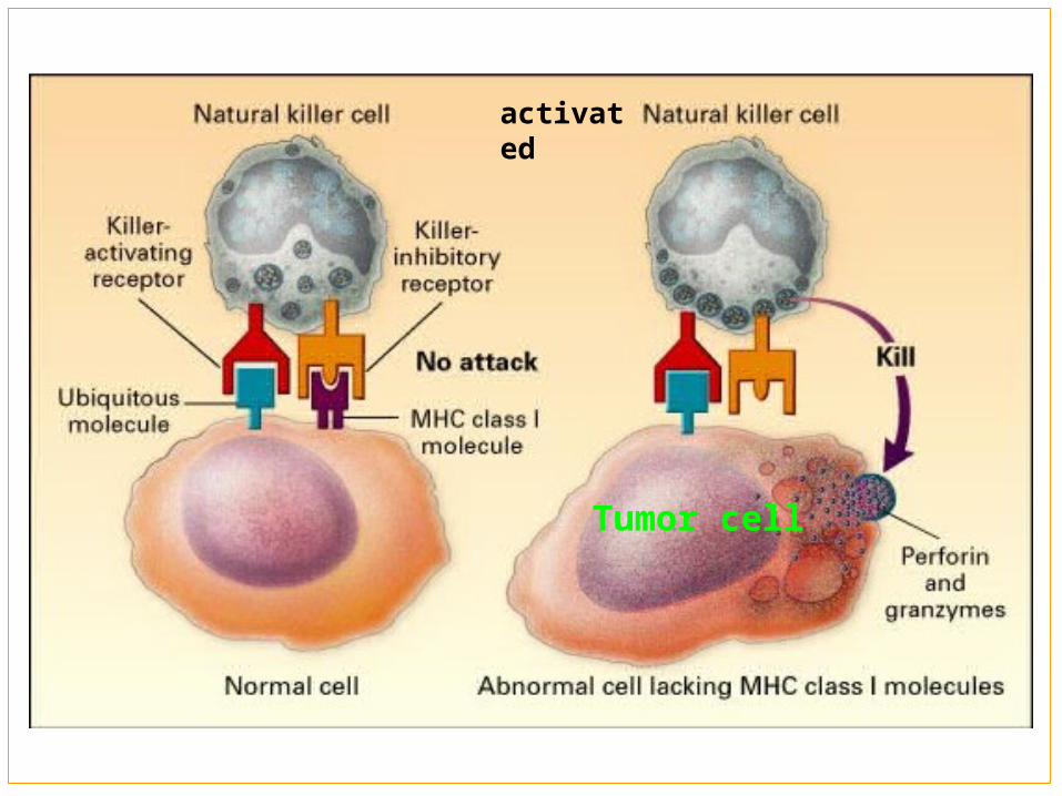
2. NK cells: NK cells are broad-spectrum killer cells It can kill target cells with low level or non
MHC class molecule. Ⅰ First line of defence of immune surveillance
Tumor cell
activated
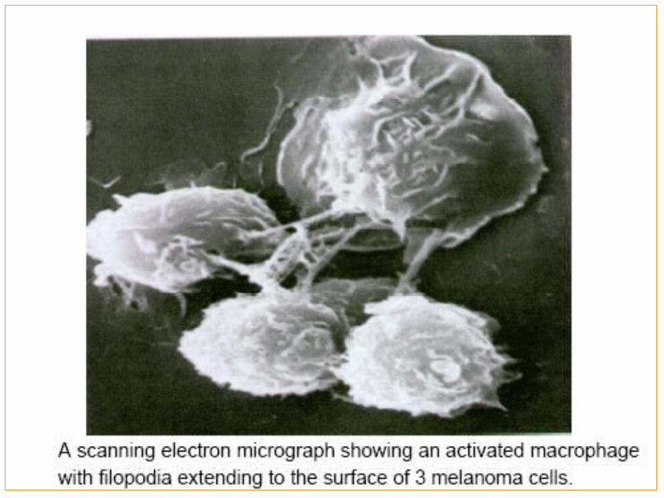
3. Macrophages(MΦ) ① APC ② release of lysosomal enzymes, reactive
oxygen intermediates, nitric oxide ③ ADCC ④ secrete cytokines
4. Dendritic cells: ① APC------Induce adaptive immune response ② Inhibit tumor growth directly
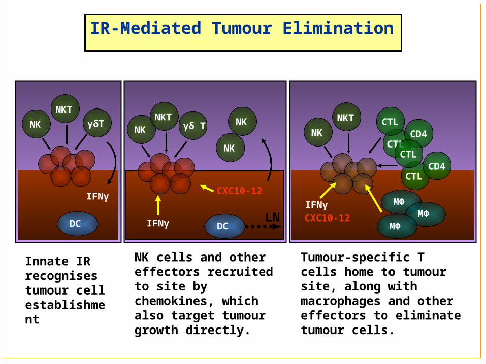
IR-Mediated Tumour Elimination
γδ TNKT
NK
IFNγ
NK
NK
DCLN
CXC10-12
NK cells and other effectors recruited to site by chemokines, which also target tumour growth directly.
γδT
NKT
NK
DC
IFNγ
Innate IR recognises tumour cell establishment
CTL
NKT
NK
MΦIFNγ
CTLCD4
CXC10-12
CTL
CTLCD4
MΦMΦ
Tumour-specific T cells home to tumour site, along with macrophages and other effectors to eliminate tumour cells.
Antibodies: Activating complement ① ADCC② Opsonization③
Ⅱ. Humoral immune responses Ⅱ. Humoral immune responses
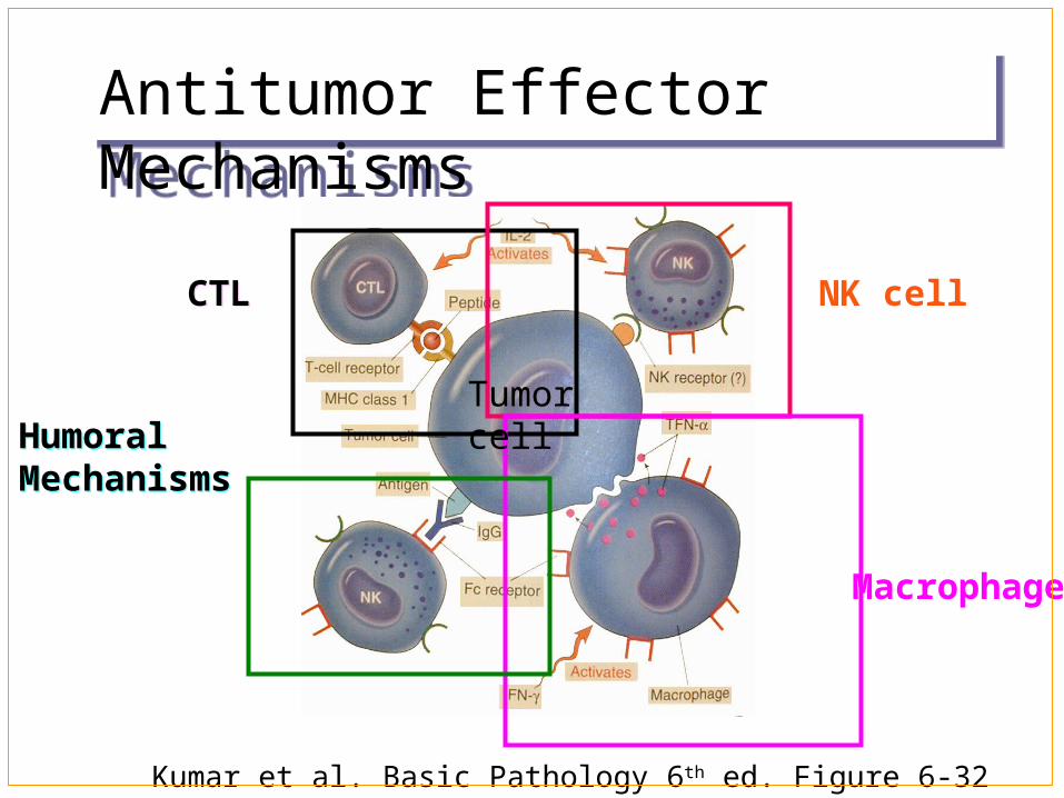
Antitumor Effector MechanismsAntitumor Effector Mechanisms
CTLCTL NK cell
Macrophage
HumoralMechanismsHumoralMechanisms
Kumar et al. Basic Pathology 6th ed. Figure 6-32
Tumor cell
Factors related to tumor cells
Factors related to the host’s
immune system
Part Mechanism of Tumor ⅢImmune Escape Part Mechanism of Tumor ⅢImmune Escape
Ⅰ. Factors related to tumor cellsⅠ. Factors related to tumor cells
1.low immunogenicity of tumor antigens and antigenic modulation
(1) low immunogenicity of tumor antigens
The failure of immunosurveillance may be the fact that in the early development of a tumor, the amount of antigen may be too small to stimulate the immune system.
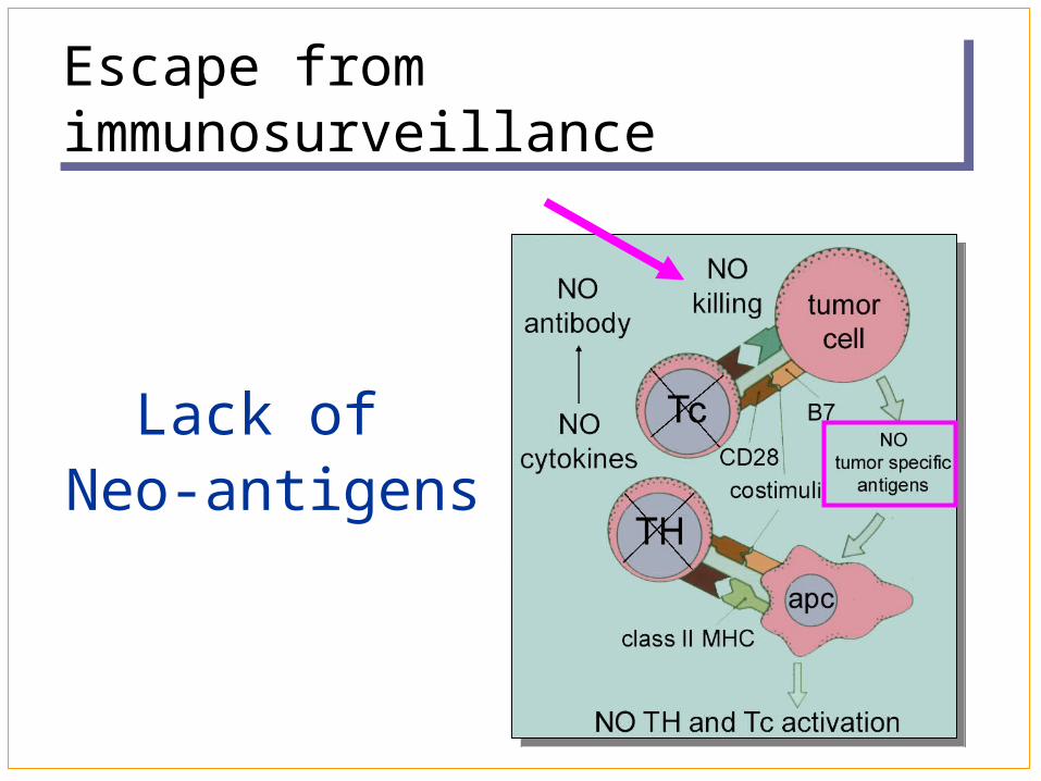
Escape from immunosurveillanceEscape from immunosurveillance
Lack of Neo-antigens
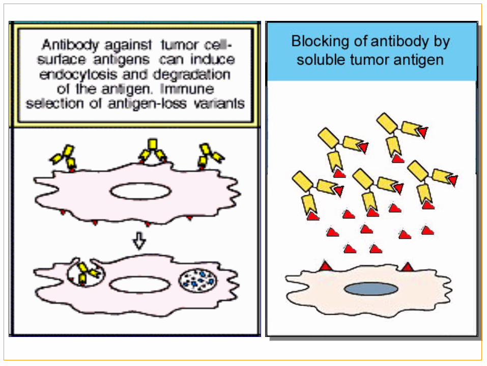
(2) antigenic modulation: is a phenomenon that cell-surface tumor antigens are decrease or lose because of attack of host’s humoral immune.
2. covering or blocking of tumor antigens on the surface of the tumor cells
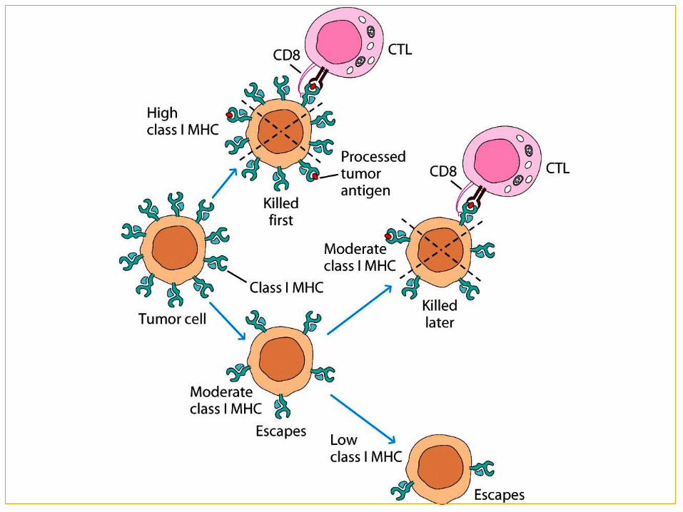
3. Diminution or absence of MHC class I molecule
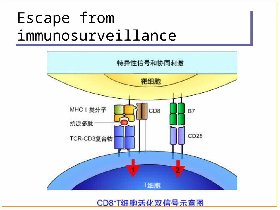
4. Lack of co-stimulatory molecule on the surface of tumor cells
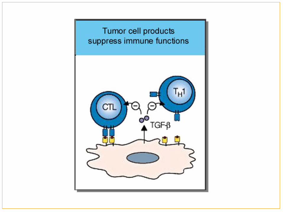
5. Immune inhibitors secreted by tumor cells
Escape from immunosurveillanceEscape from immunosurveillance
Ⅱ.Factors related to host’s immune systemⅡ.Factors related to host’s immune system
1. Immunodeficiency
2. Suppressing immune function by tumor directly or indirectly
Active immunotherapy Target immunotherapy Adoptive immunotherapy Cytokine therapy Gene therapy
Part Immunotherapy of tumorsⅤPart Immunotherapy of tumorsⅤ
Stimulation of active host immune
responses to tumors: Vaccination with tumor cells and tumor antig
ens, or with APC. Augmentation of host immunity to tumors wi
th cytokines and costimulators Nonspecific stimulation of the immune syste
m
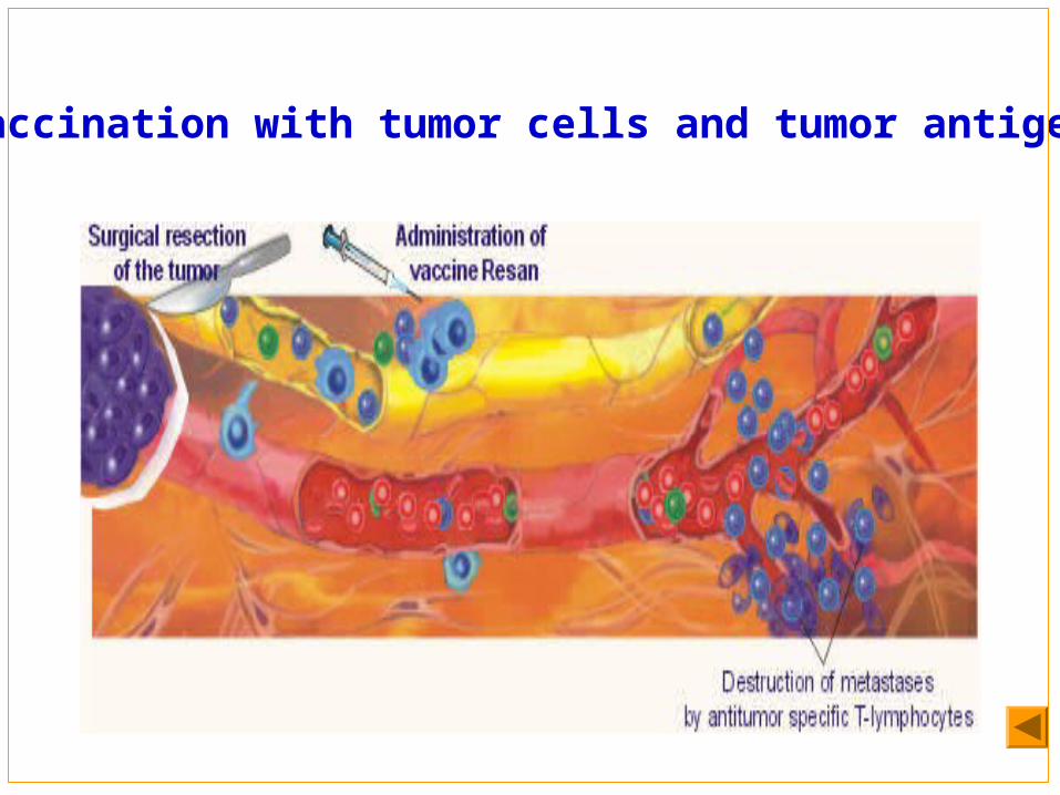
Vaccination with tumor cells and tumor antigens
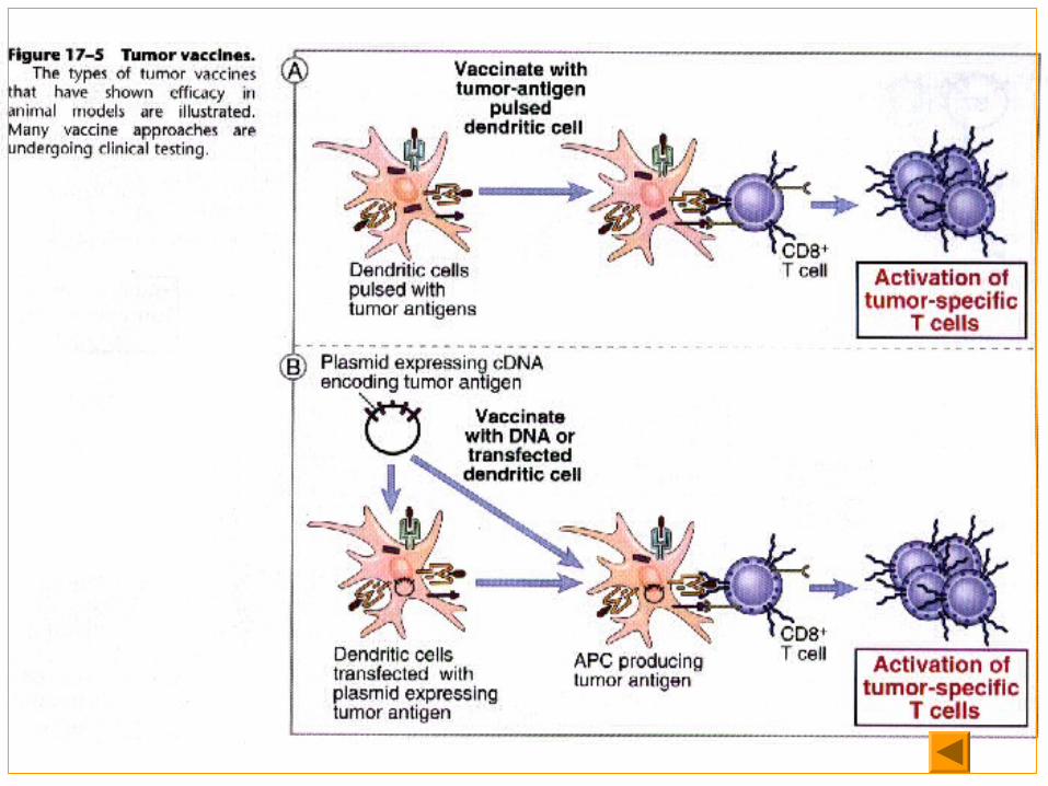
DC:
Use “primed” dendritic cells APCs can be fed tumor antigens in the laboratory and then injected into a patient. The injected cells are primed to activate T cells Alternatively, DCs can be infected with a viral vector that contains the gene for a tumor antigen.
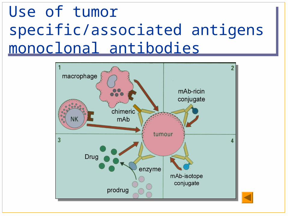
Use of tumor specific/associated antigens monoclonal antibodiesUse of tumor specific/associated antigens monoclonal antibodies
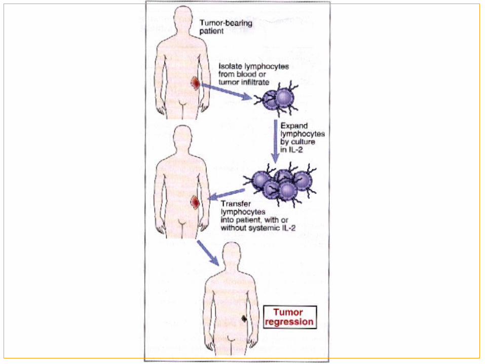
Adoptive immunotherapy Adoptive cellular immunotherapy is the transfer of
cultured immune cells that have anti-tumor reactivity into a tumor-bearing host.
LAK, TIL, CD3AK, CTL
Passive immunotherapy for tumors with
T cells and antibodies: Therapy with anti-tumor antibodies:
Monoclonal antibodies conjugated drugs Adoptive cellular therapy:
LAK,TIL,CD3AK,CTL
Cytokines may also be administered systemically for the treatment of human tumors.
IL-2 works by stimulating the proliferation and anti-tumor activity of NK cells and CTLs.
IFN-γworks by increasing the cytolytic activity of NK cells and class I MHC expression on various cell types.
Their side effects limited this treatment.
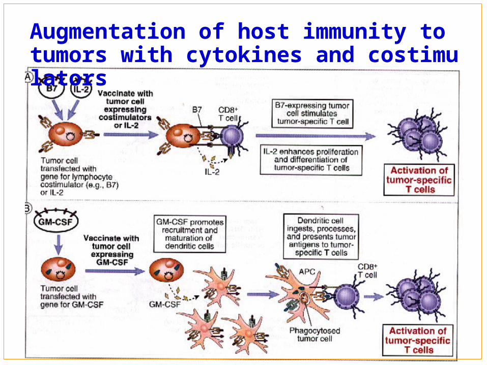
Augmentation of host immunity to tumors with cytokines and costimulators