Embed Size (px)
Citation preview
Tumor immunology is the study ofTumor immunology is the study of ::Tumor immunology is the study ofTumor immunology is the study of ::11-- The antigenic properties of the tumor cells The antigenic properties of the tumor cells 22-- The host IR to these tumor cell The host IR to these tumor cell 33-- The immunologic consequences to the hostThe immunologic consequences to the host33 The immunologic consequences to the host The immunologic consequences to the host
of the growth of the malignant cells of the growth of the malignant cells h b h h hh b h h h44-- The means by which the immune system The means by which the immune system
can be modulated to recognize tumor cells can be modulated to recognize tumor cells ggand promote tumor eradication and promote tumor eradication
TUMOR IMMUNOLOGYTUMOR IMMUNOLOGY
T tiT ti• • Tumor antigens Tumor antigens • • Effectors mechanisms in antiEffectors mechanisms in anti--tumor tumor
immunity immunity •• Mechanisms of tumor evasion of theMechanisms of tumor evasion of the• • Mechanisms of tumor evasion of the Mechanisms of tumor evasion of the
immune system immune system • • Immunotherapy for tumors Immunotherapy for tumors
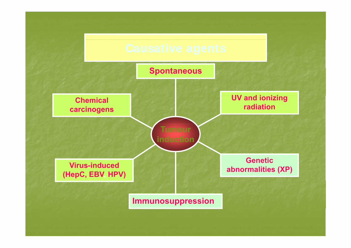
Causative agentsCausative agents
SSpontaneous
UV d i i iChemical carcinogens
UV and ionizing radiation
Tumour induction
Virus-induced (HepC, EBV, HPV)
Genetic abnormalities (XP)
Immunosuppression
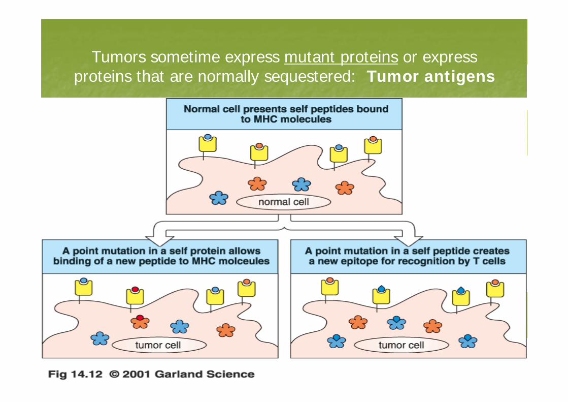
Tumors sometime express mutant proteins or express p p pproteins that are normally sequestered: Tumor antigens
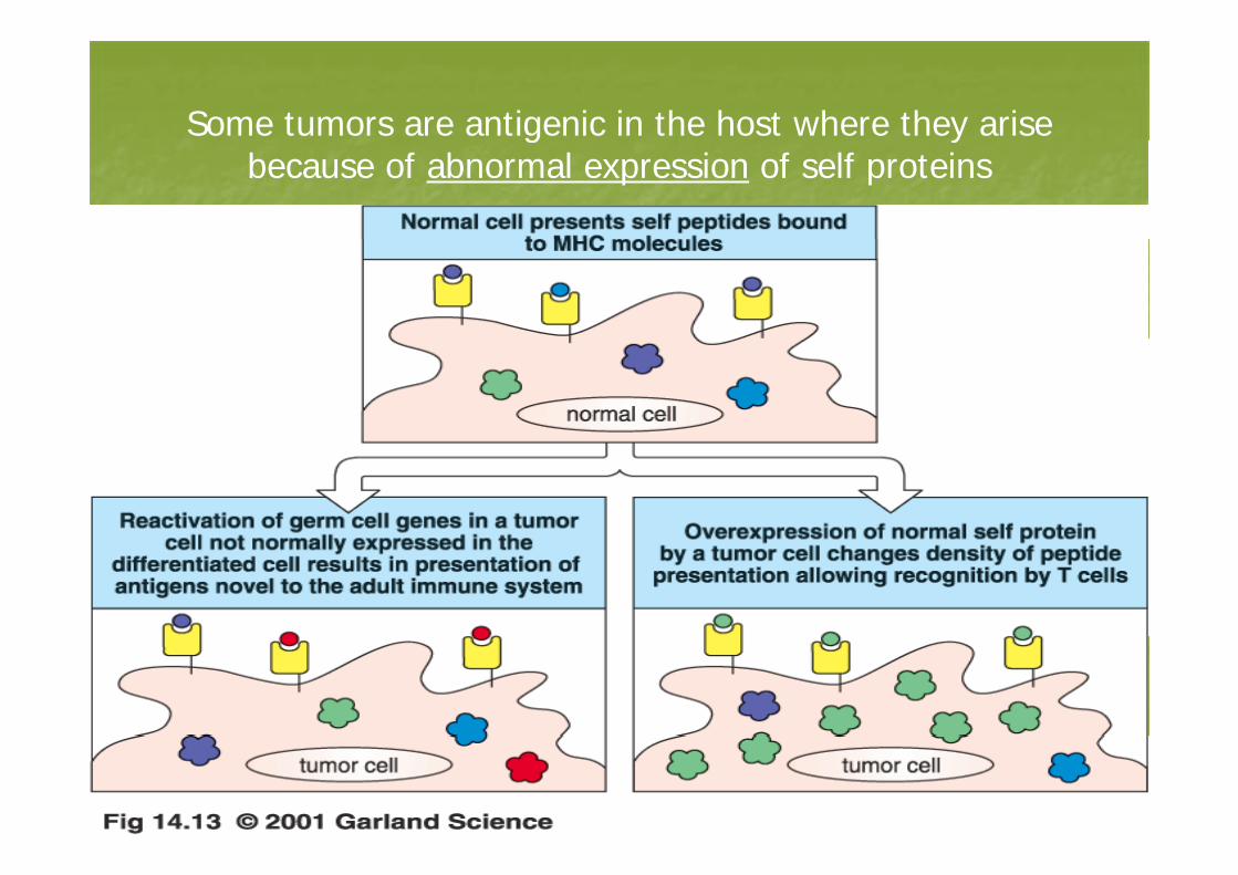
Some tumors are antigenic in the host where they arise g ybecause of abnormal expression of self proteins
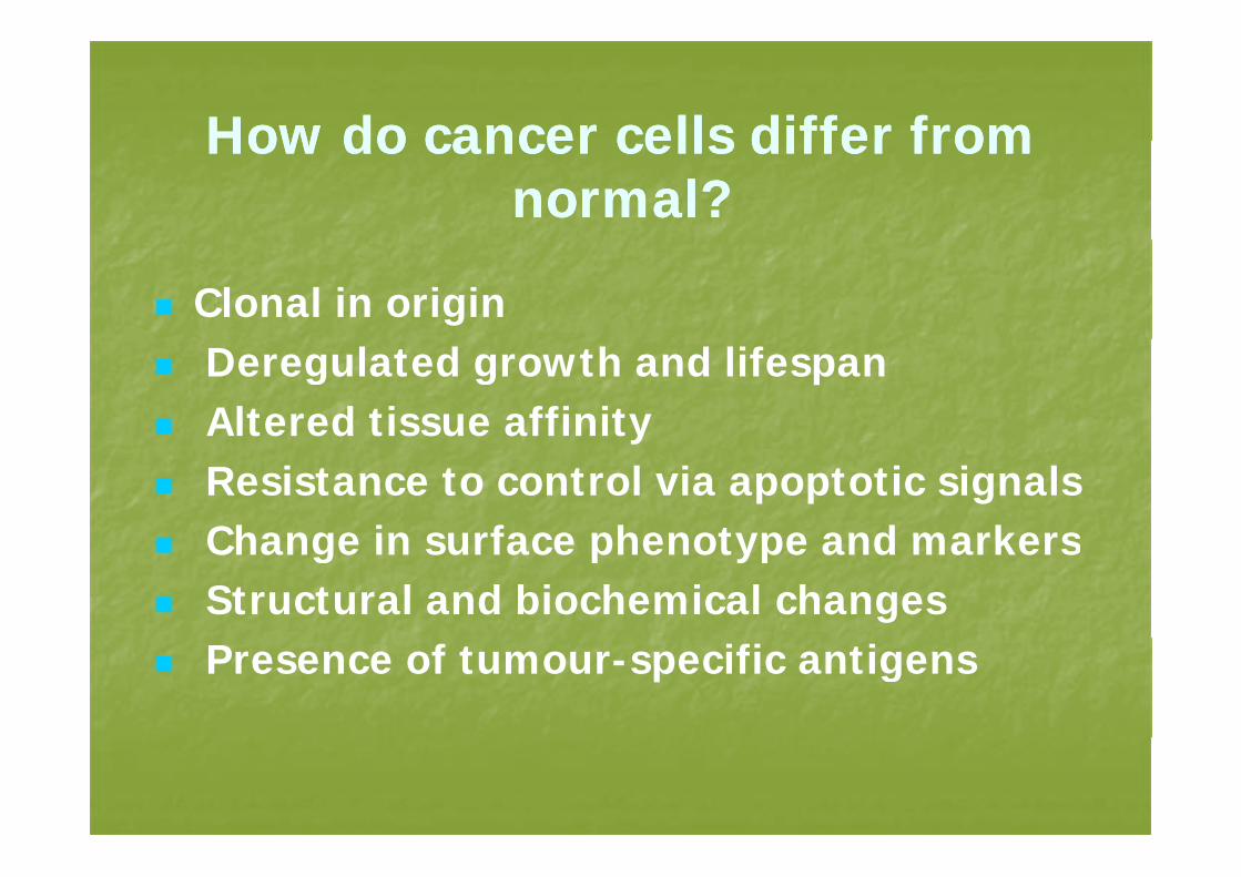
How do cancer cells differ fromHow do cancer cells differ fromHow do cancer cells differ from How do cancer cells differ from normal?normal?
Clonal in origin Deregulated growth and lifespan Altered tissue affinity yResistance to control via apoptotic signals Change in surface phenotype and markersChange in surface phenotype and markersStructural and biochemical changesPresence of tumour-specific antigens
Immunosurveillance
• An hypothesis that states that a physiologic function of the immune system is to recognize and destroy malignantly g y g ytransformed cells before they grow into tumorstumors.
• Implies that cells of the immune system recognize something “foreign” on transformed/tumor cells. /
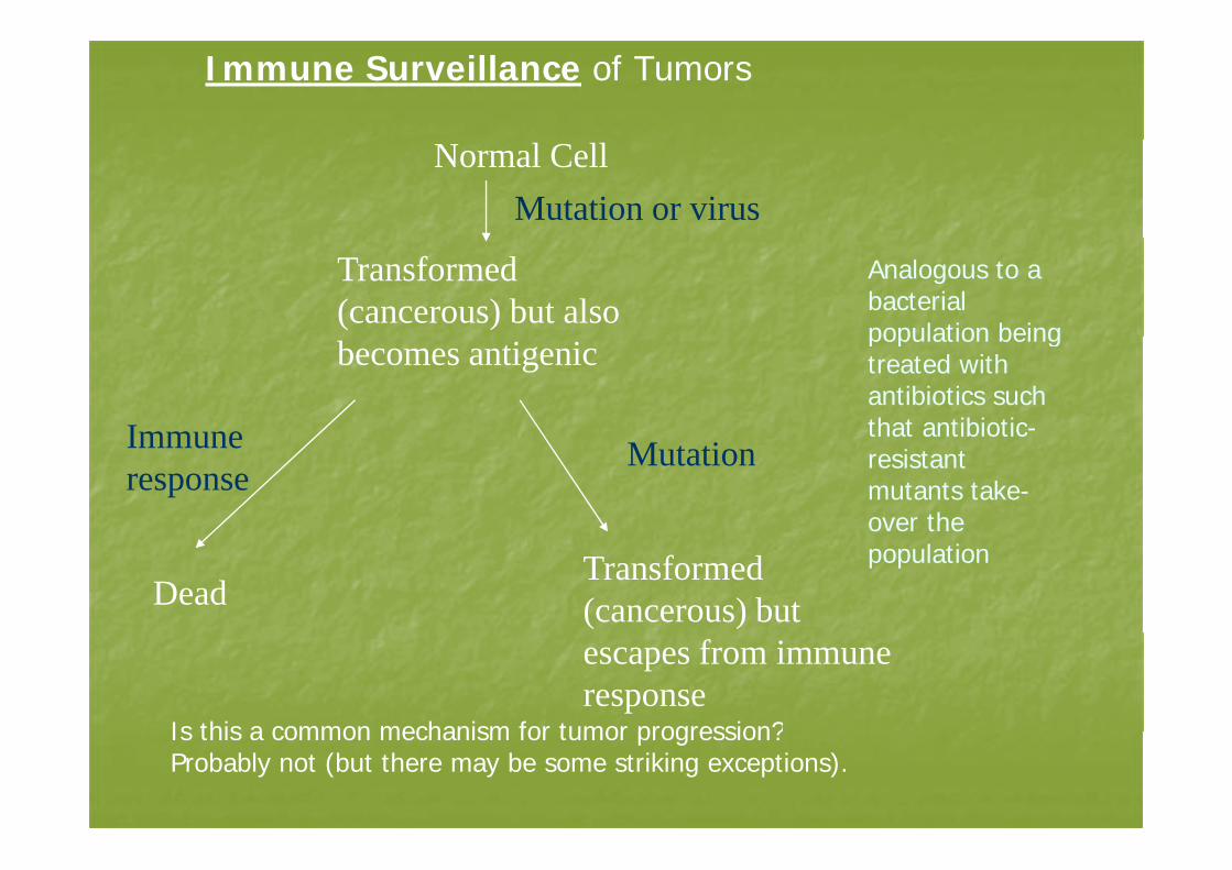
Immune Surveillance of Tumors
Normal CellMutation or virus
Transformed (cancerous) but also
Analogous to a bacterial population being
Immune
becomes antigenic population being treated with antibiotics such that antibiotic-Immune
responseMutation
that antibioticresistant mutants take-over the
DeadTransformed (cancerous) but
population
escapes from immune response
Is this a common mechanism for tumor progression?Is this a common mechanism for tumor progression?Probably not (but there may be some striking exceptions).
Evidence in Support of Immunosurveillance
Immunodeficient individuals are more likely to d l t i t f t thdevelop certain types of tumors than immunocompetent individuals.
Clinicopathologic correlations suggest that lymphocytic infiltrates in some tumors (e.g. medullary breast carcinoma, malignant melanoma) are associated with a better prognosis compared to histologically similar tumors without infiltrates
Histologic evidence indicates that activeHistologic evidence indicates that active immune responses occur within tumors or i d i i l h din draining lymph nodes.
There is ample evidence that T and BThere is ample evidence that T and Blymphocytes specific for tumor surface
l l h b ti t d dmolecules have been activated and expanded in tumor patients.
Immunosuppression leads to increasedImmunosuppression leads to increased development of viral-derived tumours (K i / NHL / HPV)(Kaposi / NHL / HPV).
Organ transplant increases malignant g p gmelanoma risk. (0.3% general paediatric pop 4% paediatric transplants)paediatric pop., 4% paediatric transplants)
High TIL presence correlates with improved survivalsurvival

Since common cancers (e g carcinomas ofSince common cancers (e.g. carcinomas of lung, colon, breast, prostate) arise f tl i i t tfrequently in immunocompetent individuals, immunosurveillance is often not effective.
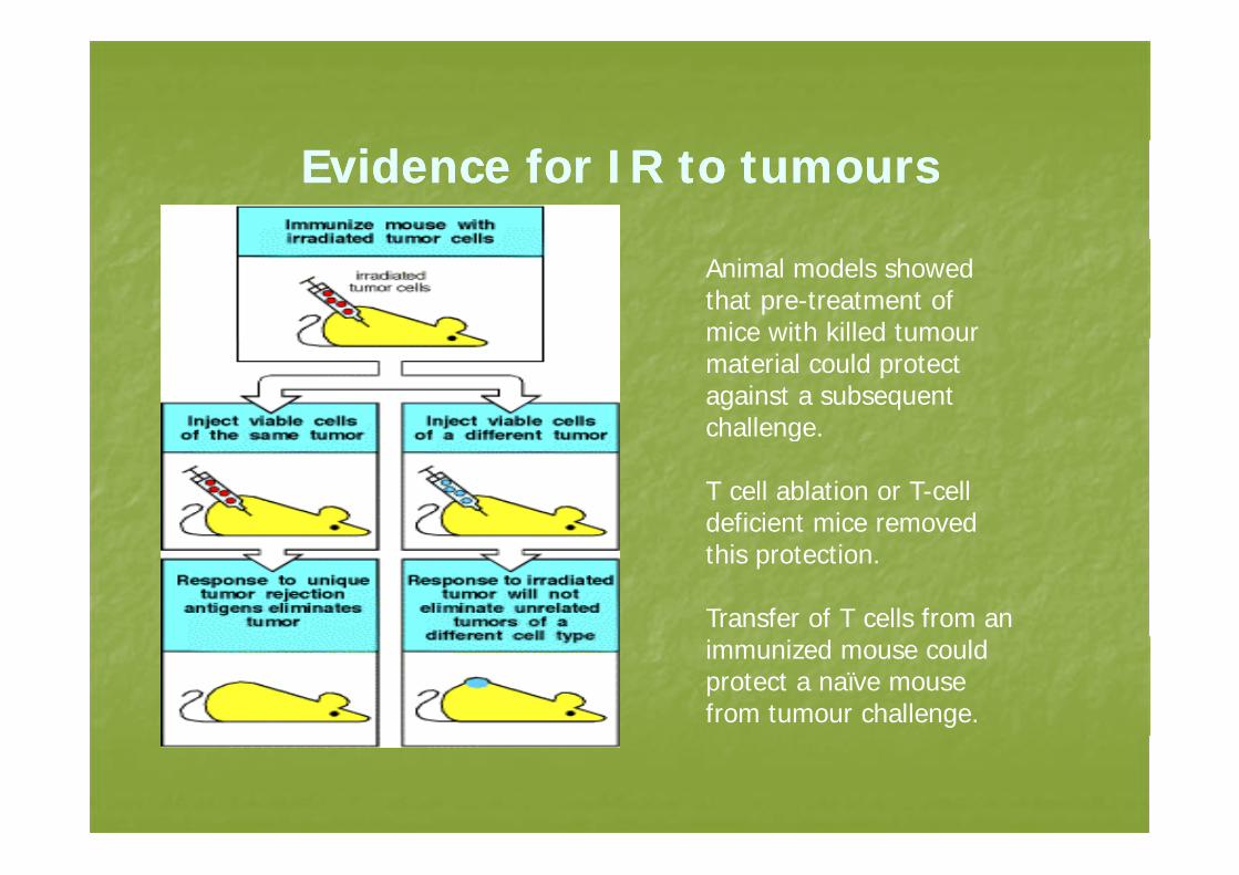
Evidence for IR to tumoursEvidence for IR to tumours
Animal models showed that pre-treatment of mice with killed tumourmice with killed tumour material could protect against a subsequent challenge.g
T cell ablation or T-cell deficient mice removed this protection.
Transfer of T cells from an immunized mouse could protect a naïve mouse from tumour challenge.
Antigens in tumor cells Antigens in tumor cells
II-- Unique tumorUnique tumor--specific Ag : are found only in tumor cells specific Ag : are found only in tumor cells and therefore represent an ideal target for an and therefore represent an ideal target for an immunologic attack immunologic attack
IIII-- Tumor associated determinants : are found in tumor Tumor associated determinants : are found in tumor cells and also in some normal cells ,but qualitative andcells and also in some normal cells ,but qualitative andcells and also in some normal cells ,but qualitative and cells and also in some normal cells ,but qualitative and quantitative differences in Ag expression permit the use quantitative differences in Ag expression permit the use of these antigens to distinguish tumor cell from normalof these antigens to distinguish tumor cell from normalof these antigens to distinguish tumor cell from normal of these antigens to distinguish tumor cell from normal cells cells
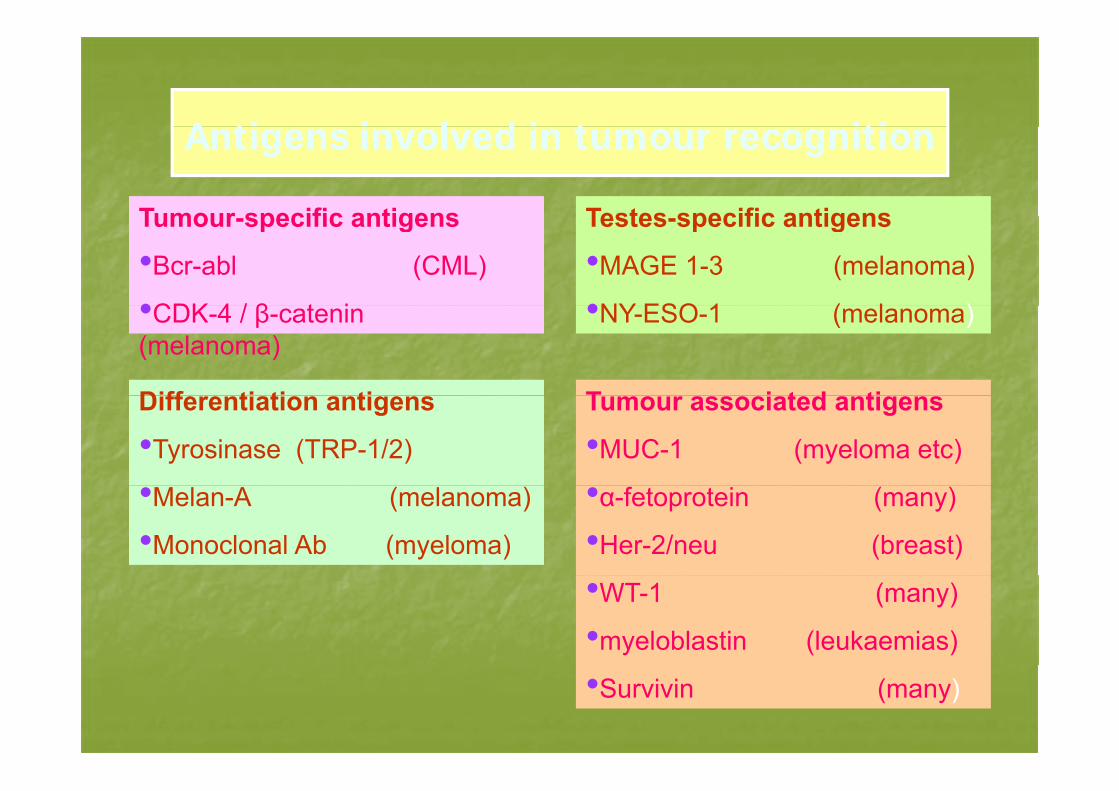
A ti i l d i t itiA ti i l d i t itiAntigens involved in tumour recognitionAntigens involved in tumour recognition
Tumour specific antigens Testes specific antigensTumour-specific antigens
•Bcr-abl (CML)
•CDK 4 / β t i
Testes-specific antigens
•MAGE 1-3 (melanoma)
•NY ESO 1 ( l )•CDK-4 / β-catenin (melanoma)
Diff ti ti ti
•NY-ESO-1 (melanoma)
T i t d tiDifferentiation antigens
•Tyrosinase (TRP-1/2)
Tumour associated antigens
•MUC-1 (myeloma etc)
•Melan-A (melanoma)
•Monoclonal Ab (myeloma)
•α-fetoprotein (many)
•Her-2/neu (breast)
•WT-1 (many)
•myeloblastin (leukaemias)
•Survivin (many)
TUMOR ANTIGENSTUMOR ANTIGENS
• Tumor Antigens Recognized by Host T L h tLymphocytes
• Tumor Antigens Recognized by Antibodiesg g y– Antibodies produced by host humoral
responsesresponses– Antibodies raised in animals used as
diagnostic therapeutic agentsdiagnostic, therapeutic agents
Examples of Tumor Antigens that Stimulate T CellExamples of Tumor Antigens that Stimulate T Cell Responses
• Viral gene products in virus-associated li imalignancies.
– SV40 T antigen (SV40-induced rat tumors)– Human papillomavirus E6 and E7 gene products
(human cervical carcinoma)( )– Epstein-Barr virus EBNA-1 gene product
(Burkitt's lymphoma and nasopharyngeal(Burkitt s, lymphoma and nasopharyngeal carcinoma)
Tumor Antigens Recognized by TTumor Antigens Recognized by T Lymphocytes
• Products of mutated normal cellular genes not related to oncogenesisrelated to oncogenesis
• Products of oncogenes and mutated tumor suppressor genessuppressor genes
• Products of normally silent genesT ti d d b f• Tumor antigens encoded by genomes of oncogenic virusesTi ifi diff ti ti ti• Tissue-specific differentiation antigens recognized
Tumor Antigens Recognized byTumor Antigens Recognized by Antibodies
• Oncofetal antigens• Altered glycolipid and glycoprotein antigensantigens
• Tissue specific differentiation antigens
Oncofetal Antigens
• Molecules normally expressed on developing (f t l) b t t d lt ti(fetal) but not adult tissues.
• Expression in adult not strictly limited to tumors; low amounts in normal tissues and increased amounts in inflammatory conditions.
• Do not induce protective immune responses.• Useful as markers that aid in the diagnosis of Useful as markers that aid in the diagnosis of
tumors.
Oncofetal Antigens: Cacinoembryoinc antigen (CEA, CD66)
• Heavily glycosylated membrane protein; may function as adhesion moleculeadhesion molecule.
• Highly expressed in developing gut, liver and pancreas (1st two trimesters).
• Expressed at low levels on granulocytes and gut epithelial cells in adult.Hi hl d b i f l• Highly expressed by carcinomas of colon, pancreas, stomach and breast, with associated elevated serum levels.levels.
• Serum levels also elevated in setting of inflammatory diseases of liver and colon.
Oncofetal Antigens: Alpha-g pfetoprotein (AFP)
l b l l d b lk d l• a-globulin glycoprotein secreted by yolk-sac and liver during fetal life; replaced by albumin in adult life.
• Serum levels elevated in patients with hepatocellular• Serum levels elevated in patients with hepatocellular carcinoma, germ-cell tumors, and some gastric and pancreatic tumors.
• Elevated levels also seen in non-neoplastic liver disease (e.g. cirrhosis.S l l f ll d t t b d ft• Serum levels followed to assess tumor burden after treatment of liver or germ-cell tumors.
• Immunohistochemical detection of AFP in sections of• Immunohistochemical detection of AFP in sections of tumors may aid in pathologic diagnosis of tumor type.
Altered Glycolipid and Glycoprotein Antigens
• Require multiple enzymes to catalyze sequential addition of carbohydrate groups to protein or lipid cores. y g p p p• Due to abnormal expression of these enzymes, many tumors express high levels and/or abnormal forms of surface glycoproteins or glycolipids. (Gangliosides, blood su ace g ycop ote s o g yco p ds (Ga g os des, b oodgroup antigens, mucins.)• These abnormal cell surface glyco-molecules may contribute to some aspects of the malignant phenotypecontribute to some aspects of the malignant phenotype. • Xenogenic antibodies have been raised against many of these molecules.
Thi l f TAA i f d t t f tib d• This class of TAAs is a preferred target for antibody-based cancer-therapy.
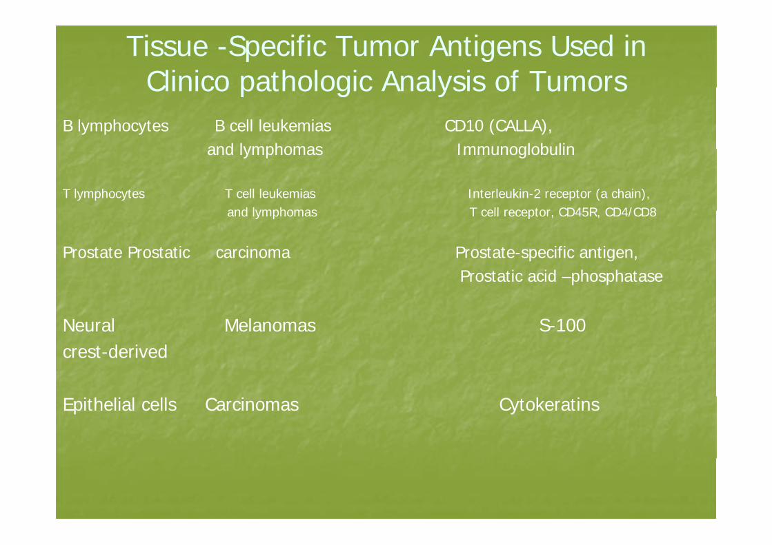
Tissue -Specific Tumor Antigens Used in Clinico pathologic Analysis of TumorsClinico pathologic Analysis of Tumors
B lymphocytes B cell leukemias CD10 (CALLA), and lymphomas Immunoglobulinand lymphomas Immunoglobulin
T lymphocytes T cell leukemias Interleukin-2 receptor (a chain), and lymphomas T cell receptor CD45R CD4/CD8and lymphomas T cell receptor, CD45R, CD4/CD8
Prostate Prostatic carcinoma Prostate-specific antigen, P ostatic acid phosphataseProstatic acid –phosphatase
Neural Melanomas S-100 crest-derived
Epithelial cells Carcinomas Cytokeratins
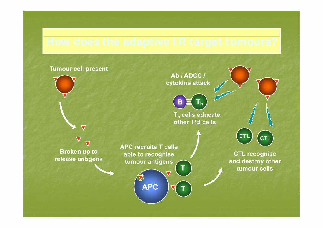
How does the adaptive IR target tumours?How does the adaptive IR target tumours?
Tumour cell presentAb / ADCC /
cytokine attack
Th
T cells educate
B
CTL CTL
Th cells educate other T/B cells
Broken up to release antigens
APC recruits T cells able to recognise tumour antigens
T
CTL recognise and destroy other
CTL
APC
T
T
tumour cells
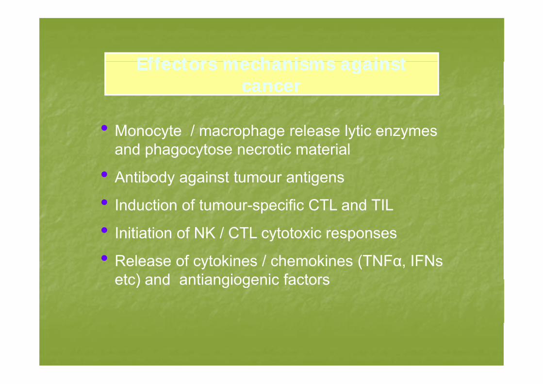
Effectors mechanisms againstEffectors mechanisms againstEffectors mechanisms against Effectors mechanisms against cancercancer
• Monocyte / macrophage release lytic enzymes and phagocytose necrotic materialand phagocytose necrotic material
• Antibody against tumour antigens
• Induction of tumour-specific CTL and TIL
• Initiation of NK / CTL cytotoxic responsesInitiation of NK / CTL cytotoxic responses
• Release of cytokines / chemokines (TNFα, IFNs etc) and antiangiogenic factorsetc) and antiangiogenic factors
ImmunoeditingImmunoediting-- The Great Escape!The Great Escape!
•Strong evidence that IR controls andStrong evidence that IR controls and eradicates nascent cancer cells
•“Immunoediting” eventually produces low• Immunoediting eventually produces low antigenicity tumour cells
•P f i t l d ith•Pressure from immune system coupled with genomic instability selects for escape
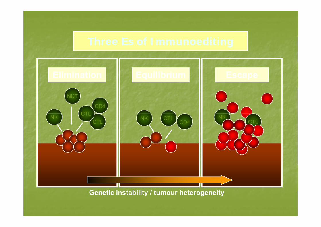
Three Es of ImmunoeditingThree Es of Immunoediting
Elimination Equilibrium Escape
CTL
NKT
CD4
CTLCTLNK NK CTL
CD4NK
CTL
G ti i t bilit / t h t itGenetic instability / tumour heterogeneity
I it i t tI it i t tImmunity against tumorImmunity against tumor
All components, specific and nonspecific, All components, specific and nonspecific, humoral and cellular affect tumor humoral and cellular affect tumor progression and growthprogression and growthp g gp g g
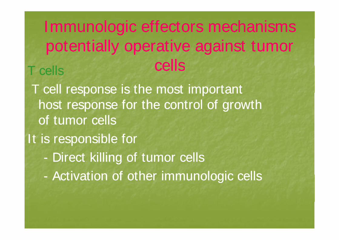
Immunologic effectors mechanisms Immunologic effectors mechanisms potentially operative against tumor potentially operative against tumor
cellscellsT llT ll cellscellsT cells T cells T cell response is the most importantT cell response is the most importantT cell response is the most important T cell response is the most important host response for the control of growth host response for the control of growth of tumor cellsof tumor cellsof tumor cells of tumor cells
It is responsible for It is responsible for -- Direct killing of tumor cells Direct killing of tumor cells
Activation of other immunologic cellsActivation of other immunologic cells-- Activation of other immunologic cells Activation of other immunologic cells
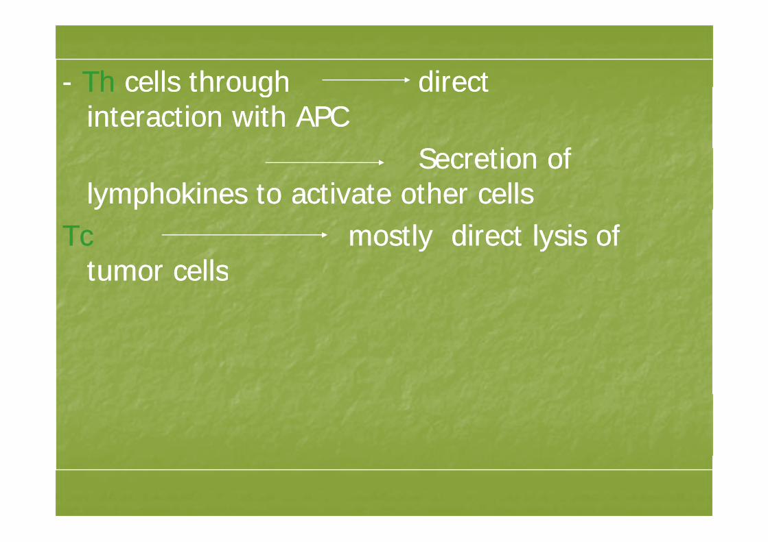
-- ThTh cells through directcells through directThTh cells through direct cells through direct interaction with APC interaction with APC
S ti fS ti fSecretion of Secretion of lymphokines to activate other cells lymphokines to activate other cells
Tc Tc mostly direct lysis ofmostly direct lysis oftumor cellstumor cellstumor cellstumor cells
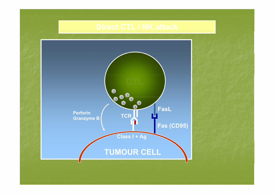
Direct CTL / NK attack
CTL
FasLTCRPerforin
Granzyme BFas (CD95)
Class I + Ag
Granzyme B
TUMOUR CELL
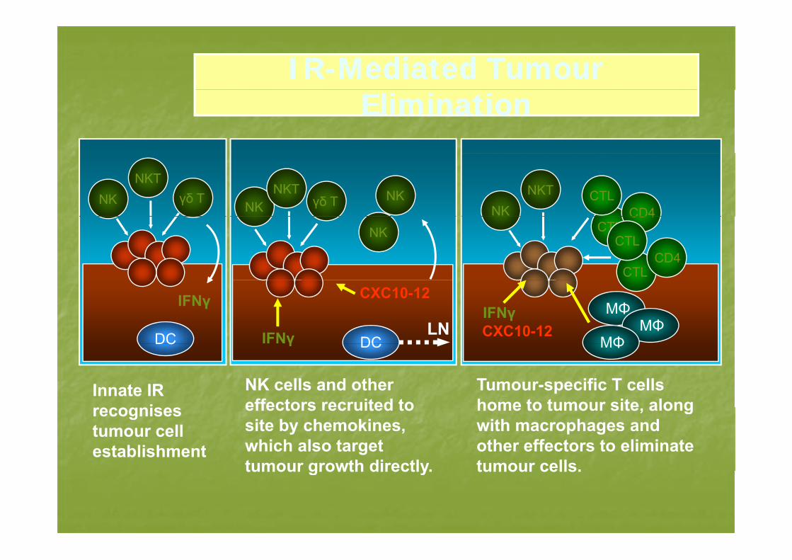
IRIR--Mediated Tumour Mediated Tumour EliminationElimination
γδ TNKT
NKNKγδ T
NKTNK
NKTNK
CTLCD4
NK CTLCD4
CTL
CTLCD4
IFNγ DCLN
CXC10-12
DC
IFNγ MΦIFNγCXC10-12 MΦ
MΦγ DC
NK cells and other effectors recruited to
DC
Innate IR i
MΦ
Tumour-specific T cells home to tumour site alongeffectors recruited to
site by chemokines, which also target tumour growth directly.
recognises tumour cell establishment
home to tumour site, along with macrophages and other effectors to eliminate tumour cells.tumour growth directly. tumour cells.
B cells and antibody dependant killingB cells and antibody dependant killingce s a d a t body depe da t gce s a d a t body depe da t gantibody to surface Ag eg Herantibody to surface Ag eg Her2 2 neu neu oncogen protein or to intracellularoncogen protein or to intracellularoncogen protein or to intracellular oncogen protein or to intracellular proteins and could facilitate T cell proteins and could facilitate T cell response by enhancing processing response by enhancing processing and presentation by APCand presentation by APCp yp y
Antibodies may act through Antibodies may act through C l t fi tiC l t fi ti-- Complement fixation Complement fixation
-- Antibody dependant cell mediatedAntibody dependant cell mediatedAntibody dependant cell mediated Antibody dependant cell mediated cytotoxicity (ADCC) cytotoxicity (ADCC)
NK cells NK cells Cytolysis by NK cells is mediated byCytolysis by NK cells is mediated by-- Cytolysis by NK cells is mediated by Cytolysis by NK cells is mediated by the release of cytotoxic factor and the the release of cytotoxic factor and the
f f i h l if f i h l iuse of perforins to puncture holes in use of perforins to puncture holes in the target cell membrane the target cell membrane gg
-- This can be augmented by ILThis can be augmented by IL2 2 and and interferoninterferoninterferon interferon
-- Also NK cells enhance resistance Also NK cells enhance resistance against metastases against metastases
-- lymphokine activated killer (LAK)lymphokine activated killer (LAK)lymphokine activated killer (LAK) lymphokine activated killer (LAK) which produced by high dose of ILwhich produced by high dose of IL2 2
Recognize lack of normal self class I MHC on some tumorssome tumors
– May be defense against tumors which h d C killihave escaped CTL killing
MHC-I recognition by NK cells is due to the surface expression of inhibitory receptors that bind MHC Iinhibitory receptors that bind MHC-I.
• Human NK cells express two families of MHC-Ibinding inhibitory receptors,p ,
– Killer cell inhibitory receptors (KIR) are type I transmembrane Ig superfamily proteins that bind to classical HLA-A, -B and -C molecules
– CD94/NKG2A receptors are heterodimeric type II transmembrane proteins with C-type lectin domains that bind to the non-classicalproteins with C-type lectin domains that bind to the non-classical HLA-E
– Engagement of KIR and CD94/NKG2A inhibitory receptors with g g y pMHC-I dominantly engages SHP-1 tyrosine phospahatses and arrests activation signals derived from numerous receptors interacting with cell surfaces such as CD2 CD16 NKR P1interacting with cell surfaces, such as CD2, CD16, NKR-P1, integrins, and several recently identified receptors.
Macrophages Macrophages APCAPC-- APCAPC
-- SStimulate the IR timulate the IR -- Potential effector cells to mediate tumor lysisPotential effector cells to mediate tumor lysis
R ti h t t l ti t t ll i it b tR ti h t t l ti t t ll i it b tResting macrophages are not cytolytic to tumor cell in vitro but Resting macrophages are not cytolytic to tumor cell in vitro but become cytolytic if activated with macrophage activating become cytolytic if activated with macrophage activating factor (MAF), MAF secreted by T cellfactor (MAF), MAF secreted by T cellfactor (MAF), MAF secreted by T cell factor (MAF), MAF secreted by T cell
Activated macrophages may produce cytotoxic factor that Activated macrophages may produce cytotoxic factor that mediate killing as well as bind to and lyse transformed cells mediate killing as well as bind to and lyse transformed cells
-- Intercellular transfer of lysosomal products ,superoxide Intercellular transfer of lysosomal products ,superoxide production. Release of neutral proteinases and secretion of production. Release of neutral proteinases and secretion of TNFTNFTNFTNF
Also production of nitric oxide may be the most effector Also production of nitric oxide may be the most effector mechanism employed by macrophage mechanism employed by macrophage p y y p gp y y p g
Potential mechanisms by which tumor cells Potential mechanisms by which tumor cells may escape from immune responsemay escape from immune responsemay escape from immune responsemay escape from immune response
II-- Immunoselection of variant cellsImmunoselection of variant cellsThis suggested by analysis of the tumor cells present in a lesion This suggested by analysis of the tumor cells present in a lesion
which often reveals heterogeneity with respect to morphologywhich often reveals heterogeneity with respect to morphologywhich often reveals heterogeneity with respect to morphology which often reveals heterogeneity with respect to morphology
and phenotype.and phenotype.
Eg. Melanoma ,in which the generation of a T cell response to one Eg. Melanoma ,in which the generation of a T cell response to one
melanosomal protein ha been associated with the outgrowth of melanosomal protein ha been associated with the outgrowth of p gp g
tumors lacking this protein tumors lacking this protein
The presence of such escapeThe presence of such escape varaint cells is probably due to thevaraint cells is probably due to theThe presence of such escape The presence of such escape ––varaint cells is probably due to the varaint cells is probably due to the
inherent genomic instability of transformed cells ( muttated inherent genomic instability of transformed cells ( muttated
oncogene )oncogene )
IIII-- Antigen modulationAntigen modulationImmune response to a tumor cells selects Immune response to a tumor cells selects
for the growth of antigen negative cellsfor the growth of antigen negative cellsfor the growth of antigen negative cells for the growth of antigen negative cells ,antigen loss reflects only a phenotypic ,antigen loss reflects only a phenotypic h i th t ll d if th IRh i th t ll d if th IRchange in the tuomr cells and if the IR change in the tuomr cells and if the IR
ablated the antigen is reexpressed ablated the antigen is reexpressed Some tumor cells have defective antigen Some tumor cells have defective antigen
processing machinary with the resultsprocessing machinary with the resultsprocessing machinary ,with the results processing machinary ,with the results that class I molecule do not get loaded that class I molecule do not get loaded with peptideswith peptideswith peptides with peptides
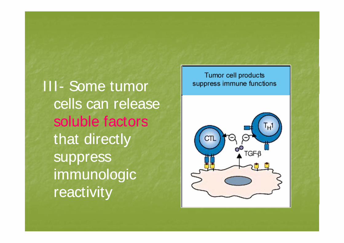
IIIIII-- Some tumor Some tumor cells can release cells can release soluble factorssoluble factorssoluble factorssoluble factorsthat directly that directly suppress suppress immunologicimmunologicimmunologic immunologic reactivity reactivity
IVIV-- Increase susceptibility to opportunisticIncrease susceptibility to opportunisticIVIV-- Increase susceptibility to opportunistic Increase susceptibility to opportunistic infection and can exhibit global infection and can exhibit global depression of depression of T llT llT cell response T cell response
VV-- Presences of tumor Presences of tumor –– specific suppressor T specific suppressor T (Ts)(Ts)(Ts)(Ts)
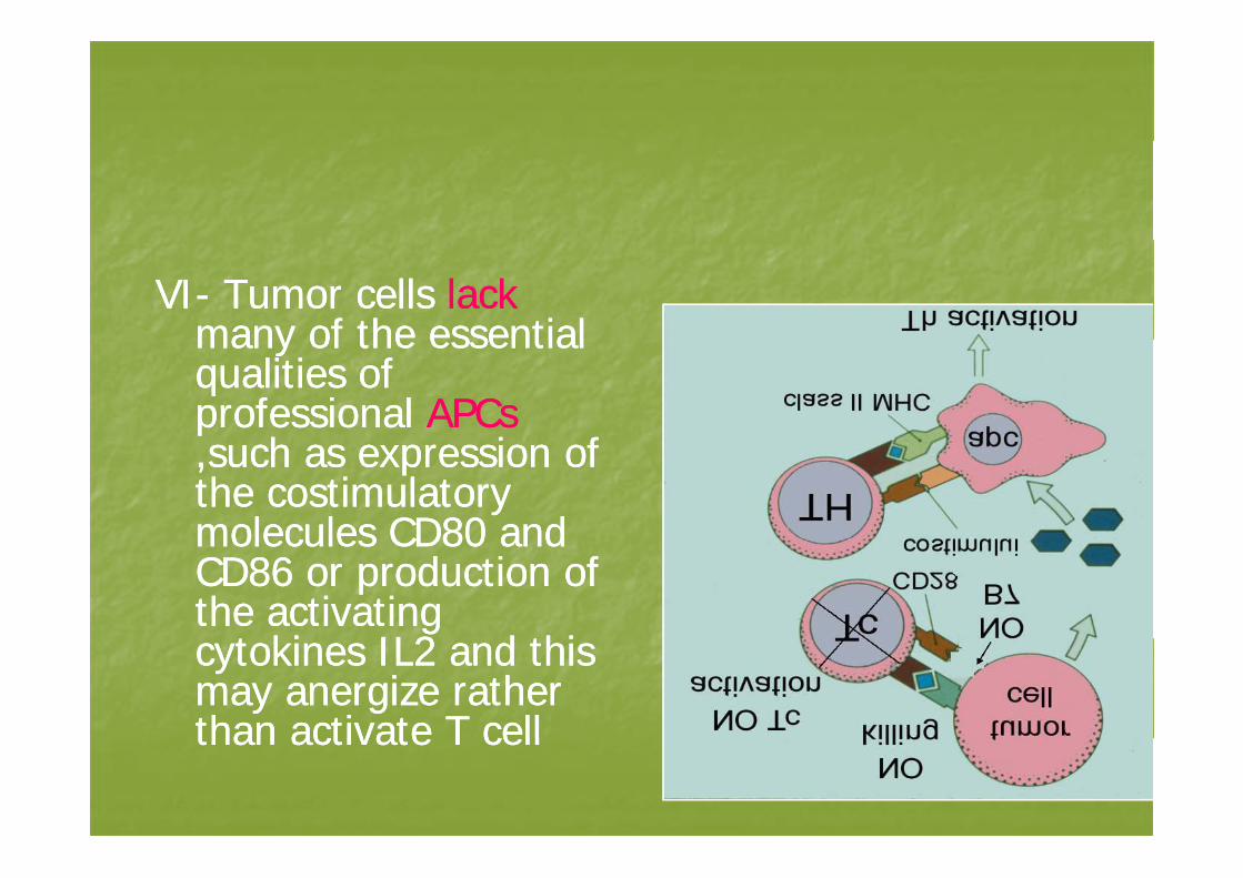
VIVI-- Tumor cells Tumor cells lacklackmany of the essentialmany of the essentialmany of the essential many of the essential qualities of qualities of professional professional APCsAPCs
h i fh i f,,such as expression of such as expression of the costimulatory the costimulatory molecules CDmolecules CD8080 andandmolecules CDmolecules CD80 80 and and CDCD86 86 or production of or production of the activating the activating
t ki ILt ki IL22 d thid thicytokines ILcytokines IL2 2 and this and this may anergize rather may anergize rather than activate T cellthan activate T cellthan activate T cell than activate T cell
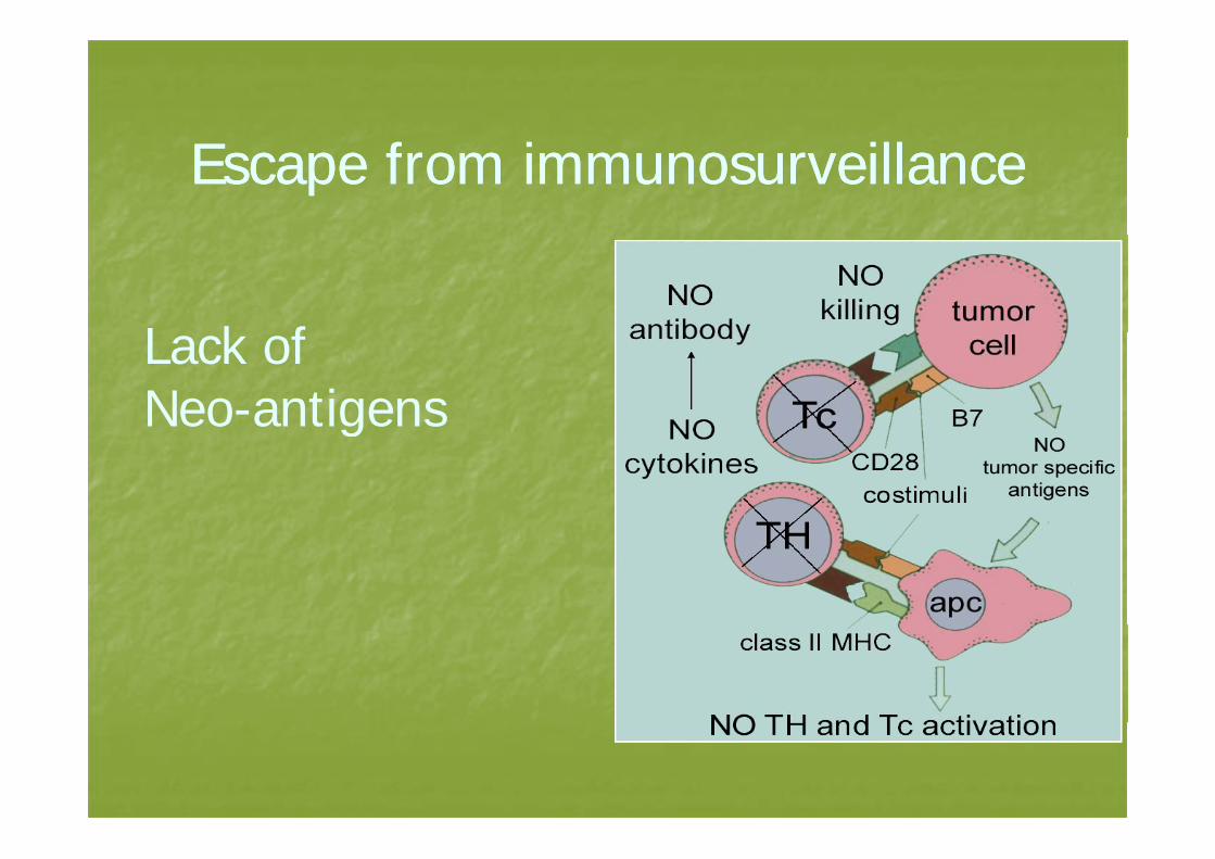
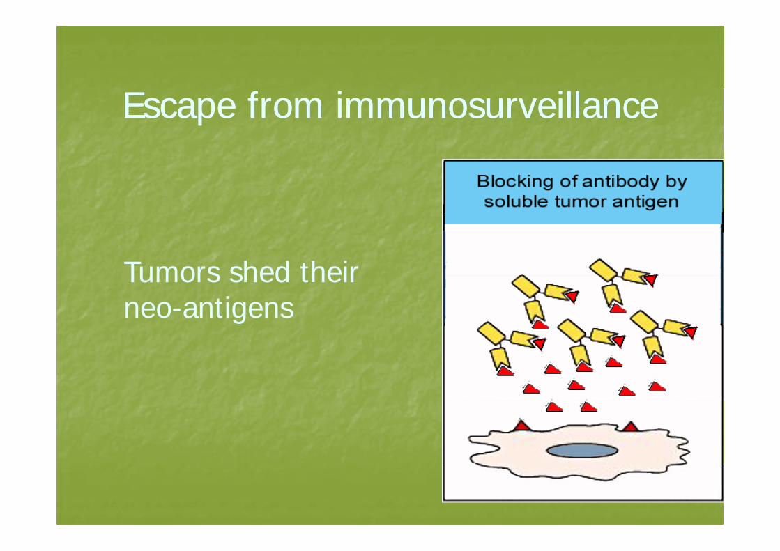
Escape from immunosurveillanceEscape from immunosurveillance
L k fLack of Neo-antigensg
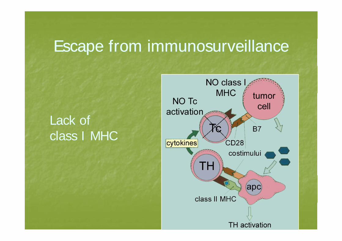
Escape from immunosurveillanceEscape from immunosurveillance
Lack ofLack of class I MHC
Escape from immunosurveillanceEscape from immunosurveillance
Tumors shed theirTumors shed their neo-antigens
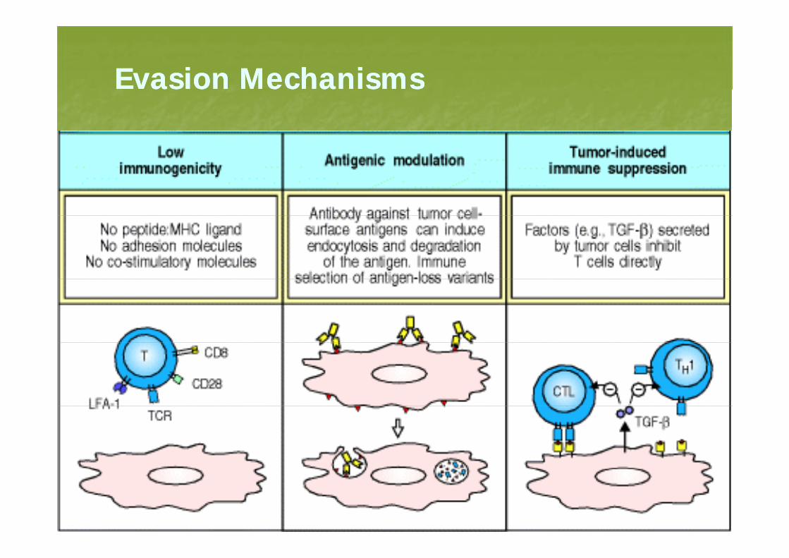
Evasion MechanismsEvasion Mechanismsas o ec a s sas o ec a s s