Embed Size (px)
Citation preview
K
CH
uoJen Tsao, MD ����� Ke��n ������� �a�����, MD����� Ke��n ������� �a�����, MD Ke��n ������� �a�����, MD
ONGENITAL DIAPHRAGMATIC ERNIA AND EVENTRATION
chapter 24
304
he management and treatment of posterolateral congenital diaphragmatic hernia (CDH) remains
a challenge for pediatric surgeons. Despite tremendous advances in prenatal diagnosis, surgical treatment, and neonatal critical care, CDH remains a significant cause of mortality and long-term disability. The clini-cal challenges lie in the broad spectrum of disease and the relative small volume of patients at individual in-stitutions, resulting in a limited experience with the most severe CDH infants. Although some institutions have become leaders in the treatment, management, and research of CDH, the vast majority will treat fewer than 10 infants with CDH per year.1 Thus, broad clini-cal experience is difficult to achieve.
In 1946, Gross reported the first successful repair of CDH in an infant younger than 24 hours old.2 CDH was considered a surgical emergency, and immedi-ate repair was the practice at that time, with excel-lent survival rates of greater than 90%.3 Since then, it has become apparent that such survival rates only included infants who lived to have an operation and did not include those who died before birth or soon thereafter. Accounting for this “hidden mortality” of CDH,4 actual survival may be closer to 50%.
Although surgical intervention is necessary to repair a CDH, the most recent advances have been in non-surgical therapies. Lung-protective ventilator strate-gies, extracorporeal membrane oxygenation (ECMO), and preoperative stabilization before surgery have all led to a significant decrease in mortality. In addition, improved understanding of pulmonary hypoplasia and hypertension associated with CDH has led to innova-tive therapies.
EPIDEMIOLOGY
The incidence of CDH has been reported between 1 in 2000 to 5000 births.5-7 In the United States, approxi-mately 1000 infants are born with CDH with a preva-lence of 2.4 per 10,000 live births.8,9 The incidence in
stillborn infants is less clear. An estimated one third of infants with CDH are stillborn and often have other associated congenital anomalies.10 Presumed to have the most severe form of CDH, infants who die in utero contribute to the “hidden mortality” of the disorder.11,12
Isolated CDH infants are typically male with one third having a major congenital anomaly.8 However, when stillbirths are included, females with CDH tend to be more prevalent than males.5,13 Approximately 80% of CDHs are left sided. Bilateral defects are rare and are often associated with other major anomalies.14 Although the exact cause remains unknown, mothers who are thin or underweight may have an increased risk of bearing an infant with CDH.15
GENETICS
Once thought to be only a sporadic isolated anomaly, it is becoming increasingly understood that genetics plays an important role in the development of CDH. A first-degree relative has an expected disease rate of approximately 2%.16 Several structural chromosomal abnormalities including deletions, translocations, and trisomies have been identified.17,18 Genetic abnormal-ities associated with CDH include both anomalies of chromosome number (Turner’s syndrome; trisomies 13, 18, 21, 22, and 23) and conditions with specific chromosomal aberrations (i.e., 15q24-q26 deletions).19 A gene distal to the 15q21 locus may be important for normal development of the diaphragm.19,20 Several specific genes have been implicated in the development of CDH based on human and animal studies, includ-ing chick ovalbumin upstream promoter-transcription factor II (COUP-TFII), Wilms’ tumor 1 gene (WT1), homolog of Drosophila slit 3 (SLIT3), and Friend of GATA2 (FOG2).21
CDH may present as an isolated defect or in associa-tion with other non–CDH-related anomalies. Several associated anomalies such as lung hypoplasia, intestinal
T
chapter 24 n CONGENITA� DIA�HRAGMATIC HERNIA AND EVENTRATION 305
malrotation, some cardiac malformations, and pat-ent ductus arteriosus are considered to be sequelae of isolated CDH. Non–CDH-related defects involving the cardiovascular, central nervous, gastrointestinal, and genitourinary systems may be a consequence of an underlying field defect of unknown etiology. Non– hernia-related anomalies associated with CDH have been estimated to occur in 40% to 60% of cases.22,23
CDH has been associated with over 50 syndromes.24 In some cases, the diaphragmatic malformation is the predominant defect as in Fryns and Donnai-Barrow syndromes, in which CDH occurs in a high proportion of these patients.25,26 In other cases, diaphragmatic defects occur in a small percentage but are still greater than the general population as in Simpson- Golabi-Behmel and Beckwith-Wiedemann syndromes. The inheritance patterns for these syndromes include dominant and recessive as well as autosomal and X-linked varieties.21 Identifying the patterns of non–hernia-related anomalies with CDH and recognizing genetic syndromes are vital in determining the prog-nosis, treatments, counseling, and outcomes for these infants and their families.14,18 Other syndromic pat-terns have been described, including Brachmann-de Lange and Pallister-Killian syndromes.27-29 If an ante-natal diagnosis of CDH is made by fetal ultrasonogra-phy, amniocentesis with karyotype and chromosomal analysis is warranted.
ASSOCIATED ANOMALIES
The impact of associated anomalies with CDH on prog-nosis and outcome cannot be overstated. Ninety-five percent of stillborn infants with CDH have an associ-ated major anomaly.5 In addition, more than 60% of infants who do not survive the immediate neonatal period have associated anomalies.30 Of those infants who survive preoperative stabilization and come to operative repair, less than 10% have additional anom-alies.30 Although the severity of pulmonary hypoplasia and hypertension are the major determinants of overall survival, infants with CDH with another major defect have a greater morbidity and mortality. The survival advantage for infants with isolated CDH is significant compared with those with associated non–hernia-related anomalies (43.7% vs. 7.1%).22 Because of this dismal outcome, the emphasis on detailed and accurate prenatal diagnosis has influenced the management and treatment of CDH. Twenty percent of prenatally diagnosed CDH infants have a chromosomal anomaly, with 70% having an associated structural malforma-tion. In contrast, only 35% of postnatally diagnosed CDH infants have an associated anomaly.22
Non–hernia-related anomalies have been associated with many different organ systems. Cardiac anomalies account for 63% of those identified.31 Hypoplastic left ventricle with hypoplasia of the aortic arch is the most common cardiac anomaly. Often confused with hypo-plastic left heart syndrome, this defect does not have the same impact as hypoplastic left heart syndrome but may exacerbate pulmonary hypertension, right-to-left
shunting, and hemodynamic instability. In addition, other common cardiac defects include atrial septal defects, ventricular septal defects, and other outflow tract anomalies (transposition of the great vessels, tetralogy of Fallot, double-outlet right ventricle, and aortic coarctation).30,32,33 In a review of 2636 infants with CDH, approximately 15% of infants had an asso-ciated cardiac defect.34 After eliminating those lesions that were deemed hemodynamically insignificant (patent foramen ovale, atrial septal defects, patent ductus arteriosus), only 10.6% of the cohort had sig-nificant cardiac lesions in which overall survival was 41.1%, compared with the group with nonsignificant findings (70.2%). Defects within the tracheobronchial tree have been reported, including tracheal stenosis, trifurcated trachea, and tracheal bronchus.35 Neural tube defects are the most common central nervous system anomaly.36
EMBRYOLOGY
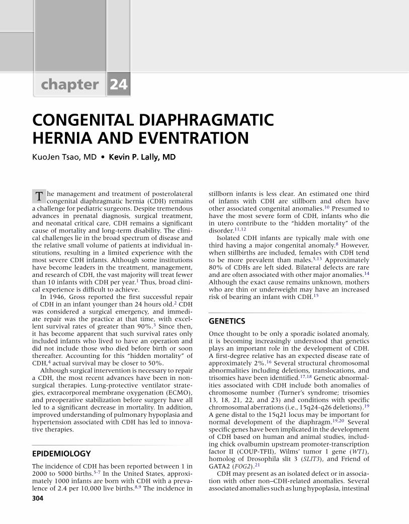
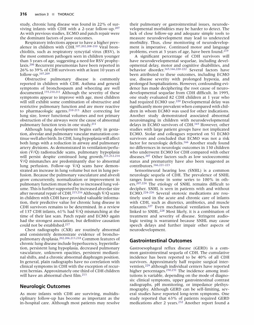
Diaphragm DevelopmentThe embryologic development of the human dia-phragm is a combination of complex, multicellular, multiple-tissue interactions that are poorly understood. Precursors to the diaphragm begin to form during the fourth week of gestation. Historically, the diaphragm has been thought to develop from the fusion of four embryonic components: anteriorly by the septum transversum, dorsolaterally by the pleuroperitoneal folds, dorsally by the crura of the esophageal mesen-tery, and posteriorly by the body wall mesoderm (Fig. 24-1).37,38 As the embryo begins to develop, the sep-tum transversum migrates dorsally and separates the pleuropericardial cavity from the peritoneal cavity. At this point there is still a communication between the pleural and peritoneal cavities but the pleural and pericardial cavities are separated. The septum trans-versum interacts with the pleuropericardial folds and mesodermal tissue surrounding the developing esoph-agus and other foregut structures, resulting in the for-mation of a primitive diaphragmatic structure known as the pleuroperitoneal fold (PPF). Bound by peri-cardial, pleural, and peritoneal folds, the paired PPFs separate the pleuropericardial and peritoneal cavities. Eventually, the septum transversum develops into the central tendon.37-41 Phrenic axons and myogenic cells destined for neuromuscularization migrate to the PPF and form the mature diaphragm.42
However, the exact mechanism in which the dia-phragm becomes muscularized remains poorly under-stood. As the PPF develops during the sixth week of gestation, the pleuroperitoneal membranes close concurrently and separate the pleural and abdominal cavities by the eighth week of gestation. Typically, the right side completes closure before the left. Histori-cally, the primitive fetal diaphragm has been thought to become muscularized by the inner thoracic mus-culature as the diaphragm closes.43 Some have sug-gested that the posthepatic mesenchymal tissue
sect�on 3 n THORACIC306
interacting with the PPF contributes to the diaphrag-matic musculature development.44 Others implicate a progressive development of the pleuroperitoneal membrane.45
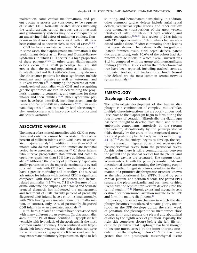
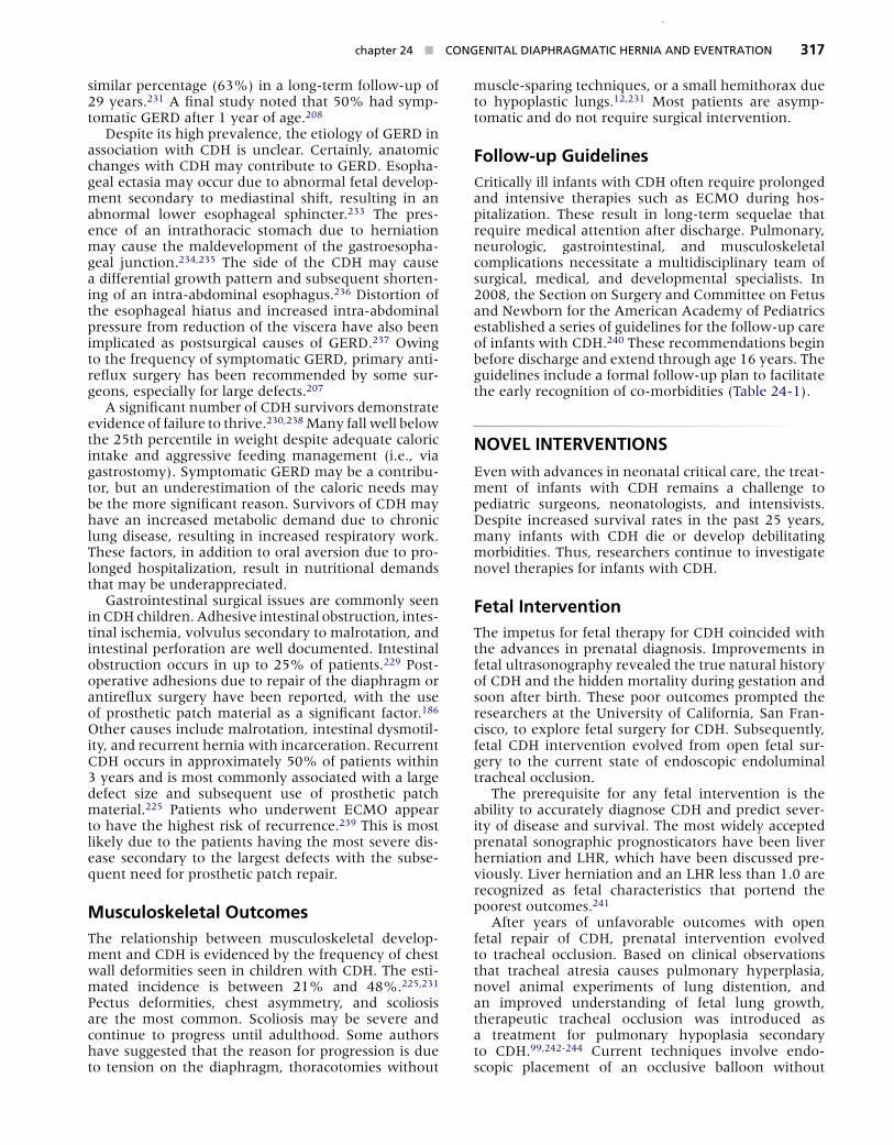
An improved understanding of diaphragmatic mus-cularization has supported an alternative hypothesis for its embryogenesis. Inactivation of c-met, a tyro-sine kinase receptor in mice, has demonstrated that the musculature of the diaphragm has a distinct origin other than the thoracic body wall.46 C-met encodes for a receptor protein that is responsible for the delamina-tion and migration of muscle cell precursors (MPCs) from somites.47 The body wall theory of diaphragm muscularization is challenged by c-met null mutant mice that have an amuscular diaphragm and a nor-mal body wall musculature. This hypothesis is further supported by the expression of Lbx1 on diaphragmatic MPCs, a transcription factor only found on migrating MPCs and not seen on body wall myocytes.48 Further-more, immunologic staining of the diaphragm muscu-lature revealed no MPCs from the body wall.42 Thus, it appears that during the process of embryologic fold-ing, a primitive diaphragmatic structure is formed, the PPF. Subsequently, the phrenic nerve and MPCs migrate from somatic origins, populate, and muscu-larize the newly formed PPF (Fig. 24-2). This process corresponds to week 10 of human gestation.
Lung DevelopmentFetal lung development is divided into five overlap-ping stages49,50: 1. The embryonic stage of lung development begins
during the 3rd week of gestation as a diverticular formation of the caudal end of the laryngotra-cheal groove. The primary lung buds and trachea
form from this diverticulum by the 4th week, and lobar structures are seen by the 6th week.
2. The pseudoglandular stage occurs between the 5th and 17th weeks of gestation with the formation of formal lung buds as well as the main and ter-minal bronchi.
Pleural canals
Foregut
Sinusvenosus
Septumtransversum
Livercords
Vitellineduct
Allantois
Cloaca
Body wall
Muscular ingrowthfrom body wall Septum transversum
Inferiorvena cava
Pleuro-peritoneal
membrane
Esophagus
Mesentery ofesophagus
Aorta
Figure 24-1. H�stor�ca�����, the d�aphragm has been thought to de�e�op from fus�on of �ts four embr���o�og�c components������ Accord�ng to th�s theor���, the septum trans�ersum fuses poster�or���� w�th the med�ast�na� mesench���me������ The p�europer�tonea� cana�s (white arrow) a�-�ow free commun�cat�on between the p�eura� and per�tonea� ca��t�es������ C�osure of these cana�s �s comp�eted as the p�europer�tonea� mem-branes de�e�op������ The four embr���o�og�c components of the de�e�op�ng d�aphragm are shown �n cross sect�on������ (From Skandalakis IJ, Colborn GL, Skandalakis JE: In Nyhus LM, Baker RJ, Fischer JE [eds]: Mastery of Surgery, 3rd ed. Boston, Little, Brown, 1996.)
*
1
2
3 4
1. Muscle precursor cells delaminate from the lateral dermo- myotomal lip. 2. Delaminated cells migrate through the lateral body wall.3. MPCs arrive in the PPF, from which point they spread out to populate the growing diaphragm.4. In CDH, the dorsolateral region of the PPF is missing (*) and MPCs are concentrated in the remaining tissue, ultimately leading to thickening of the diaphragm around the defect.
Figure 24-2. Th�s schemat�c dep�cts a d�fferent embr���o�og�c pathwa��� for d�aphragmat�c de�e�opment and CDH format�on than seen �n F�gure 24-1������ On the �eft s�de (1-3) �s the proposed norma� pathwa��� for d�aphragm de�e�opment������ On the r�ght s�de (4) �s the pathwa��� for CDH format�on������ M�C, musc�e precursor ce��s; ��F, p�europer�tonea� fo�d������ (From Clugston RD, Greer JJ: Dia-phragm development and congenital diaphragmatic hernia. Semin Pe-diatr Surg 16:94-100, 2007.)
chapter 24 n CONGENITA� DIA�HRAGMATIC HERNIA AND EVENTRATION 307
3. The canalicular stage is highlighted by the devel-opment of pulmonary vessels, respiratory bron-chioles, and alveolar ducts between weeks 16 and 25 of gestation. Type 1 pneumocytes begin to appear as well as the precursors for type 2 pneumocytes. At this stage, functional gas ex-change is possible.
4. The saccular stage continues from 24 weeks’ ges-tation to term with the maturation of alveolar sacs. Airway dimensions and surfactant synthe-sis capabilities continue to mature as well.
5. The alveolar stage begins right after birth with the appearance of mature alveoli. This stage extends well after birth with continued increase and de-velopment of functional alveoli.
Fetal pulmonary vascular development occurs in concordance with the associated lung development and follows the pattern of airway and alveolar matu-ration. A functional unit known as the acinus consists of the alveoli, alveolar ducts, and respiratory bron-chioli. The pulmonary vasculature develops as these units multiply and evolve during the canalicular stage. The preacinar structure consists of the trachea, major bronchi, lobar bronchi, and terminal bronchioles. The pulmonary vascular development for the preacinus is typically completed by the end of the pseudoglandular stage.51-53 In theory, any impedance to normal pul-monary development will concurrently hinder proper pulmonary vascular development.
Pulmonary development is recognized as a complex series of interlocking programmed events regulated by genetic signals, transcription factors, growth factors, and hormones. These events regulate the temporal and spatial interactions between epithelium and endothe-lium. Early transcription signals, such as thyroid tran-scription factor-1 and hepatocyte nuclear factor-3β, regulate pulmonary development from the primitive foregut mesenchyme. Other pathways of pulmonary development include Sonic hedgehog, transforming growth factor-β, Notch-delta pathway, and Wingless-Int.21,54 In addition, glucocorticoids, thyroid hormone, and retinoic acid have all been shown to regulate pul-monary organogenesis.55
PATHOGENESIS
The development of CDH has been attributed to a defective fusion of the pleuroperitoneal membrane, to improper development of the PPF, or to a consequence of abnormal development of the posthepatic mesen-chymal plate.37,38,56 Others suggest that the muscular defect in CDH occurs before the complete closure of the canal as a result of abnormal formation of the dia-phragm or PPFs.39
Traditionally, herniation of abdominal viscera through the diaphragmatic hernia was thought to impair lung development secondary to direct pulmo-nary compression resulting in pulmonary hypoplasia. Other evidence suggests that lung hypoplasia may occur early during embryogenesis before visceral her-niation.57-59 Still others have postulated a corollary
theory that primary lung maldevelopment leads to the fetal diaphragmatic defect, although this is not widely accepted.37 This latter theory has been refuted by the transgenic mice with the Fgf10 gene.60 Inactivated Fgf10 null-mutant mice do not develop lung tissue but have normal diaphragms suggesting independent pathways to diaphragm and lung development. This phenomenon is also seen in humans where infants with lung agenesis have normal diaphragms.61,62
Pulmonary hypoplasia is characterized by a decrease in bronchial divisions, bronchioles, and alveoli. The alveoli and terminal saccules exhibit abnormal septa-tions that impair the air-capillary interface and its ability for gas exchange.57 At birth, the alveoli are thick walled with intra-alveolar septations. These immature alveoli have increased glycogen content leading to thickened secretions that further limit gas exchange. Animal mod-els of CDH have demonstrated pulmonary hypoplasia with decreased levels of total lung DNA and total lung protein. In addition, the pulmonary vasculature appears to be less compliant with abnormally thick-walled arte-rioles.63 Surfactant levels are also decreased that may result in immature functioning lungs.64-66 Abnormali-ties in pulmonary development are not limited to the ipsilateral lung of the CDH. The contralateral lung also exhibits structural abnormalities of pulmonary hypo-plasia. These sequelae are most profound in cases in which mediastinal shift leads to contralateral lung com-pression. This supports the theory that lung compres-sion may be the major cause of pulmonary hypoplasia.
The understanding of abnormal pulmonary devel-opment in CDH has been widely aided by the nitrofen murine model.67 Nitrofen (2,4-dichloro-phenyl- p-nitrophenyl ether) is an environmental teratogen that is relatively harmless to adult mice. However, if given during pregnancy, it can cause pulmonary, car-diac, skeletal, and diaphragmatic abnormalities.68,69 Diaphragmatic defects resulting from the administra-tion of nitrofen in mice are very similar to those seen in humans with regard to size, location, and herniation of abdominal viscera.33,70,71 Depending on the time of exposure during gestation, resultant pups will develop either right or left CDH. In addition, the offspring will exhibit features of pulmonary hypoplasia, including reduced airway branching, surfactant deficiency, pul-monary vascular abnormalities, and respiratory fail-ure at birth. These mutations are the result of many alterations in developmental pathways of embryonic mice.55,72 The nitrofen model of CDH also suggests that not all of the pulmonary hypoplasia in CDH is due to lung compression. The effect of nitrofen in causing CDH is consistent with many theories that congenital anomalies are the result of environmental insults.
Other structurally similar teratogens to nitro-fen have been shown to induce CDH in animal models. Bisdiamine (N,N’-bis [dichloroacetyl]-l,8- octamethylenediamine) is a spermatogenesis inhibi-tor. BPCA (4-biphenyl carboxylic acid) is a breakdown product of a thromboxane A2 receptor antagonist. SB-210661 is a benzofuranyl urea derivative and 5-lipoxygenase inhibitor. All these agents cause similar diaphragmatic defects during the same embryologic
sect�on 3 n THORACIC308
periods in rodents. Although the exact etiology of CDH is unknown, all these compounds commonly affect the retinoic acid synthesis pathway by inhibit-ing retinol dehydrogenase-2 (RALDH2).73 The direct correlation between nitrofen and the retinoic acid system is supported by studies using transgenic mice with a LacZ reporter linked to a retinoic acid response element (RARE). The expression of RARE is reduced with the administration of nitrofen.74
Although the etiology is not fully understood, an alternative hypothesis to traditional CDH formation has been suggested by the nitrofen model. The primary defect appears to affect only the development of theonly the development of thethe development of the PPF.39 In nitrofen-exposed fetuses, a defect is clearly seen in the posterolateral portions of the PPF.75 This is further supported by observations of posterolateral-type CDH in vitamin A–deficient rats and mice with an inactivated Wt1 gene.75 In addition, nitrofen exposure does appear to affect muscularization of the PPF.76 Studies in MPC migration and myoblast proliferation and differentiation suggest that muscularization con-tinues and a thickened diaphragm is formed in the setting of an abnormal PPF (see Fig. 24-2).42,76 This concept of abnormal PPF formation with intact mus-cularization as the primary etiology of CDH is in con-trast to other theories that CDH is a result of failed closure of the pleuroperitoneal membranes or a defect in diaphragmatic muscularization.
Several clinical observations and molecular stud-ies have supported the importance of the retinoic acid pathways in the development of CDH. Vitamin A–deficient rodents will produce offspring with CDH of variable severity.77 Retinoic acid receptor knockout mice produce fetuses with CDH.78 Lower plasma levels of retinoic acid and retinol binding protein have been found in infants with CDH compared with controls.74 The conversion of retinoic acid to retinaldehyde has been prevented by inhibiting RALDH2 with nitrofen with the development of posterolateral diaphragmatic defects in rats.73
DIAGNOSIS
Prenatal DiagnosisOwing to the wide discrepancy of disease severity and potential fetal therapies, accurate and timely prenatal diagnosis of CDH has become paramount. The diagno-sis of CDH can be made by ultrasonography in 50% to 60% of pregnancies (Fig. 24-3).44 Typically diagnosed at 24 weeks’ gestation, CDH has been reported as early as 11 weeks’ gestation.79 In some tertiary medical cen-ters, up to 93% of the neonates with CDH may have a prenatal diagnosis.80 Fetal ultrasound findings include polyhydramnios, bowel loops within the chest, an echogenic chest mass, and an absent or intrathoracic gastric bubble. Severe or advanced CDH may exhibit intrathoracic “liver up,” mediastinal shift, and hydrops fetalis.81 Although most CDH can be detected during the second trimester, some sonographic features may not be exhibited until later in pregnancy.82
Independent sonographic features of CDH have not been shown to be accurate prognosticators for poor prognosis. However, two distinct features in combi-nation have been utilized to risk stratify: (1) a low lung-to-head ratio (LHR) and (2) liver herniation into the chest. The LHR is calculated by the cross-sectional area of the contralateral lung at the level of the cardiac atria divided by the head circumference (see Fig. 24-2). Survival of CDH fetuses based on LHR has been sta-tistically supported. In one series, there was 100% survival with LHR greater than 1.35, 61% with LHR between 1.35 and 0.6, and no survival with LHR less than 0.6.83 Survival based on liver herniation alone is 56%, compared with 100% survival without liver herniation.84 The combination of liver herniation and low LHR (LHR < 1.0) has a demonstrated 60% mor-tality for infants with prenatally diagnosed CDH.85-87 Although fetal ultrasonography has been adopted as the most reliable prognostic test for CDH, its wide and inconsistent utilization has created variable results among centers. An LHR of 1.0 in one institution may be consistently different than the same results in another institution. Subsequently, LHR and liver her-niation as prognosticators should be approached with some caution.88
Recently, fetal magnetic resonance imaging (MRI) of infants with CDH has been introduced as an adjunc-tive tool to evaluate fetal lung volume and liver loca-tion (Fig. 24-4). MRI-based fetal lung volume has been utilized in a manner similar to fetal ultrasonography. Several studies have touted the predictive value of fetal MRI in assessing risk. Lung volumes of CDH are calculated and compared with the predicted fetal lung volumes as a ratio. When the measured-to-expected fetal lung volumes ratio is less than 25%, there is a significant decrease in postnatal survival.89 Fetal lung volumes predicting the need for ECMO or degree of severity are less precise.90,91 Because of the nonstan-dardized approach to postnatal care for infants with CDH, conclusions regarding predictive outcomes using
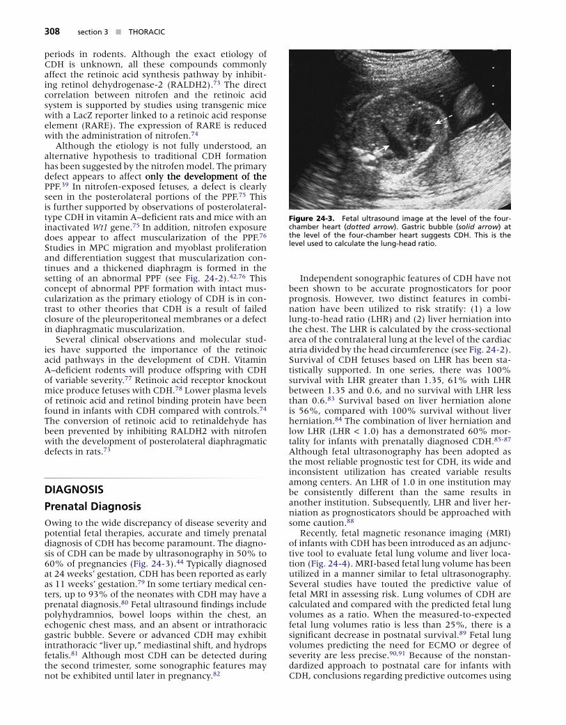
Figure 24-3. Feta� u�trasound �mage at the �e�e� of the four-chamber heart (dotted arrow)������ Gastr�c bubb�e (solid arrow) at the �e�e� of the four-chamber heart suggests CDH������ Th�s �s the �e�e� used to ca�cu�ate the �ung-head rat�o������
chapter 24 n CONGENITA� DIA�HRAGMATIC HERNIA AND EVENTRATION 309
all prenatal imaging modalities should be used with caution. Nonetheless, prenatal assessment can clearly identify the most severe cases.
Prenatal diagnosis in early pregnancy provides an opportunity to optimize the prenatal and postnatal care of the fetus and mother by providing accurate prenatal counseling, discussion of possible fetal inter-vention, and/or termination of pregnancy. Although prenatal imaging provides valuable information, it
does not consider the influences of postnatal care, especially in tertiary medical centers, on survival or overall outcome.
Clinical PresentationNewborns with CDH typically present with respiratory distress. Clinical scenarios may range from immediate respiratory distress with associated low Apgar scores to an initial stable period and a delay in respiratory dis-tress for 24 to 48 hours to late presentations that may occur years into life. Initial signs include tachypnea, chest wall retractions, grunting, cyanosis, and pallor. There may be clinical signs of ongoing fetal circulation and subsequent shunting. On physical examination, infants will often have a scaphoid abdomen and an increased chest diameter. The point of maximal car-diac impulse is often displaced (contralateral to the diaphragmatic defect), suggesting mediastinal shift. Bowel sounds may be auscultated within the chest cavity with a decrease in breath sounds bilaterally. Chest excursion may be low, suggesting a lower tidal volume.
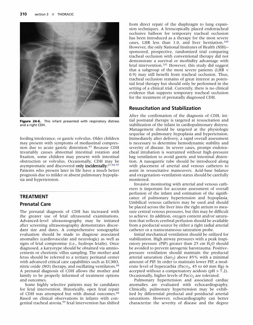
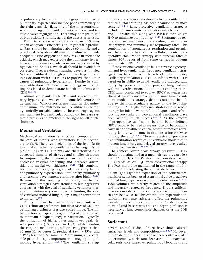
The diagnosis of CDH is typically made by chest radiography demonstrating intestinal loops within the thorax (Fig. 24-5). The location of the gastric bubble should be determined with the placement of an oro-gastric tube. In rare circumstances, a contrast radio-graph is necessary. The abdominal cavity may have minimal to no intestinal gas. Right-sided CDH may be more difficult to diagnose (Fig. 24-6). Salient fea-tures such as intestinal and gastric herniation may not occur. Rather, features of lobar compression may be the only radiographic sign and may be confused with congenital cystic adenomatoid malformations, pulmo-nary sequestrations, bronchopulmonary cysts, neuro-genic cysts, or cystic teratomas.
Although most infants with CDH will be diagnosed within the first 24 hours, as many as 20% may present beyond the neonatal period.92 These infants usually present with milder respiratory symptoms, chronic pulmonary infections, pleural effusions, pneumonias,
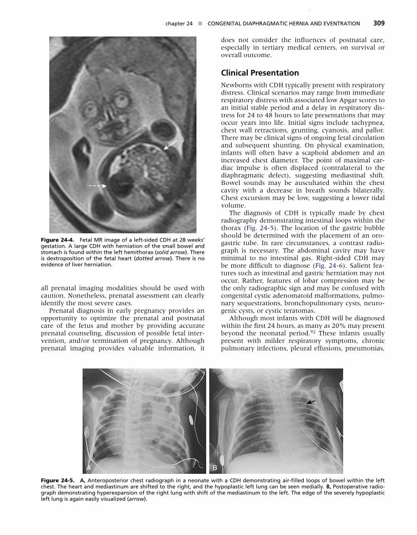
Figure 24-4. Feta� MR �mage of a �eft-s�ded CDH at 28 weeks’ gestat�on������ A �arge CDH w�th hern�at�on of the sma�� bowe� and stomach �s found w�th�n the �eft hem�thorax (solid arrow)������ There �s dextropos�t�on of the feta� heart (dotted arrow)������ There �s no e��dence of ���er hern�at�on������
A B
Figure 24-5. A, Anteroposter�or chest rad�ograph �n a neonate w�th a CDH demonstrat�ng a�r-fi��ed �oops of bowe� w�th�n the �eft chest������ The heart and med�ast�num are sh�fted to the r�ght, and the h���pop�ast�c �eft �ung can be seen med�a����������� B, �ostoperat��e rad�o-graph demonstrat�ng h���perexpans�on of the r�ght �ung w�th sh�ft of the med�ast�num to the �eft������ The edge of the se�ere���� h���pop�ast�c �eft �ung �s aga�n eas����� ��sua��zed (arrow)������
sect�on 3 n THORACIC310
feeding intolerance, or gastric volvulus. Older children may present with symptoms of mediastinal compres-sion due to acute gastric distention.93 Because CDH invariably causes abnormal intestinal rotation and fixation, some children may present with intestinal obstruction or volvulus. Occasionally, CDH may be asymptomatic and discovered only incidentally.only incidentally. incidentally.67,94-97 Patients who present later in life have a much better prognosis due to milder or absent pulmonary hypopla-sia and hypertension.
TREATMENT
Prenatal CareThe prenatal diagnosis of CDH has increased with the greater use of fetal ultrasound examinations. Advanced-level ultrasonography may be initiated after screening ultrasonography demonstrates discor-dant size and dates. A comprehensive sonographic evaluation should be made to diagnose associated anomalies (cardiovascular and neurologic) as well as signs of fetal compromise (i.e., hydrops fetalis). Once diagnosed, a karyotype should be obtained via amnio-centesis or chorionic villus sampling. The mother and fetus should be referred to a tertiary perinatal center with advanced critical care capabilities such as ECMO, nitric oxide (NO) therapy, and oscillating ventilators.98 A prenatal diagnosis of CDH allows the mother and family to be properly informed of treatment options and outcomes.
Some highly selective patients may be candidates for fetal intervention. Historically, open fetal repair of CDH was attempted but with dismal outcomes.84 Based on clinical observations in infants with con-genital tracheal atresia,99 fetal intervention has shifted
from direct repair of the diaphragm to lung expan-sion techniques. A fetoscopically placed endotracheal occlusive balloon for temporary tracheal occlusion has been introduced as a therapy for the most severe cases, LHR less than 1.0, and liver herniation.100 However, the only National Institutes of Health (NIH)– sponsored, prospective, randomized trial comparing tracheal occlusion with conventional therapy did not demonstrate a survival or morbidity advantage with fetal intervention.101 However, this study did suggest that a subgroup of the most severe patients (LHR < 0.9) may still benefit from tracheal occlusion. Thus, tracheal occlusion remains of great interest as poten-tial fetal therapy but should only be performed in the setting of a clinical trial. Currently, there is no clinical evidence that supports temporary tracheal occlusion for the treatment of prenatally diagnosed CDH.
Resuscitation and StabilizationAfter the confirmation of the diagnosis of CDH, ini-tial postnatal therapy is targeted at resuscitation and stabilization of the infant in cardiopulmonary distress. Management should be targeted at the physiologic sequelae of pulmonary hypoplasia and hypertension. Immediately after delivery, a rapid overall assessment is necessary to determine hemodynamic stability and severity of disease. In severe cases, prompt endotra-cheal intubation is warranted without high-pressure bag ventilation to avoid gastric and intestinal disten-tion. A nasogastric tube should be introduced along with placement of arterial and venous catheters to assist in resuscitative maneuvers. Acid-base balance and oxygenation-ventilation status should be carefully monitored.
Invasive monitoring with arterial and venous cath-eters is important for accurate assessment of overall perfusion of the infant and estimation of the signifi-cance of pulmonary hypertension and hypoplasia. Umbilical venous catheters may be used and should be placed across the liver into the right atrium to mea-sure central venous pressures, but this may be difficult to achieve. In addition, oxygen content and/or satura-tion that reflects cerebral perfusion should be available from a preductal source by either a right radial arterial catheter or a transcutaneous saturation probe.
Initial mechanical ventilation should be utilized for stabilization. High airway pressures with a peak inspi-ratory pressure (PIP) greater than 25 cm H2O should be avoided to prevent iatrogenic barotrauma. Positive-pressure ventilation should maintain the preductal arterial saturation (Sao2) above 85% with a minimal amount of PIP. In order to maintain lower PIP, a mod-erate level of hypercarbia (Paco2, 45 to 60 mm Hg) is accepted without a compensatory acidosis (pH > 7.2). Occasionally, higher levels of Paco2 are tolerated.
Pulmonary hypertension and associated cardiac anomalies are evaluated with echocardiography. Clinically, pulmonary hypertension may be exhib-ited by differential preductal and postductal arterial saturations. However, echocardiography can better characterize the severity of disease and the degree
Figure 24-6. Th�s �nfant presented w�th resp�rator��� d�stress and a r�ght CDH������
chapter 24 n CONGENITA� DIA�HRAGMATIC HERNIA AND EVENTRATION 311
of pulmonary hypertension. Sonographic findings of pulmonary hypertension include poor contractility of the right ventricle, flattening of the interventricular septum, enlarged right-sided heart chambers, and tri-cuspid valve regurgitation. There may be right-to-left or bidirectional shunting across the ductus arteriosus.
Preductal oxygen saturations less than 85% may impair adequate tissue perfusion. In general, a preduc-tal Pao2 should be maintained above 60 mm Hg and a postductal Pao2 above 40 mm Hg. Failure to provide adequate tissue oxygenation may result in metabolic acidosis, which may exacerbate the pulmonary hyper-tension. Pulmonary vascular resistance is increased by hypoxia and acidosis, which all should be avoided or corrected. If severe ductal shunting develops, inhaled NO can be utilized, although pulmonary hypertension in association with CDH is less responsive than other causes of pulmonary hypertension. Despite its com-mon utilization, NO as a rescue strategy in this set-ting has failed to demonstrate benefit in infants with CDH.102,103
Almost all infants with CDH and severe pulmo-nary hypertension will exhibit some left ventricular dysfunction. Vasopressor agents such as dopamine, dobutamine, and milrinone may be utilized in hemo-dynamically unstable patients. These inotropic agents may augment left ventricular output and increase sys-temic pressures to ameliorate the right-to-left ductal shunting.
Mechanical VentilationMechanical ventilation is a critical component in the care of infants with respiratory failure second-ary to CDH. The physiologic limits of the hypoplastic lung make mechanical ventilation a challenge. Hypo-plastic lungs in CDH infants are characterized by a decreased number of airways and smaller airspaces. In conjunction, the pulmonary vasculature exhibits decreased vascular branching and increased adven-titial and medial wall thickness.104,105 This combina-tion results in varying degrees of respiratory failure and pulmonary hypertension. Fortunately, pulmonary and vascular development continues after birth.106,107 Because of this ongoing maturation, mechanical ventilation strategies have trended to less aggressive approaches with the goal of stabilizing ventilator ther-apy to maintain oxygenation while limiting the risks of ventilator-induced lung injury, a major contributor to mortality.108
The type of mechanical ventilator in infants with CDH is clinician preference, but most cases of CDH can be managed using a pressure-cycled mode. The ini-tial fraction of inspired oxygen (Fio2) of 1.0 is utilized to maintain adequate oxygen saturation. Typically, the utilization of higher rates and lower peak air-way pressures (18 to 22 cm H2O) while titrating the Fio2 can maintain a preductal Pao2 greater than 60 mm Hg or better (a preductal Sao2 > 85%) and a Pco2 less than 60 mm Hg. Maintaining an accept-able pH and Pco2 is important in managing the pul-monary hypertension.109,110 The ventilation strategy
of induced respiratory alkalosis by hyperventilation to reduce ductal shunting has been abandoned by most centers.111-114 Lung-protective ventilation strategies include pressure-limited ventilation rates between 30 and 60 breaths/min along with PIP less than 25 cm H2O to minimize barotrauma.111,115 Spontaneous res-pirations are maintained by avoiding neuromuscu-lar paralysis and minimally set respiratory rates. This combination of spontaneous respiration and permis-sive hypercapnia has been a well-documented pre-operative stabilization strategy with survival rates of almost 90% reported from some centers in patients with isolated CDH.111-113
If conventional ventilation fails to reverse hypercap-nia and hypoxemia, high-frequency ventilation strat-egies may be employed. The role of high-frequency oscillatory ventilation (HFOV) in infants with CDH is based on its ability to avoid ventilatory-induced lung injury by preserving end-expiratory lung volume without overdistention. As the understanding of the CDH lungs continued to evolve, HFOV strategies also changed. Initially used in a high-pressure lung recruit-ment mode, this strategy demonstrated no benefit due to the nonrecruitable nature of the hypoplas-tic lungs.116,117 High-frequency strategies as a rescue therapy for infants with profound hypoxia and refrac-tory hypercapnia on conventional ventilators have been without much success.118,119 As the concept of preoperative stabilization became better defined, HFOV began to be used as means to avoid barotrauma early in the treatment course before refractory respi-ratory failure, with some institutions using HFOV as primary therapy.120-122 These strategies of preopera-tive stabilization regardless of ventilator modalities to prevent lung injury and delayed surgery have resulted in improved survival.108,120-123
To achieve lower peak airway pressures, HFOV should be maintained at a mean airway pressure less than 16 cm H2O. HFOV should be considered when PIP exceeds 25 cm H2O with conventional therapy. The Pco2 should be maintained in the range of 40 to 55 mm Hg by adjusting the amplitude between 35 to 45 cm H2O. Eight rib expansion of the contralateral hemithorax has been used as an initial guide to achieve optimal lung expansion without overdistention.120,122 Tidal volumes are directly related to the amplitude and inversely related to frequency. Thus, significant increases in tidal volume can be seen when frequen-cies are below 10 Hz. This can result in hyperinflation, which in turn may adversely affect the pulmonary vasculature, including venous return. Constant assess-ment of acid-base status and end-organ perfusion is necessary as lung compliance changes, or as the CDH is repaired.
SurfactantSeveral animal studies of CDH have shown altered surfactant levels and composition.63,124-129 However, human data to support this have been controversial.65 Experimentally, surfactant decreases pulmonary vas-cular resistance, improves pulmonary blood flow, and
sect�on 3 n THORACIC312
reduces ductal shunting.130 In addition, surfactant administration has been shown to augment NO deliv-ery and improve gas exchange.131 These findings led clinicians to utilize exogenous surfactant empirically. However, clinical evidence in term and preterm infants with CDH has not shown benefit. In one study, it was suggested that surfactant therapy in term infants with CDH was associated with an increased use of ECMO, an increased incidence of chronic lung disease, and higher mortality.128 Similar findings were found in preterm infants despite adjusting for Apgar scores and gestational age.127 Even as a rescue therapy in CDH infants on ECMO, surfactant failed to demonstrate a survival advantage.64,126 Despite the lack of proven efficacy, exogenous surfactant therapy continues to be used with unknown risk and unclear benefit. Propo-nents of this therapy argue that the clinical evidence is based on a heterogeneous population of disease sever-ity and that failed therapy may be due to its use in the most severe CDH infants. Given the clinical data, sur-factant therapy should only be used in a randomized, controlled fashion.
Nitric OxideInhaled NO is a potent pulmonary vasodilator that has been shown to have tremendous benefit in the treatment of persistent pulmonary hypertension of the neonate (PPHN).118,132-135 In clinical studies, NO improved oxygenation and decreased the need for ECMO in infants with respiratory failure secondary to PPHN.135,136 However, the efficacy of NO for pul-monary hypertension secondary to CDH has not been as well supported. The need for ECMO or a reduc-tion in mortality has not been shown with NO use in CDH infants.137 In the Neonatal Inhaled Nitric Oxide Study Group trial, the CDH subgroup had a greater likelihood for ECMO or death.102 The response to NO is variable and unpredictable in infants with CDH. Some may demonstrate a rebound pulmonary hypertension that is more difficult to control than the initial disease.138 Furthermore, the effect appears to be transient and does not prevent progression toward ECMO use.139,140 Thus, the role of inhaled NO has not been clearly defined and should not be routinely used. NO should be reserved for infants who persistently demonstrate suprasystemic pulmo-nary pressures in the setting of optimized pulmo-nary and cardiac function to reduce right ventricular pressures.115,140
Other pulmonary vasodilator agents have been utilized in the treatment of pulmonary hypertension in infants with CDH. Sildenafil is a 5-phosphodies-terase (5-PDE) inhibitor that has been utilized as a transitional medication to wean off NO for unresolv-ing pulmonary hypertension. Widely used for PPHN and pulmonary hypertension secondary to congenital heart disease, sildenafil can be used in both oral or intravenous forms.140,141 Epoprostenol (intravenous prostacyclin) and bosentan (a nonspecific endothelin-1 receptor inhibitor) have all been used in infants with CDH but the experience to date is limited.142-144
Extracorporeal Membrane OxygenationECMO for infants with CDH was first introduced as a potential therapy for infants who likely had severe ventilator-associated lung injury.145 Since then, ECMO strategies have evolved as understanding of CDH with its associated pulmonary hypoplasia and hyperten-sion has improved. CDH accounts for approximately one fourth of all infants requiring ECMO for respira-tory failure. Almost 30% of CDH infants will receive ECMO during their hospital course.146-149 Prior to the era of preoperative stabilization, ECMO was associated with only a modest improvement in survival in high-risk CDH infants.150-152
Initially, strict criteria were established as indications for ECMO use in CDH. These included an oxygenation index greater than 40 and persistent alveolar-arterial gradient greater than 610 mm Hg.148,153,154 Today, those criteria have been softened and the most com-mon indication for ECMO is a “failure to respond” to therapy. In efforts to maintain lung-protective ven-tilation, clinicians have opted for ECMO rather than escalation of positive airway pressures. Relative con-traindications include significant congenital anoma-lies, lethal chromosomal anomalies, intracranial hemorrhage, and gestational age less than 34 weeks. Ideally, ECMO candidates should have a birth weight greater than 2 kg owing to increased risk of intracra-nial hemorrhage, but some have reported benefit in infants weighing less than 2 kg.
ECMO was initially utilized as a postoperative res-cue therapy, but its role has evolved.155,156 In combi-nation with other adjunctive modalities such as HFOV, ECMO is now routinely used as a component of pre-operative stabilization. The strategy of stabilization with ECMO and delay in surgery has been shown to be beneficial, with a reported survival of 67% in high-risk patients.10,157 These findings were further sup-ported by the CDH study group that reported 85% of infants had ECMO before repair. Of those, 54% were repaired while still on ECMO and 29% repaired after ECMO with 16% never making it to repair. Infants who underwent repair while on ECMO had a 50% mortality, compared with 17% after ECMO.
The optimal timing of CDH repair is controversial and surgeon dependent. With more repairs of CDH on ECMO, bleeding became an increasingly recognized complication.157,158 The introduction of aminocaproic acid, a fibrinolysis inhibitor, has significantly decreased bleeding complications.147 Bleeding may also be reduced with early repair on ECMO before the develop-ment of coagulopathy and significant edema.148 Some surgeons have recommended repair when pulmonary hypertension has resolved but prior to decannula-tion.66 This allows reinstitution of ECMO if respiratory failure and/or pulmonary hypertension recurs. Others have requested that infants be decannulated and on conventional ventilation before repair. Although rare, recurrent pulmonary hypertension can occur after sur-gery, requiring a second run of ECMO.159 Survival for second-run ECMO patients is approximately 50%.160
chapter 24 n CONGENITA� DIA�HRAGMATIC HERNIA AND EVENTRATION 313
Today, ECMO is routinely used to stabilize patients before operation. Although results are dependent on patient selection and entry criteria, survival has been reported between 60% and 90%.98,111,146,161,162 With-out ECMO, the predicted mortality in the high-risk cohort reaches 80%.146 Despite these data, ECMO for CDH is not universally accepted. Some authors report no survival advantage with ECMO.163,164 Others cite an increased neurologic complication rate with ECMO use in CDH infants compared with other indications such as PPHN.165,166 Some institutions report an 80% sur-vival of infants with CDH without use of ECMO.167,168 Nonetheless, in most centers ECMO remains a compo-nent in the armamentarium for treating CDH infants with refractory cardiopulmonary failure.
SurgeryThe surgical approach to the repair of CDH has changed dramatically in the past 25 years. Historically, neonates with CDH were brought to the operating room almost immediately after birth to emergently relieve the com-pressed lung by reducing the intra-abdominal contents from the chest. Often, this maneuver was followed by a transient period of cardiopulmonary stability or improvement.169,170 However, frequently, the infant would develop progressive respiratory failure, elevated pulmonary vascular pressures, right-to-left ductal shunting, unrecoverable hypoxemia, and eventually death. With improved understanding of the pathophys-iology of CDH, repair of CDH is no longer considered a surgical emergency. Nonetheless, timing of the surgical repair of the diaphragm remains controversial.
The shift toward a delay in surgery after preopera-tive stabilization occurred after evidence was intro-duced that pulmonary compliance was actually worse after repair.171 Several factors have been implicated, including increased intra-abdominal pressure and dis-tortion of the repaired diaphragm. In addition, surgical repair has not been shown to improve gas exchange. Several studies have evaluated early versus delayed repair of CDH without demonstrating a difference in mortality or need for ECMO.172-174 These include two randomized trials of early (<12 hours) versus delayed repair after 24 hours174 and after 96 hours.172 Since the adoption of the delayed repair strategy, several cen-ters have reported improved survival rates.111,174-177 Although modifications of surgical techniques and timing have contributed to this improvement, con-comitant advances in neonatal critical care as well as a better understanding of the disease and outcomes have been the greater contributors to decreased mortality. Importantly, there has been no clinical evidence that specifically attributes a delay in surgery to improved outcomes over immediate repair.178
Timing of operation remains with clinical judgment and surgeon discretion. Some clinicians will opt for repair once the respiratory failure has stabilized (low peak airway pressures and inspired oxygen require-ments) and after echocardiography demonstrates a resolution or stability of the pulmonary hyperten-sion. This typically occurs 24 to 48 hours after clinical
evidence of ductal shunting has resolved. Although CDH can be repaired on HFOV, most surgeons will wait until the infant is transitioned to a conventional mechanical ventilator.
The repair of a CDH may be as variable as the clini-cal management. A subcostal incision on the ipsilat-eral side of the hernia is the traditional approach. Less than 10% of surgeons prefer a thoracic approach.179 The intra-abdominal contents should be reduced out of the thorax with careful attention to the spleen, which can be caught and torn on the rudimentary rim of diaphragm. A true hernia sac, which is only present less than 20% of the time, should be identified and excised.180 The thoracic and abdominal cavities should be inspected for associated pulmonary sequestrations. The extent of the diaphragmatic hernia should be examined and assessed.
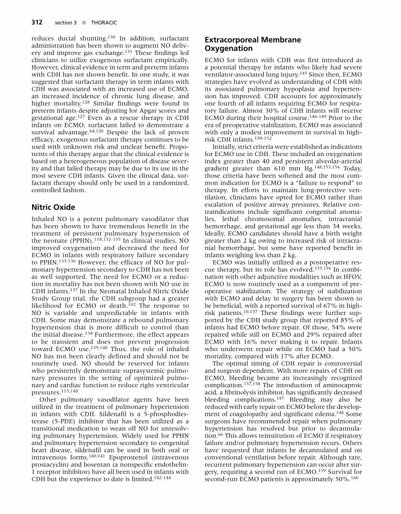
The type of repair is dependent on the size of the diaphragmatic defect. If the defect is small, a tension-free primary closure should be performed with nonab-sorbable sutures with or without pledgets. If the defect is large, attempted primary closure may cause signifi-cant tension or flattening of the diaphragm, resulting in decreased pulmonary compliance. Alternatively, a diaphragmatic replacement may be employed to restore a tension-free, diaphragmatic shape (Fig. 24-7). Several types of prosthetic and biosynthetic patch materials have been utilized. Traditionally, a polytet-rafluoroethylene (PTFE or Gore-Tex, W. L. Gore, New-ark, DE) patch has been used.179 With the use of a 1-mm thickness patch, the prosthesis is cut slightly larger than the diaphragmatic defect and is sewn to the residual diaphragm or surrounding tissue with nonabsorbable suture. The disadvantage of prosthetic material is the lack of material growth, resulting in a significant recurrence rate of nearly 50%.181 To pre-vent this, the patch is tailored in a “cone shape” that may resemble the native diaphragm. This technique has demonstrated a reduction in recurrence rate from 26% to 9%.182 Large defects with an adequate
Figure 24-7. A �arge �eft postero�atera� d�aphragmat�c hern�a was approached through a subcosta� �nc�s�on������ After reduct�on of the abdom�na� ��scera, the �arge defect was c�osed us�ng a b�o-s���nthet�c patch (A��oderm)������
sect�on 3 n THORACIC314
posterior rim of diaphragm may require suturing the patch to the abdominal wall or encircling the ribs for adequate fixation.
Outcomes regarding the type of prosthetic patch material are mixed. Opponents of prosthetic patch material, such as PTFE, cite an unacceptable recurrence rate, chest wall deformity, and intra-abdominal com-plications, such as small bowel obstruction.181,183-186 With a recurrence rate as high as 50%, alterna-tive biosynthetic materials have been introduced to repair CDH. Currently, the most popular material is small intestine submucosa (Surgisis or SIS, Cook, Inc., Bloomington, IN) or human acellular dermal matrix (Alloderm, LifeCell, Branchburg, NJ). These bioactive patches may serve as a matrix for permanent tissue ingrowth.187 Some institutions have adopted bioactive materials as the primary patch for repair of large dia-phragmatic hernias. Proponents of biosynthetic patches claim the patch material is less adherent to small bowel and allows for tissue engraftment that grows with the patient. However, the largest series comparing SIS to Gore-Tex demonstrated similar recurrence rates and no difference in small bowel obstruction.187 Long-term outcomes with biosynthetic patches for CDH remain to be seen. Certainly, the type of CDH patch material needs to be studied in a prospective fashion.
Recurrences after CDH repairs have prompted investigation into alternatives to prosthetic and biosyn-thetic materials. Some surgeons have utilized muscle flaps for primary and recurrence repairs. In small case series, an abdominal wall muscle flap using internal oblique and transversalis abdominal muscles has been described to cover very large primary or recurrent dia-phragmatic defects.188-191 Thoracic wall musculature has also been widely utilized. Latissimus dorsi and serratus anterior muscles have been described with isolated success.33,188,192 Local advancement with a reversed latissimus dorsi flap with microneural anasto-mosis of the thoracodorsal nerve to the phrenic nerve has been described for recurrent herniations.192 With this technique, the neodiaphragm was able to dem-onstrate nonparadoxical motion by fluoroscopy and ultrasonography. Muscle flaps may be advantageous in very large defects and in diaphragmatic agenesis.
Postoperative tube thoracostomy is not necessary and probably not indicated.111,114 An exception may be CDH repair on ECMO because bleeding compli-cations should be expected. The thoracic space will
eventually fill with air and fluid, the lung will gradu-ally grow, and the rib spaces may narrow to decrease the overall thoracic cavity. Tube thoracostomy is used only for postoperative chylothorax or accumulation of pleural fluid causing hemodynamic compromise.193 If a chest tube is warranted, the tube is placed in the thoracic cavity before final closure of the diaphragm. Chest tubes should be placed to water seal rather than suction. Aggressive negative pressure can shift the mediastinum to the ipsilateral hemithorax creating overdistention of the contralateral lung. Symptomatic pleural fluid may be treated with repeated thoracen-tesis. If used, chest tubes should be removed early to avoid infection.
If the CDH is repaired via laparotomy, the abdominal domain may not be able to accommodate the reduc-tion of the intestinal contents. Closure of the abdomi-nal wall may result in an abdominal compartment syndrome. A “tight abdomen” may cause respiratory compromise in an already tenuous infant. Careful attention should be paid to the peak airway pressures as the abdominal fascia is closed. Respiratory com-promise should prompt the abdomen to be left open. This scenario is more often seen with CDH infants on ECMO.194 Temporary closure can be achieved with skin, prosthetic silo, or abdominoplasty.195 Delayed closure, especially in those infants on ECMO, should be attempted after the general edema has resolved or the intra-abdominal domain has enlarged.196
Special operative considerations should be made for CDH infants on ECMO. In addition to the abdominal compartment issues, hemorrhagic complications are more likely due to the anticoagulation required for ECMO. Fibrin or thrombin sealants should be liberally used to reduce suture line hemorrhage. Aminocaproic acid should be given before operation and for 2 to 3 days postoperatively to prevent bleeding complications.
The therapeutic goals of postoperative ventila-tion should be similar to the preoperative goals, that is, to minimize barotrauma. Infants may experience a transient period of cardiopulmonary improvement that may allow clinicians to wean ventilatory support. However, rapid decreases in Fio2 should be avoided to prevent triggering pulmonary vasospasm and recur-rent pulmonary hypertension.
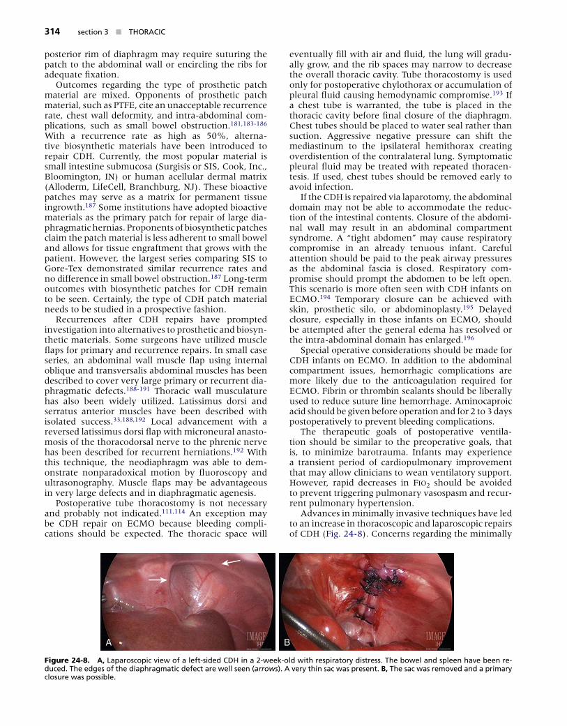
Advances in minimally invasive techniques have led to an increase in thoracoscopic and laparoscopic repairs of CDH (Fig. 24-8). Concerns regarding the minimally
A B
Figure 24-8. A, �aparoscop�c ��ew of a �eft-s�ded CDH �n a 2-week-o�d w�th resp�rator��� d�stress������ The bowe� and sp�een ha�e been re-duced������ The edges of the d�aphragmat�c defect are we�� seen (arrows)������ A �er��� th�n sac was present������ B, The sac was remo�ed and a pr�mar��� c�osure was poss�b�e������
chapter 24 n CONGENITA� DIA�HRAGMATIC HERNIA AND EVENTRATION 315
invasive approaches have centered around prolonged operations precipitating respiratory complications, trig-gering or exacerbating pulmonary hypertension, and resulting in an unnecessary increase in mortality. With careful patient selection, minimally invasive repairs have been successfully performed, both laparoscopi-cally and thoracoscopically.197-199 This approach has been utilized with primary repair as well as prosthetic patch closure of the CDH.200 Laparoscopic and tho-racoscopic operations are feasible without a demon-strable advantage to either. However, patient selection remains the main determinant of success. A preopera-tive patient selection criteria for thoracoscopic repair of CDH has been proposed.199 Preoperative requirements included minimal ventilatory support (PIP < 24 mm Hg), no clinical or sonographic evidence of pulmonary hypertension, and an intra-abdominal stomach. In a small series of seven patients, results were similar to open CDH repairs. The long-term outcomes with mini-mally invasive approaches to CDH remain to be seen.
OUTCOMES
Clinical outcomes in regard to the treatment and man-agement of infants with CDH have traditionally been difficult to interpret. Because the estimated incidence of CDH is low (1 in 2500 to 4000 live births), it has become evident that single institutions, regardless of their volume, would have difficulty in generating meaningful, evidence-based outcomes.9,10 In addition, patient characteristics and clinical practices are very different and each institution’s ability to offer state-of-the-art neonatal critical care is highly variable. Furthermore, data from a very heterogeneous disease population, such as CDH, are often pooled to generate single reports.
Congenital Diaphragmatic Hernia Registry (CDHR)The Congenital Diaphragmatic Hernia Study Group (CDHSG) was established in 1995 as an international group of clinicians and researchers interested in devel-oping a registry of all infants born with CDH. The CDHSG recognized the limitations of individual insti-tutions in producing meaningful data on their own. As an observational database, the CDHR was created by a network of voluntary centers to ask specific clini-cal questions, recommend specific therapy as needed, and monitor outcomes of various management and treatment strategies. Since 1995, more than 90 centers from 10 countries have submitted data on more than 4000 infants with CDH. Participation among centers has varied, but currently there are over 50 active cen-ters in the CDHSG with an average total submission of 340 infants per year.1
Data from the CDHR must be used with caution as with all outcome studies of CDH. There is a wide spectrum of patients within institutions and a wide spectrum of institutions within the CDHSG. Patients are only followed until hospital discharge or death.
Thus, long-term follow-up outcomes are not available. Nonetheless, large international registries such as the CDHR offer some advantages. The CDHR is a vehicle to collect data from a large number of patients. The CDHR reflects changes in management and outcomes over time. Finally, individual institutions can compare their risk-adjusted outcomes to other institutions as a method of quality assurance.
SurvivalAs with all outcomes studies regarding CDH, survival analysis remains difficult to interpret due to the tre-mendous variations in patient disease, management strategies, and operative techniques. Unique to individ-ual institutions are differences in ventilation strategies, availability and entry criteria for ECMO, and operative timing. One method to achieve better comparisons of outcomes is the development of risk stratification of patient disease. In 2001, the CDHSG published the first attempt at predicting outcome based on risk stratifica-tion.201 The study examined 1054 infants with CDH by applying a risk assessment tool created by a logistical equation. Patients were categorized into low-, moder-ate-, and high-risk groups, which correlated with actual mortality. Overall survival was 64% in which birth weight and 5-minute Apgar scores had the strongest correlation. Survival rates for CDH among institutions are highly variable even when risk adjusted. Although the overall survival of liveborn infants has been reported around 60%, individual centers have varied from 25% to 83%.9 This variation may be a reflection of the increasing prenatal diagnosis of CDH in which the most severe cases are transferred to tertiary care centers.
Pulmonary OutcomesFor many infants who survive the neonatal period with CDH, pulmonary function is excellent. Prior to the era of ECMO and lung-protective ventilation, long-term survivors were routinely reported as healthy children without respiratory disease.53,202-204 However, with improved survival, more severe CDH infants are avail-able for follow-up. Children with a history of CDH have demonstrated both functional and radiographic evidence of chronic lung disease.
Pulmonary function has been reported to be nor-mal in 50% to 70% of CDH survivors.53,202,205,206 The remainder exhibit some form of restrictive or obstruc-tive respiratory symptoms. Approximately 25% of children beyond the age of 5 years have demonstra-ble signs of obstructive airway disease. One hundred consecutive CDH survivors were recently evaluated. ECMO and the need for patch repair were independent predictors of pulmonary outcome.206 The use of ECMO significantly correlated with diuretic dependence at the time of discharge.81 While only 16% required oxy-gen at discharge, 53% required at least transient usage of bronchodilators within the first year. Similarly, 41% were either dependent on or intermittently needing corticosteroids during the first year. Others have dem-onstrated similar pulmonary morbidities. In another
sect�on 3 n THORACIC316
study, chronic lung disease was found in 22% of sur-viving infants with CDH with a 2-year follow-up.207 As with previous studies, ECMO and patch repair were the dominant factors of poor outcomes.
Respiratory infections appear to have a higher prev-alence in children with CDH.107,202,208-210 Viral bron-chiolitis, such as respiratory syncytial virus (RSV), is the most common pathogen seen in children younger than 3 years of age, suggesting a need for RSV prophy-laxis.206 Recurrent pneumonias have been reported in 26% to 39% of CDH survivors with at least 10 years of follow-up.107,209
Obstructive pulmonary disease is commonly reported in children with CDH. Asthma and general symptoms of bronchospasm and wheezing are well documented.155,210,211 Although the severity of these symptoms appear to improve with age, most children will still exhibit some combination of obstructive and restrictive pulmonary function and are more reactive to pharmacologic agents.210-213 After correcting for lung size, lower functional volumes and not primary obstruction of the airways were the cause of abnormal pulmonary function tests in one study.211
Although lung development begins early in gesta-tion, alveolar and pulmonary vascular maturation con-tinue well after birth. In CDH, lung hypoplasia will affect both lungs with a reduction in airway and pulmonary artery divisions. As demonstrated in ventilation/perfu-sion ( V/Q) radionuclide scans, pulmonary hypoplasia will persist despite continued lung growth.211,212,214 V/Q mismatches are predominantly due to abnormal lung perfusion. Follow-up V/Q scans have demon-strated an increase in lung volume but not in lung per-fusion. Because the pulmonary vasculature and alveoli grow concurrently, normalization or improvement in pulmonary function must be due to increased lung vol-ume. This is further supported by increased alveolar size after neonatal repair of CDH.215,216 Although V/Q scans in children with CDH have provided valuable informa-tion, their predictive value for chronic lung disease in CDH survivors remains to be determined. In a review of 137 CDH infants, 61% had V/Q mismatching at the time of their last scan. Patch repair and ECMO again had the strongest association, but definitive causation could not be established.217
Chest radiographs (CXR) are routinely abnormal and consistently demonstrate evidence of broncho-pulmonary dysplasia.202,206,213,218 Common features of chronic lung disease include hyperlucency, hyperinfla-tion, persistent lung hypoplasia, decreased pulmonary vasculature, unknown opacities, persistent mediasti-nal shifts, and a chronic abnormal diaphragm position. In general, plain radiographs have no correlation with clinical symptoms in CDH with the exception of recur-rent hernias. Approximately one third of CDH children will have an abnormal chest film.213
Neurologic OutcomesAs more infants with CDH are surviving, multidis-ciplinary follow-up has become as important as the in-hospital care. Although most patients may resolve
their pulmonary or gastrointestinal issues, neurode-velopmental morbidities may be harder to detect. The lack of close follow-up and adequate simple tools to measure neurodevelopment may lead to undetected morbidity. Thus, close monitoring of neurodevelop-ment is imperative. Continued motor and language problems, even at 3 years of age, have been found.219
A significant percentage of CDH survivors will have neurodevelopmental sequelae, including devel-opmental delay, motor and cognitive disabilities, and behavior disorder.165,166,220-222 Several factors have been attributed to these outcomes, including ECMO use, disease severity with prolonged hypoxia, and prolonged hospitalizations. However, confounding evi-dence has made deciphering the root cause of neuro-developmental sequelae from CDH difficult. In 1995, one study evaluated 82 CDH children at 1 year who had required ECMO use.220 Developmental delay was significantly more prevalent when compared with chil-dren in whom ECMO was used for other indications. Another study demonstrated associated abnormal neuroimaging in children with neurodevelopmental delays in ECMO survivors of CDH.165 However, other studies with large patient groups have not implicated ECMO. Stolar and colleagues reported on 51 ECMO survivors and concluded that ECMO was not a risk factor for neurologic deficits.166 Another study found no differences in neurologic outcomes in 130 children who underwent ECMO for CDH compared with other diseases.207 Other factors such as low socioeconomic status and prematurity have also been suggested as contributors.166,221-223
Sensorineural hearing loss (SNHL) is a common neurologic sequela of CDH. The prevalence of SNHL ranges from none in some series to 100% in oth-ers.207,224 The etiology of SNHL remains difficult to decipher. SNHL is seen in patients with and without ECMO.224-226 Several ototoxic medications are rou-tinely used in the acute and chronic care of infants with CDH, such as diuretics, antibiotics, and muscle relaxants.227 Even mechanical ventilation has been linked to SNHL.228 Most likely, it is a combination of treatment and severity of disease. Stringent audio-logic testing is warranted because SNHL may cause speech delays and further impair other aspects of neurodevelopment.
Gastrointestinal OutcomesGastroesophageal reflux disease (GERD) is a com-mon gastrointestinal sequela of CDH. The cumulative incidence has been reported to be 40% of all CDH survivors. Approximately half require surgical inter-vention,229 although individual centers have reported higher percentages.230,231 The incidence among insti-tutions is variable, depending on the mode of diagno-sis: clinical symptoms, upper gastrointestinal contrast radiographs, pH monitoring, or impedance plethys-mography. Although GERD can be self-limiting, sev-eral studies have reported long-term symptoms. One study reported that 63% of patients required GERD medications after 2 years.232 Another report found a
chapter 24 n CONGENITA� DIA�HRAGMATIC HERNIA AND EVENTRATION 317
similar percentage (63%) in a long-term follow-up of 29 years.231 A final study noted that 50% had symp-tomatic GERD after 1 year of age.208
Despite its high prevalence, the etiology of GERD in association with CDH is unclear. Certainly, anatomic changes with CDH may contribute to GERD. Esopha-geal ectasia may occur due to abnormal fetal develop-ment secondary to mediastinal shift, resulting in an abnormal lower esophageal sphincter.233 The pres-ence of an intrathoracic stomach due to herniation may cause the maldevelopment of the gastroesopha-geal junction.234,235 The side of the CDH may cause a differential growth pattern and subsequent shorten-ing of an intra-abdominal esophagus.236 Distortion of the esophageal hiatus and increased intra-abdominal pressure from reduction of the viscera have also been implicated as postsurgical causes of GERD.237 Owing to the frequency of symptomatic GERD, primary anti-reflux surgery has been recommended by some sur-geons, especially for large defects.207
A significant number of CDH survivors demonstrate evidence of failure to thrive.230,238 Many fall well below the 25th percentile in weight despite adequate caloric intake and aggressive feeding management (i.e., via gastrostomy). Symptomatic GERD may be a contribu-tor, but an underestimation of the caloric needs may be the more significant reason. Survivors of CDH may have an increased metabolic demand due to chronic lung disease, resulting in increased respiratory work. These factors, in addition to oral aversion due to pro-longed hospitalization, result in nutritional demands that may be underappreciated.
Gastrointestinal surgical issues are commonly seen in CDH children. Adhesive intestinal obstruction, intes-tinal ischemia, volvulus secondary to malrotation, and intestinal perforation are well documented. Intestinal obstruction occurs in up to 25% of patients.229 Post-operative adhesions due to repair of the diaphragm or antireflux surgery have been reported, with the use of prosthetic patch material as a significant factor.186 Other causes include malrotation, intestinal dysmotil-ity, and recurrent hernia with incarceration. Recurrent CDH occurs in approximately 50% of patients within 3 years and is most commonly associated with a large defect size and subsequent use of prosthetic patch material.225 Patients who underwent ECMO appear to have the highest risk of recurrence.239 This is most likely due to the patients having the most severe dis-ease secondary to the largest defects with the subse-quent need for prosthetic patch repair.
Musculoskeletal OutcomesThe relationship between musculoskeletal develop-ment and CDH is evidenced by the frequency of chest wall deformities seen in children with CDH. The esti-mated incidence is between 21% and 48%.225,231 Pectus deformities, chest asymmetry, and scoliosis are the most common. Scoliosis may be severe and continue to progress until adulthood. Some authors have suggested that the reason for progression is due to tension on the diaphragm, thoracotomies without
muscle-sparing techniques, or a small hemithorax due to hypoplastic lungs.12,231 Most patients are asymp-tomatic and do not require surgical intervention.
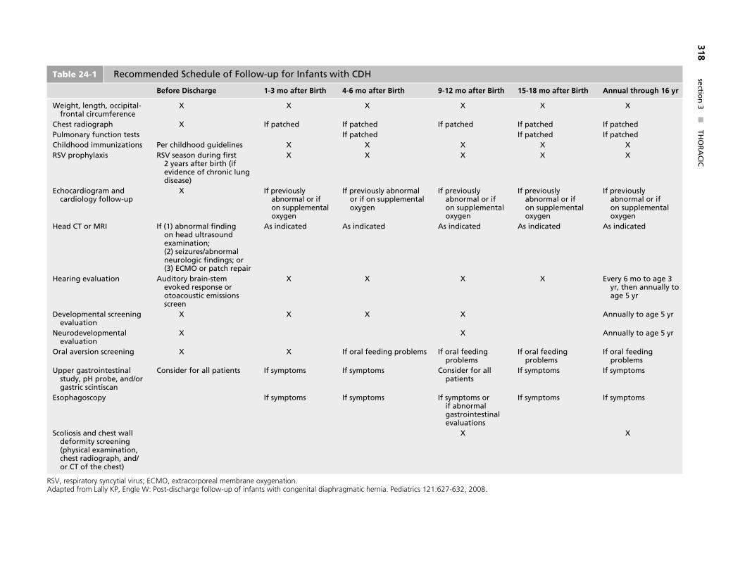
Follow-up GuidelinesCritically ill infants with CDH often require prolonged and intensive therapies such as ECMO during hos-pitalization. These result in long-term sequelae that require medical attention after discharge. Pulmonary, neurologic, gastrointestinal, and musculoskeletal complications necessitate a multidisciplinary team of surgical, medical, and developmental specialists. In 2008, the Section on Surgery and Committee on Fetus and Newborn for the American Academy of Pediatrics established a series of guidelines for the follow-up care of infants with CDH.240 These recommendations begin before discharge and extend through age 16 years. The guidelines include a formal follow-up plan to facilitate the early recognition of co-morbidities (Table 24-1).
NOVEL INTERVENTIONS
Even with advances in neonatal critical care, the treat-ment of infants with CDH remains a challenge to pediatric surgeons, neonatologists, and intensivists. Despite increased survival rates in the past 25 years, many infants with CDH die or develop debilitating morbidities. Thus, researchers continue to investigate novel therapies for infants with CDH.
Fetal InterventionThe impetus for fetal therapy for CDH coincided with the advances in prenatal diagnosis. Improvements in fetal ultrasonography revealed the true natural history of CDH and the hidden mortality during gestation and soon after birth. These poor outcomes prompted the researchers at the University of California, San Fran-cisco, to explore fetal surgery for CDH. Subsequently, fetal CDH intervention evolved from open fetal sur-gery to the current state of endoscopic endoluminal tracheal occlusion.
The prerequisite for any fetal intervention is the ability to accurately diagnose CDH and predict sever-ity of disease and survival. The most widely accepted prenatal sonographic prognosticators have been liver herniation and LHR, which have been discussed pre-viously. Liver herniation and an LHR less than 1.0 are recognized as fetal characteristics that portend the poorest outcomes.241
After years of unfavorable outcomes with open fetal repair of CDH, prenatal intervention evolved to tracheal occlusion. Based on clinical observations that tracheal atresia causes pulmonary hyperplasia, novel animal experiments of lung distention, and an improved understanding of fetal lung growth, therapeutic tracheal occlusion was introduced as a treatment for pulmonary hypoplasia secondary to CDH.99,242-244 Current techniques involve endo-scopic placement of an occlusive balloon without
sect�on
3 n
TH
OR
AC
IC318
15-18 mo after Birth Annual through 16 yr
X X
If patched If patchedIf patched If patched
X XX X
If pre��ous���� abnorma� or �f on supp�ementa� ox���gen
If pre��ous���� abnorma� or �f on supp�ementa� ox���gen
As �nd�cated As �nd�cated
X E�er��� 6 mo to age 3 ���r, then annua����� to age 5 ���r
Annua����� to age 5 ���r
Annua����� to age 5 ���r
If ora� feed�ng prob�ems
If ora� feed�ng prob�ems
If s���mptoms If s���mptoms
If s���mptoms If s���mptoms
X
Table 24-1 Recommended Schedu�e of Fo��ow-up for Infants w�th CDH
Before Discharge 1-3 mo after Birth 4-6 mo after Birth 9-12 mo after Birth
We�ght, �ength, occ�p�ta�-fronta� c�rcumference
X X X X
Chest rad�ograph X If patched If patched If patched�u�monar��� funct�on tests If patchedCh��dhood �mmun�zat�ons �er ch��dhood gu�de��nes X X XRSV proph����ax�s RSV season dur�ng first
2 ���ears after b�rth (�f e��dence of chron�c �ung d�sease)
X X X
Echocard�ogram and card�o�og��� fo��ow-up
X If pre��ous���� abnorma� or �f on supp�ementa� ox���gen
If pre��ous���� abnorma� or �f on supp�ementa� ox���gen
If pre��ous���� abnorma� or �f on supp�ementa� ox���gen
Head CT or MRI If (1) abnorma� find�ng on head u�trasound exam�nat�on; (2) se�zures/abnorma� neuro�og�c find�ngs; or (3) ECMO or patch repa�r
As �nd�cated As �nd�cated As �nd�cated
Hear�ng e�a�uat�on Aud�tor��� bra�n-stem e�oked response or otoacoust�c em�ss�ons screen
X X X
De�e�opmenta� screen�ng e�a�uat�on
X X X X
Neurode�e�opmenta� e�a�uat�on
X X
Ora� a�ers�on screen�ng X X If ora� feed�ng prob�ems If ora� feed�ng prob�ems
Upper gastro�ntest�na� stud���, pH probe, and/or gastr�c sc�nt�scan
Cons�der for a�� pat�ents If s���mptoms If s���mptoms Cons�der for a�� pat�ents
Esophagoscop��� If s���mptoms If s���mptoms If s���mptoms or �f abnorma� gastro�ntest�na� e�a�uat�ons
Sco��os�s and chest wa�� deform�t��� screen�ng (ph���s�ca� exam�nat�on, chest rad�ograph, and/or CT of the chest)
X
RSV, respiratory syncytial virus; ECMO, extracorporeal membrane oxygenation.Adapted from Lally KP, Engle W: Post-discharge follow-up of infants with congenital diaphragmatic hernia. Pediatrics 121:627-632, 2008.
chapter 24 n CONGENITA� DIA�HRAGMATIC HERNIA AND EVENTRATION 319
maternal laparotomy or general anesthesia.245 Tra-cheal balloons are placed between 24 and 28 weeks’ gestation and deflated at 34 weeks. This strategy of temporary tracheal occlusion is based on avoiding the need for an ex-utero intrapartum treatment (EXIT) procedure at delivery. In addition, prolonged tracheal occlusion has been demonstrated to differentiate type II pneumocytes into type I pneumocytes, resulting in surfactant deficiency and necessitating the need for balloon removal.246-248
An NIH-sponsored randomized trial comparing fetal tracheal occlusion versus standard postnatal care was reported in 2003.101 After 24 cases (11 by tracheal occlusion), the study was terminated early due to comparable survival outcomes (77% by postnatal care, 73% by tracheal occlusion) during interim analysis. The hazard ratio for mortality associated with tracheal occlusion, as compared with conventional therapy, was 1.2 (95% confidence interval [CI]: 0.29 to 4.67). However, when stratified based on LHR, survival was significantly better for LHR greater than 0.9. In fact, the hazard ratio for death with tracheal occlusion was 0.13 (95% CI: 0.03 to 0.64). This study recognized the wide spectrum of disease based on LHR and specu-lated that the patients with the most severe disease may still benefit from tracheal occlusion. Despite these results from the randomized trial, tracheal occlusion continues to be investigated owing to the significant mortality of infants with LHR less than 1.0 and liver herniation.
The FETO (Fetoscopic Tracheal Occlusion) task group, a European perinatology organization, created a multicenter prospective observational study.100 In an attempt to select a more severe subset based on data from the NIH trial, the study included fetuses with liver herniation and LHR less than 1.0. In the initial 21 patients (15 left-sided CDH and 6 right-sided CDH), the investigators reported a survival of 48% for tracheal occlusion compared with 8% in the non-occlusion group. In addition to the survival advan-tage, the study also demonstrated an improvement in perinatal complications such as prematurity and premature rupture of membranes. The latest series reported overall survival of 57% for tracheal occlu-sion with survival greater than 62% for LHR between 0.6 and 1.0.241 Although impressive, these results warrant further investigation in that the survival for LHR of 0.8 to 0.9 (78%) was higher than survival for LHR greater than 1.0 treated postnatally (65%). Furthermore, study limitations have prevented the widespread adoption of tracheal occlusion based on the European data. Control cases were taken from multiple centers that could not provide a standard-ized postnatal approach or reflect recent advances in postnatal care.
Long-term outcome with morbidity has yet to be reported for tracheal occlusion. However, one study examined pulmonary function in 20 patients (9 with conventional therapy and 11 with tracheal occlu-sions) from the NIH-sponsored randomized trial.249 Infants were evaluated during the first 24 hours of life, before and after operative repair, and before elective
extubation. The study demonstrated slight improve-ments in respiratory compliance and alveolar-arterial oxygenation gradients in patients who had undergone tracheal occlusion. However, other long-term outcomes remain to be seen. Based on the only randomized clini-cal trial, there appears to be no significant benefit from tracheal occlusion for the treatment of CDH. Although proponents of fetal intervention suggest that a subgroup of the most severe patients (liver herniation and LHR < 0.9) may still benefit from fetal intervention, the true efficacy of fetal tracheal occlusion for these severely affected infants will require a prospective randomized trial before its universal acceptance.
Prenatal GlucocorticoidsThe hypoplastic lungs of CDH infants are structur-ally and functionally immature. Biochemical markers for lung maturity demonstrate decreased total lung DNA, total lung protein, and desaturated phospha-tidylcholine in addition to deficiency of surfactant.63 In animal models of CDH, prenatal administration of glucocorticoids has demonstrated a reduction in alve-olar septal thickness, increased DNA synthesis, and increased total lung protein production.250,251 Thus, antenatal administration of glucocorticoids has been used to improve lung function.252,253 Initial results from small patient studies seemed promising. How-ever, a prospective randomized trial conducted by the CDHSG failed to demonstrate any benefit to antenatal corticosteroid therapy for CDH.254 In the multicenter trial, mothers received three weekly administrations of prenatal betamethasone. The study, which included 32 patients (15 on placebo and 17 on steroids), failed to show a difference in overall survival benefit, length of mechanical ventilation, length of hospitalization, or need for supplemental oxygen at 30 days. The study compared its results with a large cohort from the CDHR with comparable outcomes.
ANTERIOR HERNIAS OF MORGAGNI
Anterior diaphragmatic hernias of Morgagni account for less than 2% of all congenital diaphragmatic her-nias. The foramen of Morgagni hernia results from the failure of the crural and sternal portions of the diaphragm to fuse. This can occur on either side at the junction of the septum transversum and thoracic wall where the superior epigastric artery (internal mammary artery, intrathoracically) traverses the dia-phragm. Approximately 90% of unilateral hernias occur on the right.255 Only 7% occur bilaterally.256 Operatively, these may communicate in the midline and appear as a very large anterior hernia. Typically, a hernia sac is present with herniation of omentum, small intestine, and/or large intestine but may also contain liver and spleen. Malrotation may also be found. The majority of children with Morgagni hernia are asymptomatic and thus are rarely diagnosed dur-ing the neonatal period. When these hernias become symptomatic, pulmonary symptoms are usually not
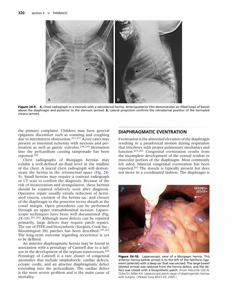
sect�on 3 n THORACIC320
the primary complaint. Children may have general epigastric discomfort such as vomiting and coughing due to intermittent obstruction.255,257 Acute cases may present as intestinal ischemia with necrosis and per-foration as well as gastric volvulus.258,259 Herniation into the pericardium causing tamponade has been reported.260
Chest radiographs of Morgagni hernias may exhibit a well-defined air-fluid level in the midline of the chest. A lateral chest radiograph will demon-strate the hernia in the retrosternal space (Fig. 24-9). Small hernias may require a contrast radiograph or CT scan to confirm the diagnosis. Because of the risk of incarceration and strangulation, these hernias should be repaired relatively soon after diagnosis. Operative repair usually entails reduction of herni-ated viscera, excision of the hernia sac, and closure of the diaphragm to the posterior rectus sheath at the costal margin. Open procedures can be performed through an upper transabdominal incision. Laparo-scopic techniques have been well documented (Fig. 24-10).261,262 Although most defects can be repaired primarily, large defects may require patch repairs. The use of PTFE and biosynthetic (Surgisis, Cook Inc., Bloomington IN) patches has been described.197,263 The long-term outcome regarding recurrence is yet to be defined.
An anterior diaphragmatic hernia may be found in association with a pentalogy of Cantrell due to a fail-ure in the development of the septum transversum.264 Pentalogy of Cantrell is a rare cluster of congenital anomalies that include omphalocele, cardiac defects, ectopic cordis, and an anterior diaphragmatic defect extending into the pericardium. The cardiac defect is the most severe problem and is the main cause of mortality.
DIAPHRAGMATIC EVENTRATION
Eventration is the abnormal elevation of the diaphragm resulting in a paradoxical motion during respiration that interferes with proper pulmonary mechanics and function.265,266 Congenital eventration results from the incomplete development of the central tendon or muscular portion of the diaphragm. Most commonly left sided, bilateral congenital eventration has been reported.267 The muscle is typically present but does not move in a coordinated fashion. The diaphragm is
A B
Figure 24-9. A, Chest rad�ograph �n a neonate w�th a retrosterna� hern�a������ Anteroposter�or fi�m demonstrates a�r-fi��ed �oops of bowe� abo�e the d�aphragm and poster�or to the sternum (arrow)������ B, �atera� project�on confirms the retrosterna� pos�t�on of the hern�ated ��scera (arrow)������
*
Figure 24-10. �aparoscop�c ��ew of a Morgagn� hern�a������ Th�s anter�or hern�a (white arrow) �s to the �eft of the fa�c�form ��ga-ment (asterisk) w�th a deep sac that was exc�sed������ The �arge bowe� (dotted arrow) was reduced from the hern�a defect, and the de-fect was c�osed w�th a b�os���nthet�c patch������ (From Holcomb GW III, Ostlie DJ, Miller KA: Laparoscopic patch repair of diaphragmatic hernias with Surgisis. J Pediatr Surg 40:e1-e5, 2005.)
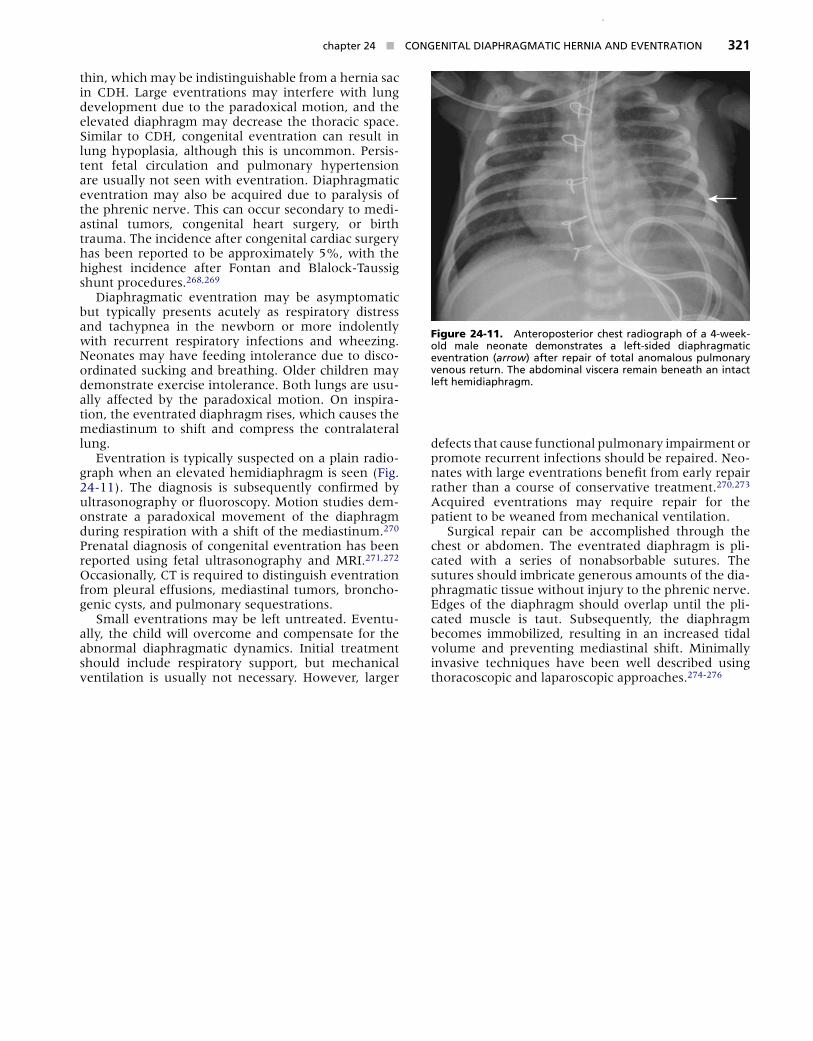
chapter 24 n CONGENITA� DIA�HRAGMATIC HERNIA AND EVENTRATION 321
thin, which may be indistinguishable from a hernia sac in CDH. Large eventrations may interfere with lung development due to the paradoxical motion, and the elevated diaphragm may decrease the thoracic space. Similar to CDH, congenital eventration can result in lung hypoplasia, although this is uncommon. Persis-tent fetal circulation and pulmonary hypertension are usually not seen with eventration. Diaphragmatic eventration may also be acquired due to paralysis of the phrenic nerve. This can occur secondary to medi-astinal tumors, congenital heart surgery, or birth trauma. The incidence after congenital cardiac surgery has been reported to be approximately 5%, with the highest incidence after Fontan and Blalock-Taussig shunt procedures.268,269
Diaphragmatic eventration may be asymptomatic but typically presents acutely as respiratory distress and tachypnea in the newborn or more indolently with recurrent respiratory infections and wheezing. Neonates may have feeding intolerance due to disco-ordinated sucking and breathing. Older children may demonstrate exercise intolerance. Both lungs are usu-ally affected by the paradoxical motion. On inspira-tion, the eventrated diaphragm rises, which causes the mediastinum to shift and compress the contralateral lung.
Eventration is typically suspected on a plain radio-graph when an elevated hemidiaphragm is seen (Fig. 24-11). The diagnosis is subsequently confirmed by ultrasonography or fluoroscopy. Motion studies dem-onstrate a paradoxical movement of the diaphragm during respiration with a shift of the mediastinum.270 Prenatal diagnosis of congenital eventration has been reported using fetal ultrasonography and MRI.271,272 Occasionally, CT is required to distinguish eventration from pleural effusions, mediastinal tumors, broncho-genic cysts, and pulmonary sequestrations.
Small eventrations may be left untreated. Eventu-ally, the child will overcome and compensate for the abnormal diaphragmatic dynamics. Initial treatment should include respiratory support, but mechanical ventilation is usually not necessary. However, larger
defects that cause functional pulmonary impairment or promote recurrent infections should be repaired. Neo-nates with large eventrations benefit from early repair rather than a course of conservative treatment.270,273 Acquired eventrations may require repair for the patient to be weaned from mechanical ventilation.
Surgical repair can be accomplished through the chest or abdomen. The eventrated diaphragm is pli-cated with a series of nonabsorbable sutures. The sutures should imbricate generous amounts of the dia-phragmatic tissue without injury to the phrenic nerve. Edges of the diaphragm should overlap until the pli-cated muscle is taut. Subsequently, the diaphragm becomes immobilized, resulting in an increased tidal volume and preventing mediastinal shift. Minimally invasive techniques have been well described using thoracoscopic and laparoscopic approaches.274-276
Figure 24-11. Anteroposter�or chest rad�ograph of a 4-week-o�d ma�e neonate demonstrates a �eft-s�ded d�aphragmat�c e�entrat�on (arrow) after repa�r of tota� anoma�ous pu�monar��� �enous return������ The abdom�na� ��scera rema�n beneath an �ntact �eft hem�d�aphragm������