Embed Size (px)
Citation preview
Copyright ©2001 McGraw-Hil l Victor, Maurice, Ropper, Allan H. Adams & Victors' Principles of Neurology, 7th Edit ion
Chapter 47 DISEASES OF THE CRANIAL NERVES
The cranial nerves are susceptible to a number of special diseases, some of which never affect the spinal peripheral nerves. For this reason alone they deserve to be considered separately. Certain of them have already been discussed: namely, disorders of olfaction, in Chap. 12; of vision and extraocular muscles, in Chap. 13 and Chap. 14; of cochlear and vestibular function, in Chap. 15; and craniofacial pain in Chap. 10. There remain to be described the disorders of the facial (seventh) nerve and of the lower cranial nerves (IX to XII) as well as certain aspects of disordered tr igeminal (f i f th) nerve function. These are considered below.
The Fifth, or Trigeminal, Nerve
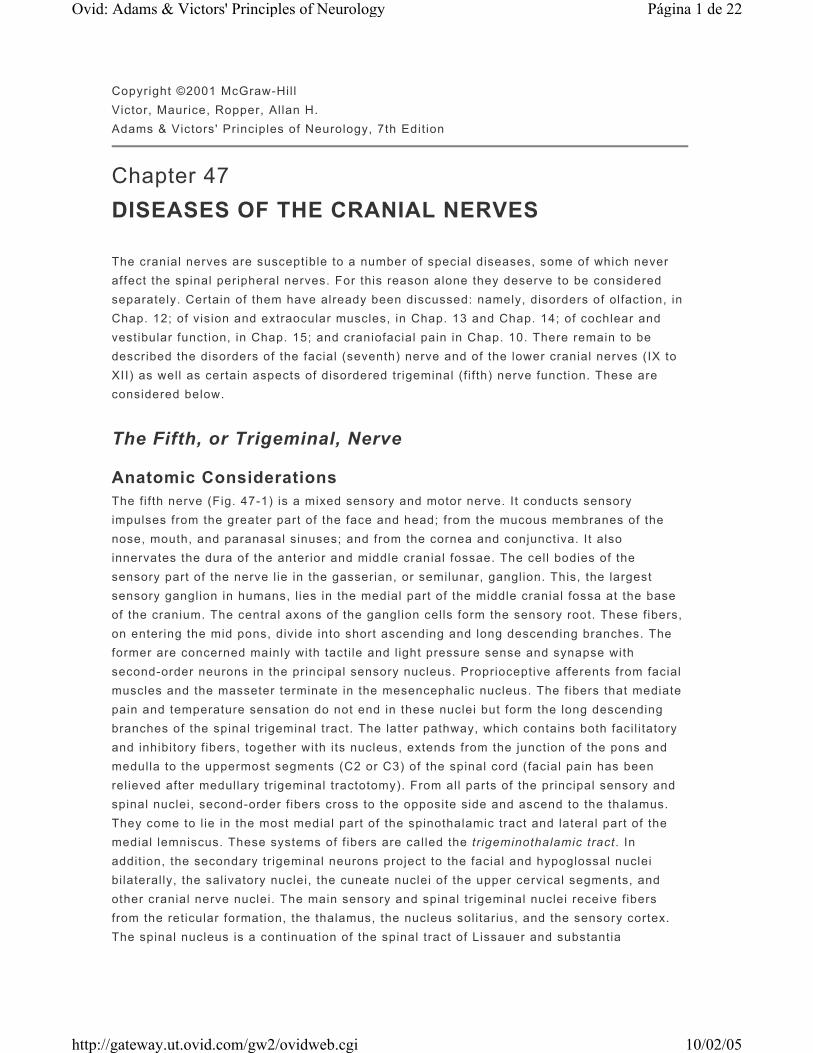
Anatomic Considerations The fifth nerve (Fig. 47-1) is a mixed sensory and motor nerve. It conducts sensory impulses from the greater part of the face and head; from the mucous membranes of the nose, mouth, and paranasal sinuses; and from the cornea and conjunctiva. It also innervates the dura of the anterior and middle cranial fossae. The cell bodies of the sensory part of the nerve l ie in the gasserian, or semilunar, ganglion. This, the largest sensory ganglion in humans, l ies in the medial part of the middle cranial fossa at the base of the cranium. The central axons of the ganglion cells form the sensory root. These fibers, on entering the mid pons, divide into short ascending and long descending branches. The former are concerned mainly with tacti le and l ight pressure sense and synapse with second-order neurons in the principal sensory nucleus. Proprioceptive afferents from facial muscles and the masseter terminate in the mesencephalic nucleus. The fibers that mediate pain and temperature sensation do not end in these nuclei but form the long descending branches of the spinal tr igeminal tract. The latter pathway, which contains both facil i tatory and inhibitory f ibers, together with its nucleus, extends from the junction of the pons and medulla to the uppermost segments (C2 or C3) of the spinal cord (facial pain has been relieved after medullary tr igeminal tractotomy). From all parts of the principal sensory and spinal nuclei, second-order f ibers cross to the opposite side and ascend to the thalamus. They come to l ie in the most medial part of the spinothalamic tract and lateral part of the medial lemniscus. These systems of f ibers are called the tr igeminothalamic tract . In addit ion, the secondary tr igeminal neurons project to the facial and hypoglossal nuclei bi laterally, the salivatory nuclei, the cuneate nuclei of the upper cervical segments, and other cranial nerve nuclei. The main sensory and spinal tr igeminal nuclei receive f ibers from the reticular formation, the thalamus, the nucleus solitarius, and the sensory cortex. The spinal nucleus is a continuation of the spinal tract of Lissauer and substantia
Página 1 de 22Ovid: Adams & Victors' Principles of Neurology
10/02/05http://gateway.ut.ovid.com/gw2/ovidweb.cgi
gelatinosa; the main sensory nucleus is a continuation of the nucleus of the medial lemniscus.
The peripheral branches of the gasserian ganglion form the three sensory divisions of the nerve. The first (ophthalmic) division passes through the superior orbital f issure; the second (maxil lary) division leaves the middle fossa through the foramen rotundum; and the third (mandibular), through the foramen ovale.
Figure 47-1 Scheme of the trigeminal nuclei and some of the trigeminal reflex arcs. I, ophthalmic division; II, maxillary division; III, mandibular division. (From Carpenter MB, Sutin J: Human Neuroanatomy, 8th ed. Baltimore, Williams & Wilkins, 1982, by permission.)
Página 2 de 22Ovid: Adams & Victors' Principles of Neurology
10/02/05http://gateway.ut.ovid.com/gw2/ovidweb.cgi
The motor portion of the f i f th nerve, which supplies the masseter and pterygoid muscles, has its origin in the tr igeminal motor nucleus in the midpons; the exit ing f ibers pass underneath the gasserian ganglion and become incorporated into the mandibular nerve. The masseter and pterygoid muscles are util ized in chewing and are implicated in a number of brainstem reflexes, the best known of which is the jaw jerk. Tapping the chin with the jaw muscles relaxed stimulates proprioceptive afferents that terminate in the mesencephalic nucleus of the midbrain, which sends collaterals to the motor nucleus of the f i f th nerve and causes the masseters to contract. This reflex is enhanced in spastic bulbar (pseudobulbar) palsy. Another pontine reflex that ut i l izes afferent tr igeminal sensory nerves is the blink reflex. Tapping of the brow or bridge of the nose evokes bilateral bl ink through activation of the orbicularis oculi muscles (facial nerve efferents). Touching the eyelids and cornea (corneal reflex) does the same.
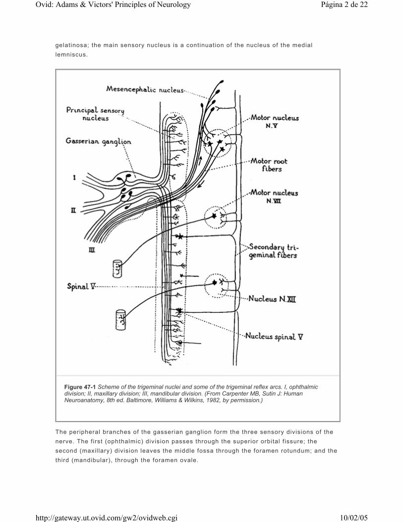
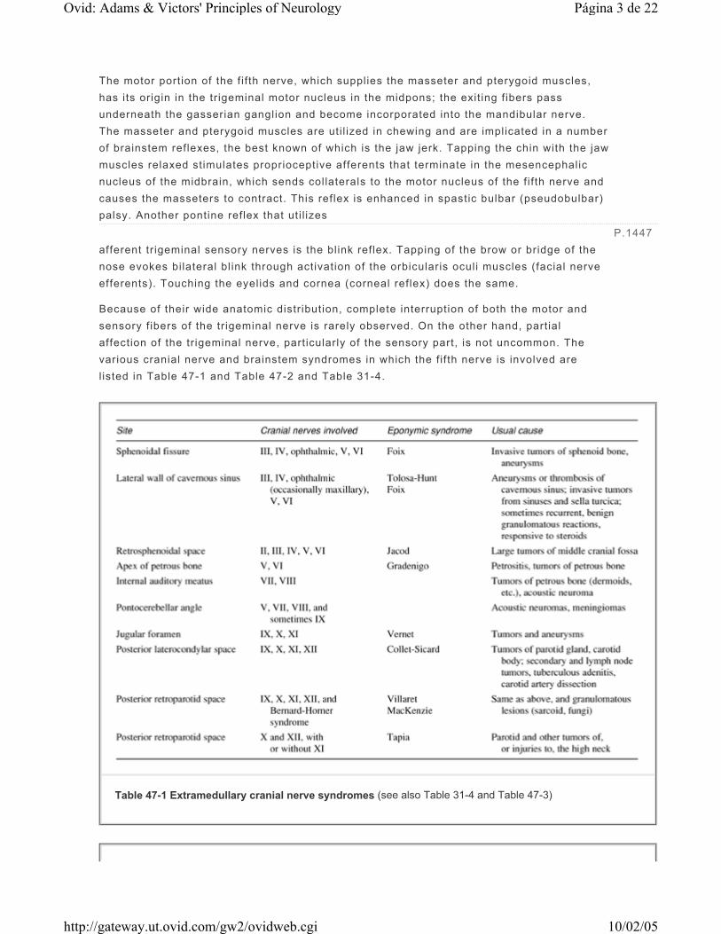
Because of their wide anatomic distribution, complete interruption of both the motor and sensory f ibers of the tr igeminal nerve is rarely observed. On the other hand, partial affection of the tr igeminal nerve, particularly of the sensory part, is not uncommon. The various cranial nerve and brainstem syndromes in which the f if th nerve is involved are listed in Table 47-1 and Table 47-2 and Table 31-4.
P.1447
Table 47-1 Extramedullary cranial nerve syndromes (see also Table 31-4 and Table 47-3)
Página 3 de 22Ovid: Adams & Victors' Principles of Neurology
10/02/05http://gateway.ut.ovid.com/gw2/ovidweb.cgi
Table 47-2 Intramedullary (brainstem) syndromes involving cranial nerves
Página 4 de 22Ovid: Adams & Victors' Principles of Neurology
10/02/05http://gateway.ut.ovid.com/gw2/ovidweb.cgi
Diseases Affecting the Fifth Nerve A variety of diseases may affect the peripheral branches of the tr igeminal nerves, the gasserian (semilunar) ganglion, and the roots (sensory and motor). These have been ably described by Hughes. The most frequent, and at the same time the most elusive from the standpoint of its pathologic basis, is tr igeminal neuralgia (t ic douloureux) , which is also discussed on page 196. This condit ion has been known since ancient t imes, having been described by Arateus in the f irst century A.D., by John Locke in 1677, by Nicolaus Andre in 1756, and by John Fothergil l in 1776 (according to Katusic et al). The overall incidence rate for both sexes combined is 4.3 per 100,000 persons per year, but i t is higher for women than for men (in a ratio of 3:2) and is much higher in the elderly. The mean age of onset is 52 to 58 years for the idiopathic form and 30 to 35 years for the symptomatic forms, the latter being caused by trauma or vascular, neoplastic, and demyelinative diseases. The nature of the pain, i ts unilaterality and tendency to involve the second and third divisions of the tr igeminal nerve, an intensity that makes the patient grimace or wince (t ic), the presence of an init iating or tr igger point, the lack of demonstrable sensory or motor deficit, and its response in more than half of the cases to carbamazepine, phenytoin, and
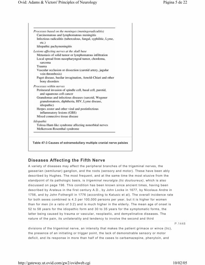
Table 47-3 Causes of extramedullary multiple cranial nerve palsies
P.1448
Página 5 de 22Ovid: Adams & Victors' Principles of Neurology
10/02/05http://gateway.ut.ovid.com/gw2/ovidweb.cgi
other drugs are characteristic. The diagnosis of the idiopathic form of tr igeminal neuralgia and its differentiation from symptomatic forms—as well as from cluster headache, dental neuralgia, temporomandibular pain, and atypical facial pain—is usually not diff icult, especially if there is a tr igger point and no demonstrable evidence of sensory or motor impairment. The treatment of idiopathic tr igeminal neuralgia is considered on page 198.
In rare instances tr igeminal neuralgia is preceded or accompanied by hemifacial spasm, a combination that Cushing called t ic convulsif . This is often indicative of a tumor (cholesteatoma), an aneurysmal dilatation of the basilar artery, or an arteriovenous malformation that compresses both the tr igeminal and facial nerves. Trigeminal neuralgia and glossopharyngeal neuralgia (pain in the tonsil lar region) may also be combined in these disease states, but these condit ions are usually idiopathic.
Of the condit ions that damage the branches of the tr igeminal nerve, cranial injuries and fractures are the most common. The most superficial branches—the supratrochlear, supraorbital, and infraorbital—are the ones usually involved. The sensory loss is present from the time of the injury, and partial regeneration may be attended by constant pain, often demanding nerve block and sectioning. Sti lbamidine and trichloroethylene are known to cause sensory loss, t ingling, burning, and itching exclusively in the tr igeminal sensory territory.
Of the various inflammatory and infectious diseases that affect the tr igeminal nerves or ganglia, herpes zoster ranks f irst (page 799). Herpes simplex virus has been isolated from the ganglion in as many as 50 percent of routine autopsies, but in nearly all patients this virus is associated only with lesions of the skin and l ips. Middle ear infections and osteomyelit is of the apex of the petrous bone may spread to the ganglion and root, also implicating the sixth cranial nerve (Gradenigo syndrome).
The trigeminal root may be compressed or invaded by intracranial meningiomas, acoustic neuromas, tr igeminal neuromas (Fig. 47-2), cholesteatomas, and chordomas. Sinus tumors and metastatic disease may also implicate the nerve, causing pain and a gradually progressive sensory loss. The ophthalmic division of the f i f th nerve may be involved in the wall of the cavernous sinus in combination with the third, fourth, and sixth nerves by a variety of processes, including thrombosis of the cavernous sinus. Tumors of the sphenoid bone (myeloma, metastatic carcinoma, squamous cell carcinoma, and lymphoepithelioma of the nasopharynx) may involve branches of the tr igeminal nerve at their foramina of entry or exit. The mandibular division may be compressed by the roots of an impacted third molar (wisdom) tooth. We have several t imes observed numbness of the chin and lower l ip ( infi l tration of the mental nerve) as the f irst indication of metastatic carcinoma of the breast and prostate and from multiple myeloma. Massey and colleagues have described 19 such cases (“numb-chin syndrome”). A perineural spread of tumor from squamous cell skin cancers of the face is discussed further on, under “Mult iple Cranial Nerve Palsies.”
P.1449P.1450
Página 6 de 22Ovid: Adams & Victors' Principles of Neurology
10/02/05http://gateway.ut.ovid.com/gw2/ovidweb.cgi
Loss of facial sensation can occur as part of a widespread sensory neuropathy that occurs as a remote effect of cancer or as part of Sjögren disease (pages 727 and 1403). Far more common is the association between isolated tr igeminal neuropathy and immune-mediated connective t issue disease. Of 22 such cases described by Lecky and colleagues, 9 had either scleroderma or mixed connective t issue disease, and a similar number had either organ- or non-organ-specif ic serum autoantibodies. The symptoms may involve the other side years later. Hughes has also reported cases of tr igeminal neuropathy with scleroderma, lupus erythematosus, and Sjögren disease. We have seen several patients with Sjögren disease in whom the tr igeminal neuropathy and the associated antibodies or inflammation of the minor salivary glands were evident well before the characteristic sicca syndrome or other systemic manifestations of the disease. The condit ion may remain troublesome for years. Pathologic data are l imited but point to an inflammatory lesion of the tr igeminal ganglion or sensory root.
Every neurologist from time to t ime encounters idiopathic instances of slowly evolving unilateral or bi lateral tr igeminal neuropathy in which there is a sensory impairment confined to the territory of the tr igeminal nerve, sometimes associated with pain and paresthesias and disturbances of taste. Spil lane and Wells stressed the importance of this isolated tr igeminal neuropathy (it has been called “Spil lane's tr igeminal neurit is” in some texts). Four of their 16 patients had an associated paranasal sinusit is, but subsequent reports have fai led to substantiate a causal relationship between sinusit is and cranial neurit is. One wonders how many of these individuals had an occult connective t issue disease. A less common form of idiopathic tr igeminal sensory neuropathy has a more acute onset and a tendency to resolve completely or partial ly, in much the same manner as Bell 's palsy, with which it is sometimes associated (Blau et al). A recurrent variety of uncertain origin has been reported in the dental l i terature. We have had experience with patients whose facial
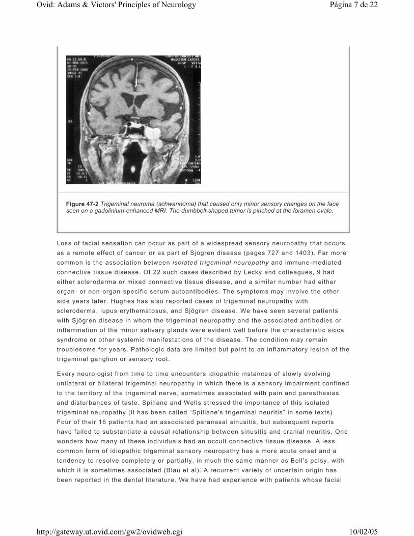
Figure 47-2 Trigeminal neuroma (schwannoma) that caused only minor sensory changes on the face seen on a gadolinium-enhanced MRI. The dumbbell-shaped tumor is pinched at the foramen ovale.
Página 7 de 22Ovid: Adams & Victors' Principles of Neurology
10/02/05http://gateway.ut.ovid.com/gw2/ovidweb.cgi
numbness was a component of an upper cervical disc syndrome that included numbness on the same side of the body. Cases such as these are reported in the l i terature.
A pure unilateral tr igeminal motor neuropathy is a cl inical rarity. Chia has described five patients in whom an aching pain in the cheek and unilateral weakness of mastication were the main features. Electromyography (EMG) showed denervation changes in the ipsilateral masseter and temporalis muscles. The outcome was apparently favorable.
In most cases of tr igeminal neuropathy except those due to tumor, the results of gadolinium-enhanced MRI are normal, as is the cerebrospinal f luid. The function of the nerve may be studied by the electrical recording of bl ink reflexes. A few laboratories have developed an evoked potential test specif ically of the trigeminal nerve.
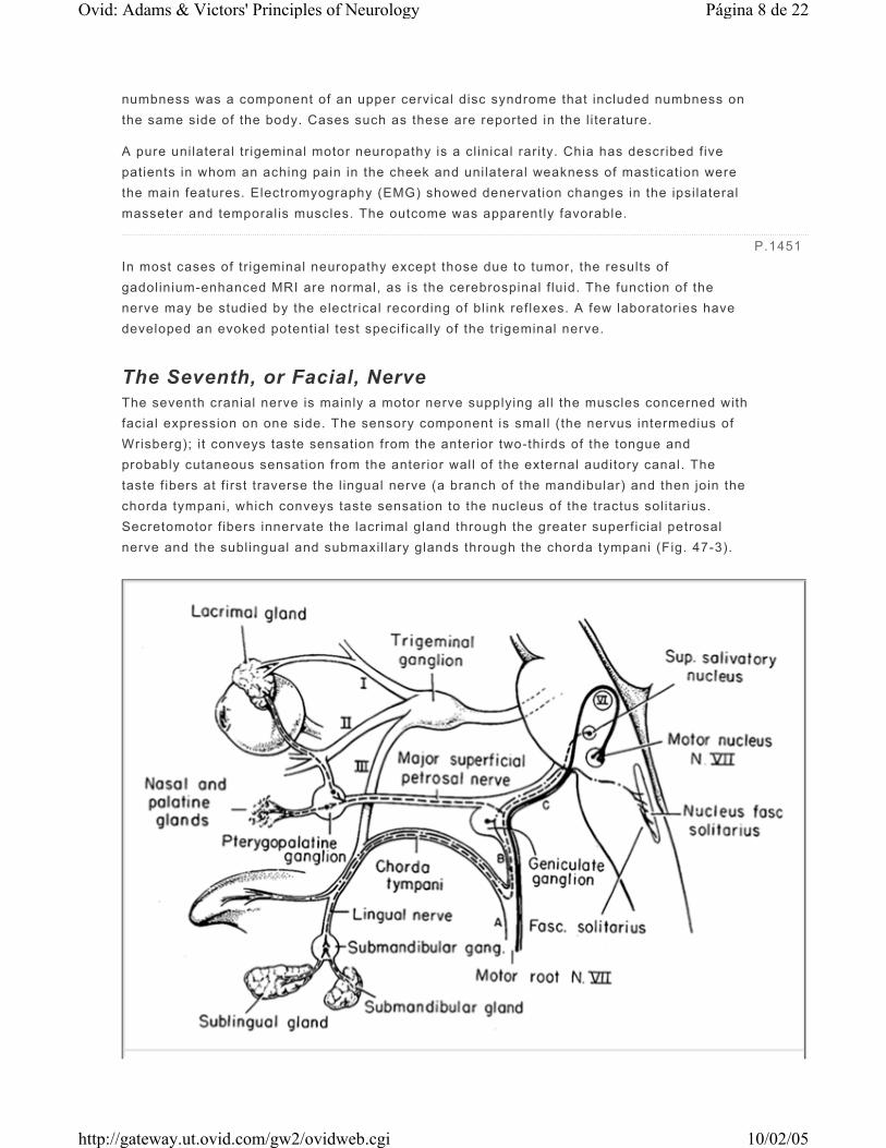
The Seventh, or Facial, Nerve The seventh cranial nerve is mainly a motor nerve supplying all the muscles concerned with facial expression on one side. The sensory component is small (the nervus intermedius of Wrisberg); it conveys taste sensation from the anterior two-thirds of the tongue and probably cutaneous sensation from the anterior wall of the external auditory canal. The taste f ibers at f irst traverse the l ingual nerve (a branch of the mandibular) and then join the chorda tympani, which conveys taste sensation to the nucleus of the tractus solitarius. Secretomotor f ibers innervate the lacrimal gland through the greater superficial petrosal nerve and the sublingual and submaxil lary glands through the chorda tympani (Fig. 47-3).
P.1451
Página 8 de 22Ovid: Adams & Victors' Principles of Neurology
10/02/05http://gateway.ut.ovid.com/gw2/ovidweb.cgi
Several other anatomic facts are worth remembering. The motor nucleus of the seventh nerve lies ventral and lateral to the abducens nucleus, and the intrapontine f ibers of the facial nerve hook around and pass ventrolaterally to the abducens nucleus before emerging from the pons, just lateral to the corticospinal tract. The facial nerve enters the internal auditory meatus with the acoustic nerve and then bends sharply forward and downward around the anterior boundary of the vestibule of the inner ear. At this angle (genu) l ies the sensory ganglion (named geniculate because of i ts proximity to the genu). The nerve continues its course in its own bony channel, the facial canal, within which, just distal to the geniculate ganglion, i t provides a branch to the pterygopalatine ganglion, i .e., the greater superficial petrosal nerve; somewhat more distally, it gives off a small branch to the stapedius muscle and is joined by the chorda tympani. It makes its exit from the skull at the stylomastoid foramen, then passes through the parotid gland and subdivides into f ive branches that supply the facial muscles, the stylomastoid muscle, and the posterior belly of the digastric muscle.
A complete interruption of the facial nerve at the stylomastoid foramen paralyzes all muscles of facial expression. The corner of the mouth droops, the creases and skin folds are effaced, the forehead is unfurrowed, the palpebral f issure is widened, and the eyelids wil l not close. Upon attempted closure of the lids, both eyes roll upward (Bell 's phenomenon), but the one on the paralyzed side remains visible. The lower l id sags also, and the punctum falls away from the conjunctiva, permitt ing tears to spil l over the cheek. Food collects between the teeth and cheek, and saliva may dribble from the corner of the mouth. The patient complains of a heaviness or numbness and sometimes an aching pain in the face, but sensory loss can usually not be demonstrated. Taste, however, is intact because the lesion is beyond the site where the chorda tympani has separated from the main trunk of the facial nerve.
If the lesion is in the facial canal above the junction with the chorda tympani but below the geniculate ganglion, all the above symptoms occur; in addit ion, taste is lost over the anterior two-thirds of the tongue on the same side. If the nerve to the stapedius muscle is involved, there is hyperacusis (painful sensit ivity to loud sounds). With a stethoscope in the patient's ears, a tuning fork at the bell is louder on the side of the paralyzed stapedius muscle. If the geniculate ganglion or the motor root proximal to it is involved, lacrimation and salivation may be reduced. Lesions at this point may also affect the adjacent eighth nerve, causing deafness, t innitus, or dizziness. Intrapontine lesions that paralyze the face often affect the abducens nucleus and the corticospinal and sensory tracts (Mil lard-Gubler syndrome; Table 47-2).
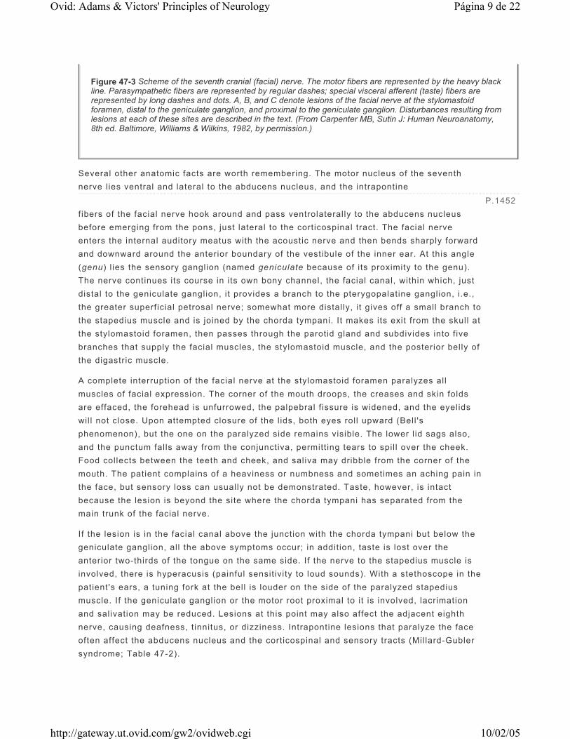
Figure 47-3 Scheme of the seventh cranial (facial) nerve. The motor fibers are represented by the heavy black line. Parasympathetic fibers are represented by regular dashes; special visceral afferent (taste) fibers are represented by long dashes and dots. A, B, and C denote lesions of the facial nerve at the stylomastoid foramen, distal to the geniculate ganglion, and proximal to the geniculate ganglion. Disturbances resulting from lesions at each of these sites are described in the text. (From Carpenter MB, Sutin J: Human Neuroanatomy, 8th ed. Baltimore, Williams & Wilkins, 1982, by permission.)
P.1452
Página 9 de 22Ovid: Adams & Victors' Principles of Neurology
10/02/05http://gateway.ut.ovid.com/gw2/ovidweb.cgi
I f the peripheral facial paralysis has existed for some time and return of motor function has begun but is incomplete, a kind of contracture (in reality a continuous diffuse myokymic contraction) may appear. The palpebral f issure becomes narrowed and the nasolabial fold deepens. Attempts to move one group of facial muscles result in contraction of al l of them (associated movements, or synkinesis). Spasms of facial muscles may develop and persist indefinitely, being init iated by every facial movement. However, this condit ion, called hemifacial spasm , occurs most often in adults who have never had a facial palsy and may be related to compression of the nerve by a tortuous blood vessel on the surface of the ventral pons as discussed further on (page 1455).
Anomalous regeneration of the seventh nerve f ibers, fol lowing a Bell 's palsy or other injury, may result in other curious disorders. The most common is the “jaw-winking” phenomenon (also called Wartenberg's or inverse Marcus-Gunn sign), in which jaw movements, especially lateral movements (pterygoid muscle), cause an involuntary closure of the eyelid. If regenerating f ibers originally connected with the orbicularis oculi become connected with the orbicularis oris, closure of the l ids may cause a retraction of the corner of the mouth; or i f visceromotor f ibers originally innervating the salivary glands later come to innervate the lacrimal gland, anomalous tearing (crocodile tears) occurs whenever the patient salivates. A similar mechanism explains gustatory sweating of the cheek and upper l ip. With the passage of t ime, the corner of the mouth and even the t ip of the nose may become pulled to the affected side.
Bell's Palsy The most common disease of the facial nerve is Bell 's palsy (incidence rate of 23 per 100,000 annually, according to Hauser et al). This disorder affects men and women more or less equally and occurs at al l ages and all t imes of the year. There is controversy regarding an increased incidence in women during the third tr imester of pregnancy, particularly in the 2 weeks preceding delivery and in the f irst 2 weeks postpartum; up to a threefold increase has been cited by some authors, but others have fai led to f ind this disproportionate number of cases. Supporting such a proclivity for facial palsy are scattered reports of a recurrence with each pregnancy. Bell 's palsy is probably more common in diabetics and possibly in hypertensives than in the normal population.
As one might expect, the opportunity to examine the facial nerve in the course of Bell 's palsy occurs very rarely. Only a handful of such cases are on record, al l showing varying degrees of degeneration of nerve f ibers. One case was said to show inflammatory changes, but these may have been misinterpreted (see Karnes).
Regarding causation of Bell 's palsy, a viral agent has long been suspected (Baringer). Only in the past few years, however, has such a mechanism been established with a reasonable degree of certainty for the majority of cases formerly considered to be idiopathic. Burgess and colleagues identif ied the genome of herpes simplex virus (HSV) in the geniculate ganglion of an elderly man who died 6 weeks after the onset of Bell 's palsy. More recently, Murakami and coworkers, using the polymerase chain reaction (PCR) technique to amplify viral genomic sequences, identif ied HSV type I in the endoneurial f luid surrounding the
P.1453
Página 10 de 22Ovid: Adams & Victors' Principles of Neurology
10/02/05http://gateway.ut.ovid.com/gw2/ovidweb.cgi
seventh nerve in 11 of 14 cases of Bell 's palsy; the f luid was obtained during surgical decompression of the nerve in severe cases. The same investigators have produced facial paralysis by inoculating HSV into the ears and tongues of mice; virus antigens were then found in the facial nerve and geniculate ganglion. Varicella zoster virus (VZV) was not found in any of the Bell 's palsy patients but was isolated from patients with the Ramsay-Hunt syndrome (page 799 and further on). Control patients with fracture or infections of the temporal bone yielded neither HSV or VZV gene sequences. In the l ight of these findings, the term idiopathic facial paralysis , unti l now the accepted synonym for Bell 's palsy, is not entirely appropriate and should perhaps be changed to herpes simplex or herpetic facial paralysis .
The onset of Bell 's palsy is acute; about one-half of the cases attain maximum paralysis in 48 h and practically all cases within 5 days. Pain behind the ear may precede the paralysis by a day or two and in a few patients is quite intense and persistent. In a small proportion of patients, a hypesthesia in one or more branches of the tr igeminal nerve can be demonstrated. The explanation of this f inding is not clear. Impairment of taste is present to some degree in almost al l patients but rarely persists beyond the second week of paralysis. As indicated above, it means that the lesion has extended to or above the point where the chorda tympani f ibers join the facial nerve. Hyperacusis or distort ion of sound in the ipsilateral ear indicates paralysis of the stapedius muscle. Unlike the f i f th nerve in idiopathic tr igeminal neurit is, the facial nerve in Bell 's palsy is often seen to display enhancement on the gadolinium-enhanced MRI. Correspondingly, there is a mild increase of lymphocytes and mononuclear cells in the CSF in a few of the cases.
Fully 80 percent of patients recover within a few weeks or in a month or two. Recovery of taste precedes recovery of motor function; i f the former occurs in the f irst week, it is a good prognostic sign. Early recovery of some motor function in the f irst 5 to 7 days is the most favorable prognostic sign. Electromyography may be of value in distinguishing temporary conduction defects from a pathologic interruption of nerve fibers; i f there is evidence of denervation after 10 days, one may expect a long delay in the onset of recovery (3 months, on the average). Recovery then proceeds by regeneration of nerve, a process that may take 2 years or longer and is often incomplete.
Idiopathic Bell 's palsy is said to recur in 8 percent of cases (van Amstel and Devriese; Pitts et al), presumably as a result of reactivation of the herpesvirus. The palsy reemerges during an infection or pregnancy or for no apparent reason. The interval between episodes is unpredictable but has been 10 years, on average. Other recurrent forms of facial paralysis occur with Lyme disease and sarcoidosis, and in a famil ial variety as mentioned below.
Protection of the eye during sleep, massage of the weakened muscles, and a splint to prevent drooping of the lower part of the face are the measures generally employed in the management of such cases. There is no evidence that surgical decompression of the facial nerve is effective, and it may be harmful. The administration of prednisone (40 to 60 mg/day) during the f irst week to 10 days after onset may be beneficial; i t is thought to decrease the possibil i ty of permanent paralysis from swell ing of the nerve in the t ight facial canal. The recent f inding of viral genome surrounding the seventh nerve suggests that antiviral agents may be useful in the management of Bell 's palsy. Several small tr ials
Página 11 de 22Ovid: Adams & Victors' Principles of Neurology
10/02/05http://gateway.ut.ovid.com/gw2/ovidweb.cgi
suggest that acyclovir, used alone, is no more effective than corticosteroids. The use of both medications together is under study. The treatment of facial palsy due to VZV (Ramsay-Hunt syndrome) with these drugs is discussed below.
Other Causes of Facial Palsy Lyme disease commonly involves the facial nerve, as indicated on pages 768 and 1403. The mechanism is not thought to be a direct spirochetal infection. The diagnosis is l ikely when there has been a t ick bite with erythema marginatum or arthrit is. Several of our cases have had almost simultaneous facial palsy and sensory neuropathy. Human immunodeficiency virus (HIV) infection is an infrequent cause of facial palsy. The facial palsy of both Lyme and HIV infections is associated with a pleocytosis; serologic and CSF examination may be useful i f there is a suspicion of either process. Rarely, chickenpox in children may be followed in 1 to 2 weeks by facial paralysis. Tuberculous infection of the mastoid and middle ear or of the petrous bone is a cause of facial paralysis in parts of the world where tuberculosis is particularly common. Facial palsy may occur in infectious mononucleosis and was observed occasionally in poliomyelit is. The facial nerve is frequently involved in leprosy. Bilateral involvement of the facial nerve is commented on below.
The Ramsay Hunt syndrome , due presumably to herpes zoster of the geniculate ganglion, consists of a facial palsy associated with a vesicular eruption in the external auditory canal, other parts of the cranial integument, and mucous membrane of the oropharynx. Often the eighth cranial nerve is affected as well, causing vertigo and deafness (see pages 189 and 799). Murakami et al have shown that the virus can be detected even before the emergence of typical vesicles by collecting exudate from the skin of the pinna on a Schirmer strip (otherwise used to quantitate tearing) and applying PCR techniques. In this way, in a manner of a few hours, they documented VZV infection in 71 percent of patients with Ramsay-Hunt syndrome without vesicles. Treatment with a combination of corticosteroids and acyclovir has been recommended based on a randomized tr ial (Whitley et al).
Tumors of the parotid gland or ones that invade the temporal bone (carotid body, cholesteatoma, and dermoid) or granulomatosis at the base of the brain (histiocytosis) may produce a facial palsy; the onset is insidious and the course progressive. Fracture of the temporal bone (usually with damage to the middle or internal ear), otit is media, and middle ear surgery are relatively uncommon causes. Acoustic neuromas, neurofibromas, glomus jugulare tumors, and aneurysmal dilatations of the vertebral or basilar artery may involve the facial nerve. Pontine lesions, most often vascular or neoplastic, may cause facial palsy, usually in conjunction with other neurologic signs. Amyloidosis of the type associated with crystal latt ice deposits in the cornea may involve the facial nerves. An autosomal dominant form of facial palsy, mult iple truncal café-au-lait spots, and mild developmental delay have been described by Johnson and colleagues. Weakness of only a portion of the facial musculature, associated with numbness in the same region, may be the result of perineural tumor invasion by squamous cell or other skin cancers (see further on, under “Multiple Cranial Nerve Palsies”).
P.1454
Página 12 de 22Ovid: Adams & Victors' Principles of Neurology
10/02/05http://gateway.ut.ovid.com/gw2/ovidweb.cgi
Bell 's palsy may be bilateral, but rarely is the involvement on the two sides simultaneous. Contemporaneous bilateral facial paralysis (facial diplegia) is most often a manifestation of the Guil lain-Barré syndrome and may also occur in Lyme disease (page 1472). There are numerous other causes of bilateral facial palsy, al l of them infrequent (Keane). It is reported in approximately 7 of every 1000 patients with sarcoidosis (uveoparotid fever , or Heerfordt syndrome), where the paralysis on each side tends to be separated by weeks or longer. It is a feature of the Möbius syndrome; even less common is the Melkersson-Rosenthal syndrome , consisting of the tr iad of recurrent facial paralysis, facial (particularly labial) edema, and, less constantly, pl ication of the tongue. The syndrome begins in childhood or adolescence and may be famil ial. Biopsy of the l ip may reveal a granulomatous inflammation. Causes of recurrent Bell 's palsy have been l isted above and are summarized by Pitts and colleagues.
Muscles innervated by the facial nerve may be affected by lesions of the supranuclear pathways, which disinhibit or otherwise derange brainstem reflex activity. In the condit ion referred to as “apraxia of the eyelids,” the patients cannot close the eyelids voluntari ly, but they wil l st i l l close reflexively in response to stimulation of the supraorbital branch of the tr igeminal nerve (by a tap on the brow or bridge of the nose or by touching the cornea). As described in Chap. 45, under “Blink Reflexes,” these are tr igeminofacial reflexes. Actually, the blink reflex is expressed by two electrical responses, one early and mainly ipsilateral (termed R1) and the other late and bilateral (R2). The late response (to a tap on the brow), which is lost in Parkinson disease and is enhanced in pseudobulbar palsy, uti l izes large f iber bundles in the supraorbital nerves; the early response (corneal reflex) uti l izes the small f iber bundles in the long ci l iary nerves. Derangement of one of these reflexes or of the jaw jerk is found in 25 percent of patients with multiple sclerosis. In pseudobulbar palsy, tapping the tendinous insertions into the orbicularis oris el icits a buccal (tr igeminofacial) reflex, which may spread to cause closure of the eyes.
All forms of nuclear or peripheral facial palsy must be distinguished from the supranuclear type. In the latter, the frontalis and orbicularis oculi muscles are involved less than those of the lower part of the face or not at al l , since the corticopontine innervation of the upper facial muscles is bilateral, and that of the lower facial muscles, mainly contralateral (a f inding attr ibuted to Broadbent). In supranuclear lesions there may be a dissociation of emotional and voluntary facial movements (page 56); often, some degree of paralysis of the arm and leg or an aphasia (in dominant hemisphere lesions) is conjoined. A developmental malformation of the perisylvian regions of the cortex may present as facial diplegia and pharyngeal paralysis, essential ly a pseudobulbar palsy.
An obscure disorder is the facial hemiatrophy of Romberg . It occurs mainly in females and is characterized by a disappearance of fat in the dermal and subcutaneous tissues on one or both sides of the face. It usually begins in adolescence or early adulthood and is slowly progressive. In its advanced form, the affected side of the face is gaunt and the skin thin, wrinkled, and rather dark; the hair may turn white and fall out, and the sebaceous glands become atrophic; the muscles and bones are not involved as a rule. The condit ion is a form of l ipodystrophy , but the localization within a dermatome indicates the operation of some neural factor (possibly a growth factor) of unknown nature. A variegated coloration of the
P.1455
Página 13 de 22Ovid: Adams & Victors' Principles of Neurology
10/02/05http://gateway.ut.ovid.com/gw2/ovidweb.cgi
ir is and a congenital oculosympathetic paralysis are found in some cases. Rarely certain central nervous system abnormalit ies (mainly focal seizures and ventricular di latation) referable to the homolateral hemisphere are conjoined (Hosten). The signif icance of these associations is unclear. Wilson and Hoxie have pointed out the frequent coexistence of facial asymmetry in adults with congenital or early-onset superior oblique palsy and compensatory head ti l t or tort icoll is.
Hemifacial Spasm The facial muscles on one side may be involved in painless irregular clonic contractions of varying degree (hemifacial spasm). This condit ion develops in the f i f th and sixth decades, affects women more than men, and is usually without known antecedent cause. It usually proves to be due to a compressive lesion of the facial nerve (most often by a tortuous branch of the basilar artery that l ies on the ventral surface of the pons and forms a loop under the proximal nerve, and rarely by a basilar artery aneurysm, or by a acoustic nerve tumor or meningioma). Less often it is a transient or permanent sequela of a Bell 's palsy, as stated earlier. The spasm usually begins in the orbicularis oculi muscle and gradually spreads to other muscles on that side of the face, including the platysma. The paroxysm may be induced or aggravated by voluntary and reflexive movements of the face.
There was for a t ime controversy concerning the pathogenesis of “ idiopathic” hemifacial spasm. Jannetta attr ibutes all cases to a compression of the root of the facial nerve by an aberrant looped blood vessel. Microsurgical decompression of the root with the interposit ion of a pledget between the vessel and the root has relieved the facial spasm in most of his cases. These results were corroborated by Barker and associates in a series of 705 patients, followed postoperatively for an average period of 8 years; 84 percent achieved an excellent result. Similar results were obtained in a prospective series of 83 patients by I l l ingworth and colleagues (cure of 81 of 83 patients).
The pathophysiology of the spasm is believed to be nerve root compression and segmental demyelination. The demyelinated axon is capable of activating adjacent nerve fibers by ephaptic transmission (“art i f icial synapse” of Granit et al). Another possible source of the spasm is ectopic excitation arising in injured f ibers. Nielsen and Jannetta, who offer the same explanation (and treatment) for tr igeminal neuralgia, have shown that ephaptic transmission disappears after the nerve is decompressed.
Surgical decompression of a vascular loop, which involves exploration of the posterior fossa, is not without r isk. The facial muscles may be weakened, sometimes permanently. Another complication has been deafness due to injury of the adjacent eighth nerve. Also, there is a modest risk of recurrence of the spasms, usually within 2 years of the operation (Piatt and Wilkins). Operative success depends on tight dural closure to prevent CSF leakage from the posterior fossa.
The authors believe that patients with idiopathic hemifacial spasm should f irst be treated medically. Alexander and Moses noted that carbamazepine (Tegretol), in a dosage of 600 to 1200 mg/day, controlled the spasm in two-thirds of the patients. Baclofen or gabapentin can be tried if carbamazepine fai ls. Some patients cannot tolerate these drugs, have only brief remissions, or fai l to respond; they may be treated with botulinum toxin injected into
Página 14 de 22Ovid: Adams & Victors' Principles of Neurology
10/02/05http://gateway.ut.ovid.com/gw2/ovidweb.cgi
the orbicularis oculi and other facial muscles. The hemifacial spasms are relieved for 4 to 5 months and injections can be repeated without danger. Some patients have been injected repeatedly for more than 5 years without apparent adverse effects.
Other Disorders of the Facial Nerve Facial myokymia is a f ine rippling activity of al l the muscles of one side of the face. It develops most often in the course of multiple sclerosis or a brainstem glioma. It has also been seen in the course of diseases of the facial nerve, e.g., in Guil lain-Barré syndrome (GBS), in which case it is usually bilateral. We have seen it more often in the recovery stage than in the early phase of GBS. The fibri l lary nature of the involuntary movements and their arrhythmicity tend to distinguish them from the coarser intermittent facial spasms and contracture, t ics, tardive dyskinesia, and clonus. The EMG pattern is one of spontaneous asynchronous discharge of adjacent motor units, appearing singly or in doublets or tr iplets at a rate varying from 30 to 70 cycles per second. Demyelination of the intrapontine part of the facial nerve and possibly supranuclear disinhibit ion of the facial nucleus have been the postulated mechanisms. But the observation of facial myokymia in GBS informs us that the abnormal movement may have its origin in a lesion at any point along the nerve (see pages 1467 and 1568 for further discussion of myokymia).
A clonic or tonic contraction of one side of the face may be the sole manifestation of a cerebral cortical seizure. An involuntary recurrent spasm of both eyelids (blepharospasm) may occur with dystonia but is most frequent in elderly persons as an isolated phenomenon, and there may be varying degrees of spasm of the other facial muscles (see page 114). Relaxant and tranquil izing drugs are of l i t t le help in this disorder, but injections of botulinum toxin into the orbicularis oculi muscles give temporary or lasting relief. A few of our patients have been helped (paradoxically) by L-dopa; baclofen, clonazepam, and tetrabenazine in increasing doses may be helpful. In the past, fai l ing these measures, the periorbital muscles were destroyed by injections of doxorubicin or surgical myectomy (Hallett and Daroff). With the advent of botulinum treatment, there is no longer a need to resort to extreme surgical measures. In some cases, blepharospasm subsides spontaneously. Rhythmic unilateral myoclonia, akin to palatal myoclonus, may be restricted to facial, l ingual, or laryngeal muscles.
Hypersensit ivity of the facial nerve occurs in tetany; spasm of the facial muscles is elicited by tapping in front of the ear (Chvostek sign).
The Ninth, or Glossopharyngeal, Nerve This nerve arises from the lateral surface of the medulla by a series of small roots that l ie just rostral to those of the vagus nerve. The glossopharyngeal, vagus, and spinal accessory nerves leave the skull together through the jugular foramen and are then distributed peripherally. The ninth nerve is mainly sensory, with cell bodies in the inferior, or petrosal, ganglion (the central processes of which end in the nucleus solitarius) and the small superior ganglion (the central f ibers of which enter the spinal tr igeminal tract and nucleus). Within the nerve are afferent f ibers from baroreceptors in the wall of the carotid sinus and
P.1456
Página 15 de 22Ovid: Adams & Victors' Principles of Neurology
10/02/05http://gateway.ut.ovid.com/gw2/ovidweb.cgi
from chemoreceptors in the carotid body. The baroreceptors are involved in the regulation of blood pressure, and chemoreceptors are responsible for the venti latory responses to hypoxia. The somatic efferent f ibers of the ninth nerve are derived from the nucleus ambiguus, and the visceral efferent (secretory) f ibers, from the inferior salivatory nucleus. These fibers contribute in a l imited way to the motor innervation of the striated musculature of the pharynx (mainly of the stylopharyngeus, which elevates the pharynx), the parotid gland, and the glands in the pharyngeal mucosa (see the discussion of swallowing on page 581).
It is commonly stated that this nerve mediates sensory impulses from the faucial tonsils, posterior wall of the pharynx, and part of the soft palate as well as taste sensation from the posterior third of the tongue. However, an isolated lesion of the ninth cranial nerve is a rarity, and the effects are not ful ly known. In one personally observed case of bi lateral surgical interruption of the ninth nerves, verif ied at autopsy, there had been no demonstrable loss of taste or other sensory or motor impairment. This suggests that the tenth nerve may be responsible for these functions, at least in some individuals. The role of the ninth nerve in the reflex control of blood pressure and venti lation has been mentioned above.
One may occasionally observe a glossopharyngeal palsy in conjunction with vagus and accessory nerve involvement due to a tumor in the posterior fossa or an aneurysm of the vertebral artery. The nerves are compressed as they pass through the jugular foramen. Hoarseness due to vocal cord paralysis, some diff iculty in swallowing, deviation of the soft palate to the sound side, anesthesia of the posterior wall of the pharynx, and weakness of the upper trapezius and sternomastoid muscles make up the cl inical picture (see Table 47-1, jugular foramen syndrome). On leaving the skull, the ninth, tenth, and eleventh nerves lie adjacent to the internal carotid artery, where they can be damaged by a dissection of that vessel.
Glossopharyngeal neuralgia , f irst described by Weisenburg in 1910, is a syndrome that resembles tr igeminal neuralgia in many respects except that the unilateral stabbing pain is localized to the root of the tongue and throat. It is far less common than trigeminal neuralgia. Sometimes the pain overlaps the vagal territory beneath the angle of the jaw and external auditory meatus. It may be tr iggered by coughing, sneezing, swallowing, and pressure on the tragus of the ear (see page 198). Temporary blocking of the pain by anesthetizing the tonsil lar fauces and posterior pharynx with 10% lidocaine spray is diagnostic. Rarely, herpes zoster may involve the glossopharyngeal nerve. Fainting as a manifestation of vagoglossopharyngeal neuralgia is described on page 395.
The same drugs that have been found helpful in the treatment of t ic douloureux may be used to treat glossopharyngeal neuralgia, but their eff icacy has been diff icult to judge. Resnick and colleagues have reported the results of microvascular decompression of the ninth nerve in 40 patients; in 32 of these, rel ief of symptoms was complete and was sustained during an average fol low-up of 4 years; 3 patients remained with permanent weakness of structures thought to be innervated by the ninth nerve. A similar high rate of success has been achieved by others. If syncope is associated with the pain, i t can be expected to cease with abolit ion of the attacks of pain.
P.1457
Página 16 de 22Ovid: Adams & Victors' Principles of Neurology
10/02/05http://gateway.ut.ovid.com/gw2/ovidweb.cgi
Syncope can also occur when the ninth nerve is involved by tumors of the parapharyngeal space; most of these are squamous cell carcinomas and both the ninth and tenth nerves are implicated. Section of rootlets of the ninth nerve has reportedly reduced or abolished the episodes of fainting.
The Tenth, or Vagus, Nerve This nerve has an extensive sensory and motor distribution. It has two ganglia: the jugular , which contains the cell bodies of the somatic sensory nerves (innervating the skin in the concha of the ear), and the nodose , which contains the cell bodies of the afferent f ibers from the pharynx, larynx, trachea, esophagus, and the thoracic and abdominal viscera. The central processes of these two ganglia terminate in relation to the nucleus of the spinal tr igeminal tract and the tractus solitarius, respectively. The motor f ibers of the vagus are derived from two nuclei in the medulla—the nucleus ambiguus and the dorsal motor nucleus. The former supplies somatic motor f ibers to the striated muscles of the larynx, pharynx, and palate; the latter supplies visceral motor f ibers to the heart and other thoracic and abdominal organs. The distribution of vagal f ibers is i l lustrated in Fig. 47-4, and their participation in swallowing is described on page 581.
Página 17 de 22Ovid: Adams & Victors' Principles of Neurology
10/02/05http://gateway.ut.ovid.com/gw2/ovidweb.cgi
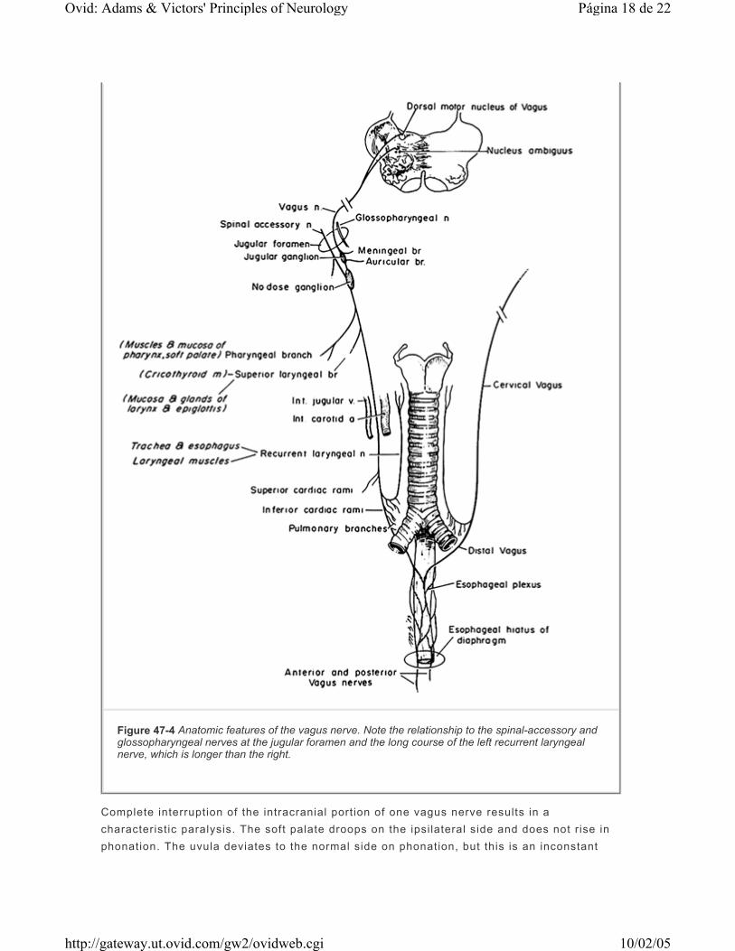
Complete interruption of the intracranial portion of one vagus nerve results in a characteristic paralysis. The soft palate droops on the ipsilateral side and does not r ise in phonation. The uvula deviates to the normal side on phonation, but this is an inconstant
Figure 47-4 Anatomic features of the vagus nerve. Note the relationship to the spinal-accessory and glossopharyngeal nerves at the jugular foramen and the long course of the left recurrent laryngeal nerve, which is longer than the right.
Página 18 de 22Ovid: Adams & Victors' Principles of Neurology
10/02/05http://gateway.ut.ovid.com/gw2/ovidweb.cgi
sign. There is loss of the gag reflex on the affected side and of the curtain movement of the lateral wall of the pharynx, whereby the faucial pil lars move medially as the palate rises in saying “ah.” The voice is hoarse, often nasal, and the vocal cord on the affected side l ies immobile in a “cadaveric” posit ion, i .e., midway between abduction and adduction. With partial lesions, movements of abduction are affected more than those of adduction (Semon's law). There may also be a loss of sensation at the external auditory meatus and back of the pinna. Usually no change in visceral function can be demonstrated. If the pharyngeal branches of both vagi are affected, as in diphtheria, the voice has a nasal quality, and regurgitation of l iquids through the nose occurs during the act of swallowing.
Complete bilateral paralysis is said to be incompatible with l i fe, and this is probably true if the nuclei are completely destroyed in the medulla by poliomyelit is or some other disease. However, in the cervical region, both vagi have been blocked with procaine in the treatment of intractable asthma, without mishap. Moreover, Johnson and Stern report a case of bi lateral vocal cord paralysis in association with familial hypertrophic polyneuropathy, and Plott relates the history of three brothers with congenital laryngeal abductor paralysis due to bilateral dysgenesis of the nucleus ambiguus. Bannister and Oppenheimer have called attention to defects of phonation and laryngeal stridor as early features of autonomic failure in multiple system atrophy (page 1137). We have seen several such patients in whom stridor was a prominent feature of the il lness and required tracheostomy for survival.
The vagus nerve may be implicated at the meningeal level by tumors and infectious processes and within the medulla by vascular lesions (e.g., the lateral medullary syndrome of Wallenberg or a cavernous angioma), by motor system disease, and occasionally by tumors. Dysphagia is then invariably present. Herpes zoster may attack this nerve, either alone or together with the ninth nerve as part of a jugular foramen syndrome. The vagus is often affected along with the glossopharyngeal nerve by dissection of the carotid artery at the base of the skull. The vagus nerves may be damaged in the course of thyroid surgery and may be involved in cases of advanced alcoholic or diabetic neuropathy. Polymyosit is and dermatomyosit is, which cause hoarseness and dysphagia by direct involvement of laryngeal and pharyngeal muscles, may simulate disease of the vagus nerves.
A fact of some importance is that the left recurrent laryngeal nerve may be damaged as a result of thoracic disease. There is no dysphagia with lesions at this point in the nerve since the branches to the pharynx (but not to the larynx) have already been given off. An aneurysm of the aortic arch, an enlarged left atrium, mediastinal lymph nodes from bronchial carcinoma, and a mediastinal or superior sulcus lung tumor are much more frequent causes of an isolated vocal cord palsy than are intracranial diseases.
It is estimated that in one-quarter to one-third of al l cases of paralysis of the recurrent laryngeal nerve no cause can be established, i.e., they are idiopathic. The highest incidence is in the third decade, and males are more susceptible than females. Of the 21 cases reported by Blau and Kapadia, 5 recovered completely and 5 partial ly within a few months; no other disease appeared in the 8-year period that followed. Palsies of the superior and recurrent laryngeal nerves, occurring as part of isolated vagal neuropathies, have been described by Berry and Blair. A few were bilateral, and again the majority of the cases were idiopathic and had much the same prognosis as isolated palsies
P.1458
Página 19 de 22Ovid: Adams & Victors' Principles of Neurology
10/02/05http://gateway.ut.ovid.com/gw2/ovidweb.cgi
of the recurrent laryngeal nerve.
Laryngeal neuralgia is a rare entity, in which paroxysms of pain are localized over the upper portion of the thyroid carti lage or hyoid bone on one or both sides. It is evoked by coughing, yawning, talking, or sneezing. In the case reported by Brownstone and coworkers it was relieved by carbamazepine.
When confronted with a case of vocal cord palsy, the physician must attempt to determine the site of the lesion. If it is intramedullary, there are usually ipsilateral cerebellar signs, loss of pain and temperature sensation over the ipsilateral face and contralateral arm and leg, and an ipsilateral Bernard-Horner syndrome. If the lesion is extramedullary but intracranial, the glossopharyngeal and spinal accessory nerves are frequently involved (jugular foramen syndrome, Table 47-1). If i t is extracranial in the posterior laterocondylar or retroparotid space, there may be a combination of ninth, tenth, eleventh, and twelfth cranial nerve palsies and a Bernard-Horner syndrome. Combinations of these lower cranial nerve palsies, which have a variety of eponymic designations (see Table 47-1), are caused by various types of tumor, both primary and metastatic, or by chronic inflammations or granulomas involving lymph nodes at the base of the skull. I f there is no palatal weakness and no pharyngeal or palatal sensory loss, the lesion is below the origin of the pharyngeal branches, which leave the vagus nerve high in the cervical region. The usual site of disease is then the mediastinum.
The 11th, or Accessory, Nerve This is a purely motor nerve, actually of spinal rather than cranial origin. Its f ibers arise from the anterior horn cells of the upper four or f ive cervical segments and enter the skull through the foramen magnum. Intracranially, the accessory nerve travels for a short distance with the part of the 10th nerve that is derived from the caudalmost cells of the nucleus ambiguus; together, the two roots are referred to as the vagal-accessory nerve or cranial root of the accessory nerve. The two roots leave the skull through the jugular foramen. The aberrant vagus fibers then rejoin the main trunk of the vagus, and the f ibers derived from the cervical segments of the spinal cord form the external ramus and innervate the sternocleidomastoid and trapezius muscles. Only the latter f ibers constitute the accessory nerve in the strict sense. In patients with tort icoll is, however, division of the upper cervical motor roots or the spinal accessory nerve has often fai led to ablate completely the contraction of the sternomastoid muscle. This suggests a wider innervation of the muscle, perhaps by f ibers of apparent vagal origin that join the accessory nerve for passage through the jugular foramen.
A complete lesion of the accessory nerve results in weakness of the sternocleidomastoid muscle and upper part of the trapezius (the lower part of the trapezius is innervated by the third and fourth cervical roots through the cervical plexus). This can be demonstrated by asking the patient to shrug his shoulders; the affected trapezius wil l be found to be weaker, and there wil l often be evident atrophy of i ts upper part. With the arms at the sides, the shoulder on the affected side droops and the scapula is slightly winged; the latter defect is accentuated with lateral movement of the arm (with serratus anterior weakness, winging of
P.1459
Página 20 de 22Ovid: Adams & Victors' Principles of Neurology
10/02/05http://gateway.ut.ovid.com/gw2/ovidweb.cgi
the scapula occurs on forward elevation of the arm). When the patient turns his head forcibly against the examiner's hand, preferably from the deviated to the straight-ahead posit ion, the sternomastoid of the opposite side does not contract f irmly beneath the f ingers. This muscle can be further tested by having the patient press his head forward against resistance or l if t his head from the pil low.
Motor system disease, poliomyelit is, syringomyelia, and spinal cord tumors may involve the cells of origin of the spinal accessory nerve. In its intracranial portion, the nerve is usually affected along with the ninth and tenth cranial nerves by herpes zoster or by lesions of the jugular foramen (glomus tumors, neurofibromas, metastatic carcinoma, jugular vein thrombosis). Tumors at the foramen magnum may also damage the nerve. In the posterior tr iangle of the neck, the 11th nerve can be damaged during surgical operations and by external compression or injury. Compressive-invasive lesions of this nerve may be visualized by computed tomography (CT) or MRI of the posterior cervical space.
A benign disorder of the 11th nerve, akin to Bell 's palsy, has been described by Spillane and by Eisen and Bertrand; it begins with pain that subsides in a few days and is fol lowed by weakness and atrophy in the distribution of the nerve. Also, a recurrent form of spontaneous accessory neuropathy has been described (Chalk and Isaacs). About one-quarter to one-third of 11th nerve lesions are estimated to be of this idiopathic type; most but not al l of the patients recover. As in the case of idiopathic brachial plexopathy, ideas about its origin are speculative.
Bilateral sternomastoid and trapezius palsy, which occurs with primary disease of muscles—e.g., polymyositis and muscular dystrophy—may be diff icult to distinguish from a bilateral affection of the accessory nerves or the motor nuclei (progressive bulbar palsy).
Hypoglossal Nerve This is also a pure motor nerve, which supplies the somatic musculature of the tongue. It arises as a series of rootlets that issue from the medulla between the pyramid and inferior ol ivary complex. The nerve leaves the skull through the hypoglossal foramen and innervates the genioglossus muscle, which acts to protrude the tongue; the styloglossus, which retracts and elevates its root; and the hypoglossus, which causes the upper surface to become convex. Complete interruption of the nerve results in paralysis of one side of the tongue. The tongue curves sl ightly to the healthy side as it l ies in the mouth, but on protrusion it deviates to the affected side, owing to the unopposed contraction of the healthy genioglossus muscle. By pushing against the tongue in the cheek, one can judge the degree of one-sided weakness. The tongue cannot be moved with natural facil i ty. The denervated side becomes wrinkled and atrophied, and fasciculations and fibri l lations can be seen.
Lesions of the hypoglossal nerve roots are rare. Occasionally an intramedullary lesion damages the emerging f ibers of the hypoglossal nerve, corticospinal tract, and medial lemniscus. The result is paralysis and atrophy of one side of the tongue, together with spastic paralysis and loss of vibration and posit ion sense in the opposite arm and leg. Poliomyelit is and motor system disease may destroy the hypoglossal nuclei. Lesions of the
P.1460
Página 21 de 22Ovid: Adams & Victors' Principles of Neurology
10/02/05http://gateway.ut.ovid.com/gw2/ovidweb.cgi
basal meninges and the occipital bones (platybasia, invagination of the occipital condyles, Paget disease) may involve the nerve in its extramedullary course, and it is sometimes damaged in operations on the neck. A dissecting aneurysm of the carotid artery was shown by Goodman and coworkers to have compressed the hypoglossal nerve, with resultant weakness and atrophy. Lance and Anthony have described the simultaneous occurrence of nuchal-occipital pain and ipsilateral numbness of the tongue, provoked by the sudden, sharp turning of the head (“neck-tongue syndrome”). The phenomenon is attr ibuted to compression, in the atlantoaxial space, of the second cervical root, which carries some of the sensory f ibers from the tongue, via the hypoglossal nerve, to the C2 segment of the spinal cord.
The tongue may be red and smooth in vitamin deficiency states. Glossodynia (burning pain of the tongue), a condit ion most frequently seen in the elderly, may or may not be accompanied by redness and dryness but not by weakness. A habit of tongue-thrusting and teeth-clenching is often associated. The facile ascription of these motor abnormalit ies to a psychogenic mechanism does not accord with the authors' experience (see also Quinn).
Syndrome of Bulbar Palsy This syndrome is the result of weakness or paralysis of muscles that are supplied by the motor nuclei of the lower brainstem, i.e., the motor nuclei of the f i f th, seventh, and ninth to twelfth cranial nerves. (Strictly speaking, the motor nuclei of the f i f th and seventh nerves lie outside the “bulb,” which is the old name for the medulla oblongata.) Involved are the muscles of the jaw and face; the sternomastoids and upper parts of the trapezii; and the muscles of the tongue, pharynx, and larynx. If weakness develops rapidly, as may happen in Guil lain-Barré syndrome, diphtheria, or poliomyelit is, there is no t ime for muscle atrophy. Myasthenia gravis and polymyosit is may on rare occasions produce such a picture. The more chronic diseases—e.g., progressive bulbar palsy (a form of motor system disease) and the childhood form of Fazio-Londe—result in marked wasting and fasciculation of the facial, tongue, sternomastoid, and trapezius muscles. These disorders must be differentiated from pseudobulbar palsy (see page 517).
MULTIPLE CRANIAL NERVE PALSIES
Página 22 de 22Ovid: Adams & Victors' Principles of Neurology
10/02/05http://gateway.ut.ovid.com/gw2/ovidweb.cgi