Embed Size (px)
Citation preview
Chapter 6: A Tour Of the Cell
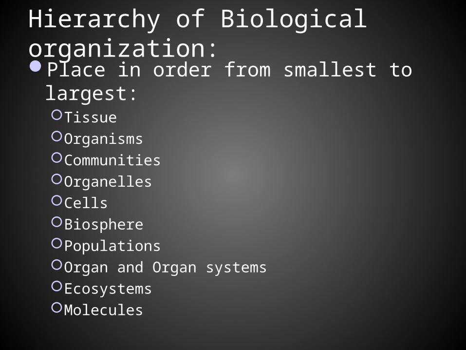
Hierarchy of Biological organization: Place in order from smallest to largest:
TissueOrganismsCommunitiesOrganellesCellsBiospherePopulationsOrgan and Organ systemsEcosystemsMolecules
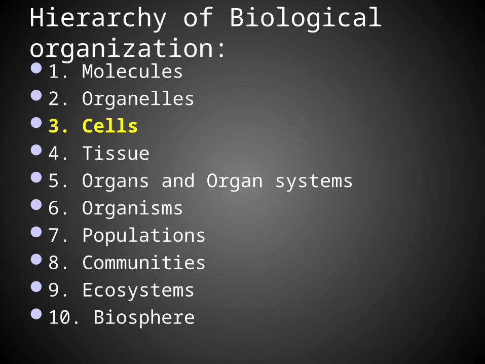
Hierarchy of Biological organization: 1. Molecules2. Organelles3. Cells4. Tissue5. Organs and Organ systems6. Organisms7. Populations8. Communities9. Ecosystems10. Biosphere
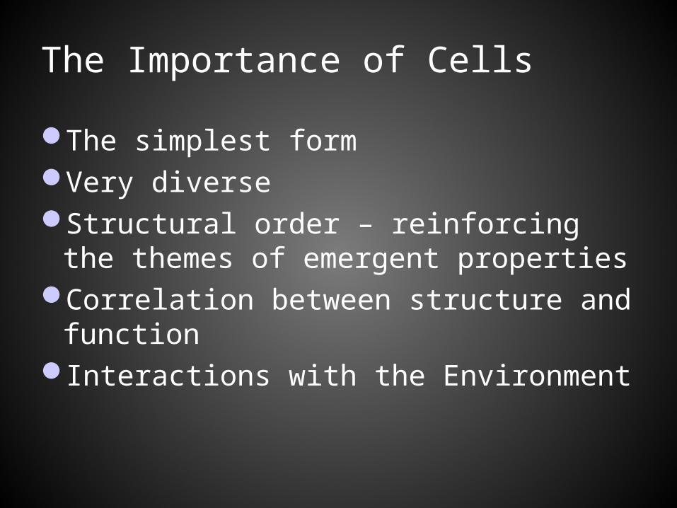
The Importance of Cells
The simplest formVery diverseStructural order – reinforcing the
themes of emergent propertiesCorrelation between structure and
functionInteractions with the Environment
How do biologists study cells:
Two methods: 1. Microscopy- investigations employing a
microscope2. Cell Fractionation- using the tools of
biochemistry
Microscopy: Investigations employing a microscope A. Two important parameters:
Magnification- the ratio of an object’s image size to its real size
Resolution- a measure of the clarity of the image; the minimum distance 2 points can be separated and still be distinguished as 2 points Resolution is inversely related to
wavelength
Microscopy: Investigations employing a microscope
B. Two types: Light microscopes (LMs) Electron Microscope (EM)
Light Microscopy:
Passing visible light through a lens, then a specimen, then to the eye. Lens refracts the light to magnify the image
Limited by the shortest wavelength of visible light used to illuminate the specimen.
Electron microscope:
Focuses a beam of electrons either on the surface or through a specimen
Much shorter wavelengths (remember the relationship between resolution and wavelength?)Resolution of about 2nm- a hundred- fold
improvement over light microscopes
Electron microscope:
Two types: Scanning Electron Microscope (SEM):Transmission Electron Microscope(TEM):
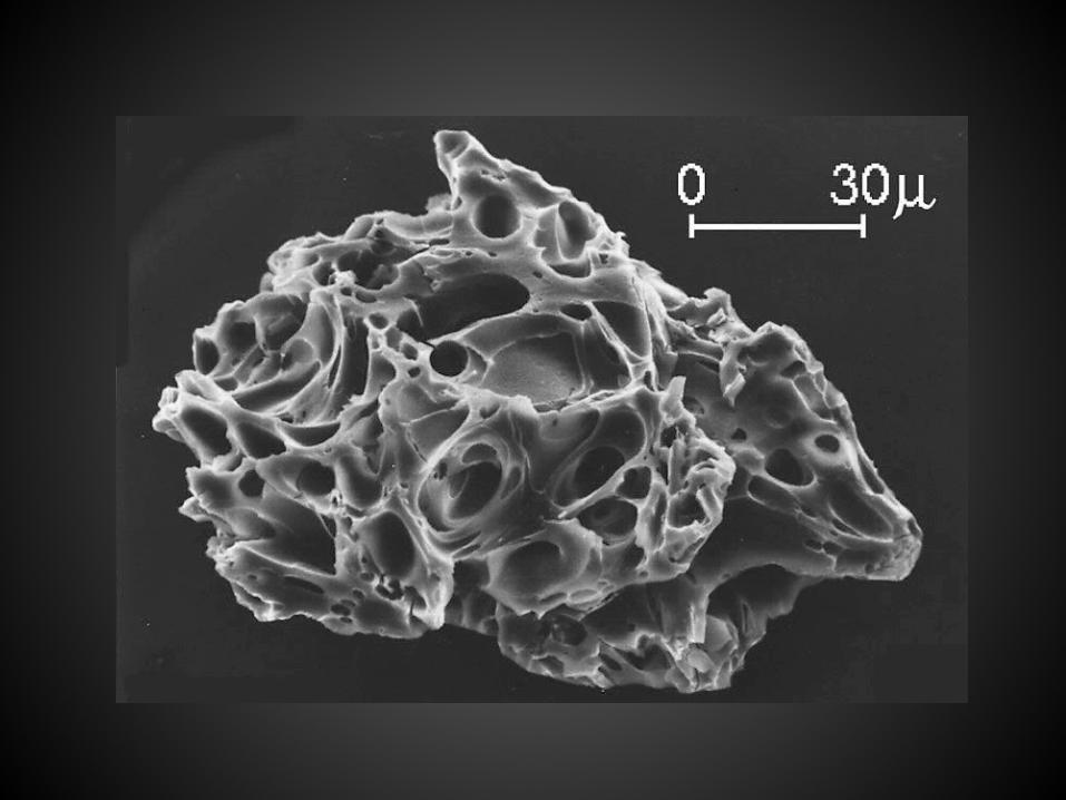
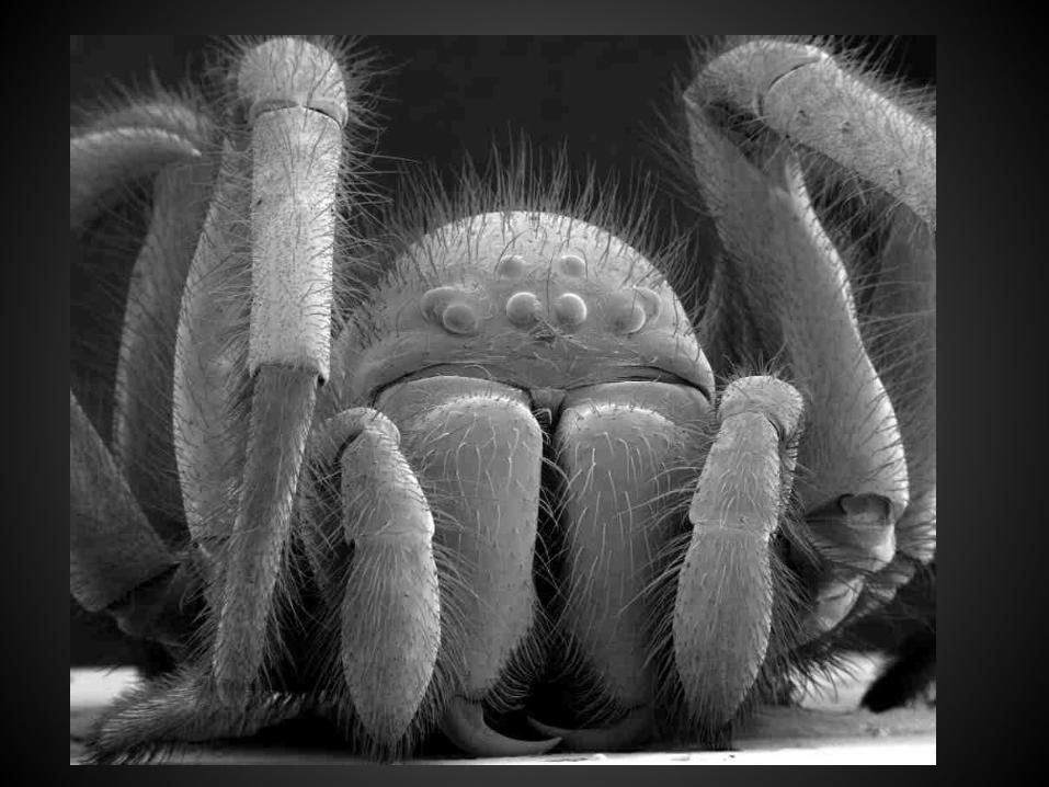
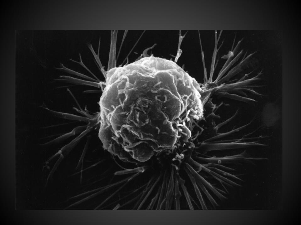
Scanning Electron Microscope(SEM)
Useful in detailed study of surface of specimenSpecimen is covered with a thin layer of goldGreat depth of field3-D like image
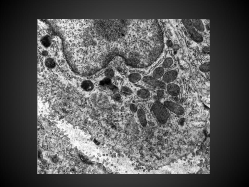
Transmission Electron Microscope (TEM):
Used to study internal structures of the cellUses electromagnets as lenses
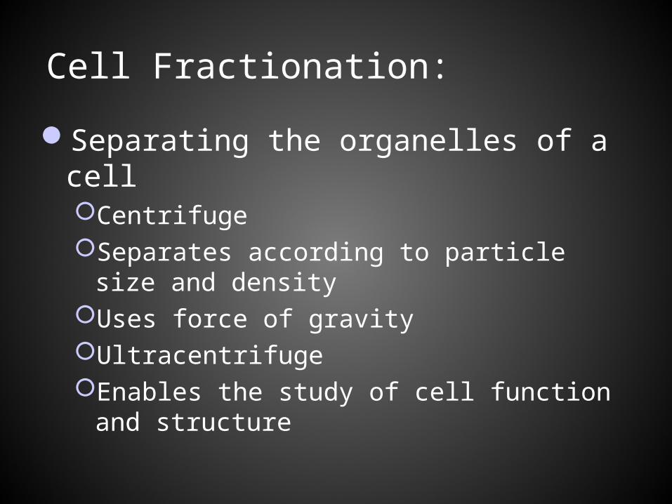
Cell Fractionation:
Separating the organelles of a cellCentrifugeSeparates according to particle size and
densityUses force of gravityUltracentrifugeEnables the study of cell function and structure
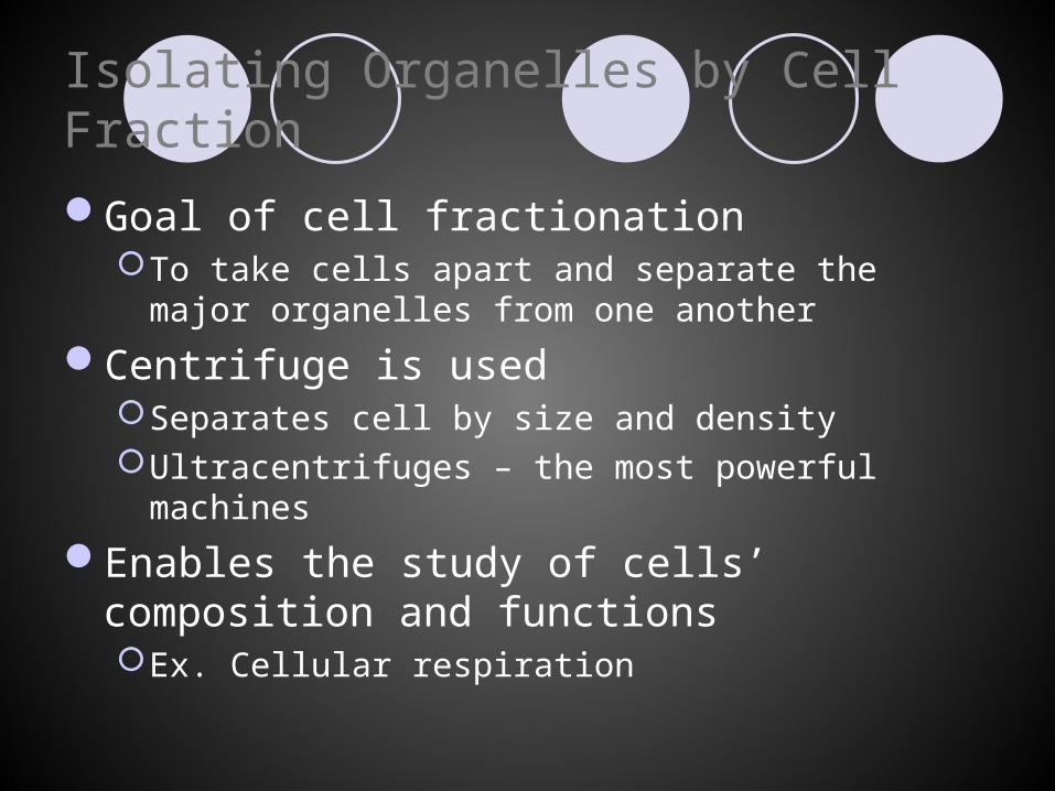
Isolating Organelles by Cell Fraction
Goal of cell fractionationTo take cells apart and separate the major organelles
from one another
Centrifuge is usedSeparates cell by size and densityUltracentrifuges – the most powerful machines
Enables the study of cells’ composition and functionsEx. Cellular respiration
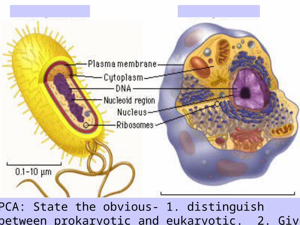
PCA: State the obvious- 1. distinguish between prokaryotic and eukaryotic. 2. Give two obvious differences.
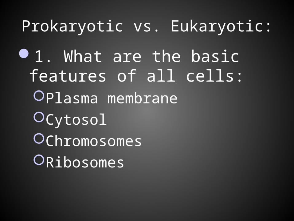
Prokaryotic vs. Eukaryotic:
1. What are the basic features of all cells: Plasma membraneCytosolChromosomesRibosomes
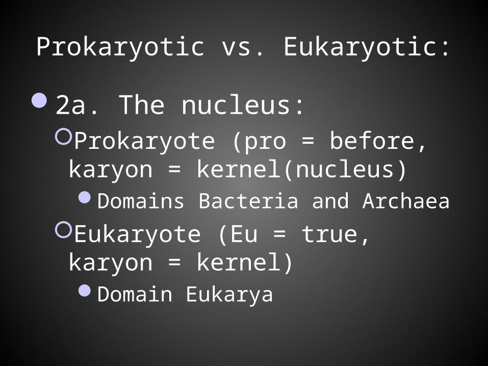
Prokaryotic vs. Eukaryotic:
2a. The nucleus: Prokaryote (pro = before, karyon =
kernel(nucleus)Domains Bacteria and Archaea
Eukaryote (Eu = true, karyon = kernel)Domain Eukarya
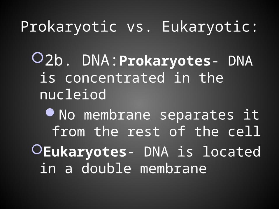
Prokaryotic vs. Eukaryotic:
2b. DNA:Prokaryotes- DNA is concentrated in the nucleiod No membrane separates it from the rest of the cell
Eukaryotes- DNA is located in a double membrane
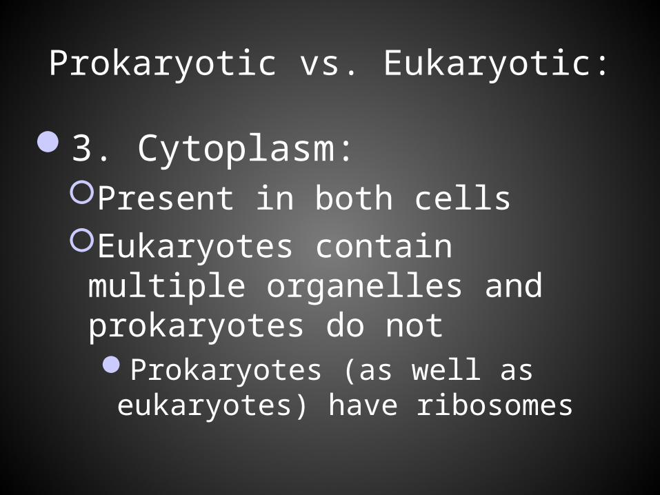
Prokaryotic vs. Eukaryotic:
3. Cytoplasm:Present in both cellsEukaryotes contain multiple
organelles and prokaryotes do not Prokaryotes (as well as eukaryotes)
have ribosomes
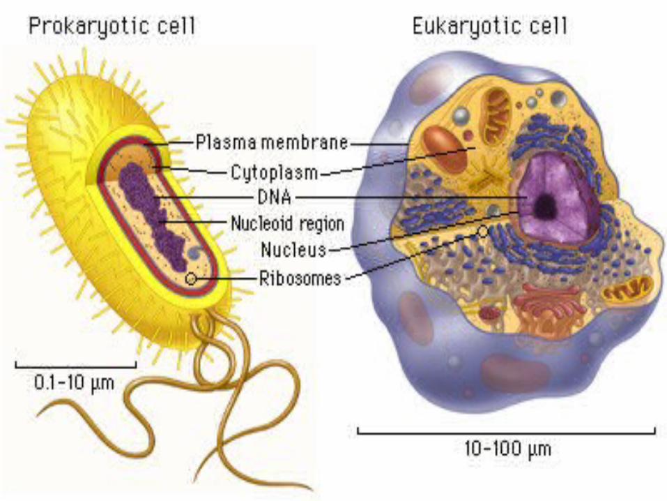
Prokaryotic vs. Eukaryotic:
For other features see diagram:
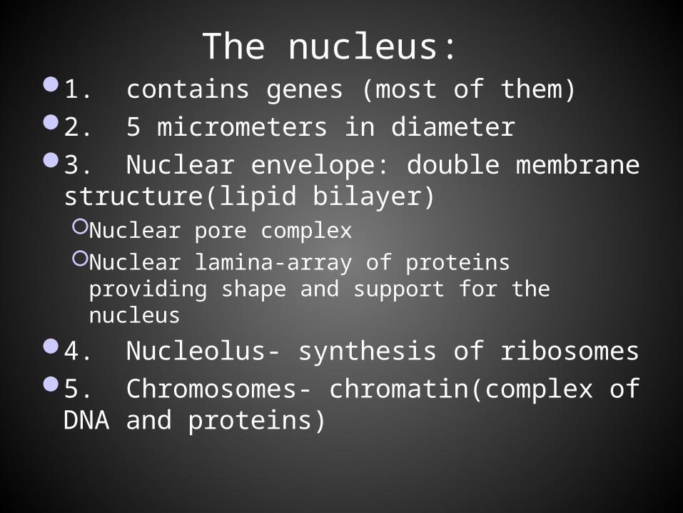
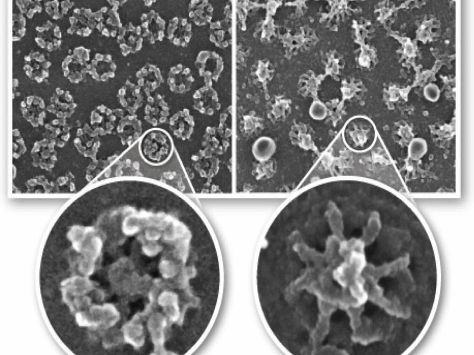
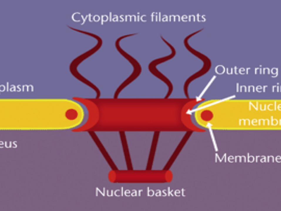
The nucleus: 1. contains genes (most of them)2. 5 micrometers in diameter3. Nuclear envelope: double membrane
structure(lipid bilayer)Nuclear pore complexNuclear lamina-array of proteins providing
shape and support for the nucleus
4. Nucleolus- synthesis of ribosomes5. Chromosomes- chromatin(complex of
DNA and proteins)
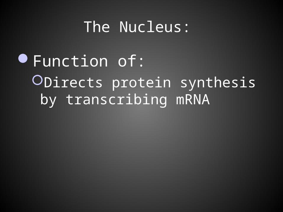
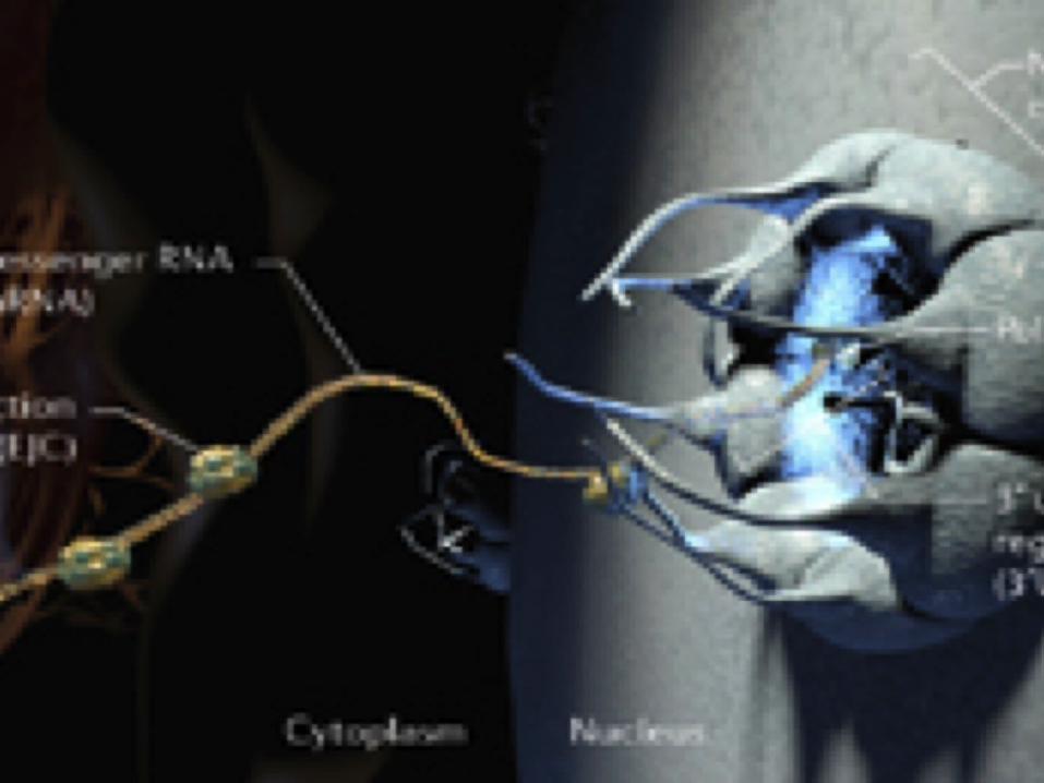
The Nucleus:
Function of: Directs protein synthesis by
transcribing mRNA
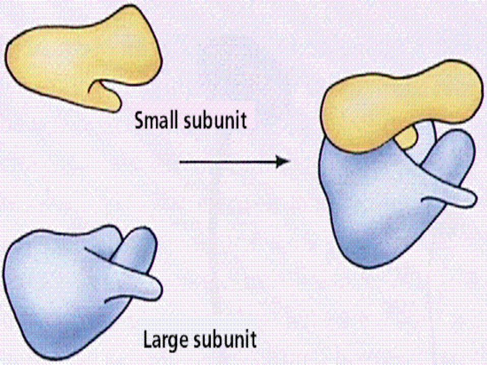
Ribosomes:
Protein factories23nm in diameterComposed of half RNA and half proteinOne large subunit and one small subunitSynthesized at the nucleolusHalf million ribosomes per cell
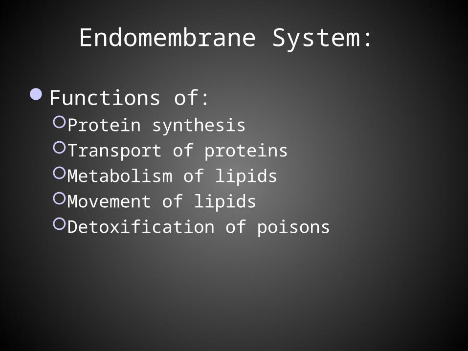
Endomembrane System:
Functions of:Protein synthesisTransport of proteinsMetabolism of lipidsMovement of lipidsDetoxification of poisons
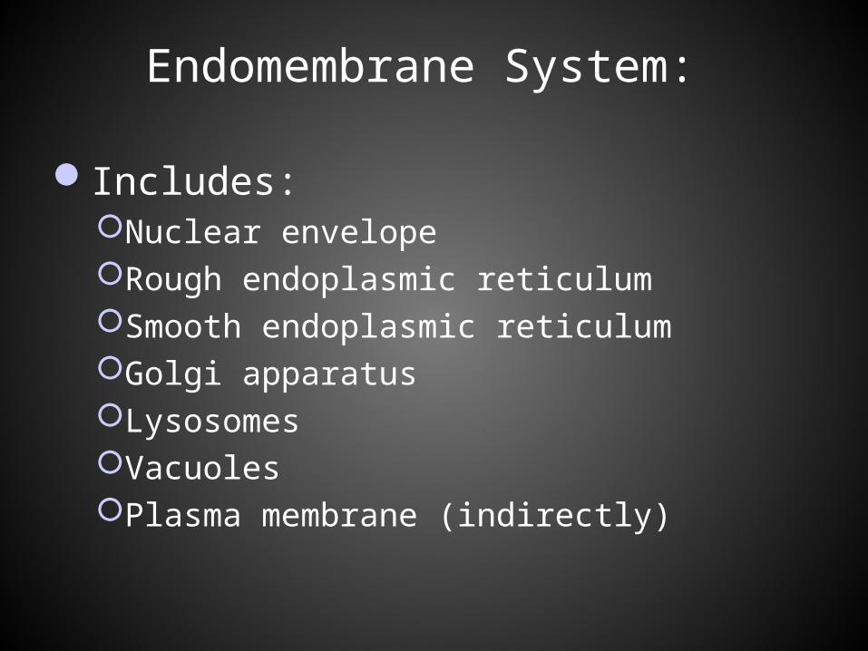
Endomembrane System:
Includes:Nuclear envelopeRough endoplasmic reticulumSmooth endoplasmic reticulumGolgi apparatusLysosomesVacuolesPlasma membrane (indirectly)
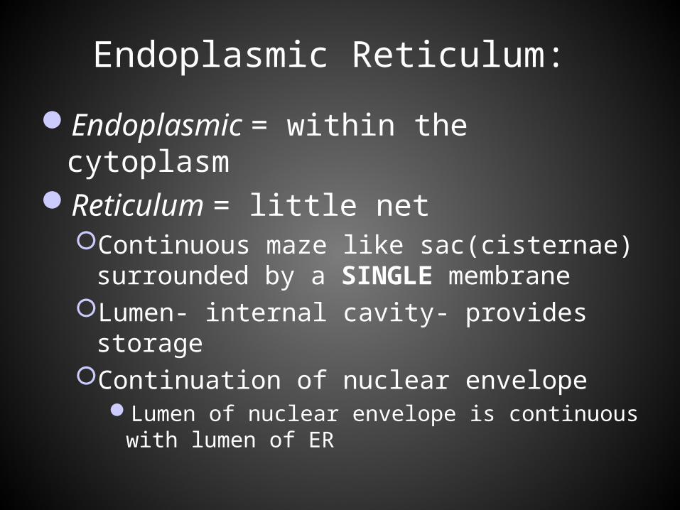
Endoplasmic Reticulum:
Endoplasmic = within the cytoplasmReticulum = little net
Continuous maze like sac(cisternae) surrounded by a SINGLE membrane
Lumen- internal cavity- provides storageContinuation of nuclear envelope
Lumen of nuclear envelope is continuous with lumen of ER
Endomembrane System:
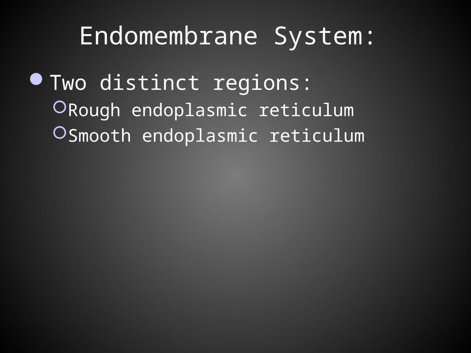
Two distinct regions:Rough endoplasmic reticulumSmooth endoplasmic reticulum
Endomembrane System:
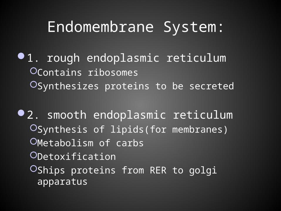
1. rough endoplasmic reticulumContains ribosomesSynthesizes proteins to be secreted
2. smooth endoplasmic reticulum Synthesis of lipids(for membranes)Metabolism of carbsDetoxificationShips proteins from RER to golgi apparatus
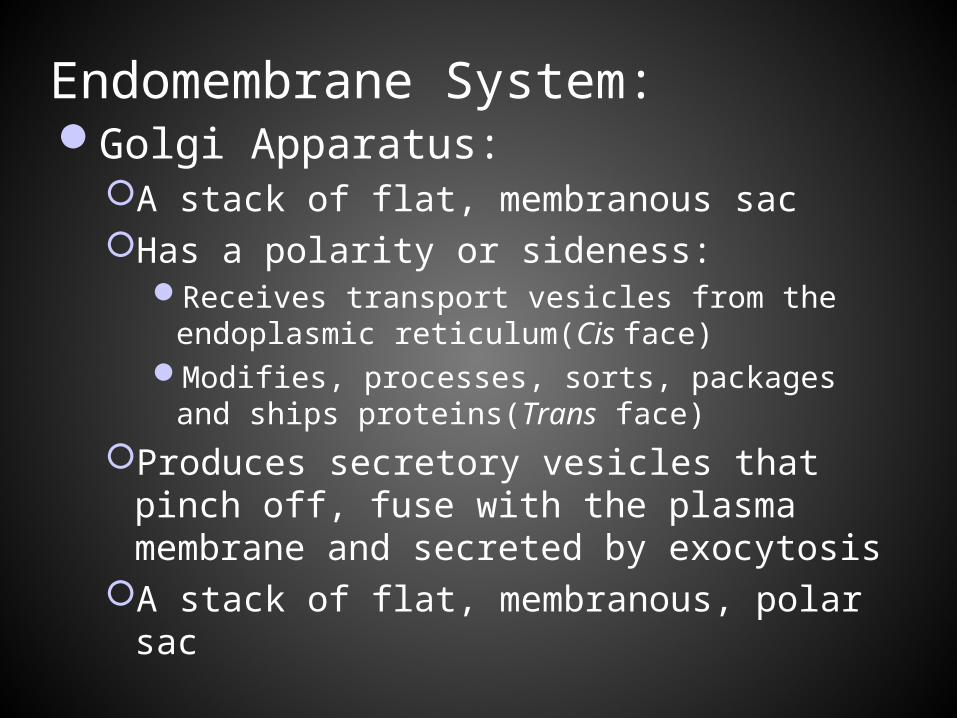
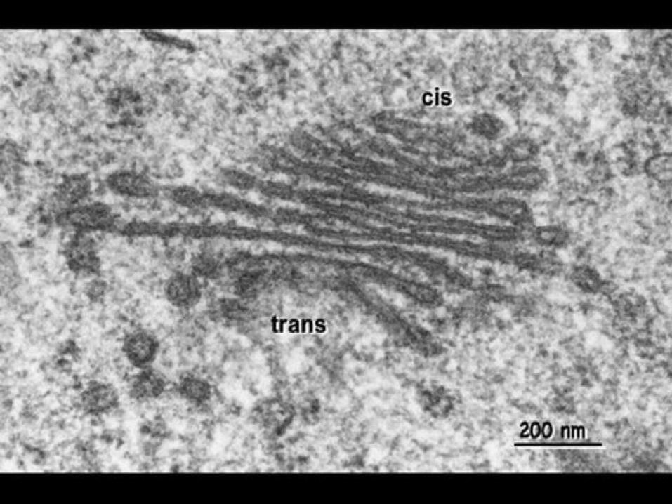
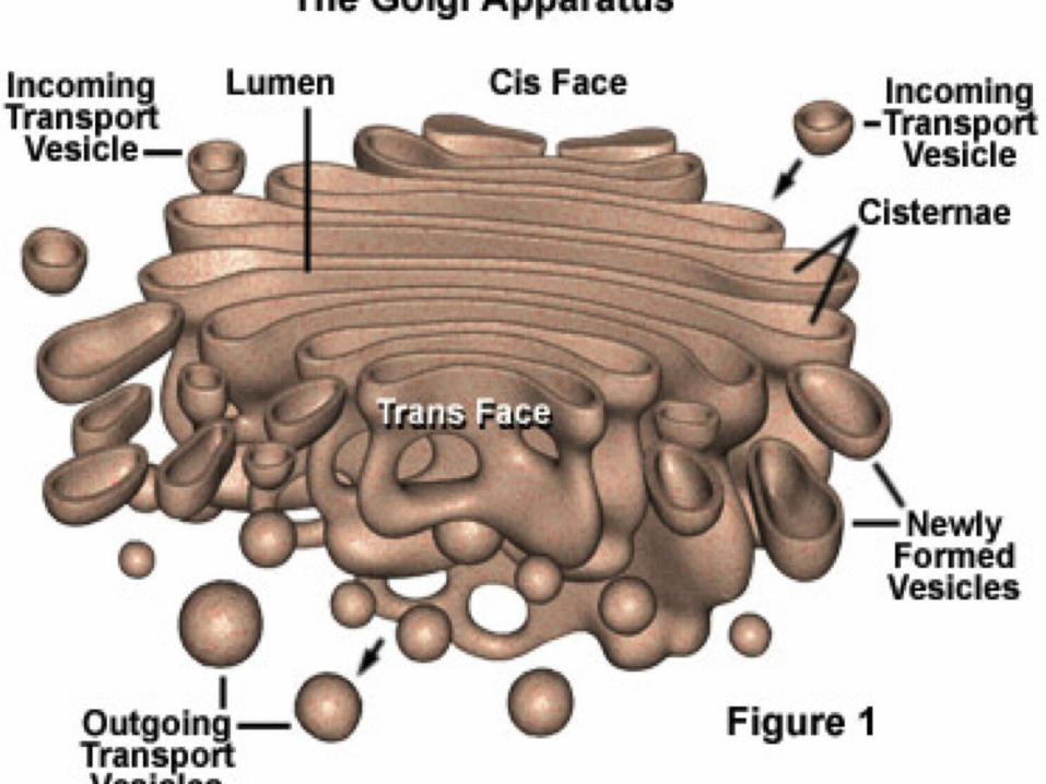
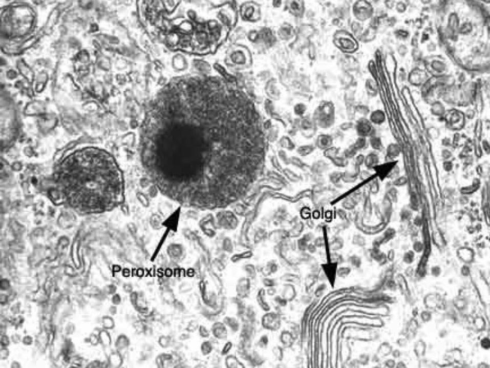
Endomembrane System: Golgi Apparatus:
A stack of flat, membranous sacHas a polarity or sideness:
Receives transport vesicles from the endoplasmic reticulum(Cis face)
Modifies, processes, sorts, packages and ships proteins(Trans face)
Produces secretory vesicles that pinch off, fuse with the plasma membrane and secreted by exocytosis
A stack of flat, membranous, polar sac
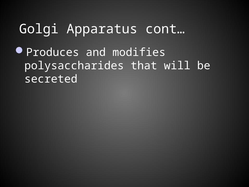
Golgi Apparatus cont…
Produces and modifies polysaccharides that will be secreted
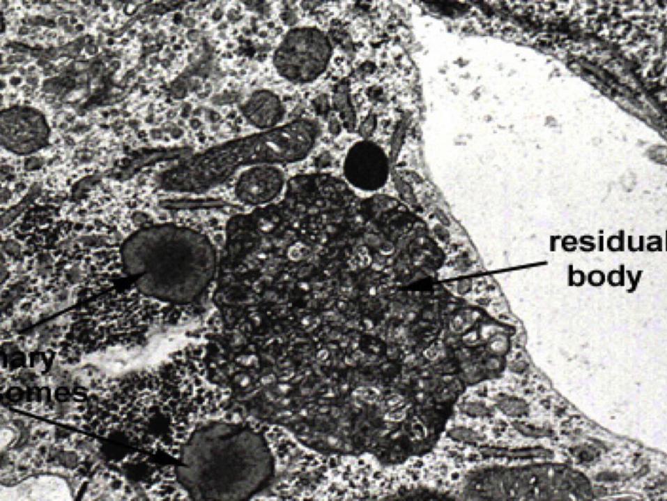
Endomembrane system: Lysosomes:
Single membrane bound sac containing hydrolytic digestive enzymes
Used to digest macromoleculesSynthesized in the rough endoplasmic
reticulum, sent to the golgi apparatus and pinched off as a vesicle with over forty digestive enzymes
Recycle the cells organic materialLipid digestion enzyme in this organelle can
lead to Tay- Sachs disease
Endomembrane System:
Peroxisomes: Single membrane organelles that function in the
synthesis of fatty acidsProduce hydrogen peroxide as a by-productFunction in detoxifying alcohol and other
poisons in the liverBreak down fatty acids into smaller molecules
which can be transferred to the mitochondria for cellular respiration
Break down molecules through oxidation
Endomembrane System:Glyoxosomes:
Specialized peroxisomesFound in fat storing tissues of plant seedsConvert fatty acids to sugars for the emerging
seedlingCapable of increasing in numbers by splitting in
two when they reach a certain size
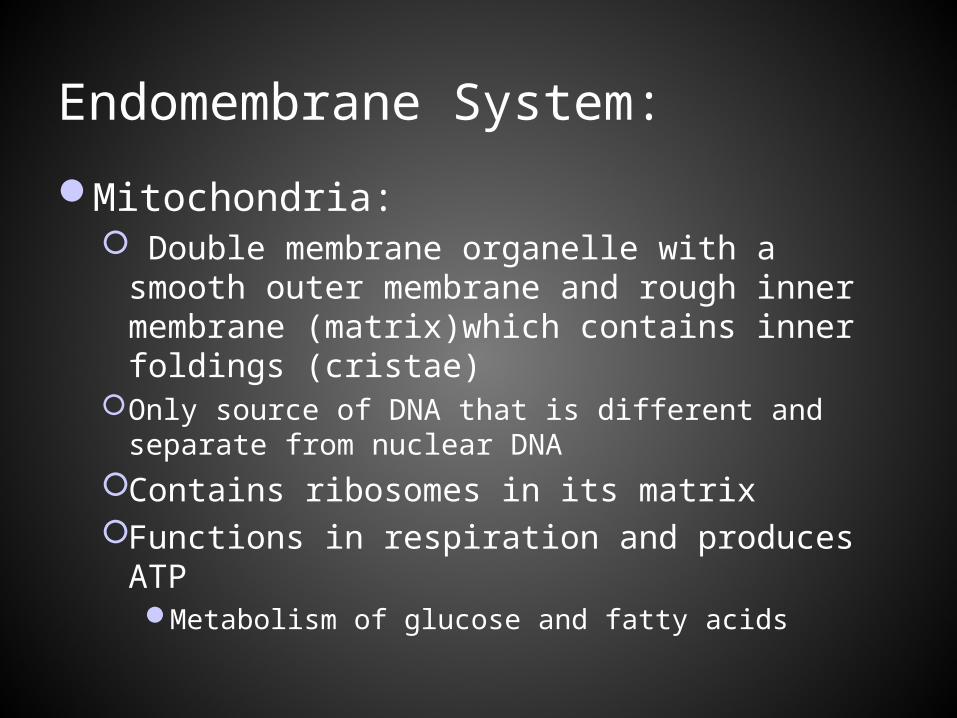
Endomembrane System:
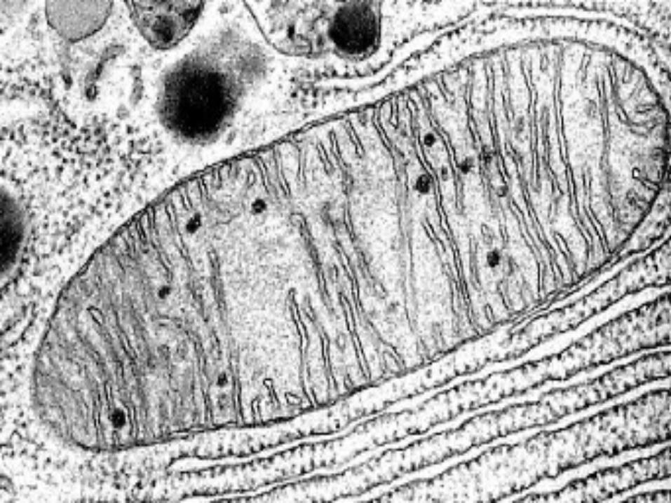
Mitochondria: Double membrane organelle with a smooth
outer membrane and rough inner membrane (matrix)which contains inner foldings (cristae)
Only source of DNA that is different and separate from nuclear DNA
Contains ribosomes in its matrixFunctions in respiration and produces ATP
Metabolism of glucose and fatty acids
Endomembrane System:
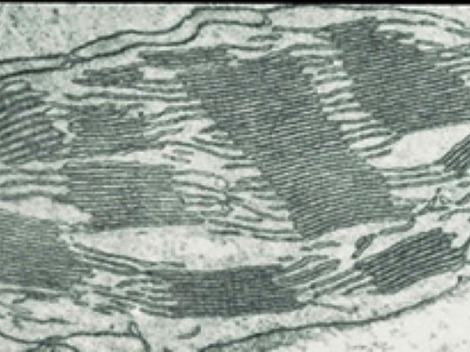
Chloroplasts: Belongs to a group of organelles called plastidsFunctions in the photosynthetic production of
glucose(used to make ATP)Convert light energy to chemical energyDouble membrane structure that contains
thylakoid sacsStacks of thylakoid membranes = granum(grana)
Stroma- fluid surrounding thylakoid, contains DNA, ribosomes and enzymes.
Endomembrane System:
Mitochondria and Chloroplasts:Contain DNA, ribosomes and enzymesCan change shape, grow and occasionally split
into twomobile
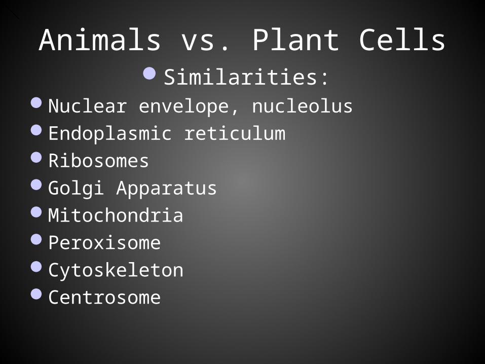
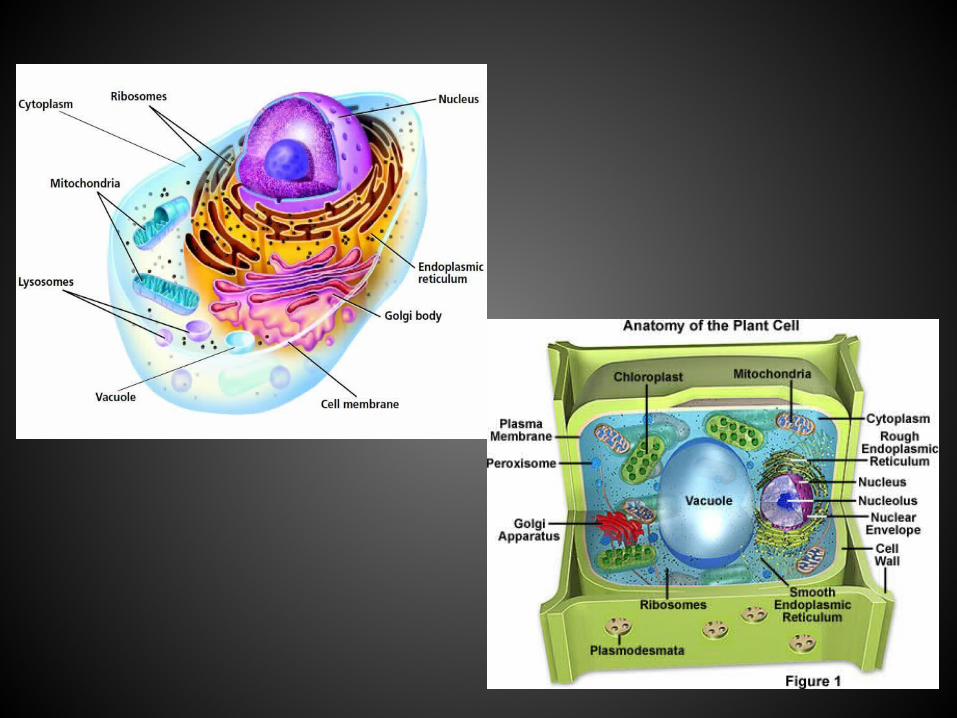
Animals vs. Plant CellsSimilarities:
Nuclear envelope, nucleolusEndoplasmic reticulumRibosomesGolgi ApparatusMitochondriaPeroxisomeCytoskeletonCentrosome
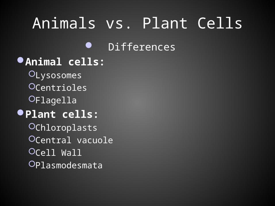
Animals vs. Plant Cells Differences
Animal cells:LysosomesCentriolesFlagella
Plant cells: ChloroplastsCentral vacuoleCell WallPlasmodesmata
Cytoskeleton:
a network of protein fibers that plays a role in organizing the structure and activities of the cell:
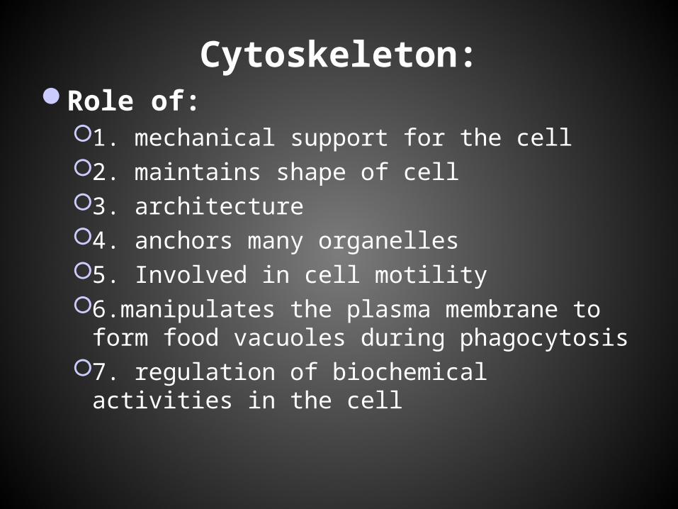
Cytoskeleton:Role of:
1. mechanical support for the cell2. maintains shape of cell3. architecture4. anchors many organelles5. Involved in cell motility6.manipulates the plasma membrane to form
food vacuoles during phagocytosis7. regulation of biochemical activities in the cell
Cytoskeleton:
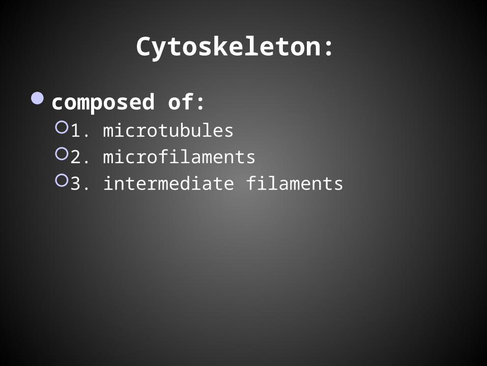
composed of:1. microtubules2. microfilaments3. intermediate filaments
Microtubules:
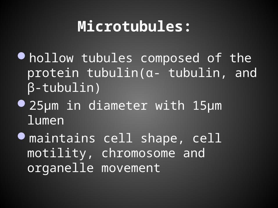
hollow tubules composed of the protein tubulin(α- tubulin, and β-tubulin)
25μm in diameter with 15μm lumenmaintains cell shape, cell motility,
chromosome and organelle movement
Types of Microtubules:
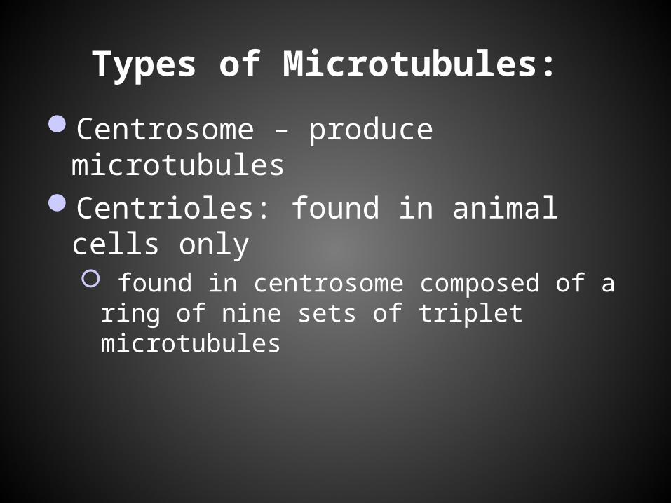
Centrosome – produce microtubulesCentrioles: found in animal cells only
found in centrosome composed of a ring of nine sets of triplet microtubules
Types of Microtubules:
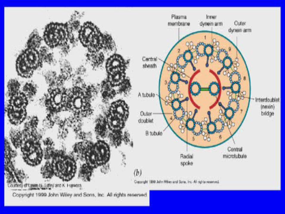
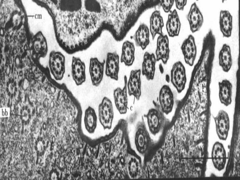
Cilia- locomotor appendage25 µm in diameter, 2- 20µm in lengthon stationary cells, cilia move objects on the
cell surfacecilia propels cell in direction perpendicular to
cilia’s axis
Types of Microtubules:
Flagella: locomotor appendages much larger than ciliasame diameter as cilia(.25µm) but much longer
10-200µmpropels cell in same direction as flagellum’s
axisundulating, snake like motion

Types of Microtubules:
Basal body: anchors flagella or cilia to the cellstructurally identical to centriole
Types of microfilaments:
Actincan form 3-D structure to help support cell
shapemake up core of microvillifound in skeletal muscle cells and used for
muscle contraction
Myosinmotor proteins
Actin/ Myosin Interactions:
slide filament theory- results in muscle contraction
cytokinesisamoeboid movement- pseudopodia-
converts cytoplasm from sol(liquid) to gel cytoplasmic streaming
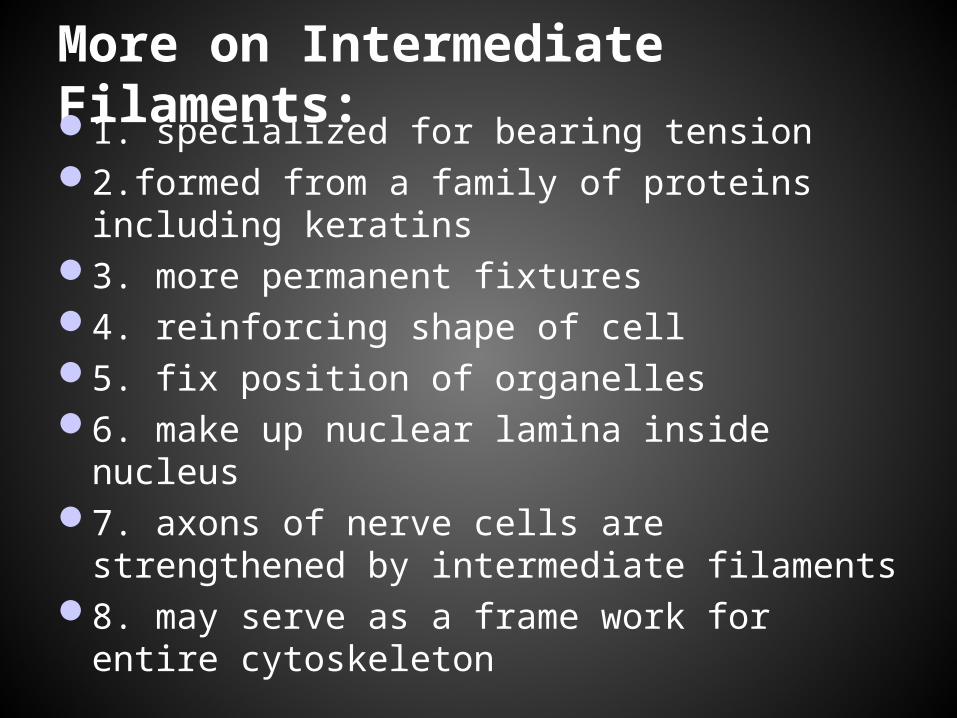
More on Intermediate Filaments: 1. specialized for bearing tension2.formed from a family of proteins including
keratins3. more permanent fixtures4. reinforcing shape of cell5. fix position of organelles6. make up nuclear lamina inside nucleus7. axons of nerve cells are strengthened by
intermediate filaments8. may serve as a frame work for entire
cytoskeleton
Extracellular components and connections
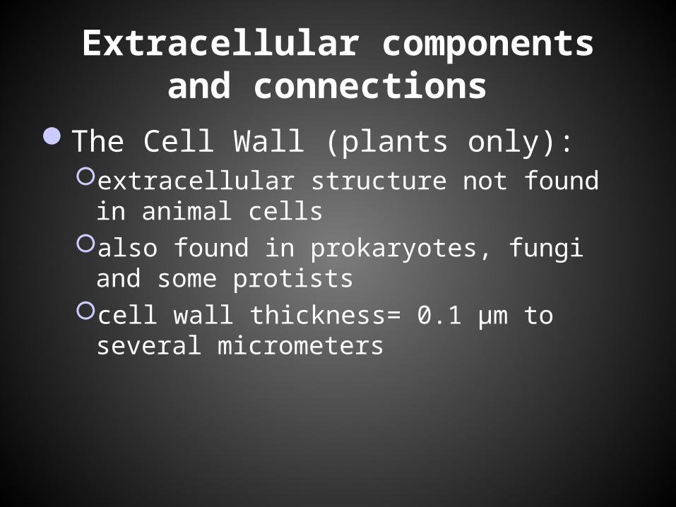
The Cell Wall (plants only): extracellular structure not found in animal cellsalso found in prokaryotes, fungi and some
protistscell wall thickness= 0.1 µm to several
micrometers
Extracellular components and connections
Role of the Cell Wall: protects the plant cellmaintains shapeprevents excessive uptake of waterresist force of gravity
Extracellular components and connections
cell wall design: polysaccharide cellulose microfibrils embedded
in a matrix of other polysaccharides and proteins
primary cell wall- cell wall secreted by the young plant
middle lamella: thin layer located between two adjacent plant cells
secondary cell wall: added between the plasma membrane and the primary cell wall
Extracellular components and connections
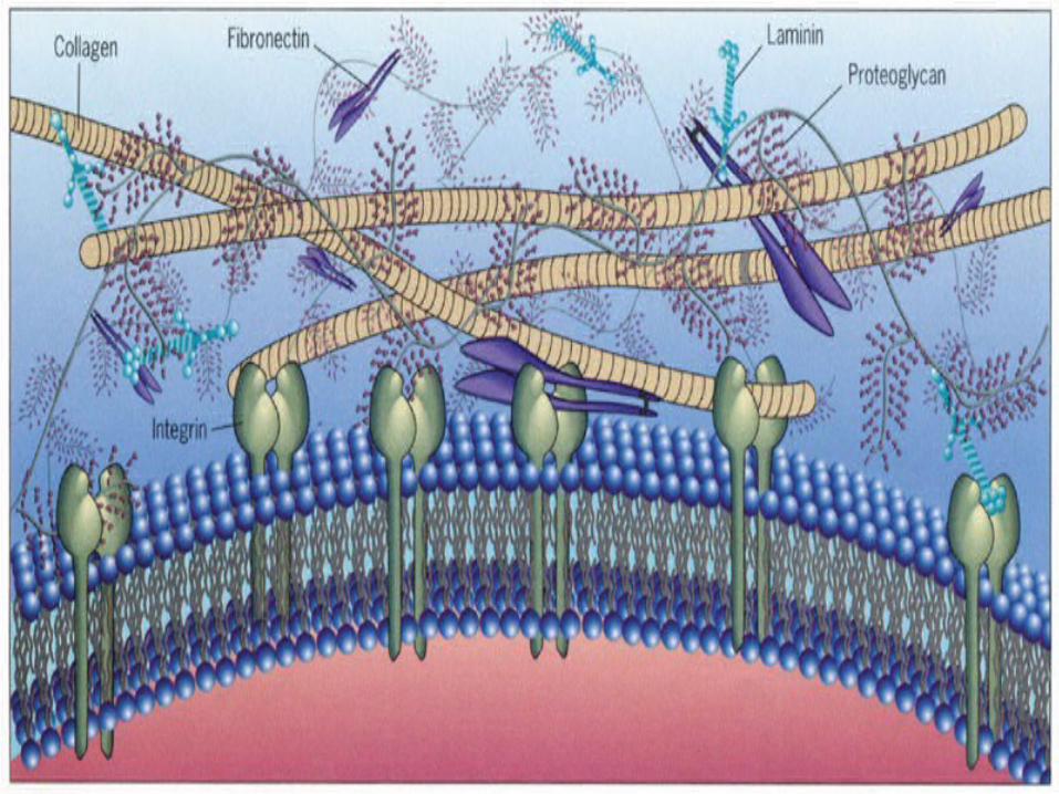
The Extracellular Matrix(ECM) of animal cellsGlycoproteins
Mainly collagenIntegrinFibronectin
functions in regulating passage of materialsExample: blood platelets
Extracellular components and connections
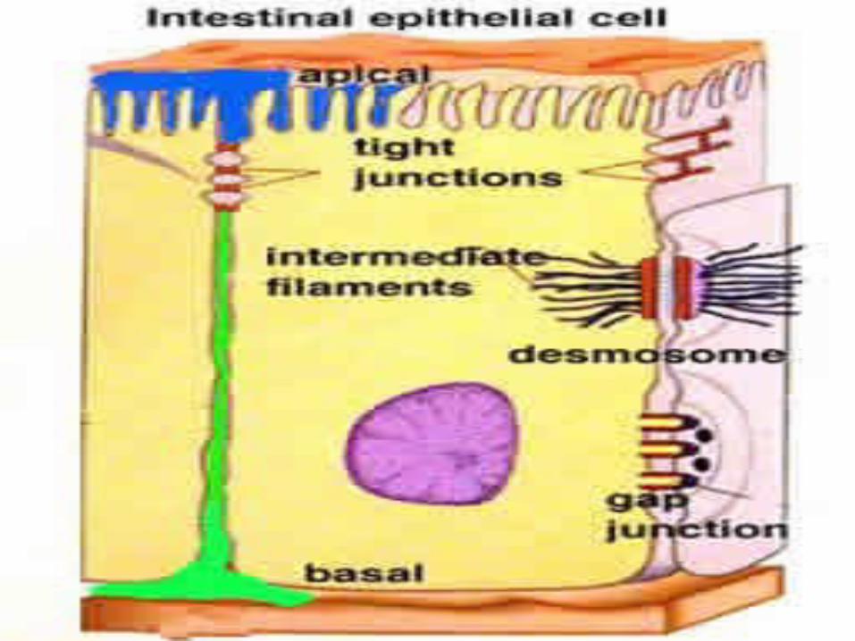
Intercellular Junctions: cell to cell communication through direct physical contactplasmodesmatatight junctionsdesmosomes:gap junctions:
Extracellular components and connections
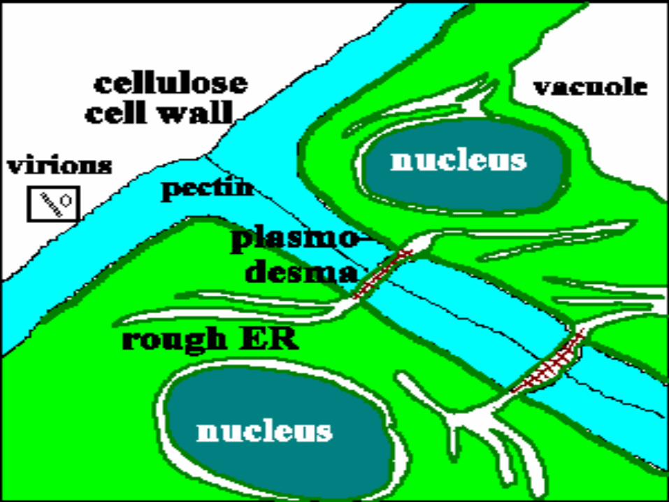
plasmodesmata: perforations of the cell wallchemical communication between the cytosol of
one cell to the nextunify plants into a living columncontinuous from one cell to the nextallows water, solutes, specific proteins and
RNA molecules to move from cell to cell
Extracellular components and connections
Tight Junctions: series of connections between two animal cells that form a criss- cross pattern Interacts with proteins between cells
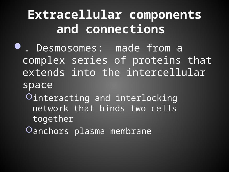
Extracellular components and connections
. Desmosomes: made from a complex series of proteins that extends into the intercellular spaceinteracting and interlocking network that binds
two cells togetheranchors plasma membrane
Extracellular components and connections
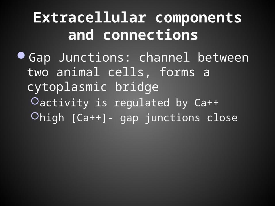
Gap Junctions: channel between two animal cells, forms a cytoplasmic bridgeactivity is regulated by Ca++high [Ca++]- gap junctions close
http://highered.mcgraw-hill.com/sites/9834092339/student_view0/chapter4/animation_-_endosymbiosis.html
Possible Essay Questions:
1. Discuss the endosymbiotic hypothesis. 2. Discuss the similarities and differences
between animal and plant cells.3. How does information flow through the
cell for proteins destined to be secreted by the cell.
4. How would you distinguish a prokaryotic cell from an animal cell. What are the similarities and differences?