Embed Size (px)
Citation preview

Chapter 7, “Cellular Respiration” from Biology by Boundless is available under a Creative Commons Attribution-ShareAlike 4.0 International license. © 2015, boundless.com.
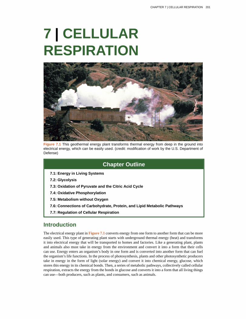
7 | CELLULARRESPIRATION
Figure 7.1 This geothermal energy plant transforms thermal energy from deep in the ground intoelectrical energy, which can be easily used. (credit: modification of work by the U.S. Department ofDefense)
Chapter Outline7.1: Energy in Living Systems
7.2: Glycolysis
7.3: Oxidation of Pyruvate and the Citric Acid Cycle
7.4: Oxidative Phosphorylation
7.5: Metabolism without Oxygen
7.6: Connections of Carbohydrate, Protein, and Lipid Metabolic Pathways
7.7: Regulation of Cellular Respiration
IntroductionThe electrical energy plant in Figure 7.1 converts energy from one form to another form that can be moreeasily used. This type of generating plant starts with underground thermal energy (heat) and transformsit into electrical energy that will be transported to homes and factories. Like a generating plant, plantsand animals also must take in energy from the environment and convert it into a form that their cellscan use. Energy enters an organism’s body in one form and is converted into another form that can fuelthe organism’s life functions. In the process of photosynthesis, plants and other photosynthetic producerstake in energy in the form of light (solar energy) and convert it into chemical energy, glucose, whichstores this energy in its chemical bonds. Then, a series of metabolic pathways, collectively called cellularrespiration, extracts the energy from the bonds in glucose and converts it into a form that all living thingscan use—both producers, such as plants, and consumers, such as animals.
CHAPTER 7 | CELLULAR RESPIRATION 201
7.1 | Energy in Living Systems
By the end of this section, you will be able to:
• Discuss the importance of electrons in the transfer of energy in living systems
• Explain how ATP is used by the cell as an energy source
Energy production within a cell involves many coordinated chemical pathways. Most of these pathwaysare combinations of oxidation and reduction reactions. Oxidation and reduction occur in tandem. Anoxidation reaction strips an electron from an atom in a compound, and the addition of this electron toanother compound is a reduction reaction. Because oxidation and reduction usually occur together, thesepairs of reactions are called oxidation reduction reactions, or redox reactions.
Electrons and EnergyThe removal of an electron from a molecule, oxidizing it, results in a decrease in potential energy in theoxidized compound. The electron (sometimes as part of a hydrogen atom), does not remain unbonded,however, in the cytoplasm of a cell. Rather, the electron is shifted to a second compound, reducingthe second compound. The shift of an electron from one compound to another removes some potentialenergy from the first compound (the oxidized compound) and increases the potential energy of thesecond compound (the reduced compound). The transfer of electrons between molecules is importantbecause most of the energy stored in atoms and used to fuel cell functions is in the form of high-energyelectrons. The transfer of energy in the form of electrons allows the cell to transfer and use energy in anincremental fashion—in small packages rather than in a single, destructive burst. This chapter focuseson the extraction of energy from food; you will see that as you track the path of the transfers, you aretracking the path of electrons moving through metabolic pathways.
Electron Carriers
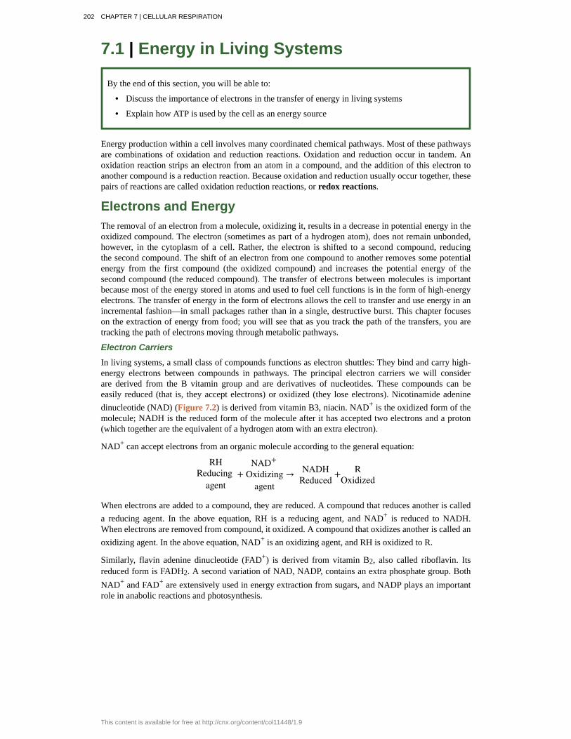
In living systems, a small class of compounds functions as electron shuttles: They bind and carry high-energy electrons between compounds in pathways. The principal electron carriers we will considerare derived from the B vitamin group and are derivatives of nucleotides. These compounds can beeasily reduced (that is, they accept electrons) or oxidized (they lose electrons). Nicotinamide adeninedinucleotide (NAD) (Figure 7.2) is derived from vitamin B3, niacin. NAD+ is the oxidized form of themolecule; NADH is the reduced form of the molecule after it has accepted two electrons and a proton(which together are the equivalent of a hydrogen atom with an extra electron).
NAD+ can accept electrons from an organic molecule according to the general equation:
RHReducing
agent +
NAD+
Oxidizingagent
→ NADHReduced
+ ROxidized
When electrons are added to a compound, they are reduced. A compound that reduces another is calleda reducing agent. In the above equation, RH is a reducing agent, and NAD+ is reduced to NADH.When electrons are removed from compound, it oxidized. A compound that oxidizes another is called anoxidizing agent. In the above equation, NAD+ is an oxidizing agent, and RH is oxidized to R.
Similarly, flavin adenine dinucleotide (FAD+) is derived from vitamin B2, also called riboflavin. Itsreduced form is FADH2. A second variation of NAD, NADP, contains an extra phosphate group. BothNAD+ and FAD+ are extensively used in energy extraction from sugars, and NADP plays an importantrole in anabolic reactions and photosynthesis.
202 CHAPTER 7 | CELLULAR RESPIRATION
This content is available for free at http://cnx.org/content/col11448/1.9
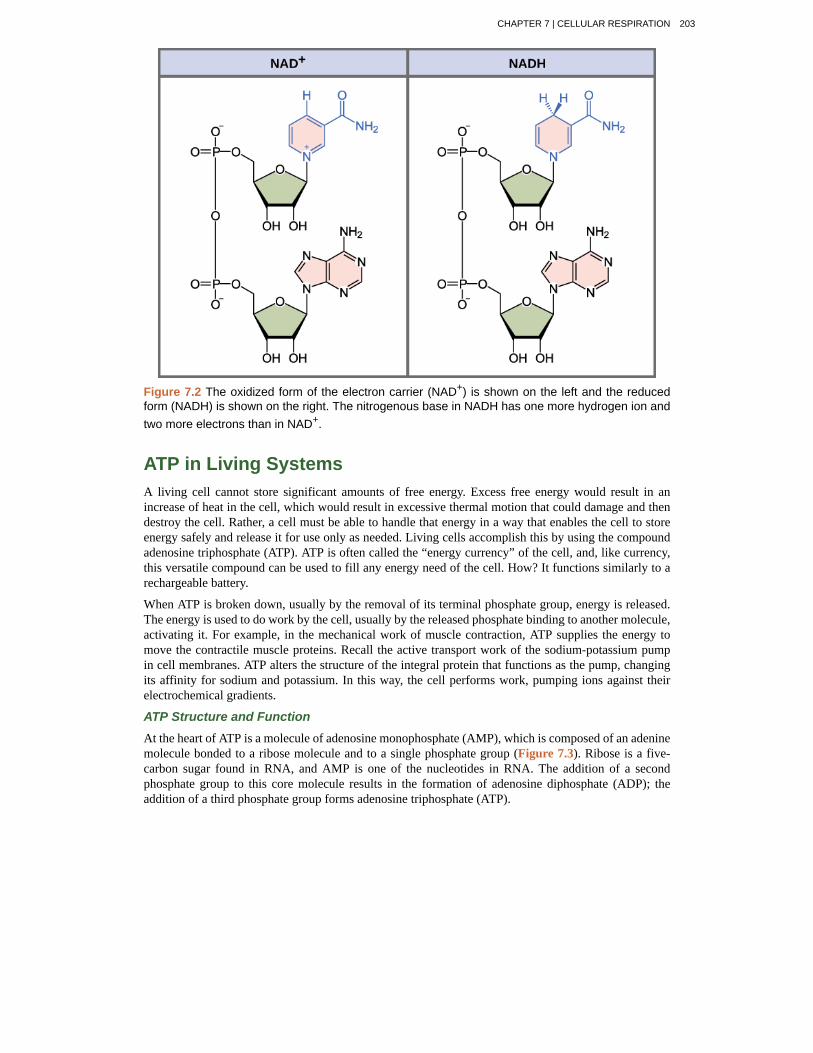
Figure 7.2 The oxidized form of the electron carrier (NAD+) is shown on the left and the reducedform (NADH) is shown on the right. The nitrogenous base in NADH has one more hydrogen ion andtwo more electrons than in NAD+.
ATP in Living SystemsA living cell cannot store significant amounts of free energy. Excess free energy would result in anincrease of heat in the cell, which would result in excessive thermal motion that could damage and thendestroy the cell. Rather, a cell must be able to handle that energy in a way that enables the cell to storeenergy safely and release it for use only as needed. Living cells accomplish this by using the compoundadenosine triphosphate (ATP). ATP is often called the “energy currency” of the cell, and, like currency,this versatile compound can be used to fill any energy need of the cell. How? It functions similarly to arechargeable battery.
When ATP is broken down, usually by the removal of its terminal phosphate group, energy is released.The energy is used to do work by the cell, usually by the released phosphate binding to another molecule,activating it. For example, in the mechanical work of muscle contraction, ATP supplies the energy tomove the contractile muscle proteins. Recall the active transport work of the sodium-potassium pumpin cell membranes. ATP alters the structure of the integral protein that functions as the pump, changingits affinity for sodium and potassium. In this way, the cell performs work, pumping ions against theirelectrochemical gradients.
ATP Structure and Function
At the heart of ATP is a molecule of adenosine monophosphate (AMP), which is composed of an adeninemolecule bonded to a ribose molecule and to a single phosphate group (Figure 7.3). Ribose is a five-carbon sugar found in RNA, and AMP is one of the nucleotides in RNA. The addition of a secondphosphate group to this core molecule results in the formation of adenosine diphosphate (ADP); theaddition of a third phosphate group forms adenosine triphosphate (ATP).
CHAPTER 7 | CELLULAR RESPIRATION 203
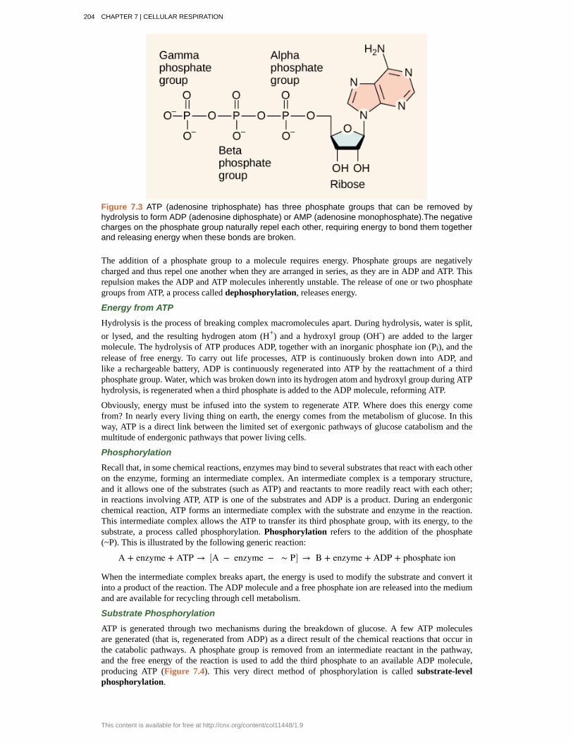
Figure 7.3 ATP (adenosine triphosphate) has three phosphate groups that can be removed byhydrolysis to form ADP (adenosine diphosphate) or AMP (adenosine monophosphate).The negativecharges on the phosphate group naturally repel each other, requiring energy to bond them togetherand releasing energy when these bonds are broken.
The addition of a phosphate group to a molecule requires energy. Phosphate groups are negativelycharged and thus repel one another when they are arranged in series, as they are in ADP and ATP. Thisrepulsion makes the ADP and ATP molecules inherently unstable. The release of one or two phosphategroups from ATP, a process called dephosphorylation, releases energy.
Energy from ATP
Hydrolysis is the process of breaking complex macromolecules apart. During hydrolysis, water is split,or lysed, and the resulting hydrogen atom (H+) and a hydroxyl group (OH-) are added to the largermolecule. The hydrolysis of ATP produces ADP, together with an inorganic phosphate ion (Pi), and therelease of free energy. To carry out life processes, ATP is continuously broken down into ADP, andlike a rechargeable battery, ADP is continuously regenerated into ATP by the reattachment of a thirdphosphate group. Water, which was broken down into its hydrogen atom and hydroxyl group during ATPhydrolysis, is regenerated when a third phosphate is added to the ADP molecule, reforming ATP.
Obviously, energy must be infused into the system to regenerate ATP. Where does this energy comefrom? In nearly every living thing on earth, the energy comes from the metabolism of glucose. In thisway, ATP is a direct link between the limited set of exergonic pathways of glucose catabolism and themultitude of endergonic pathways that power living cells.
Phosphorylation
Recall that, in some chemical reactions, enzymes may bind to several substrates that react with each otheron the enzyme, forming an intermediate complex. An intermediate complex is a temporary structure,and it allows one of the substrates (such as ATP) and reactants to more readily react with each other;in reactions involving ATP, ATP is one of the substrates and ADP is a product. During an endergonicchemical reaction, ATP forms an intermediate complex with the substrate and enzyme in the reaction.This intermediate complex allows the ATP to transfer its third phosphate group, with its energy, to thesubstrate, a process called phosphorylation. Phosphorylation refers to the addition of the phosphate(~P). This is illustrated by the following generic reaction:
A + enzyme + ATP → ⎡⎣A − enzyme − ∼ P⎤⎦ → B + enzyme + ADP + phosphate ion
When the intermediate complex breaks apart, the energy is used to modify the substrate and convert itinto a product of the reaction. The ADP molecule and a free phosphate ion are released into the mediumand are available for recycling through cell metabolism.
Substrate Phosphorylation
ATP is generated through two mechanisms during the breakdown of glucose. A few ATP moleculesare generated (that is, regenerated from ADP) as a direct result of the chemical reactions that occur inthe catabolic pathways. A phosphate group is removed from an intermediate reactant in the pathway,and the free energy of the reaction is used to add the third phosphate to an available ADP molecule,producing ATP (Figure 7.4). This very direct method of phosphorylation is called substrate-levelphosphorylation.
204 CHAPTER 7 | CELLULAR RESPIRATION
This content is available for free at http://cnx.org/content/col11448/1.9
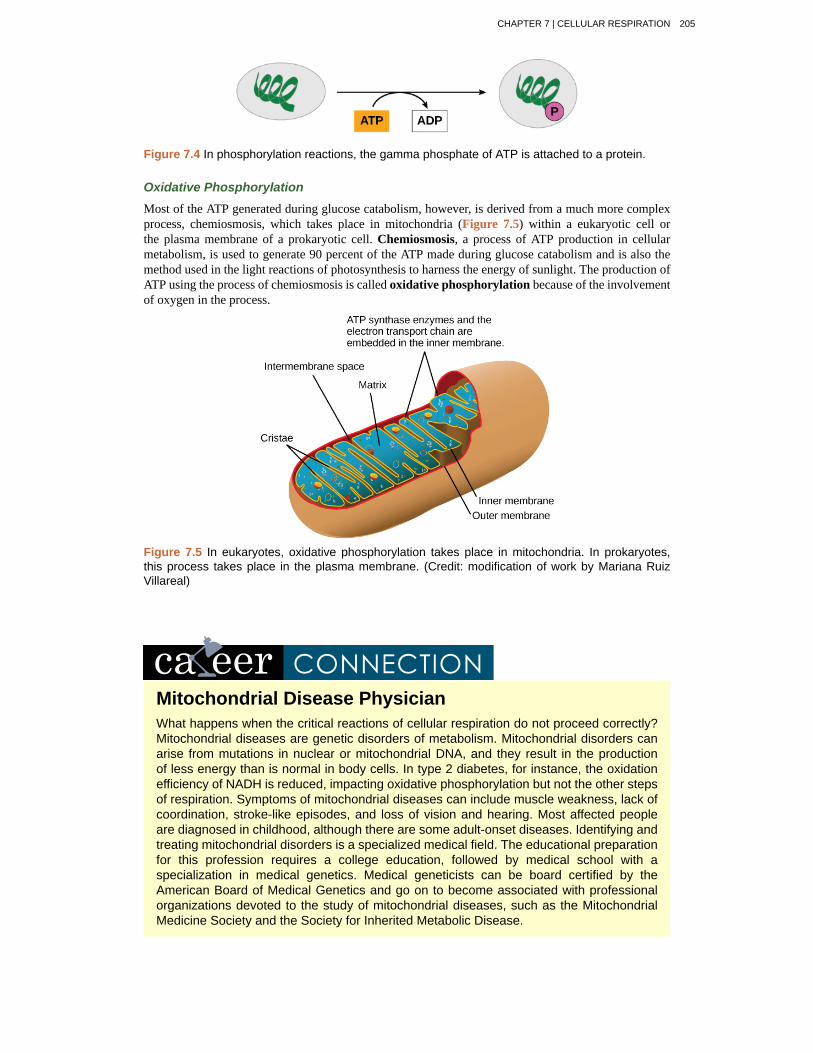
Figure 7.4 In phosphorylation reactions, the gamma phosphate of ATP is attached to a protein.
Oxidative Phosphorylation
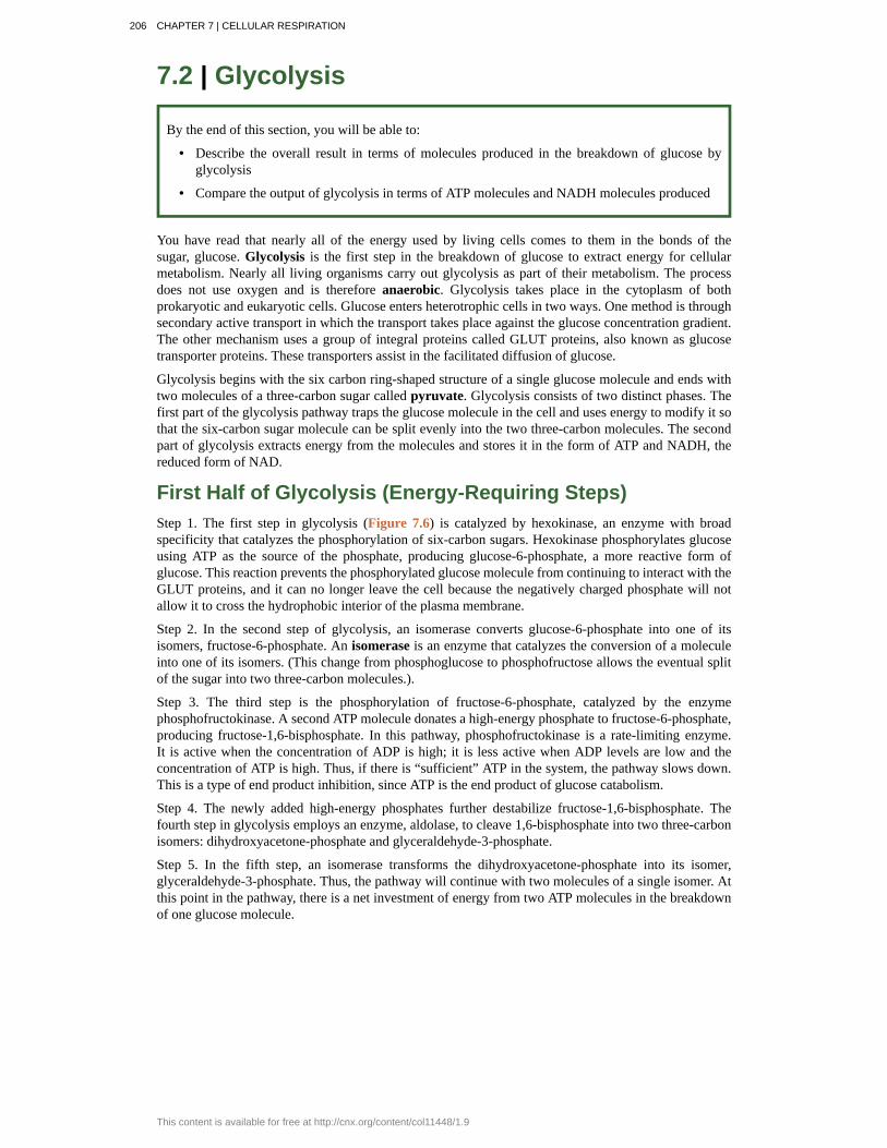
Most of the ATP generated during glucose catabolism, however, is derived from a much more complexprocess, chemiosmosis, which takes place in mitochondria (Figure 7.5) within a eukaryotic cell orthe plasma membrane of a prokaryotic cell. Chemiosmosis, a process of ATP production in cellularmetabolism, is used to generate 90 percent of the ATP made during glucose catabolism and is also themethod used in the light reactions of photosynthesis to harness the energy of sunlight. The production ofATP using the process of chemiosmosis is called oxidative phosphorylation because of the involvementof oxygen in the process.
Figure 7.5 In eukaryotes, oxidative phosphorylation takes place in mitochondria. In prokaryotes,this process takes place in the plasma membrane. (Credit: modification of work by Mariana RuizVillareal)
Mitochondrial Disease PhysicianWhat happens when the critical reactions of cellular respiration do not proceed correctly?Mitochondrial diseases are genetic disorders of metabolism. Mitochondrial disorders canarise from mutations in nuclear or mitochondrial DNA, and they result in the productionof less energy than is normal in body cells. In type 2 diabetes, for instance, the oxidationefficiency of NADH is reduced, impacting oxidative phosphorylation but not the other stepsof respiration. Symptoms of mitochondrial diseases can include muscle weakness, lack ofcoordination, stroke-like episodes, and loss of vision and hearing. Most affected peopleare diagnosed in childhood, although there are some adult-onset diseases. Identifying andtreating mitochondrial disorders is a specialized medical field. The educational preparationfor this profession requires a college education, followed by medical school with aspecialization in medical genetics. Medical geneticists can be board certified by theAmerican Board of Medical Genetics and go on to become associated with professionalorganizations devoted to the study of mitochondrial diseases, such as the MitochondrialMedicine Society and the Society for Inherited Metabolic Disease.
CHAPTER 7 | CELLULAR RESPIRATION 205
7.2 | Glycolysis
By the end of this section, you will be able to:
• Describe the overall result in terms of molecules produced in the breakdown of glucose byglycolysis
• Compare the output of glycolysis in terms of ATP molecules and NADH molecules produced
You have read that nearly all of the energy used by living cells comes to them in the bonds of thesugar, glucose. Glycolysis is the first step in the breakdown of glucose to extract energy for cellularmetabolism. Nearly all living organisms carry out glycolysis as part of their metabolism. The processdoes not use oxygen and is therefore anaerobic. Glycolysis takes place in the cytoplasm of bothprokaryotic and eukaryotic cells. Glucose enters heterotrophic cells in two ways. One method is throughsecondary active transport in which the transport takes place against the glucose concentration gradient.The other mechanism uses a group of integral proteins called GLUT proteins, also known as glucosetransporter proteins. These transporters assist in the facilitated diffusion of glucose.
Glycolysis begins with the six carbon ring-shaped structure of a single glucose molecule and ends withtwo molecules of a three-carbon sugar called pyruvate. Glycolysis consists of two distinct phases. Thefirst part of the glycolysis pathway traps the glucose molecule in the cell and uses energy to modify it sothat the six-carbon sugar molecule can be split evenly into the two three-carbon molecules. The secondpart of glycolysis extracts energy from the molecules and stores it in the form of ATP and NADH, thereduced form of NAD.
First Half of Glycolysis (Energy-Requiring Steps)Step 1. The first step in glycolysis (Figure 7.6) is catalyzed by hexokinase, an enzyme with broadspecificity that catalyzes the phosphorylation of six-carbon sugars. Hexokinase phosphorylates glucoseusing ATP as the source of the phosphate, producing glucose-6-phosphate, a more reactive form ofglucose. This reaction prevents the phosphorylated glucose molecule from continuing to interact with theGLUT proteins, and it can no longer leave the cell because the negatively charged phosphate will notallow it to cross the hydrophobic interior of the plasma membrane.
Step 2. In the second step of glycolysis, an isomerase converts glucose-6-phosphate into one of itsisomers, fructose-6-phosphate. An isomerase is an enzyme that catalyzes the conversion of a moleculeinto one of its isomers. (This change from phosphoglucose to phosphofructose allows the eventual splitof the sugar into two three-carbon molecules.).
Step 3. The third step is the phosphorylation of fructose-6-phosphate, catalyzed by the enzymephosphofructokinase. A second ATP molecule donates a high-energy phosphate to fructose-6-phosphate,producing fructose-1,6-bisphosphate. In this pathway, phosphofructokinase is a rate-limiting enzyme.It is active when the concentration of ADP is high; it is less active when ADP levels are low and theconcentration of ATP is high. Thus, if there is “sufficient” ATP in the system, the pathway slows down.This is a type of end product inhibition, since ATP is the end product of glucose catabolism.
Step 4. The newly added high-energy phosphates further destabilize fructose-1,6-bisphosphate. Thefourth step in glycolysis employs an enzyme, aldolase, to cleave 1,6-bisphosphate into two three-carbonisomers: dihydroxyacetone-phosphate and glyceraldehyde-3-phosphate.
Step 5. In the fifth step, an isomerase transforms the dihydroxyacetone-phosphate into its isomer,glyceraldehyde-3-phosphate. Thus, the pathway will continue with two molecules of a single isomer. Atthis point in the pathway, there is a net investment of energy from two ATP molecules in the breakdownof one glucose molecule.
206 CHAPTER 7 | CELLULAR RESPIRATION
This content is available for free at http://cnx.org/content/col11448/1.9
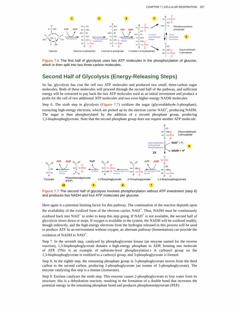
Figure 7.6 The first half of glycolysis uses two ATP molecules in the phosphorylation of glucose,which is then split into two three-carbon molecules.
Second Half of Glycolysis (Energy-Releasing Steps)So far, glycolysis has cost the cell two ATP molecules and produced two small, three-carbon sugarmolecules. Both of these molecules will proceed through the second half of the pathway, and sufficientenergy will be extracted to pay back the two ATP molecules used as an initial investment and produce aprofit for the cell of two additional ATP molecules and two even higher-energy NADH molecules.
Step 6. The sixth step in glycolysis (Figure 7.7) oxidizes the sugar (glyceraldehyde-3-phosphate),extracting high-energy electrons, which are picked up by the electron carrier NAD+, producing NADH.The sugar is then phosphorylated by the addition of a second phosphate group, producing1,3-bisphosphoglycerate. Note that the second phosphate group does not require another ATP molecule.
Figure 7.7 The second half of glycolysis involves phosphorylation without ATP investment (step 6)and produces two NADH and four ATP molecules per glucose.
Here again is a potential limiting factor for this pathway. The continuation of the reaction depends uponthe availability of the oxidized form of the electron carrier, NAD+. Thus, NADH must be continuouslyoxidized back into NAD+ in order to keep this step going. If NAD+ is not available, the second half ofglycolysis slows down or stops. If oxygen is available in the system, the NADH will be oxidized readily,though indirectly, and the high-energy electrons from the hydrogen released in this process will be usedto produce ATP. In an environment without oxygen, an alternate pathway (fermentation) can provide theoxidation of NADH to NAD+.
Step 7. In the seventh step, catalyzed by phosphoglycerate kinase (an enzyme named for the reversereaction), 1,3-bisphosphoglycerate donates a high-energy phosphate to ADP, forming one moleculeof ATP. (This is an example of substrate-level phosphorylation.) A carbonyl group on the1,3-bisphosphoglycerate is oxidized to a carboxyl group, and 3-phosphoglycerate is formed.
Step 8. In the eighth step, the remaining phosphate group in 3-phosphoglycerate moves from the thirdcarbon to the second carbon, producing 2-phosphoglycerate (an isomer of 3-phosphoglycerate). Theenzyme catalyzing this step is a mutase (isomerase).
Step 9. Enolase catalyzes the ninth step. This enzyme causes 2-phosphoglycerate to lose water from itsstructure; this is a dehydration reaction, resulting in the formation of a double bond that increases thepotential energy in the remaining phosphate bond and produces phosphoenolpyruvate (PEP).
CHAPTER 7 | CELLULAR RESPIRATION 207
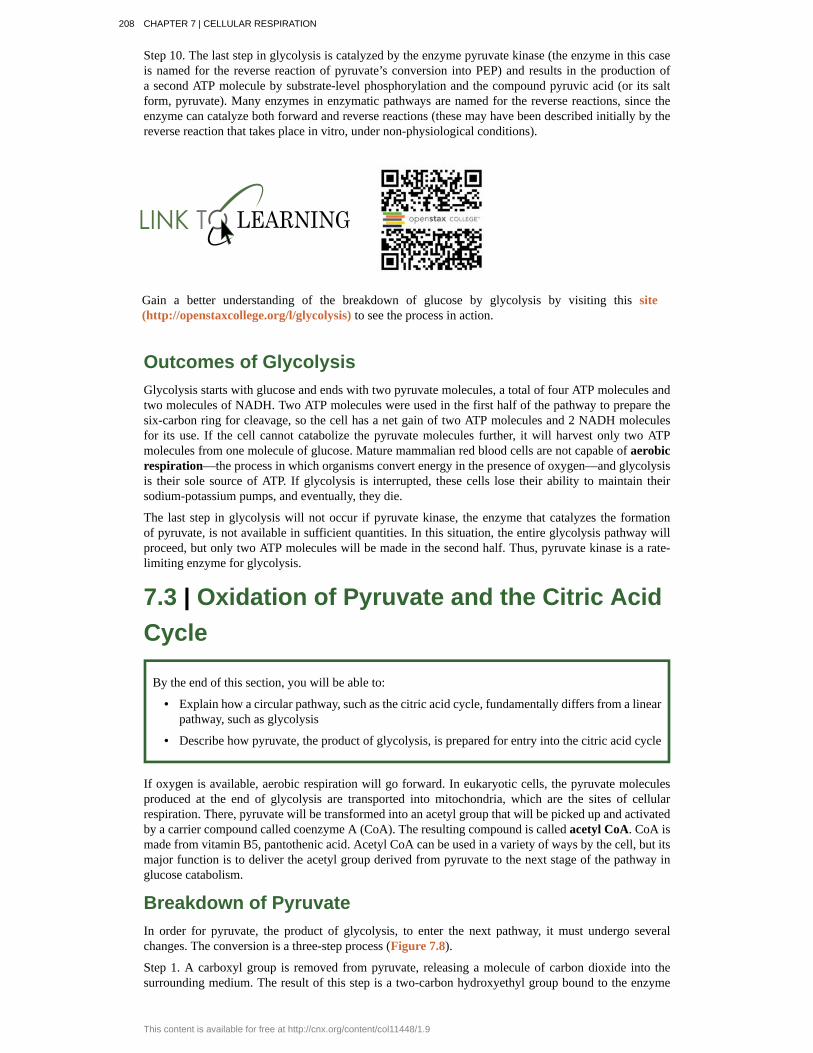
Step 10. The last step in glycolysis is catalyzed by the enzyme pyruvate kinase (the enzyme in this caseis named for the reverse reaction of pyruvate’s conversion into PEP) and results in the production ofa second ATP molecule by substrate-level phosphorylation and the compound pyruvic acid (or its saltform, pyruvate). Many enzymes in enzymatic pathways are named for the reverse reactions, since theenzyme can catalyze both forward and reverse reactions (these may have been described initially by thereverse reaction that takes place in vitro, under non-physiological conditions).
Gain a better understanding of the breakdown of glucose by glycolysis by visiting this site(http://openstaxcollege.org/l/glycolysis) to see the process in action.
Outcomes of GlycolysisGlycolysis starts with glucose and ends with two pyruvate molecules, a total of four ATP molecules andtwo molecules of NADH. Two ATP molecules were used in the first half of the pathway to prepare thesix-carbon ring for cleavage, so the cell has a net gain of two ATP molecules and 2 NADH moleculesfor its use. If the cell cannot catabolize the pyruvate molecules further, it will harvest only two ATPmolecules from one molecule of glucose. Mature mammalian red blood cells are not capable of aerobicrespiration—the process in which organisms convert energy in the presence of oxygen—and glycolysisis their sole source of ATP. If glycolysis is interrupted, these cells lose their ability to maintain theirsodium-potassium pumps, and eventually, they die.
The last step in glycolysis will not occur if pyruvate kinase, the enzyme that catalyzes the formationof pyruvate, is not available in sufficient quantities. In this situation, the entire glycolysis pathway willproceed, but only two ATP molecules will be made in the second half. Thus, pyruvate kinase is a rate-limiting enzyme for glycolysis.
7.3 | Oxidation of Pyruvate and the Citric AcidCycle
By the end of this section, you will be able to:
• Explain how a circular pathway, such as the citric acid cycle, fundamentally differs from a linearpathway, such as glycolysis
• Describe how pyruvate, the product of glycolysis, is prepared for entry into the citric acid cycle
If oxygen is available, aerobic respiration will go forward. In eukaryotic cells, the pyruvate moleculesproduced at the end of glycolysis are transported into mitochondria, which are the sites of cellularrespiration. There, pyruvate will be transformed into an acetyl group that will be picked up and activatedby a carrier compound called coenzyme A (CoA). The resulting compound is called acetyl CoA. CoA ismade from vitamin B5, pantothenic acid. Acetyl CoA can be used in a variety of ways by the cell, but itsmajor function is to deliver the acetyl group derived from pyruvate to the next stage of the pathway inglucose catabolism.
Breakdown of PyruvateIn order for pyruvate, the product of glycolysis, to enter the next pathway, it must undergo severalchanges. The conversion is a three-step process (Figure 7.8).
Step 1. A carboxyl group is removed from pyruvate, releasing a molecule of carbon dioxide into thesurrounding medium. The result of this step is a two-carbon hydroxyethyl group bound to the enzyme
208 CHAPTER 7 | CELLULAR RESPIRATION
This content is available for free at http://cnx.org/content/col11448/1.9
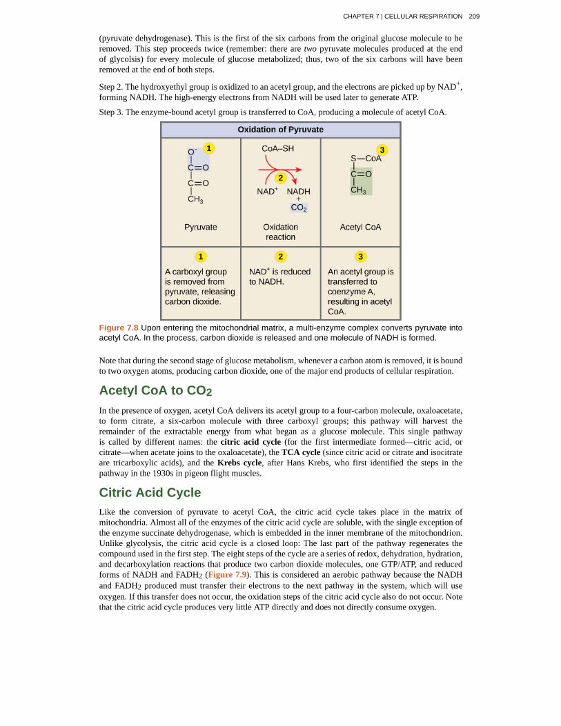
(pyruvate dehydrogenase). This is the first of the six carbons from the original glucose molecule to beremoved. This step proceeds twice (remember: there are two pyruvate molecules produced at the endof glycolsis) for every molecule of glucose metabolized; thus, two of the six carbons will have beenremoved at the end of both steps.
Step 2. The hydroxyethyl group is oxidized to an acetyl group, and the electrons are picked up by NAD+,forming NADH. The high-energy electrons from NADH will be used later to generate ATP.
Step 3. The enzyme-bound acetyl group is transferred to CoA, producing a molecule of acetyl CoA.
Figure 7.8 Upon entering the mitochondrial matrix, a multi-enzyme complex converts pyruvate intoacetyl CoA. In the process, carbon dioxide is released and one molecule of NADH is formed.
Note that during the second stage of glucose metabolism, whenever a carbon atom is removed, it is boundto two oxygen atoms, producing carbon dioxide, one of the major end products of cellular respiration.
Acetyl CoA to CO2In the presence of oxygen, acetyl CoA delivers its acetyl group to a four-carbon molecule, oxaloacetate,to form citrate, a six-carbon molecule with three carboxyl groups; this pathway will harvest theremainder of the extractable energy from what began as a glucose molecule. This single pathwayis called by different names: the citric acid cycle (for the first intermediate formed—citric acid, orcitrate—when acetate joins to the oxaloacetate), the TCA cycle (since citric acid or citrate and isocitrateare tricarboxylic acids), and the Krebs cycle, after Hans Krebs, who first identified the steps in thepathway in the 1930s in pigeon flight muscles.
Citric Acid CycleLike the conversion of pyruvate to acetyl CoA, the citric acid cycle takes place in the matrix ofmitochondria. Almost all of the enzymes of the citric acid cycle are soluble, with the single exception ofthe enzyme succinate dehydrogenase, which is embedded in the inner membrane of the mitochondrion.Unlike glycolysis, the citric acid cycle is a closed loop: The last part of the pathway regenerates thecompound used in the first step. The eight steps of the cycle are a series of redox, dehydration, hydration,and decarboxylation reactions that produce two carbon dioxide molecules, one GTP/ATP, and reducedforms of NADH and FADH2 (Figure 7.9). This is considered an aerobic pathway because the NADHand FADH2 produced must transfer their electrons to the next pathway in the system, which will useoxygen. If this transfer does not occur, the oxidation steps of the citric acid cycle also do not occur. Notethat the citric acid cycle produces very little ATP directly and does not directly consume oxygen.
CHAPTER 7 | CELLULAR RESPIRATION 209
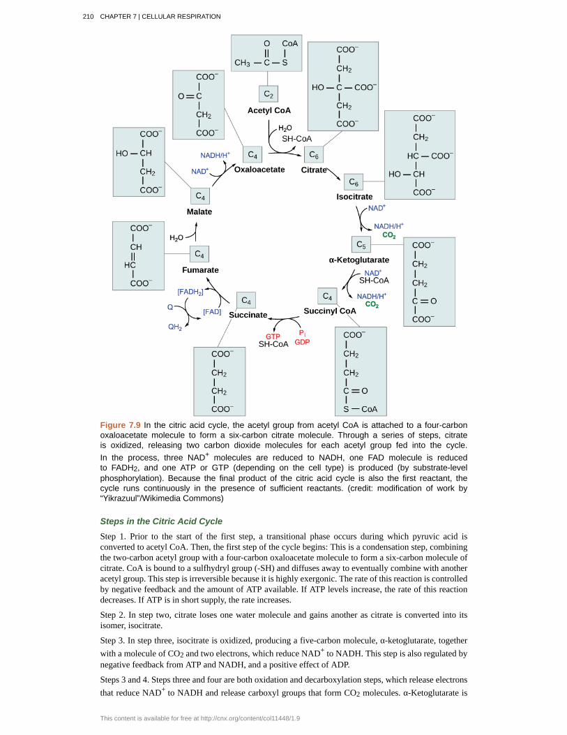
Figure 7.9 In the citric acid cycle, the acetyl group from acetyl CoA is attached to a four-carbonoxaloacetate molecule to form a six-carbon citrate molecule. Through a series of steps, citrateis oxidized, releasing two carbon dioxide molecules for each acetyl group fed into the cycle.In the process, three NAD+ molecules are reduced to NADH, one FAD molecule is reducedto FADH2, and one ATP or GTP (depending on the cell type) is produced (by substrate-levelphosphorylation). Because the final product of the citric acid cycle is also the first reactant, thecycle runs continuously in the presence of sufficient reactants. (credit: modification of work by“Yikrazuul”/Wikimedia Commons)
Steps in the Citric Acid Cycle
Step 1. Prior to the start of the first step, a transitional phase occurs during which pyruvic acid isconverted to acetyl CoA. Then, the first step of the cycle begins: This is a condensation step, combiningthe two-carbon acetyl group with a four-carbon oxaloacetate molecule to form a six-carbon molecule ofcitrate. CoA is bound to a sulfhydryl group (-SH) and diffuses away to eventually combine with anotheracetyl group. This step is irreversible because it is highly exergonic. The rate of this reaction is controlledby negative feedback and the amount of ATP available. If ATP levels increase, the rate of this reactiondecreases. If ATP is in short supply, the rate increases.
Step 2. In step two, citrate loses one water molecule and gains another as citrate is converted into itsisomer, isocitrate.
Step 3. In step three, isocitrate is oxidized, producing a five-carbon molecule, α-ketoglutarate, togetherwith a molecule of CO2 and two electrons, which reduce NAD+ to NADH. This step is also regulated bynegative feedback from ATP and NADH, and a positive effect of ADP.
Steps 3 and 4. Steps three and four are both oxidation and decarboxylation steps, which release electronsthat reduce NAD+ to NADH and release carboxyl groups that form CO2 molecules. α-Ketoglutarate is
210 CHAPTER 7 | CELLULAR RESPIRATION
This content is available for free at http://cnx.org/content/col11448/1.9
the product of step three, and a succinyl group is the product of step four. CoA binds the succinyl groupto form succinyl CoA. The enzyme that catalyzes step four is regulated by feedback inhibition of ATP,succinyl CoA, and NADH.
Step 5. In step five, a phosphate group is substituted for coenzyme A, and a high-energy bond is formed.This energy is used in substrate-level phosphorylation (during the conversion of the succinyl group tosuccinate) to form either guanine triphosphate (GTP) or ATP. There are two forms of the enzyme, calledisoenzymes, for this step, depending upon the type of animal tissue in which they are found. One formis found in tissues that use large amounts of ATP, such as heart and skeletal muscle. This form producesATP. The second form of the enzyme is found in tissues that have a high number of anabolic pathways,such as liver. This form produces GTP. GTP is energetically equivalent to ATP; however, its use is morerestricted. In particular, protein synthesis primarily uses GTP.
Step 6. Step six is a dehydration process that converts succinate into fumarate. Two hydrogen atomsare transferred to FAD, producing FADH2. The energy contained in the electrons of these atoms isinsufficient to reduce NAD+ but adequate to reduce FAD. Unlike NADH, this carrier remains attachedto the enzyme and transfers the electrons to the electron transport chain directly. This process ismade possible by the localization of the enzyme catalyzing this step inside the inner membrane of themitochondrion.
Step 7. Water is added to fumarate during step seven, and malate is produced. The last step in the citricacid cycle regenerates oxaloacetate by oxidizing malate. Another molecule of NADH is produced in theprocess.
Click through each step of the citric acid cycle here (http://openstaxcollege.org/l/krebs_cycle) .
Products of the Citric Acid Cycle
Two carbon atoms come into the citric acid cycle from each acetyl group, representing four out of thesix carbons of one glucose molecule. Two carbon dioxide molecules are released on each turn of thecycle; however, these do not necessarily contain the most recently added carbon atoms. The two acetylcarbon atoms will eventually be released on later turns of the cycle; thus, all six carbon atoms from theoriginal glucose molecule are eventually incorporated into carbon dioxide. Each turn of the cycle formsthree NADH molecules and one FADH2 molecule. These carriers will connect with the last portion ofaerobic respiration to produce ATP molecules. One GTP or ATP is also made in each cycle. Severalof the intermediate compounds in the citric acid cycle can be used in synthesizing non-essential aminoacids; therefore, the cycle is amphibolic (both catabolic and anabolic).
7.4 | Oxidative Phosphorylation
By the end of this section, you will be able to:
• Describe how electrons move through the electron transport chain and what happens to theirenergy levels
• Explain how a proton (H+) gradient is established and maintained by the electron transport chain
You have just read about two pathways in glucose catabolism—glycolysis and the citric acid cycle—thatgenerate ATP. Most of the ATP generated during the aerobic catabolism of glucose, however, is notgenerated directly from these pathways. Rather, it is derived from a process that begins with movingelectrons through a series of electron transporters that undergo redox reactions. This causes hydrogenions to accumulate within the matrix space. Therefore, a concentration gradient forms in which hydrogen
CHAPTER 7 | CELLULAR RESPIRATION 211
ions diffuse out of the matrix space by passing through ATP synthase. The current of hydrogen ionspowers the catalytic action of ATP synthase, which phosphorylates ADP, producing ATP.
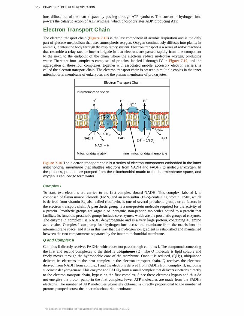
Electron Transport ChainThe electron transport chain (Figure 7.10) is the last component of aerobic respiration and is the onlypart of glucose metabolism that uses atmospheric oxygen. Oxygen continuously diffuses into plants; inanimals, it enters the body through the respiratory system. Electron transport is a series of redox reactionsthat resemble a relay race or bucket brigade in that electrons are passed rapidly from one componentto the next, to the endpoint of the chain where the electrons reduce molecular oxygen, producingwater. There are four complexes composed of proteins, labeled I through IV in Figure 7.10, and theaggregation of these four complexes, together with associated mobile, accessory electron carriers, iscalled the electron transport chain. The electron transport chain is present in multiple copies in the innermitochondrial membrane of eukaryotes and the plasma membrane of prokaryotes.
Figure 7.10 The electron transport chain is a series of electron transporters embedded in the innermitochondrial membrane that shuttles electrons from NADH and FADH2 to molecular oxygen. Inthe process, protons are pumped from the mitochondrial matrix to the intermembrane space, andoxygen is reduced to form water.
Complex I
To start, two electrons are carried to the first complex aboard NADH. This complex, labeled I, iscomposed of flavin mononucleotide (FMN) and an iron-sulfur (Fe-S)-containing protein. FMN, whichis derived from vitamin B2, also called riboflavin, is one of several prosthetic groups or co-factors inthe electron transport chain. A prosthetic group is a non-protein molecule required for the activity ofa protein. Prosthetic groups are organic or inorganic, non-peptide molecules bound to a protein thatfacilitate its function; prosthetic groups include co-enzymes, which are the prosthetic groups of enzymes.The enzyme in complex I is NADH dehydrogenase and is a very large protein, containing 45 aminoacid chains. Complex I can pump four hydrogen ions across the membrane from the matrix into theintermembrane space, and it is in this way that the hydrogen ion gradient is established and maintainedbetween the two compartments separated by the inner mitochondrial membrane.
Q and Complex II
Complex II directly receives FADH2, which does not pass through complex I. The compound connectingthe first and second complexes to the third is ubiquinone (Q). The Q molecule is lipid soluble andfreely moves through the hydrophobic core of the membrane. Once it is reduced, (QH2), ubiquinonedelivers its electrons to the next complex in the electron transport chain. Q receives the electronsderived from NADH from complex I and the electrons derived from FADH2 from complex II, includingsuccinate dehydrogenase. This enzyme and FADH2 form a small complex that delivers electrons directlyto the electron transport chain, bypassing the first complex. Since these electrons bypass and thus donot energize the proton pump in the first complex, fewer ATP molecules are made from the FADH2electrons. The number of ATP molecules ultimately obtained is directly proportional to the number ofprotons pumped across the inner mitochondrial membrane.
212 CHAPTER 7 | CELLULAR RESPIRATION
This content is available for free at http://cnx.org/content/col11448/1.9
Complex III
The third complex is composed of cytochrome b, another Fe-S protein, Rieske center (2Fe-2S center),and cytochrome c proteins; this complex is also called cytochrome oxidoreductase. Cytochrome proteinshave a prosthetic group of heme. The heme molecule is similar to the heme in hemoglobin, but it carrieselectrons, not oxygen. As a result, the iron ion at its core is reduced and oxidized as it passes theelectrons, fluctuating between different oxidation states: Fe++ (reduced) and Fe+++ (oxidized). The hememolecules in the cytochromes have slightly different characteristics due to the effects of the differentproteins binding them, giving slightly different characteristics to each complex. Complex III pumpsprotons through the membrane and passes its electrons to cytochrome c for transport to the fourthcomplex of proteins and enzymes (cytochrome c is the acceptor of electrons from Q; however, whereasQ carries pairs of electrons, cytochrome c can accept only one at a time).
Complex IV
The fourth complex is composed of cytochrome proteins c, a, and a3. This complex contains two hemegroups (one in each of the two cytochromes, a, and a3) and three copper ions (a pair of CuA and oneCuB in cytochrome a3). The cytochromes hold an oxygen molecule very tightly between the iron andcopper ions until the oxygen is completely reduced. The reduced oxygen then picks up two hydrogenions from the surrounding medium to make water (H2O). The removal of the hydrogen ions from thesystem contributes to the ion gradient used in the process of chemiosmosis.
ChemiosmosisIn chemiosmosis, the free energy from the series of redox reactions just described is used to pumphydrogen ions (protons) across the membrane. The uneven distribution of H+ ions across the membraneestablishes both concentration and electrical gradients (thus, an electrochemical gradient), owing to thehydrogen ions’ positive charge and their aggregation on one side of the membrane.
If the membrane were open to diffusion by the hydrogen ions, the ions would tend to diffuse back acrossinto the matrix, driven by their electrochemical gradient. Recall that many ions cannot diffuse throughthe nonpolar regions of phospholipid membranes without the aid of ion channels. Similarly, hydrogenions in the matrix space can only pass through the inner mitochondrial membrane through an integralmembrane protein called ATP synthase (Figure 7.11). This complex protein acts as a tiny generator,turned by the force of the hydrogen ions diffusing through it, down their electrochemical gradient. Theturning of parts of this molecular machine facilitates the addition of a phosphate to ADP, forming ATP,using the potential energy of the hydrogen ion gradient.
CHAPTER 7 | CELLULAR RESPIRATION 213

Figure 7.11 ATP synthase is a complex, molecular machine that uses a proton (H+) gradientto form ATP from ADP and inorganic phosphate (Pi). (Credit: modification of work by KlausHoffmeier)
Dinitrophenol (DNP) is an uncoupler that makes the inner mitochondrial membrane leakyto protons. It was used until 1938 as a weight-loss drug. What effect would you expectDNP to have on the change in pH across the inner mitochondrial membrane? Why do youthink this might be an effective weight-loss drug?
Chemiosmosis (Figure 7.12) is used to generate 90 percent of the ATP made during aerobic glucosecatabolism; it is also the method used in the light reactions of photosynthesis to harness the energy ofsunlight in the process of photophosphorylation. Recall that the production of ATP using the process ofchemiosmosis in mitochondria is called oxidative phosphorylation. The overall result of these reactionsis the production of ATP from the energy of the electrons removed from hydrogen atoms. These atomswere originally part of a glucose molecule. At the end of the pathway, the electrons are used to reduce anoxygen molecule to oxygen ions. The extra electrons on the oxygen attract hydrogen ions (protons) fromthe surrounding medium, and water is formed.
214 CHAPTER 7 | CELLULAR RESPIRATION
This content is available for free at http://cnx.org/content/col11448/1.9
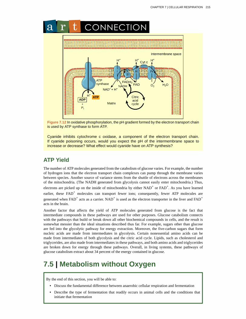
Figure 7.12 In oxidative phosphorylation, the pH gradient formed by the electron transport chainis used by ATP synthase to form ATP.
Cyanide inhibits cytochrome c oxidase, a component of the electron transport chain.If cyanide poisoning occurs, would you expect the pH of the intermembrane space toincrease or decrease? What effect would cyanide have on ATP synthesis?
ATP YieldThe number of ATP molecules generated from the catabolism of glucose varies. For example, the numberof hydrogen ions that the electron transport chain complexes can pump through the membrane variesbetween species. Another source of variance stems from the shuttle of electrons across the membranesof the mitochondria. (The NADH generated from glycolysis cannot easily enter mitochondria.) Thus,electrons are picked up on the inside of mitochondria by either NAD+ or FAD+. As you have learnedearlier, these FAD+ molecules can transport fewer ions; consequently, fewer ATP molecules aregenerated when FAD+ acts as a carrier. NAD+ is used as the electron transporter in the liver and FAD+
acts in the brain.
Another factor that affects the yield of ATP molecules generated from glucose is the fact thatintermediate compounds in these pathways are used for other purposes. Glucose catabolism connectswith the pathways that build or break down all other biochemical compounds in cells, and the result issomewhat messier than the ideal situations described thus far. For example, sugars other than glucoseare fed into the glycolytic pathway for energy extraction. Moreover, the five-carbon sugars that formnucleic acids are made from intermediates in glycolysis. Certain nonessential amino acids can bemade from intermediates of both glycolysis and the citric acid cycle. Lipids, such as cholesterol andtriglycerides, are also made from intermediates in these pathways, and both amino acids and triglyceridesare broken down for energy through these pathways. Overall, in living systems, these pathways ofglucose catabolism extract about 34 percent of the energy contained in glucose.
7.5 | Metabolism without Oxygen
By the end of this section, you will be able to:
• Discuss the fundamental difference between anaerobic cellular respiration and fermentation
• Describe the type of fermentation that readily occurs in animal cells and the conditions thatinitiate that fermentation
CHAPTER 7 | CELLULAR RESPIRATION 215
In aerobic respiration, the final electron acceptor is an oxygen molecule, O2. If aerobic respirationoccurs, then ATP will be produced using the energy of high-energy electrons carried by NADH orFADH2 to the electron transport chain. If aerobic respiration does not occur, NADH must be reoxidizedto NAD+ for reuse as an electron carrier for the glycolytic pathway to continue. How is this done?Some living systems use an organic molecule as the final electron acceptor. Processes that use an organicmolecule to regenerate NAD+ from NADH are collectively referred to as fermentation. In contrast,some living systems use an inorganic molecule as a final electron acceptor. Both methods are calledanaerobic cellular respiration in which organisms convert energy for their use in the absence ofoxygen.
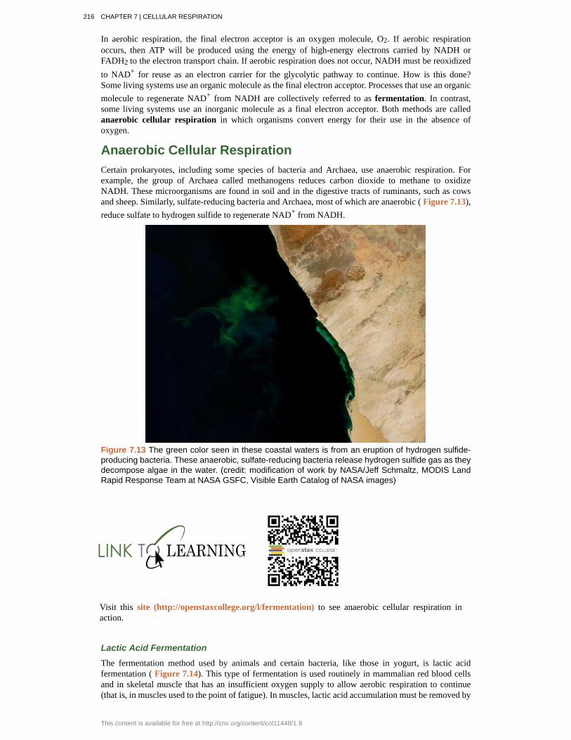
Anaerobic Cellular RespirationCertain prokaryotes, including some species of bacteria and Archaea, use anaerobic respiration. Forexample, the group of Archaea called methanogens reduces carbon dioxide to methane to oxidizeNADH. These microorganisms are found in soil and in the digestive tracts of ruminants, such as cowsand sheep. Similarly, sulfate-reducing bacteria and Archaea, most of which are anaerobic ( Figure 7.13),reduce sulfate to hydrogen sulfide to regenerate NAD+ from NADH.
Figure 7.13 The green color seen in these coastal waters is from an eruption of hydrogen sulfide-producing bacteria. These anaerobic, sulfate-reducing bacteria release hydrogen sulfide gas as theydecompose algae in the water. (credit: modification of work by NASA/Jeff Schmaltz, MODIS LandRapid Response Team at NASA GSFC, Visible Earth Catalog of NASA images)
Visit this site (http://openstaxcollege.org/l/fermentation) to see anaerobic cellular respiration inaction.
Lactic Acid Fermentation
The fermentation method used by animals and certain bacteria, like those in yogurt, is lactic acidfermentation ( Figure 7.14). This type of fermentation is used routinely in mammalian red blood cellsand in skeletal muscle that has an insufficient oxygen supply to allow aerobic respiration to continue(that is, in muscles used to the point of fatigue). In muscles, lactic acid accumulation must be removed by
216 CHAPTER 7 | CELLULAR RESPIRATION
This content is available for free at http://cnx.org/content/col11448/1.9
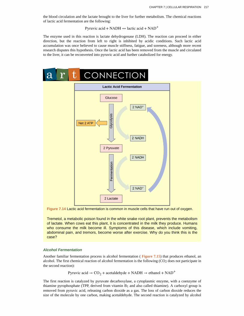
the blood circulation and the lactate brought to the liver for further metabolism. The chemical reactionsof lactic acid fermentation are the following:
Pyruvic acid + NADH ↔ lactic acid + NAD+
The enzyme used in this reaction is lactate dehydrogenase (LDH). The reaction can proceed in eitherdirection, but the reaction from left to right is inhibited by acidic conditions. Such lactic acidaccumulation was once believed to cause muscle stiffness, fatigue, and soreness, although more recentresearch disputes this hypothesis. Once the lactic acid has been removed from the muscle and circulatedto the liver, it can be reconverted into pyruvic acid and further catabolized for energy.
Figure 7.14 Lactic acid fermentation is common in muscle cells that have run out of oxygen.
Tremetol, a metabolic poison found in the white snake root plant, prevents the metabolismof lactate. When cows eat this plant, it is concentrated in the milk they produce. Humanswho consume the milk become ill. Symptoms of this disease, which include vomiting,abdominal pain, and tremors, become worse after exercise. Why do you think this is thecase?
Alcohol Fermentation
Another familiar fermentation process is alcohol fermentation ( Figure 7.15) that produces ethanol, analcohol. The first chemical reaction of alcohol fermentation is the following (CO2 does not participate inthe second reaction):
Pyruvic acid → CO2 + acetaldehyde + NADH → ethanol + NAD+
The first reaction is catalyzed by pyruvate decarboxylase, a cytoplasmic enzyme, with a coenzyme ofthiamine pyrophosphate (TPP, derived from vitamin B1 and also called thiamine). A carboxyl group isremoved from pyruvic acid, releasing carbon dioxide as a gas. The loss of carbon dioxide reduces thesize of the molecule by one carbon, making acetaldehyde. The second reaction is catalyzed by alcohol
CHAPTER 7 | CELLULAR RESPIRATION 217
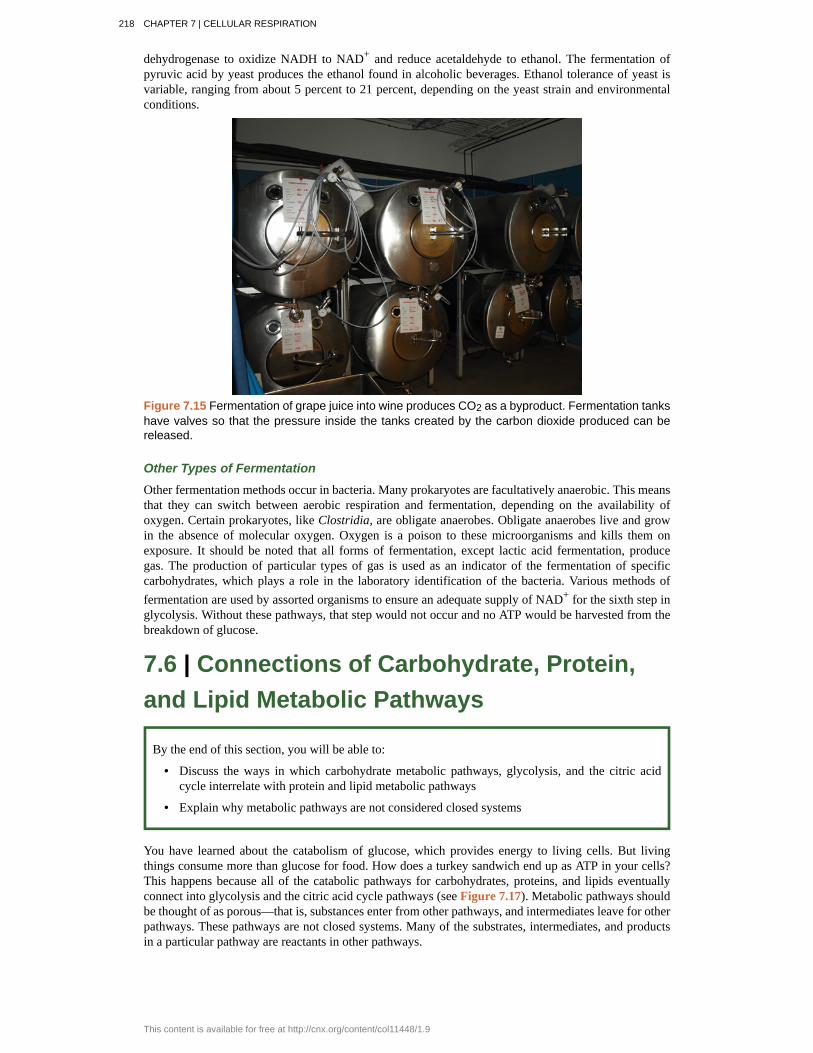
dehydrogenase to oxidize NADH to NAD+ and reduce acetaldehyde to ethanol. The fermentation ofpyruvic acid by yeast produces the ethanol found in alcoholic beverages. Ethanol tolerance of yeast isvariable, ranging from about 5 percent to 21 percent, depending on the yeast strain and environmentalconditions.
Figure 7.15 Fermentation of grape juice into wine produces CO2 as a byproduct. Fermentation tankshave valves so that the pressure inside the tanks created by the carbon dioxide produced can bereleased.
Other Types of Fermentation
Other fermentation methods occur in bacteria. Many prokaryotes are facultatively anaerobic. This meansthat they can switch between aerobic respiration and fermentation, depending on the availability ofoxygen. Certain prokaryotes, like Clostridia, are obligate anaerobes. Obligate anaerobes live and growin the absence of molecular oxygen. Oxygen is a poison to these microorganisms and kills them onexposure. It should be noted that all forms of fermentation, except lactic acid fermentation, producegas. The production of particular types of gas is used as an indicator of the fermentation of specificcarbohydrates, which plays a role in the laboratory identification of the bacteria. Various methods offermentation are used by assorted organisms to ensure an adequate supply of NAD+ for the sixth step inglycolysis. Without these pathways, that step would not occur and no ATP would be harvested from thebreakdown of glucose.
7.6 | Connections of Carbohydrate, Protein,and Lipid Metabolic Pathways
By the end of this section, you will be able to:
• Discuss the ways in which carbohydrate metabolic pathways, glycolysis, and the citric acidcycle interrelate with protein and lipid metabolic pathways
• Explain why metabolic pathways are not considered closed systems
You have learned about the catabolism of glucose, which provides energy to living cells. But livingthings consume more than glucose for food. How does a turkey sandwich end up as ATP in your cells?This happens because all of the catabolic pathways for carbohydrates, proteins, and lipids eventuallyconnect into glycolysis and the citric acid cycle pathways (see Figure 7.17). Metabolic pathways shouldbe thought of as porous—that is, substances enter from other pathways, and intermediates leave for otherpathways. These pathways are not closed systems. Many of the substrates, intermediates, and productsin a particular pathway are reactants in other pathways.
218 CHAPTER 7 | CELLULAR RESPIRATION
This content is available for free at http://cnx.org/content/col11448/1.9
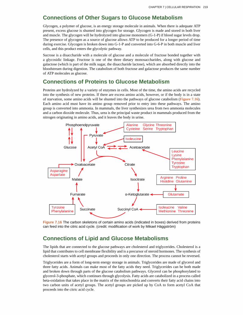
Connections of Other Sugars to Glucose MetabolismGlycogen, a polymer of glucose, is an energy storage molecule in animals. When there is adequate ATPpresent, excess glucose is shunted into glycogen for storage. Glycogen is made and stored in both liverand muscle. The glycogen will be hydrolyzed into glucose monomers (G-1-P) if blood sugar levels drop.The presence of glycogen as a source of glucose allows ATP to be produced for a longer period of timeduring exercise. Glycogen is broken down into G-1-P and converted into G-6-P in both muscle and livercells, and this product enters the glycolytic pathway.
Sucrose is a disaccharide with a molecule of glucose and a molecule of fructose bonded together witha glycosidic linkage. Fructose is one of the three dietary monosaccharides, along with glucose andgalactose (which is part of the milk sugar, the disaccharide lactose), which are absorbed directly into thebloodstream during digestion. The catabolism of both fructose and galactose produces the same numberof ATP molecules as glucose.
Connections of Proteins to Glucose MetabolismProteins are hydrolyzed by a variety of enzymes in cells. Most of the time, the amino acids are recycledinto the synthesis of new proteins. If there are excess amino acids, however, or if the body is in a stateof starvation, some amino acids will be shunted into the pathways of glucose catabolism (Figure 7.16).Each amino acid must have its amino group removed prior to entry into these pathways. The aminogroup is converted into ammonia. In mammals, the liver synthesizes urea from two ammonia moleculesand a carbon dioxide molecule. Thus, urea is the principal waste product in mammals produced from thenitrogen originating in amino acids, and it leaves the body in urine.
Figure 7.16 The carbon skeletons of certain amino acids (indicated in boxes) derived from proteinscan feed into the citric acid cycle. (credit: modification of work by Mikael Häggström)
Connections of Lipid and Glucose MetabolismsThe lipids that are connected to the glucose pathways are cholesterol and triglycerides. Cholesterol is alipid that contributes to cell membrane flexibility and is a precursor of steroid hormones. The synthesis ofcholesterol starts with acetyl groups and proceeds in only one direction. The process cannot be reversed.
Triglycerides are a form of long-term energy storage in animals. Triglycerides are made of glycerol andthree fatty acids. Animals can make most of the fatty acids they need. Triglycerides can be both madeand broken down through parts of the glucose catabolism pathways. Glycerol can be phosphorylated toglycerol-3-phosphate, which continues through glycolysis. Fatty acids are catabolized in a process calledbeta-oxidation that takes place in the matrix of the mitochondria and converts their fatty acid chains intotwo carbon units of acetyl groups. The acetyl groups are picked up by CoA to form acetyl CoA thatproceeds into the citric acid cycle.
CHAPTER 7 | CELLULAR RESPIRATION 219
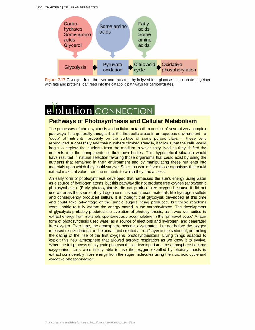
Figure 7.17 Glycogen from the liver and muscles, hydrolyzed into glucose-1-phosphate, togetherwith fats and proteins, can feed into the catabolic pathways for carbohydrates.
Pathways of Photosynthesis and Cellular MetabolismThe processes of photosynthesis and cellular metabolism consist of several very complexpathways. It is generally thought that the first cells arose in an aqueous environment—a“soup” of nutrients—probably on the surface of some porous clays. If these cellsreproduced successfully and their numbers climbed steadily, it follows that the cells wouldbegin to deplete the nutrients from the medium in which they lived as they shifted thenutrients into the components of their own bodies. This hypothetical situation wouldhave resulted in natural selection favoring those organisms that could exist by using thenutrients that remained in their environment and by manipulating these nutrients intomaterials upon which they could survive. Selection would favor those organisms that couldextract maximal value from the nutrients to which they had access.
An early form of photosynthesis developed that harnessed the sun’s energy using wateras a source of hydrogen atoms, but this pathway did not produce free oxygen (anoxygenicphotosynthesis). (Early photosynthesis did not produce free oxygen because it did notuse water as the source of hydrogen ions; instead, it used materials like hydrogen sulfideand consequently produced sulfur). It is thought that glycolysis developed at this timeand could take advantage of the simple sugars being produced, but these reactionswere unable to fully extract the energy stored in the carbohydrates. The developmentof glycolysis probably predated the evolution of photosynthesis, as it was well suited toextract energy from materials spontaneously accumulating in the “primeval soup.” A laterform of photosynthesis used water as a source of electrons and hydrogen, and generatedfree oxygen. Over time, the atmosphere became oxygenated, but not before the oxygenreleased oxidized metals in the ocean and created a “rust” layer in the sediment, permittingthe dating of the rise of the first oxygenic photosynthesizers. Living things adapted toexploit this new atmosphere that allowed aerobic respiration as we know it to evolve.When the full process of oxygenic photosynthesis developed and the atmosphere becameoxygenated, cells were finally able to use the oxygen expelled by photosynthesis toextract considerably more energy from the sugar molecules using the citric acid cycle andoxidative phosphorylation.
220 CHAPTER 7 | CELLULAR RESPIRATION
This content is available for free at http://cnx.org/content/col11448/1.9
7.7 | Regulation of Cellular Respiration
By the end of this section, you will be able to:
• Describe how feedback inhibition would affect the production of an intermediate or product ina pathway
• Identify the mechanism that controls the rate of the transport of electrons through the electrontransport chain
Cellular respiration must be regulated in order to provide balanced amounts of energy in the form ofATP. The cell also must generate a number of intermediate compounds that are used in the anabolismand catabolism of macromolecules. Without controls, metabolic reactions would quickly come to a standstill as the forward and backward reactions reached a state of equilibrium. Resources would be usedinappropriately. A cell does not need the maximum amount of ATP that it can make all the time: Attimes, the cell needs to shunt some of the intermediates to pathways for amino acid, protein, glycogen,lipid, and nucleic acid production. In short, the cell needs to control its metabolism.
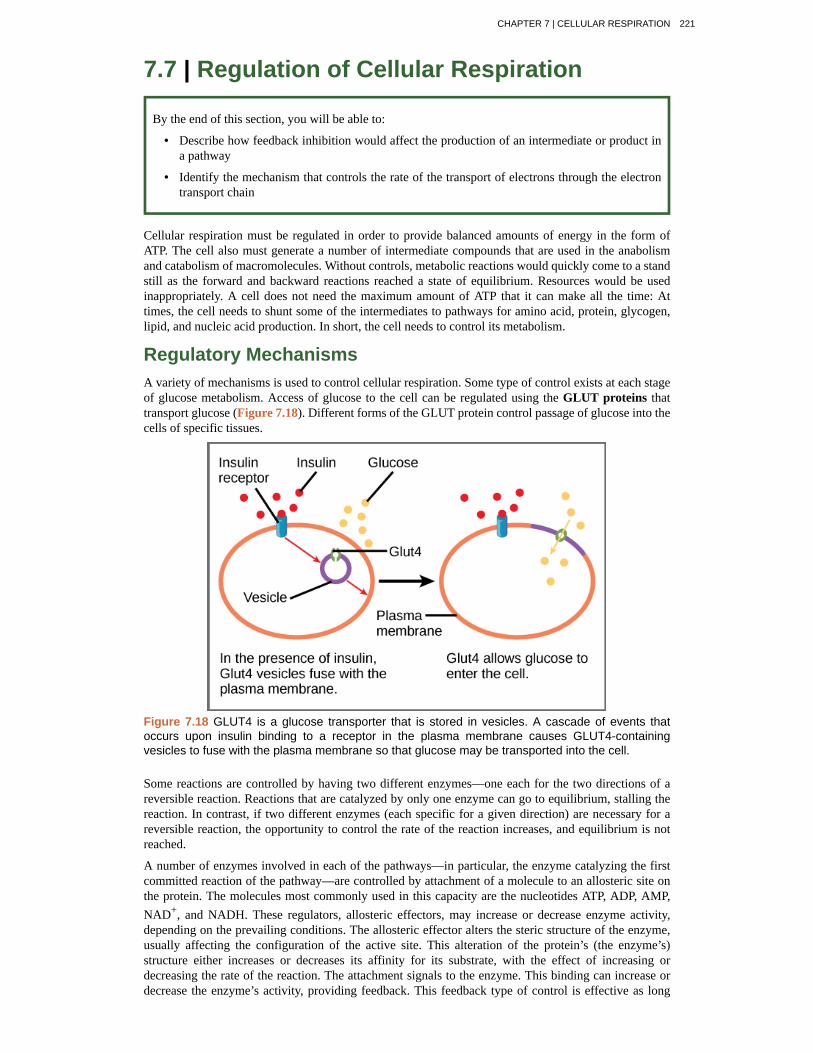
Regulatory MechanismsA variety of mechanisms is used to control cellular respiration. Some type of control exists at each stageof glucose metabolism. Access of glucose to the cell can be regulated using the GLUT proteins thattransport glucose (Figure 7.18). Different forms of the GLUT protein control passage of glucose into thecells of specific tissues.
Figure 7.18 GLUT4 is a glucose transporter that is stored in vesicles. A cascade of events thatoccurs upon insulin binding to a receptor in the plasma membrane causes GLUT4-containingvesicles to fuse with the plasma membrane so that glucose may be transported into the cell.
Some reactions are controlled by having two different enzymes—one each for the two directions of areversible reaction. Reactions that are catalyzed by only one enzyme can go to equilibrium, stalling thereaction. In contrast, if two different enzymes (each specific for a given direction) are necessary for areversible reaction, the opportunity to control the rate of the reaction increases, and equilibrium is notreached.
A number of enzymes involved in each of the pathways—in particular, the enzyme catalyzing the firstcommitted reaction of the pathway—are controlled by attachment of a molecule to an allosteric site onthe protein. The molecules most commonly used in this capacity are the nucleotides ATP, ADP, AMP,NAD+, and NADH. These regulators, allosteric effectors, may increase or decrease enzyme activity,depending on the prevailing conditions. The allosteric effector alters the steric structure of the enzyme,usually affecting the configuration of the active site. This alteration of the protein’s (the enzyme’s)structure either increases or decreases its affinity for its substrate, with the effect of increasing ordecreasing the rate of the reaction. The attachment signals to the enzyme. This binding can increase ordecrease the enzyme’s activity, providing feedback. This feedback type of control is effective as long
CHAPTER 7 | CELLULAR RESPIRATION 221
as the chemical affecting it is attached to the enzyme. Once the overall concentration of the chemicaldecreases, it will diffuse away from the protein, and the control is relaxed.
Control of Catabolic PathwaysEnzymes, proteins, electron carriers, and pumps that play roles in glycolysis, the citric acid cycle, and theelectron transport chain tend to catalyze non-reversible reactions. In other words, if the initial reactiontakes place, the pathway is committed to proceeding with the remaining reactions. Whether a particularenzyme activity is released depends upon the energy needs of the cell (as reflected by the levels of ATP,ADP, and AMP).
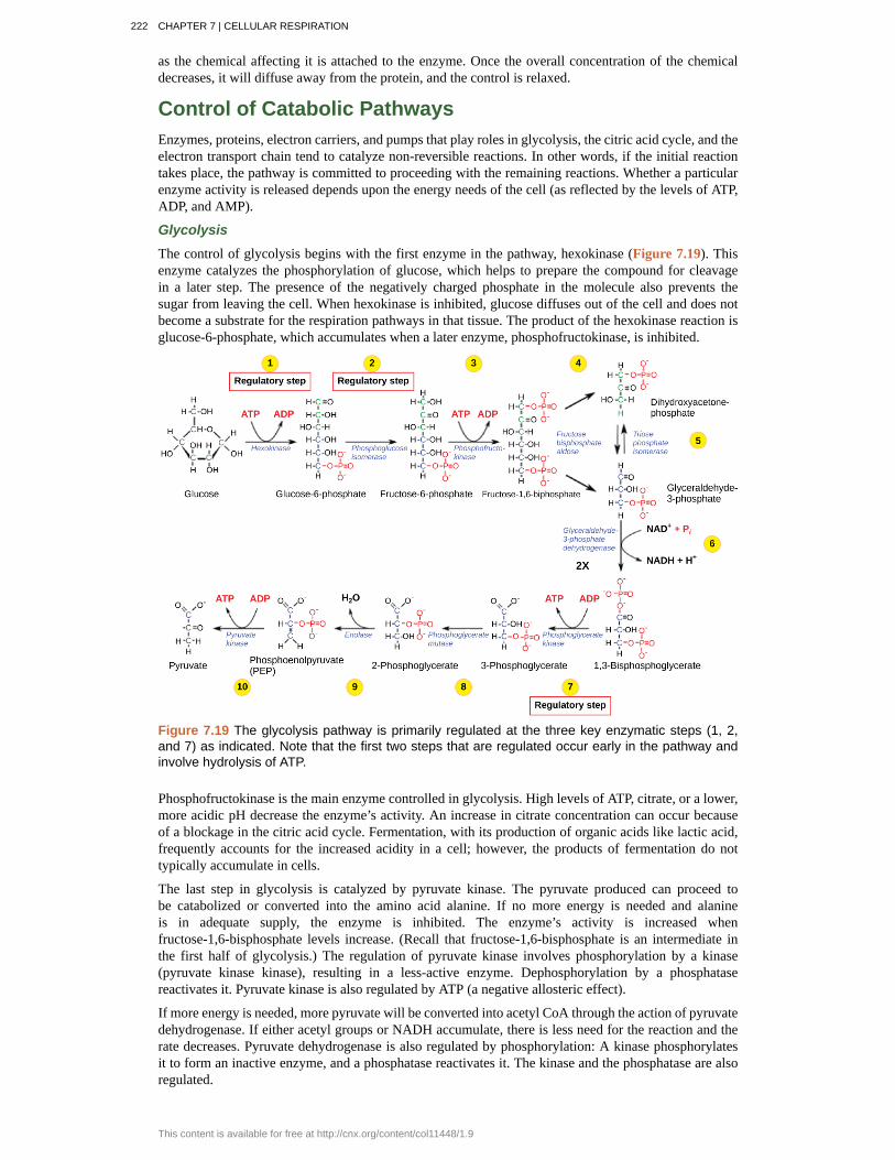
Glycolysis
The control of glycolysis begins with the first enzyme in the pathway, hexokinase (Figure 7.19). Thisenzyme catalyzes the phosphorylation of glucose, which helps to prepare the compound for cleavagein a later step. The presence of the negatively charged phosphate in the molecule also prevents thesugar from leaving the cell. When hexokinase is inhibited, glucose diffuses out of the cell and does notbecome a substrate for the respiration pathways in that tissue. The product of the hexokinase reaction isglucose-6-phosphate, which accumulates when a later enzyme, phosphofructokinase, is inhibited.
Figure 7.19 The glycolysis pathway is primarily regulated at the three key enzymatic steps (1, 2,and 7) as indicated. Note that the first two steps that are regulated occur early in the pathway andinvolve hydrolysis of ATP.
Phosphofructokinase is the main enzyme controlled in glycolysis. High levels of ATP, citrate, or a lower,more acidic pH decrease the enzyme’s activity. An increase in citrate concentration can occur becauseof a blockage in the citric acid cycle. Fermentation, with its production of organic acids like lactic acid,frequently accounts for the increased acidity in a cell; however, the products of fermentation do nottypically accumulate in cells.
The last step in glycolysis is catalyzed by pyruvate kinase. The pyruvate produced can proceed tobe catabolized or converted into the amino acid alanine. If no more energy is needed and alanineis in adequate supply, the enzyme is inhibited. The enzyme’s activity is increased whenfructose-1,6-bisphosphate levels increase. (Recall that fructose-1,6-bisphosphate is an intermediate inthe first half of glycolysis.) The regulation of pyruvate kinase involves phosphorylation by a kinase(pyruvate kinase kinase), resulting in a less-active enzyme. Dephosphorylation by a phosphatasereactivates it. Pyruvate kinase is also regulated by ATP (a negative allosteric effect).
If more energy is needed, more pyruvate will be converted into acetyl CoA through the action of pyruvatedehydrogenase. If either acetyl groups or NADH accumulate, there is less need for the reaction and therate decreases. Pyruvate dehydrogenase is also regulated by phosphorylation: A kinase phosphorylatesit to form an inactive enzyme, and a phosphatase reactivates it. The kinase and the phosphatase are alsoregulated.
222 CHAPTER 7 | CELLULAR RESPIRATION
This content is available for free at http://cnx.org/content/col11448/1.9
Citric Acid Cycle
The citric acid cycle is controlled through the enzymes that catalyze the reactions that make the firsttwo molecules of NADH (Figure 7.9). These enzymes are isocitrate dehydrogenase and α-ketoglutaratedehydrogenase. When adequate ATP and NADH levels are available, the rates of these reactionsdecrease. When more ATP is needed, as reflected in rising ADP levels, the rate increases. α-Ketoglutarate dehydrogenase will also be affected by the levels of succinyl CoA—a subsequentintermediate in the cycle—causing a decrease in activity. A decrease in the rate of operation of thepathway at this point is not necessarily negative, as the increased levels of the α-ketoglutarate not usedby the citric acid cycle can be used by the cell for amino acid (glutamate) synthesis.
Electron Transport Chain
Specific enzymes of the electron transport chain are unaffected by feedback inhibition, but the rateof electron transport through the pathway is affected by the levels of ADP and ATP. Greater ATPconsumption by a cell is indicated by a buildup of ADP. As ATP usage decreases, the concentration ofADP decreases, and now, ATP begins to build up in the cell. This change is the relative concentration ofADP to ATP triggers the cell to slow down the electron transport chain.
Visit this site (http://openstaxcollege.org/l/electron_transp) to see an animation of the electrontransport chain and ATP synthesis.
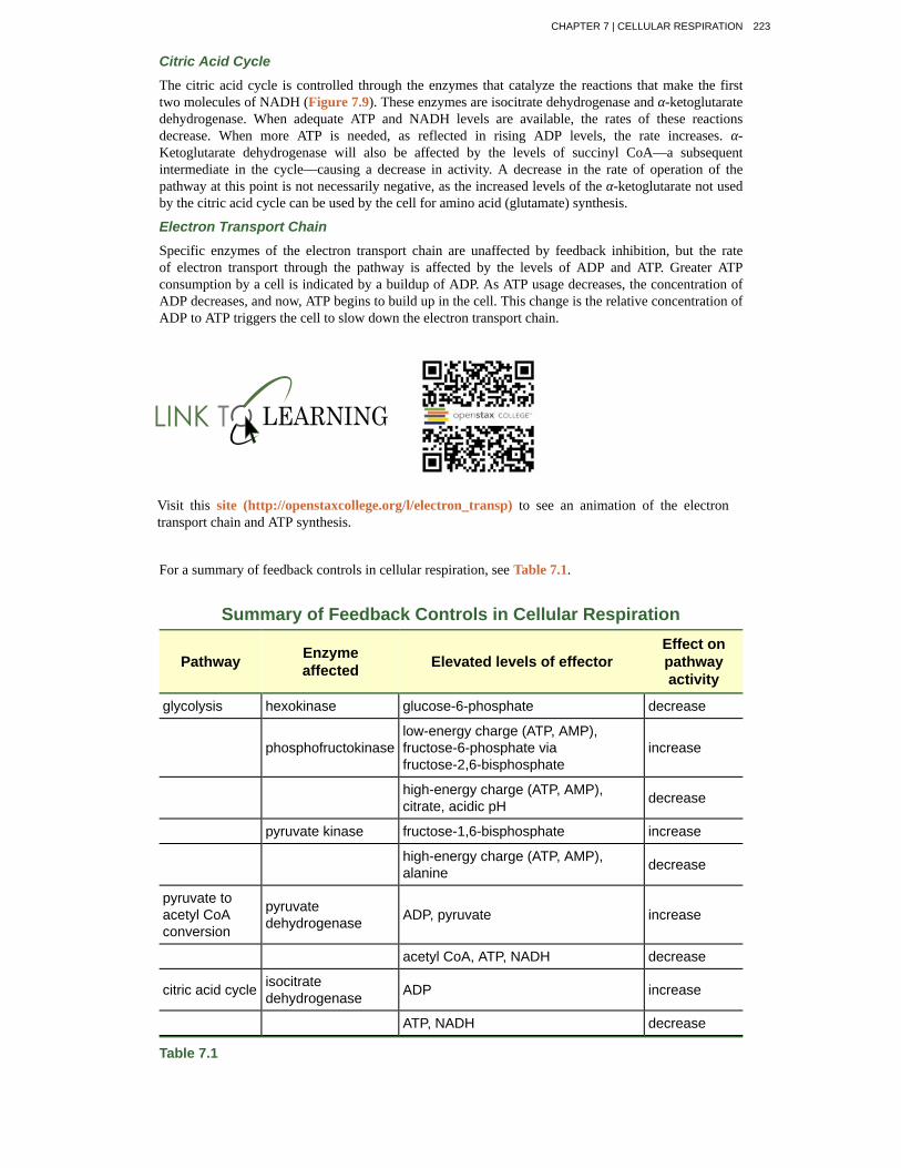
For a summary of feedback controls in cellular respiration, see Table 7.1.
Summary of Feedback Controls in Cellular Respiration
Pathway Enzymeaffected Elevated levels of effector
Effect onpathwayactivity
glycolysis hexokinase glucose-6-phosphate decrease
phosphofructokinaselow-energy charge (ATP, AMP),fructose-6-phosphate viafructose-2,6-bisphosphate
increase
high-energy charge (ATP, AMP),citrate, acidic pH decrease
pyruvate kinase fructose-1,6-bisphosphate increase
high-energy charge (ATP, AMP),alanine decrease
pyruvate toacetyl CoAconversion
pyruvatedehydrogenase ADP, pyruvate increase
acetyl CoA, ATP, NADH decrease
citric acid cycle isocitratedehydrogenase ADP increase
ATP, NADH decrease
Table 7.1
CHAPTER 7 | CELLULAR RESPIRATION 223
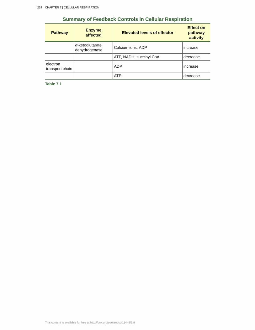
Summary of Feedback Controls in Cellular Respiration
Pathway Enzymeaffected Elevated levels of effector
Effect onpathwayactivity
α-ketoglutaratedehydrogenase Calcium ions, ADP increase
ATP, NADH, succinyl CoA decrease
electrontransport chain ADP increase
ATP decrease
Table 7.1
224 CHAPTER 7 | CELLULAR RESPIRATION
This content is available for free at http://cnx.org/content/col11448/1.9
ATP synthase
acetyl CoA
aerobic respiration
anaerobic cellular respiration
anaerobic
chemiosmosis
citric acid cycle
dephosphorylation
fermentation
GLUT protein
glycolysis
isomerase
Krebs cycle
oxidative phosphorylation
phosphorylation
prosthetic group
pyruvate
redox reaction
substrate-level phosphorylation
TCA cycle
ubiquinone
KEY TERMS(also, F1F0 ATP synthase) membrane-embedded protein complex that adds a
phosphate to ADP with energy from protons diffusing through it
combination of an acetyl group derived from pyruvic acid and coenzyme A, which ismade from pantothenic acid (a B-group vitamin)
process in which organisms convert energy in the presence of oxygen
process in which organisms convert energy for their use in theabsence of oxygen
process that does not use oxygen
process in which there is a production of adenosine triphosphate (ATP) in cellularmetabolism by the involvement of a proton gradient across a membrane
(also, Krebs cycle) series of enzyme-catalyzed chemical reactions of centralimportance in all living cells
removal of a phosphate group from a molecule
process of regenerating NAD+ with either an inorganic or organic compound servingas the final electron accceptor, occurs in the absence; occurs in the absence of oxygen
integral membrane protein that transports glucose
process of breaking glucose into two three-carbon molecules with the production of ATPand NADH
enzyme that converts a molecule into its isomer
(also, citric acid cycle) alternate name for the citric acid cycle, named after Hans Krebswho first identified the steps in the pathway in the 1930s in pigeon flight muscles; see citric acidcycle
production of ATP using the process of chemiosmosis and oxygen
addition of a high-energy phosphate to a compound, usually a metabolicintermediate, a protein, or ADP
(also, prosthetic cofactor) molecule bound to a protein that facilitates the functionof the protein
three-carbon sugar that can be decarboxylated and oxidized to make acetyl CoA, whichenters the citric acid cycle under aerobic conditions; the end product of glycolysis
chemical reaction that consists of the coupling of an oxidation reaction and areduction reaction
production of ATP from ADP using the excess energy from achemical reaction and a phosphate group from a reactant
(also, citric acid cycle) alternate name for the citric acid cycle, named after the groupname for citric acid, tricarboxylic acid (TCA); see citric acid cycle
soluble electron transporter in the electron transport chain that connects the first orsecond complex to the third
CHAPTER SUMMARY
CHAPTER 7 | CELLULAR RESPIRATION 225
7.1 Energy in Living Systems
ATP functions as the energy currency for cells. It allows the cell to store energy briefly and transport itwithin the cell to support endergonic chemical reactions. The structure of ATP is that of an RNAnucleotide with three phosphates attached. As ATP is used for energy, a phosphate group or two aredetached, and either ADP or AMP is produced. Energy derived from glucose catabolism is used toconvert ADP into ATP. When ATP is used in a reaction, the third phosphate is temporarily attached to asubstrate in a process called phosphorylation. The two processes of ATP regeneration that are used inconjunction with glucose catabolism are substrate-level phosphorylation and oxidative phosphorylationthrough the process of chemiosmosis.
7.2 Glycolysis
Glycolysis is the first pathway used in the breakdown of glucose to extract energy. It was probably oneof the earliest metabolic pathways to evolve and is used by nearly all of the organisms on earth.Glycolysis consists of two parts: The first part prepares the six-carbon ring of glucose for cleavage intotwo three-carbon sugars. ATP is invested in the process during this half to energize the separation. Thesecond half of glycolysis extracts ATP and high-energy electrons from hydrogen atoms and attachesthem to NAD+. Two ATP molecules are invested in the first half and four ATP molecules are formed bysubstrate phosphorylation during the second half. This produces a net gain of two ATP and two NADHmolecules for the cell.
7.3 Oxidation of Pyruvate and the Citric Acid Cycle
In the presence of oxygen, pyruvate is transformed into an acetyl group attached to a carrier moleculeof coenzyme A. The resulting acetyl CoA can enter several pathways, but most often, the acetyl groupis delivered to the citric acid cycle for further catabolism. During the conversion of pyruvate into theacetyl group, a molecule of carbon dioxide and two high-energy electrons are removed. The carbondioxide accounts for two (conversion of two pyruvate molecules) of the six carbons of the originalglucose molecule. The electrons are picked up by NAD+, and the NADH carries the electrons to a laterpathway for ATP production. At this point, the glucose molecule that originally entered cellularrespiration has been completely oxidized. Chemical potential energy stored within the glucose moleculehas been transferred to electron carriers or has been used to synthesize a few ATPs.
The citric acid cycle is a series of redox and decarboxylation reactions that remove high-energyelectrons and carbon dioxide. The electrons temporarily stored in molecules of NADH and FADH2 areused to generate ATP in a subsequent pathway. One molecule of either GTP or ATP is produced bysubstrate-level phosphorylation on each turn of the cycle. There is no comparison of the cyclic pathwaywith a linear one.
7.4 Oxidative Phosphorylation
The electron transport chain is the portion of aerobic respiration that uses free oxygen as the finalelectron acceptor of the electrons removed from the intermediate compounds in glucose catabolism.The electron transport chain is composed of four large, multiprotein complexes embedded in the innermitochondrial membrane and two small diffusible electron carriers shuttling electrons between them.The electrons are passed through a series of redox reactions, with a small amount of free energy used atthree points to transport hydrogen ions across a membrane. This process contributes to the gradientused in chemiosmosis. The electrons passing through the electron transport chain gradually lose energy,High-energy electrons donated to the chain by either NADH or FADH2 complete the chain, as low-energy electrons reduce oxygen molecules and form water. The level of free energy of the electronsdrops from about 60 kcal/mol in NADH or 45 kcal/mol in FADH2 to about 0 kcal/mol in water. The endproducts of the electron transport chain are water and ATP. A number of intermediate compounds of thecitric acid cycle can be diverted into the anabolism of other biochemical molecules, such asnonessential amino acids, sugars, and lipids. These same molecules can serve as energy sources for theglucose pathways.
7.5 Metabolism without Oxygen
If NADH cannot be oxidized through aerobic respiration, another electron acceptor is used. Mostorganisms will use some form of fermentation to accomplish the regeneration of NAD+, ensuring the
226 CHAPTER 7 | CELLULAR RESPIRATION
This content is available for free at http://cnx.org/content/col11448/1.9
continuation of glycolysis. The regeneration of NAD+ in fermentation is not accompanied by ATPproduction; therefore, the potential of NADH to produce ATP using an electron transport chain is notutilized.
7.6 Connections of Carbohydrate, Protein, and Lipid Metabolic Pathways
The breakdown and synthesis of carbohydrates, proteins, and lipids connect with the pathways ofglucose catabolism. The simple sugars are galactose, fructose, glycogen, and pentose. These arecatabolized during glycolysis. The amino acids from proteins connect with glucose catabolism throughpyruvate, acetyl CoA, and components of the citric acid cycle. Cholesterol synthesis starts with acetylgroups, and the components of triglycerides come from glycerol-3-phosphate from glycolysis andacetyl groups produced in the mitochondria from pyruvate.
7.7 Regulation of Cellular Respiration
Cellular respiration is controlled by a variety of means. The entry of glucose into a cell is controlled bythe transport proteins that aid glucose passage through the cell membrane. Most of the control of therespiration processes is accomplished through the control of specific enzymes in the pathways. This is atype of negative feedback, turning the enzymes off. The enzymes respond most often to the levels of theavailable nucleosides ATP, ADP, AMP, NAD+, and FAD. Other intermediates of the pathway also affectcertain enzymes in the systems.
ART CONNECTION QUESTIONS1. Figure 7.11 Dinitrophenol (DNP) is anuncoupler that makes the inner mitochondrialmembrane leaky to protons. It was used until 1938as a weight-loss drug. What effect would youexpect DNP to have on the change in pH acrossthe inner mitochondrial membrane? Why do youthink this might be an effective weight-loss drug?
2. Figure 7.12 Cyanide inhibits cytochrome coxidase, a component of the electron transportchain. If cyanide poisoning occurs, would youexpect the pH of the intermembrane space to
increase or decrease? What effect would cyanidehave on ATP synthesis?
3. Figure 7.14 Tremetol, a metabolic poisonfound in the white snake root plant, prevents themetabolism of lactate. When cows eat this plant, itis concentrated in the milk they produce. Humanswho consume the milk become ill. Symptoms ofthis disease, which include vomiting, abdominalpain, and tremors, become worse after exercise.Why do you think this is the case?
REVIEW QUESTIONS4. The energy currency used by cells is ________.
a. ATPb. ADPc. AMPd. adenosine
5. A reducing chemical reaction ________.
a. reduces the compound to a simpler formb. adds an electron to the substratec. removes a hydrogen atom from the
substrated. is a catabolic reaction
6. During the second half of glycolysis, whatoccurs?
a. ATP is used up.b. Fructose is split in two.c. ATP is made.d. Glucose becomes fructose.
7. What is removed from pyruvate during itsconversion into an acetyl group?
a. oxygenb. ATPc. B vitamind. carbon dioxide
8. What do the electrons added to NAD+ do?
a. They become part of a fermentationpathway.
b. They go to another pathway for ATPproduction.
c. They energize the entry of the acetylgroup into the citric acid cycle.
d. They are converted to NADP.
9. GTP or ATP is produced during the conversionof ________.
a. isocitrate into α-ketoglutarateb. succinyl CoA into succinatec. fumarate into malated. malate into oxaloacetate
10. How many NADH molecules are produced oneach turn of the citric acid cycle?
CHAPTER 7 | CELLULAR RESPIRATION 227

a. oneb. twoc. threed. four
11. What compound receives electrons fromNADH?
a. FMNb. ubiquinonec. cytochrome c1d. oxygen
12. Chemiosmosis involves ________.a. the movement of electrons across the
cell membraneb. the movement of hydrogen atoms across
a mitochondrial membranec. the movement of hydrogen ions across a
mitochondrial membraned. the movement of glucose through the
cell membrane
13. Which of the following fermentation methodscan occur in animal skeletal muscles?
a. lactic acid fermentationb. alcohol fermentationc. mixed acid fermentationd. propionic fermentation
14. A major connection for sugars in glycolysis is________.
a. glucose-6-phosphateb. fructose-1,6-bisphosphatec. dihydroxyacetone phosphated. phosphoenolpyruvate
15. Beta-oxidation is ________.a. the breakdown of sugarsb. the assembly of sugarsc. the breakdown of fatty acidsd. the removal of amino groups from
amino acids
16. The effect of high levels of ADP is to________.
a. increase the activity of the enzymeb. decrease the activity of the enzymec. have no effect on the activity of the
enzymed. slow down the pathway
17. The control of which enzyme exerts the mostcontrol on glycolysis?
a. hexokinaseb. phosphofructokinasec. glucose-6-phosphatased. aldolase
CRITICAL THINKING QUESTIONS18. Why is it beneficial for cells to use ATP ratherthan energy directly from the bonds ofcarbohydrates? What are the greatest drawbacks toharnessing energy directly from the bonds ofseveral different compounds?
19. Nearly all organisms on earth carry out someform of glycolysis. How does that fact support ornot support the assertion that glycolysis is one ofthe oldest metabolic pathways?
20. Red blood cells do not perform aerobicrespiration, but they do perform glycolysis. Whydo all cells need an energy source, and whatwould happen if glycolysis were blocked in a redblood cell?
21. What is the primary difference between acircular pathway and a linear pathway?
22. How do the roles of ubiquinone andcytochrome c differ from the other components ofthe electron transport chain?
23. What accounts for the different number ofATP molecules that are formed through cellularrespiration?
24. What is the primary difference betweenfermentation and anaerobic respiration?
25. Would you describe metabolic pathways asinherently wasteful or inherently economical, andwhy?
26. How does citrate from the citric acid cycleaffect glycolysis?
27. Why might negative feedback mechanisms bemore common than positive feedback mechanismsin living cells?
228 CHAPTER 7 | CELLULAR RESPIRATION
This content is available for free at http://cnx.org/content/col11448/1.9