Embed Size (px)
Citation preview

www.ck12.org
CHAPTER 5 The Cell Cycle, Mitosis, andMeiosis
Chapter Outline5.1 CELL DIVISION AND THE CELL CYCLE
5.2 CHROMOSOMES AND MITOSIS
5.3 REPRODUCTION AND MEIOSIS
5.4 REFERENCES
What do you think this colorful picture shows? If you guessed that it’s a picture of a cell undergoing cell division, youare right. In fact, the picture is an image of a lung cell stained with fluorescent dyes undergoing mitosis, specificallyduring early anaphase. You will read about mitosis, a type of cell division, in this chapter.
Cell division is just one of the stages that all cells go through during their life. This includes cells that are harmful,such as cancer cells. Cancer cells divide more often than normal cells, and grow out of control. In fact, this is howcancer cells cause illness. In this chapter, you will read about how cells divide, what other stages cells go through,and what causes cancer cells to divide out of control and harm the body.Courtesy o f Conly Rieder/National Institutes o f Health. commons.wikimedia.org/wiki/File:Mitosis� f luorescent. jpg. Public Domain.
108

www.ck12.org Chapter 5. The Cell Cycle, Mitosis, and Meiosis
5.1 Cell Division and the Cell Cycle
Lesson Objectives
• Contrast cell division in prokaryotes and eukaryotes.• Identify the phases of the eukaryotic cell cycle.• Explain how the cell cycle is controlled.• Define cancer, and relate it to the cell cycle.
Vocabulary
• binary fission• cancer• cell cycle• cell division• cytokinesis• DNA replication• interphase• mitosis• tumor
Introduction
You consist of a great many cells, but like all other organisms, you started life as a single cell. How did youdevelop from a single cell into an organism with trillions of cells? The answer is cell division. After cells grow totheir maximum size, they divide into two new cells. These new cells are small at first, but they grow quickly andeventually divide and produce more new cells. This process keeps repeating in a continuous cycle.
Cell Division
Cell division is the process in which one cell, called the parent cell, divides to form two new cells, referred to asdaughter cells. How this happens depends on whether the cell is prokaryotic or eukaryotic.
Cell division is simpler in prokaryotes than eukaryotes because prokaryotic cells themselves are simpler. Prokaryoticcells have a single circular chromosome, no nucleus, and few other organelles. Eukaryotic cells, in contrast, havemultiple chromosomes contained within a nucleus and many other organelles. All of these cell parts must beduplicated and then separated when the cell divides.
109

5.1. Cell Division and the Cell Cycle www.ck12.org
Cell Division in Prokaryotes
Most prokaryotic cells divide by the process of binary fission. A bacterial cell dividing this way is depicted inFigure 5.1. You can also watch an animation of binary fission at this link: http://en.wikipedia.org/wiki/File:Binary_fission_anim.gif .
FIGURE 5.1Binary Fission in a Bacterial Cell. Celldivision is relatively simple in prokaryoticcells. The two cells are dividing by binaryfission. Green and orange lines indicateold and newly-generated bacterial cellwalls, respectively. Eventually the parentcell will pinch apart to form two identicaldaughter cells. Left, growth at the centerof bacterial body. Right, apical growthfrom the ends of the bacterial body.
Binary fission can be described as a series of steps, although it is actually a continuous process. The steps aredescribed below and also illustrated in Figure 5.2. They include DNA replication, chromosome segregation, andfinally the separation into two daughter cells.
• Step 1: DNA Replication. Just before the cell divides, its DNA is copied in a process called DNA replication.This results in two identical chromosomes instead of just one. This step is necessary so that when the celldivides, each daughter cell will have its own chromosome.
• Step 2: Chromosome Segregation. The two chromosomes segregate, or separate, and move to opposite ends(known as “poles”) of the cell. This occurs as each copy of DNA attaches to different parts of the cellmembrane.
• Step 3: Separation. A new plasma membrane starts growing into the center of the cell, and the cytoplasm splitsapart, forming two daughter cells. As the cell begins to pull apart, the new and the original chromosomes areseparated. The two daughter cells that result are genetically identical to each other and to the parent cell. Newcell wall must also form around the two cells.
Cell Division in Eukaryotes
Cell division is more complex in eukaryotes than prokaryotes. Prior to dividing, all the DNA in a eukaryotic cell’smultiple chromosomes is replicated. Its organelles are also duplicated. Then, when the cell divides, it occurs in twomajor steps:
• The first step is mitosis, a multi-phase process in which the nucleus of the cell divides. During mitosis, thenuclear membrane breaks down and later reforms. The chromosomes are also sorted and separated to ensurethat each daughter cell receives a complete set of chromosomes. Mitosis is described in greater detail in thelesson “Chromosomes and Mitosis.”
110

www.ck12.org Chapter 5. The Cell Cycle, Mitosis, and Meiosis
FIGURE 5.2Steps of Binary Fission. Prokaryotic cellsdivide by binary fission. This is alsohow many single-celled organisms repro-duce.
• The second major step is cytokinesis. As in prokaryotic cells, during this step the cytoplasm divides and twodaughter cells form.
The Cell Cycle
Cell division is just one of several stages that a cell goes through during its lifetime. The cell cycle is a repeatingseries of events that include growth, DNA synthesis, and cell division. The cell cycle in prokaryotes is quite simple:the cell grows, its DNA replicates, and the cell divides. In eukaryotes, the cell cycle is more complicated.
Eukaryotic Cell Cycle
The diagram in Figure 5.3 represents the cell cycle of a eukaryotic cell. As you can see, the eukaryotic cell cyclehas several phases. The mitotic phase (M) actually includes both mitosis and cytokinesis. This is when the nucleusand then the cytoplasm divide. The other three phases (G1, S, and G2) are generally grouped together as interphase.During interphase, the cell grows, performs routine life processes, and prepares to divide. These phases are discussedbelow. You can watch a eukaryotic cell going through these phases of the cell cycle at the following link: http://www.cellsalive.com/cell_cycle.htm .
Interphase
Interphase of the eukaryotic cell cycle can be subdivided into the following three phases, which are represented inFigure 5.3:
• Growth Phase 1 (G1): during this phase, the cell grows rapidly, while performing routine metabolic pro-cesses. It also makes proteins needed for DNA replication and copies some of its organelles in preparation forcell division. A cell typically spends most of its life in this phase. This phase is also known as gap phase 1.
111

5.1. Cell Division and the Cell Cycle www.ck12.org
FIGURE 5.3Eukaryotic Cell Cycle. This diagram rep-resents the cell cycle in eukaryotes. TheFirst Gap, Synthesis, and Second Gapphases make up interphase (I). The M(mitotic) phase includes mitosis and cy-tokinesis. After the M phase, two cellsresult.
• Synthesis Phase (S): during this phase, the cell’s DNA is copied in the process of DNA replication.• Growth Phase 2 (G2): during this phase, the cell makes final preparations to divide. For example, it makes
additional proteins and organelles. This phase is also known as gap phase 2.
Control of the Cell Cycle
If the cell cycle occurred without regulation, cells might go from one phase to the next before they were ready.What controls the cell cycle? How does the cell know when to grow, synthesize DNA, and divide? The cell cycle iscontrolled mainly by regulatory proteins. These proteins control the cycle by signaling the cell to either start or delaythe next phase of the cycle. They ensure that the cell completes the previous phase before moving on. Regulatoryproteins control the cell cycle at key checkpoints, which are shown in Figure 5.4. There are a number of maincheckpoints.
• The G1 checkpoint, just before entry into S phase, makes the key decision of whether the cell should divide.• The S checkpoint determines if the DNA has been replicated properly.• The mitotic spindle checkpoint occurs at the point in metaphase where all the chromosomes should have
aligned at the mitotic plate.
Cancer and the Cell Cycle
Cancer is a disease that occurs when the cell cycle is no longer regulated. This may happen because a cell’s DNAbecomes damaged. Damage can occur due to exposure to hazards such as radiation or toxic chemicals. Cancerouscells generally divide much faster than normal cells. They may form a mass of abnormal cells called a tumor (seeFigure 5.5). The rapidly dividing cells take up nutrients and space that normal cells need. This can damage tissuesand organs and eventually lead to death.
Cancer is discussed in the video at http://www.youtube.com/watch?v=RZhL7LDPk8w .
112
www.ck12.org Chapter 5. The Cell Cycle, Mitosis, and Meiosis
FIGURE 5.4Checkpoints in the eukaryotic cell cycle ensure that the cell is ready to proceed before it moves on to the nextphase of the cycle.
MEDIAClick image to the left for more content.
Lesson Summary
• Cell division is part of the life cycle of virtually all cells. It is a more complicated process in eukaryotic thanprokaryotic cells because eukaryotic cells have multiple chromosomes and a nucleus.
113

5.1. Cell Division and the Cell Cycle www.ck12.org
FIGURE 5.5These cells are cancer cells, growing outof control and forming a tumor.
• The cell cycle is a repeating series of events that cells go through. It includes growth, DNA synthesis, and celldivision. In eukaryotic cells, there are two growth phases, and cell division includes mitosis.
• The cell cycle is controlled by regulatory proteins at three key checkpoints in the cycle. The proteins signalthe cell to either start or delay the next phase of the cycle.
• Cancer is a disease that occurs when the cell cycle is no longer regulated. Cancer cells grow rapidly and mayform a mass of abnormal cells called a tumor.
Lesson Review Questions
Recall
1. Describe binary fission.
2. What is mitosis?
3. Identify the phases of the eukaryotic cell cycle.
4. What happens during interphase?
5. Define cancer.
Apply Concepts
6. How might the relationship between cancer and the cell cycle be used in the search for causes of cancer?
Think Critically
7. Cells go through a series of events that include growth, DNA synthesis, and cell division. Why are these eventsbest represented by a cycle diagram?
8. Contrast cell division in prokaryotes and eukaryotes. Why are the two types of cell division different?
114

www.ck12.org Chapter 5. The Cell Cycle, Mitosis, and Meiosis
9. Explain how the cell cycle is regulated.
10. Why is DNA replication essential to the cell cycle?
Points to Consider
When a eukaryotic cell divides, the nucleus divides first in the process of mitosis.
• What do you think happens during mitosis? Can you predict what molecules and cell structures are involvedin this process?
• How do you think mitosis might differ from binary fission? What steps might be involved in mitosis?
115

5.2. Chromosomes and Mitosis www.ck12.org
5.2 Chromosomes and Mitosis
Lesson Objectives
• Describe chromosomes and their role in mitosis.• Outline the phases of mitosis.
Vocabulary
• anaphase• centromere• chromatid• chromatin• chromosome• gene• homologous chromosomes• metaphase• prophase• telophase
Introduction
In eukaryotic cells, the nucleus divides before the cell itself divides. The process in which the nucleus divides iscalled mitosis. Before mitosis occurs, a cell’s DNA is replicated. This is necessary so that each daughter cell willhave a complete copy of the genetic material from the parent cell. How is the replicated DNA sorted and separatedso that each daughter cell gets a complete set of the genetic material? To understand how this happens, you need toknow more chromosomes.
Chromosomes
Chromosomes are coiled structures made of DNA and proteins. Chromosomes are the form of the genetic materialof a cell during cell division. During other phases of the cell cycle, DNA is not coiled into chromosomes. Instead, itexists as a grainy material called chromatin.
The vocabulary of DNA: chromosomes, chromatids, chromatin, transcription, translation, and replication is dis-cussed at http://www.youtube.com/watch?v=s9HPNwXd9fk (18:23).
116

www.ck12.org Chapter 5. The Cell Cycle, Mitosis, and Meiosis
MEDIAClick image to the left for more content.
Chromatids and the Centromere
DNA condenses and coils into the familiar X-shaped form of a chromosome, shown in Figure 5.6, only after it hasreplicated. (You can watch DNA coiling into a chromosome at the link below.) Because DNA has already replicated,each chromosome actually consists of two identical copies. The two copies are called sister chromatids. They areattached to one another at a region called the centromere. A remarkable animation can be viewed at http://www.hhmi.org/biointeractive/dna-packaging .
FIGURE 5.6Chromosome. After DNA replicates, itforms chromosomes like the one shownhere.
Chromosomes and Genes
The DNA of a chromosome is encoded with genetic instructions for making proteins. These instructions areorganized into units called genes. Most genes contain the instructions for a single protein. There may be hundredsor even thousands of genes on a single chromosome.
Human Chromosomes
Human cells normally have two sets of chromosomes, one set inherited from each parent. There are 23 chromosomesin each set, for a total of 46 chromosomes per cell. Each chromosome in one set is matched by a chromosome of thesame type in the other set, so there are actually 23 pairs of chromosomes per cell. Each pair consists of chromosomesof the same size and shape that also contain the same genes. The chromosomes in a pair are known as homologouschromosomes.
117
5.2. Chromosomes and Mitosis www.ck12.org
Mitosis and Cytokinesis
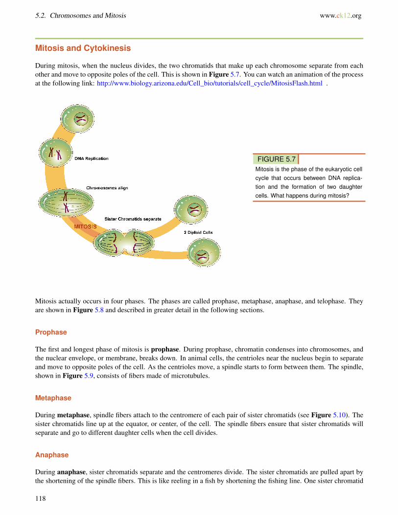
During mitosis, when the nucleus divides, the two chromatids that make up each chromosome separate from eachother and move to opposite poles of the cell. This is shown in Figure 5.7. You can watch an animation of the processat the following link: http://www.biology.arizona.edu/Cell_bio/tutorials/cell_cycle/MitosisFlash.html .
FIGURE 5.7Mitosis is the phase of the eukaryotic cellcycle that occurs between DNA replica-tion and the formation of two daughtercells. What happens during mitosis?
Mitosis actually occurs in four phases. The phases are called prophase, metaphase, anaphase, and telophase. Theyare shown in Figure 5.8 and described in greater detail in the following sections.
Prophase
The first and longest phase of mitosis is prophase. During prophase, chromatin condenses into chromosomes, andthe nuclear envelope, or membrane, breaks down. In animal cells, the centrioles near the nucleus begin to separateand move to opposite poles of the cell. As the centrioles move, a spindle starts to form between them. The spindle,shown in Figure 5.9, consists of fibers made of microtubules.
Metaphase
During metaphase, spindle fibers attach to the centromere of each pair of sister chromatids (see Figure 5.10). Thesister chromatids line up at the equator, or center, of the cell. The spindle fibers ensure that sister chromatids willseparate and go to different daughter cells when the cell divides.
Anaphase
During anaphase, sister chromatids separate and the centromeres divide. The sister chromatids are pulled apart bythe shortening of the spindle fibers. This is like reeling in a fish by shortening the fishing line. One sister chromatid
118

www.ck12.org Chapter 5. The Cell Cycle, Mitosis, and Meiosis
FIGURE 5.8Mitosis in the Eukaryotic Cell Cycle. Mitosis is the multi-phase process in which the nucleus of a eukaryotic celldivides.
moves to one pole of the cell, and the other sister chromatid moves to the opposite pole. At the end of anaphase,each pole of the cell has a complete set of chromosomes.
Telophase
During telophase, the chromosomes begin to uncoil and form chromatin. This prepares the genetic material fordirecting the metabolic activities of the new cells. The spindle also breaks down, and new nuclear membranes form.
Cytokinesis
Cytokinesis is the final stage of cell division in eukaryotes as well as prokaryotes. During cytokinesis, the cytoplasmsplits in two and the cell divides. Cytokinesis occurs somewhat differently in plant and animal cells, as shown inFigure 5.11. In animal cells, the plasma membrane of the parent cell pinches inward along the cell’s equator untiltwo daughter cells form. In plant cells, a cell plate forms along the equator of the parent cell. Then, a new plasmamembrane and cell wall form along each side of the cell plate.
119

5.2. Chromosomes and Mitosis www.ck12.org
FIGURE 5.9Spindle. The spindle starts to form duringprophase of mitosis. Kinetochores on thespindle attach to the centromeres of sisterchromatids.
FIGURE 5.10Chromosomes, consisting of sister chro-matids, line up at the equator or middle ofthe cell during metaphase.
The phases of mitosis are discussed in the video: http://www.youtube.com/watch?v=LLKX_4DHE3I .
MEDIAClick image to the left for more content.
120

www.ck12.org Chapter 5. The Cell Cycle, Mitosis, and Meiosis
FIGURE 5.11Cytokinesis is the final stage of eukaryoticcell division. It occurs differently in animal(left) and plant (right) cells.
Lesson Summary
• Chromosomes are coiled structures made of DNA and proteins. They form after DNA replicates and are theform in which the genetic material goes through cell division. Chromosomes contain genes, which code forproteins.
• Cell division in eukaryotic cells includes mitosis, in which the nucleus divides, and cytokinesis, in which thecytoplasm divides and daughter cells form.
• Mitosis occurs in four phases, called prophase, metaphase, anaphase, and telophase.
Lesson Review Questions
Recall
1. What are chromosomes? When do they form?
2. Identify the chromatids and the centromere of a chromosome.
3. List the phases of mitosis.
4. What happens during prophase of mitosis?
5. During which phase of mitosis do sister chromatids separate?
6. Describe what happens during cytokinesis in animal cells.
Apply Concepts
7. If a cell skipped metaphase during mitosis, how might this affect the two daughter cells?
121

5.2. Chromosomes and Mitosis www.ck12.org
Think Critically
8. Explain how chromosomes are related to chromatin. Why are chromosomes important for mitosis?
9. Explain the significance of the spindle in mitosis.
Points to Consider
Cell division occurs not only as organisms grow. It also occurs when they reproduce.
• What role do you think cell division plays when prokaryotes such as bacteria reproduce?• How do you think cell division is involved in the reproduction of eukaryotes such as humans?
122

www.ck12.org Chapter 5. The Cell Cycle, Mitosis, and Meiosis
5.3 Reproduction and Meiosis
Lesson Objectives
• Compare and contrast asexual and sexual reproduction.• Give an overview of sexual reproduction, and outline the phases of meiosis.• Explain why sexual reproduction leads to variation in offspring.• Define life cycle, and identify different types of sexual life cycles.
Vocabulary
• asexual reproduction• crossing-over• diploid• egg• fertilization• gamete• gametogenesis• haploid• independent assortment• life cycle• meiosis• sexual reproduction• sperm• zygote
Introduction
Cell division is how organisms grow and repair themselves. It is also how they produce offspring. Many single-celled organisms reproduce by binary fission. The parent cell simply divides to form two daughter cells that areidentical to the parent. In many other organisms, two parents are involved, and the offspring are not identical to theparents. In fact, each offspring is unique. Look at the family in Figure 5.12. The children resemble their parents, butthey are not identical to them. Instead, each has a unique combination of characteristics inherited from both parents.In this lesson, you will learn how this happens.
Reproduction: Asexual vs. Sexual
Reproduction is the process by which organisms give rise to offspring. It is one of the defining characteristics ofliving things. There are two basic types of reproduction: asexual reproduction and sexual reproduction.
123

5.3. Reproduction and Meiosis www.ck12.org
FIGURE 5.12Family Portrait: Mother, Daughter, Father, and Son. Children resembletheir parents, but they are never identical to them. Do you know why thisis the case?
Asexual Reproduction
Asexual reproduction involves a single parent. It results in offspring that are genetically identical to each otherand to the parent. All prokaryotes and some eukaryotes reproduce this way. There are several different methods ofasexual reproduction. They include binary fission, fragmentation, and budding.
• Binary fission occurs when a parent cell splits into two identical daughter cells of the same size ( Figure 5.13).This process was described in detail in the lesson “Cell Division and the Cell Cycle.”
• Fragmentation occurs when a parent organism breaks into fragments, or pieces, and each fragment developsinto a new organism. Starfish, like the one in Figure 5.14, reproduce this way. A new starfish can developfrom a single ray, or arm.
• Budding occurs when a parent cell forms a bubble-like bud. The bud stays attached to the parent cell whileit grows and develops. When the bud is fully developed, it breaks away from the parent cell and forms a neworganism. Budding in yeast is shown in Figure 5.15.
Asexual reproduction can be very rapid. This is an advantage for many organisms. It allows them to crowd outother organisms that reproduce more slowly. Bacteria, for example, may divide several times per hour. Under idealconditions, 100 bacteria can divide to produce millions of bacterial cells in just a few hours! However, most bacteriado not live under ideal conditions. If they did, the entire surface of the planet would soon be covered with them.Instead, their reproduction is kept in check by limited resources, predators, and their own wastes. This is true ofmost other organisms as well.
Sexual Reproduction
Sexual reproduction involves two parents. As you can see from Figure 5.16, in sexual reproduction, parentsproduce reproductive cells—called gametes—that unite to form an offspring. Gametes are haploid cells. This meansthey contain only half the number of chromosomes found in other cells of the organism. Gametes are produced by atype of cell division called meiosis, which is described in detail below. The process in which two gametes unite iscalled fertilization. The fertilized cell that results is referred to as a zygote. A zygote is diploid cell, which meansthat it has twice the number of chromosomes as a gamete.
Mitosis, Meiosis, and Sexual Reproduction is discussed at http://www.youtube.com/watch?v=kaSIjIzAtYA .
124
www.ck12.org Chapter 5. The Cell Cycle, Mitosis, and Meiosis
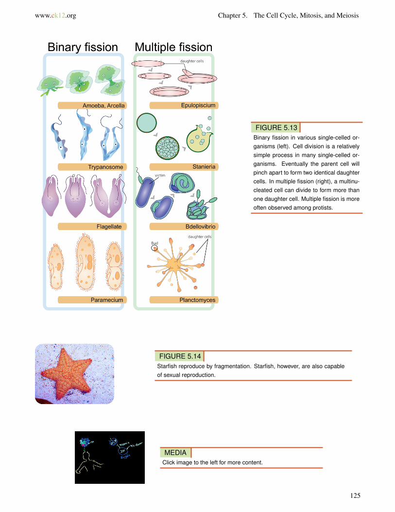
FIGURE 5.13Binary fission in various single-celled or-ganisms (left). Cell division is a relativelysimple process in many single-celled or-ganisms. Eventually the parent cell willpinch apart to form two identical daughtercells. In multiple fission (right), a multinu-cleated cell can divide to form more thanone daughter cell. Multiple fission is moreoften observed among protists.
FIGURE 5.14Starfish reproduce by fragmentation. Starfish, however, are also capableof sexual reproduction.
MEDIAClick image to the left for more content.
125
5.3. Reproduction and Meiosis www.ck12.org
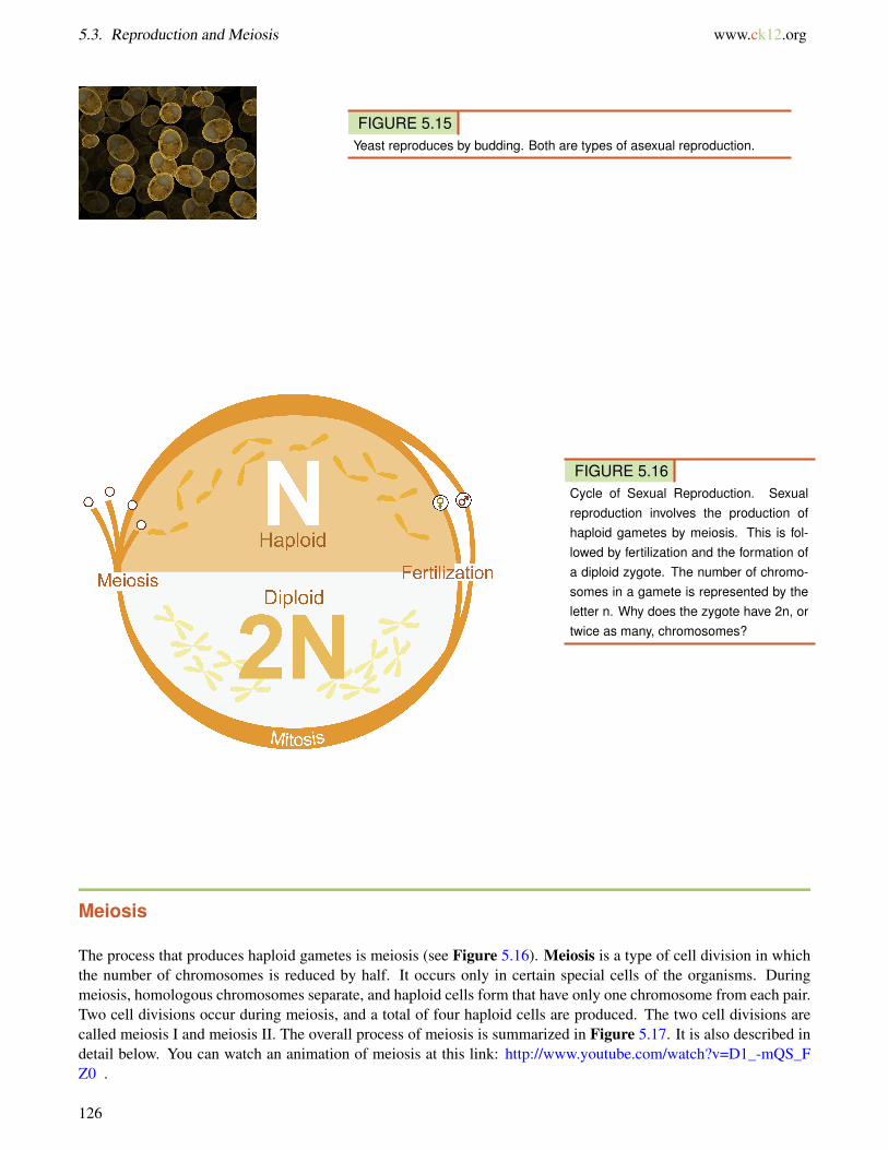
FIGURE 5.15Yeast reproduces by budding. Both are types of asexual reproduction.
FIGURE 5.16Cycle of Sexual Reproduction. Sexualreproduction involves the production ofhaploid gametes by meiosis. This is fol-lowed by fertilization and the formation ofa diploid zygote. The number of chromo-somes in a gamete is represented by theletter n. Why does the zygote have 2n, ortwice as many, chromosomes?
Meiosis
The process that produces haploid gametes is meiosis (see Figure 5.16). Meiosis is a type of cell division in whichthe number of chromosomes is reduced by half. It occurs only in certain special cells of the organisms. Duringmeiosis, homologous chromosomes separate, and haploid cells form that have only one chromosome from each pair.Two cell divisions occur during meiosis, and a total of four haploid cells are produced. The two cell divisions arecalled meiosis I and meiosis II. The overall process of meiosis is summarized in Figure 5.17. It is also described indetail below. You can watch an animation of meiosis at this link: http://www.youtube.com/watch?v=D1_-mQS_FZ0 .
126

www.ck12.org Chapter 5. The Cell Cycle, Mitosis, and Meiosis
FIGURE 5.17Overview of Meiosis. During meiosis,homologous chromosomes separate andgo to different daughter cells. This dia-gram shows just the nuclei of the cells.Notice the exchange of genetic materialthat occurs prior to the first cell division.
Phases of Meiosis
Meiosis I begins after DNA replicates during interphase. In both meiosis I and meiosis II, cells go through the samefour phases as mitosis. However, there are important differences between meiosis I and mitosis. The flowchart inFigure 5.18 shows what happens in both meiosis I and II. You can follow the changes in the flowchart as you readabout them below.
The phases of meiosis are discussed at http://www.youtube.com/watch?v=ijLc52LmFQg (27:23).
MEDIAClick image to the left for more content.
Meiosis I
1. Prophase I: The nuclear envelope begins to break down, and the chromosomes condense. Centrioles startmoving to opposite poles of the cell, and a spindle begins to form. Importantly, homologous chromosomespair up, which is unique to prophase I. In prophase of mitosis and meiosis II, homologous chromosomes donot form pairs in this way. During prophase I, crossing-over occurs (see below).
2. Metaphase I: Spindle fibers attach to the paired homologous chromosomes. The paired chromosomes line upalong the equator of the cell. This occurs only in metaphase I. In metaphase of mitosis and meiosis II, it issister chromatids that line up along the equator of the cell.
3. Anaphase I: Spindle fibers shorten, and the chromosomes of each homologous pair start to separate from eachother. One chromosome of each pair moves toward one pole of the cell, and the other chromosome movestoward the opposite pole.
4. Telophase I and Cytokinesis: The spindle breaks down, and new nuclear membranes form. The cytoplasm of
127

5.3. Reproduction and Meiosis www.ck12.org
FIGURE 5.18Phases of Meiosis. This flowchart ofmeiosis shows meiosis I in greater detailthan meiosis II. Meiosis I—but not meiosisII—differs somewhat from mitosis. Com-pare meiosis I in this flowchart with theearlier figure featuring mitosis. How doesmeiosis I differ from mitosis?
the cell divides, and two haploid daughter cells result. The daughter cells each have a random assortment ofchromosomes, with one from each homologous pair. Both daughter cells go on to meiosis II.
Meiosis II
1. Prophase II: The nuclear envelope breaks down and the spindle begins to form in each haploid daughter cellfrom meiosis I. The centrioles also start to separate.
2. Metaphase II: Spindle fibers line up the sister chromatids of each chromosome along the equator of the cell.3. Anaphase II: Sister chromatids separate and move to opposite poles.4. Telophase II and Cytokinesis: The spindle breaks down, and new nuclear membranes form. The cytoplasm of
each cell divides, and four haploid cells result. Each cell has a unique combination of chromosomes.
Mitosis, Meiosis, and Sexual Reproduction is discussed at http://www.youtube.com/watch?v=kaSIjIzAtYA (18:23).
MEDIAClick image to the left for more content.
128
www.ck12.org Chapter 5. The Cell Cycle, Mitosis, and Meiosis
Gametogenesis
At the end of meiosis, four haploid cells have been produced, but the cells are not yet gametes. The cells need todevelop before they become mature gametes capable of fertilization. The development of haploid cells into gametesis called gametogenesis.
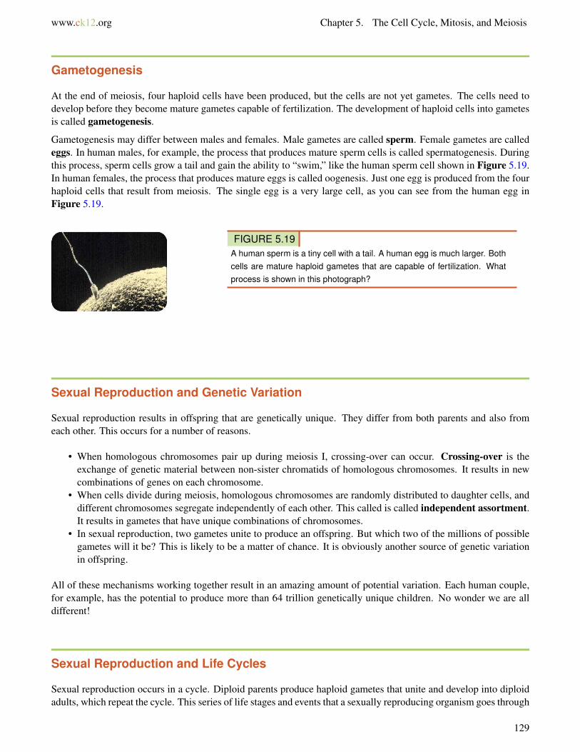
Gametogenesis may differ between males and females. Male gametes are called sperm. Female gametes are calledeggs. In human males, for example, the process that produces mature sperm cells is called spermatogenesis. Duringthis process, sperm cells grow a tail and gain the ability to “swim,” like the human sperm cell shown in Figure 5.19.In human females, the process that produces mature eggs is called oogenesis. Just one egg is produced from the fourhaploid cells that result from meiosis. The single egg is a very large cell, as you can see from the human egg inFigure 5.19.
FIGURE 5.19A human sperm is a tiny cell with a tail. A human egg is much larger. Bothcells are mature haploid gametes that are capable of fertilization. Whatprocess is shown in this photograph?
Sexual Reproduction and Genetic Variation
Sexual reproduction results in offspring that are genetically unique. They differ from both parents and also fromeach other. This occurs for a number of reasons.
• When homologous chromosomes pair up during meiosis I, crossing-over can occur. Crossing-over is theexchange of genetic material between non-sister chromatids of homologous chromosomes. It results in newcombinations of genes on each chromosome.
• When cells divide during meiosis, homologous chromosomes are randomly distributed to daughter cells, anddifferent chromosomes segregate independently of each other. This called is called independent assortment.It results in gametes that have unique combinations of chromosomes.
• In sexual reproduction, two gametes unite to produce an offspring. But which two of the millions of possiblegametes will it be? This is likely to be a matter of chance. It is obviously another source of genetic variationin offspring.
All of these mechanisms working together result in an amazing amount of potential variation. Each human couple,for example, has the potential to produce more than 64 trillion genetically unique children. No wonder we are alldifferent!
Sexual Reproduction and Life Cycles
Sexual reproduction occurs in a cycle. Diploid parents produce haploid gametes that unite and develop into diploidadults, which repeat the cycle. This series of life stages and events that a sexually reproducing organism goes through
129

5.3. Reproduction and Meiosis www.ck12.org
is called its life cycle. Sexually reproducing organisms can have different types of life cycles. Three are describedin the following sections.
Haploid Life Cycle
The haploid life cycle ( Figure 5.20) is the simplest life cycle. It is found in many single-celled organisms.Organisms with a haploid life cycle spend the majority of their lives as haploid gametes. When the haploid gametesfuse, they form a diploid zygote. It quickly undergoes meiosis to produce more haploid gametes that repeat the lifecycle.
FIGURE 5.20Haploid Life Cycle. The letter N indicateshaploid stages of the life cycles, and 2Nindicates diploid stages.
Diploid Life Cycle
Organisms with a diploid life cycle ( Figure 5.21) spend the majority of their lives as diploid adults. When they areready to reproduce, they undergo meiosis and produce haploid gametes. Gametes then unite in fertilization and forma diploid zygote. The zygote develops into a diploid adult that repeats the life cycle. Can you think of an organismwith a diploid life cycle? (Hint: What type of life cycle do humans have?)
Alternation of Generations
Organisms that have a life cycle with alternating generations ( Figure 5.22) switch back and forth between diploidand haploid stages. Organisms with this type of life cycle include plants, algae, and some protists. These life cyclesmay be quite complicated. You can read about them in later chapters.
130

www.ck12.org Chapter 5. The Cell Cycle, Mitosis, and Meiosis
FIGURE 5.21Diploid Life Cycle. The letter N indicateshaploid stages of the life cycles, and 2Nindicates diploid stages.
FIGURE 5.22Alternation of Generations. The letter Nindicates haploid stages of the life cycles,and 2N indicates diploid stages.
131

5.3. Reproduction and Meiosis www.ck12.org
Lesson Summary
• Asexual reproduction involves one parent and produces offspring that are genetically identical to each otherand to the parent. Sexual reproduction involves two parents and produces offspring that are genetically unique.
• During sexual reproduction, two haploid gametes join in the process of fertilization to produce a diploidzygote. Meiosis is the type of cell division that produces gametes. It involves two cell divisions and producesfour haploid cells.
• Sexual reproduction has the potential to produce tremendous genetic variation in offspring. This variationis due to independent assortment and crossing-over during meiosis and random union of gametes duringfertilization.
• A life cycle is the sequence of stages an organisms goes through from one generation to the next. Organismsthat reproduce sexually can have different types of life cycles, such as haploid or diploid life cycles.
Lesson Review Questions
Recall
1. What are three types of asexual reproduction?
2. Define gamete and zygote. What number of chromosomes does each have?
3. What happens during fertilization?
4. Outline the phases of meiosis.
5. What is a life cycle?
6. What is gametogenesis, and when does it occur?
Apply Concepts
7. Create a diagram to show how crossing-over occurs and how it creates new gene combinations on each chromo-some.
8. An adult organism produces gametes that quickly go through fertilization and form diploid zygotes. The zygotesmature into adults, which live for many years. Eventually the adults produce gametes and the cycle repeats. Whattype of life cycle does this organism have? Explain your answer.
Think Critically
9. Compare and contrast asexual and sexual reproduction.
10. Explain why sexual reproduction results in genetically unique offspring.
11. Explain how meiosis I differs from mitosis.
132

www.ck12.org Chapter 5. The Cell Cycle, Mitosis, and Meiosis
Points to Consider
In sexually reproducing organisms, parents pass a copy of each type of chromosome to their offspring by producinggametes. When gametes are fertilized and form offspring, each has a unique combination of chromosomes and genesfrom both parents. The inherited gene combination determines the characteristics of the offspring.
• Is it possible to predict possible gene combinations in offspring from the genes of their parents?• Can the characteristics of offspring be predicted from the characteristics of their parents?
133
5.4. References www.ck12.org
5.4 References
1. Zachary Wilson. CK-12 Foundation . CC BY-NC 3.02. Mariana Ruiz Villarreal (LadyofHats) for CK-12 Foundation. CK-12 Foundation . CC BY-NC 3.03. Mariana Ruiz Villarreal (LadyofHats) for CK1-12 Foundation. CK-12 Foundation . CC BY-NC 3.04. Mariana Ruiz Villarreal (LadyofHats) for CK-12 Foundation. CK-12 Foundation . CC BY-NC 3.05. Ed Uthman, MD. http://web2.airmail.net/uthman/specimens/images/tvp.html . Public Domain6. Zappy’s. CK-12 Foundation . CC BY-NC 3.07. Mariana Ruiz Villarreal (LadyofHats) for CK-12 Foundation. CK-12 Foundation . CC BY-NC 3.08. Mariana Ruiz Villarreal (LadyofHats) for CK-12 Foundation. CK-12 Foundation . CC BY-NC 3.09. Courtesy of Nogales group and Lawrence Berkeley National Laboratory. http://www.lbl.gov/Science-Artic
les/Archive/sabl/2007/Oct/onering.html . Public Domain10. Mariana Ruiz Villarreal (LadyofHats) for CK-12 Foundation. CK-12 Foundation . CC BY-NC 3.011. Mariana Ruiz Villarreal (LadyofHats) for CK-12 Foundation. CK-12 Foundation . CC BY-NC 3.012. Image copyright Juan Carlos Tinjaca, 2014. http://www.shutterstock.com . Used under license from Shutter-
stock.com13. Mariana Ruiz Villarreal (LadyofHats) for CK-12 Foundation. CK-12 Foundation . CC BY-NC 3.014. Flickr:ShyViolet09. http://www.flickr.com/photos/allthestarsthatshine/7356697446/ . CC BY 2.015. Zappy’s. CK-12 Foundation . CC BY-NC 3.016. Mariana Ruiz Villarreal (LadyofHats) for CK-12 Foundation. CK-12 Foundation . CC BY-NC 3.017. Mariana Ruiz Villarreal (LadyofHats) for CK-12 Foundation. CK-12 Foundation . CC BY-NC 3.018. Mariana Ruiz Villarreal (LadyofHats) for CK-12 Foundation. CK-12 Foundation . CC BY-NC 3.019. Courtesy of www.PDImages.com. http://www.pdimages.com/03709.html-ssi . Public Domain20. Mariana Ruiz Villarreal (LadyofHats) for CK-12 Foundation. CK-12 Foundation . CC BY-NC 3.021. Mariana Ruiz Villarreal (LadyofHats) for CK-12 Foundation. CK-12 Foundation . CC BY-NC 3.022. Mariana Ruiz Villarreal (LadyofHats) for CK-12 Foundation. CK-12 Foundation . CC BY-NC 3.0
134