Embed Size (px)
Citation preview
23
Chapter Three
Vestibular System
3.1 Introduction
The vestibular, visual, and proprioceptive systems work together to maintain
postural equilibrium and visual stability while stationary and when moving. The
peripheral vestibular apparatus, a component of the inner ear, has the ability to detect a
wide range of angular and linear accelerations of the head, as well as changes in static
head position with respect to gravity. The information obtained from the peripheral
vestibular apparatus is utilized in central pathways to help generate appropriate postural
and eye movement responses to changes in head position.
The vestibulo-ocular pathways produce a number of reflexive eye movement
responses to help maintain clear vision during head movement (see figure 3.1). These
responses include “compensatory” responses to angular head accelerations (generated by
the angular vestibulo-ocular reflex or aVOR) and linear head accelerations (generated by
the linear vestibulo-ocular reflex or lVOR) (Raphan and Cohen 2002). The vestibulo-
ocular pathways also produce “orienting” responses, serving to align the z-axis of the eye
with the gravito-inertial acceleration (GIA) vector (Raphan and Cohen 2002).
Vestibular-evoked eye movements have short latencies following the onset of
head movement, reported to be as low as 5ms in some studies of the aVOR (Tabak et al.
1997). Compensatory smooth eye movements resulting from stimulation of the visual
system or the neck proprioceptors are not seen until ≥80ms after the onset of head
movement (Bronstein and Hood 1986; Carl and Gellman 1987), making the vestibular
system critical for the generation of an early eye movement response to head movement
stimuli. The aVOR has an additional advantage, in that it functions well in response to
short duration, high frequency head movements, ensuring clear vision during commonly
performed activities such as walking and running. The physician J.C. highlighted the
importance of these functions by describing how he continuously experienced severe

24
oscillopsia while walking, following the complete loss of vestibular function after
treatment with an aminoglycoside antibiotic (J.C. 1952).
The anatomy and physiology of the peripheral and central components of the
vestibular system are considered in this chapter. Emphasis is placed on the structure and
function of the peripheral vestibular apparatus and on the central structures and pathways
influencing the aVOR, as relevant to the current study.
Figure 3.1 The vestibulo-ocular pathways produce reflexive eye movements that either
compensate for head movement or change the orientation of the eye so that its z-axis aligns with
the gravito-inertial acceleration (GIA) vector. A. The aVOR produces a rotational eye movement
that compensates for rotation of the head. B. The lVOR produces a rotational eye movement to
compensate for head translation. C. With the head upright, the head and eye z-axes (denoted by
arrows on top of the head and eyes, respectively) align with the GIA, which in turn aligns with the
vector representing acceleration due to gravity. D. When the head is tilted, the eyes roll so that
their z-axes align with the GIA. E. During horizontal linear acceleration (LINEAR ACC), which
tilts the GIA relative to the head vertical, the eyes will again roll to align their z-axes with the
GIA (from Raphan and Cohen 2002).

25
3.2 Anatomy of the Peripheral Vestibular System
The inner ear lies deep within the petrous temporal bone, in a chamber of
communicating ducts and cavities known as the bony labyrinth. The inner ear, or
membranous labyrinth, a fluid-filled membranous structure with a shape similar to that of
the bony labyrinth, is suspended within the bony labyrinth by a supportive network of
connective tissue. The space between the bony labyrinth and the membranous labyrinth is
filled with perilymph, a fluid with a high concentration of sodium ions and a low
concentration of potassium ions (Smith et al. 1954), making it similar in composition to
extracellular fluid. The membranous labyrinth, on the other hand, is filled with
endolymph, a fluid with a high concentration of potassium ions and a low concentration
of sodium ions, making it similar to intracellular fluid in its ionic composition (Smith et al.
1954). The ionic composition of endolymph is vital to the normal functioning of the
sensory cells of the inner ear (Corey and Hudspeth 1979), as discussed below in section
3.3.
The membranous labyrinth may be functionally and anatomically divided into two
main portions: the peripheral auditory apparatus, or cochlea, and the peripheral vestibular
apparatus. The peripheral vestibular apparatus incorporates five structures: the three
semicircular canals (SCCs), and two otolith organs, the utricle and saccule (figure 3.2).
The three SCCs (anterior, posterior, and lateral) are named for their positions in the head.
The anterior SCC is also known as the superior SCC, while the lateral SCC is also known
as the horizontal SCC.
Neural signals from the sensory tissue of the SCCs and otolith organs pass to the
brainstem in the primary (first-order) vestibular afferents, which comprise the vestibular
nerve, one of the two nerves making up the eighth cranial nerve (CN VIII). The vestibular
nerve may be divided into superior and inferior divisions. The primary vestibular
afferents from the anterior SCC, lateral SCC, utricle, and part of the saccule pass to the
brainstem in the superior division of the vestibular nerve, while those from the posterior
SCC and remainder of the saccule pass to the brainstem in the inferior division. The
nuclei of the afferents comprising the superior and inferior divisions of the vestibular
nerve are found in the superior and inferior vestibular ganglia of Scarpa, respectively.

26
Figure 3.2 A line drawing of the left inner ear, viewed from the lateral aspect. The semicircular
canals, otolith organs (utricle and saccule), and cochlea are illustrated, as are the nerves and other
structures closely associated with the inner ear. A narrow channel, the ductus reuniens
(unlabelled), joins the cochlea and the peripheral vestibular apparatus (from Kandel et al. 1991).
3.3 Sensory Epithelium and Vestibular Afferents
The sensory epithelium of the peripheral vestibular apparatus consists of highly
specialized hair cells (Wersäll 1956). Each hair cell has a hexagonal array of 60-100
stereocilia and one kinocilium protruding from its apical surface. The stereocilia are
asymmetrically arranged, gradually increasing in height in a staircase fashion, with the
tallest stereocilia being located near the kinocilium and the shortest being located at the
opposite end of the cell surface. The kinocilium is the longest apical process extending
from the hair cell.
The arrangement of the stereocilia with respect to the kinocilium results in each
hair cell having a morphological axis of polarity (Flock 1964). When the stereocilia of the
hair cell are in their resting position, a certain number of mechanically-gated transduction
channels on the stereocilia are open, allowing potassium ions from the endolymph to
move into the cell and depolarize it. Consequently, the cell has a resting discharge of
transmitter. When the stereocilia are deflected towards the kinocilium, more transduction
27
channels on the stereocilia open by a mechanical process that is mediated by tip links
connecting the stereocilia (Corey and Hudspeth 1983), leading to further depolarization
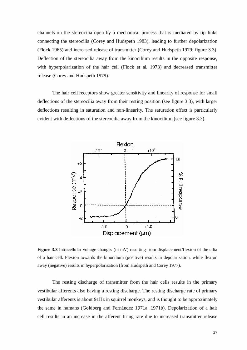
(Flock 1965) and increased release of transmitter (Corey and Hudspeth 1979; figure 3.3).
Deflection of the stereocilia away from the kinocilium results in the opposite response,
with hyperpolarization of the hair cell (Flock et al. 1973) and decreased transmitter
release (Corey and Hudspeth 1979).
The hair cell receptors show greater sensitivity and linearity of response for small
deflections of the stereocilia away from their resting position (see figure 3.3), with larger
deflections resulting in saturation and non-linearity. The saturation effect is particularly
evident with deflections of the stereocilia away from the kinocilium (see figure 3.3).
Figure 3.3 Intracellular voltage changes (in mV) resulting from displacement/flexion of the cilia
of a hair cell. Flexion towards the kinocilium (positive) results in depolarization, while flexion
away (negative) results in hyperpolarization (from Hudspeth and Corey 1977).
The resting discharge of transmitter from the hair cells results in the primary
vestibular afferents also having a resting discharge. The resting discharge rate of primary
vestibular afferents is about 91Hz in squirrel monkeys, and is thought to be approximately
the same in humans (Goldberg and Fernández 1971a, 1971b). Depolarization of a hair
cell results in an increase in the afferent firing rate due to increased transmitter release

28
from the hair cell, while hyperpolarization of the cell will produce a decrease in the
afferent firing rate due to decreased transmitter release (see figure 3.4). Thus, neural
signals coding deflections of the stereocilia both toward and away from the kinocilium
can be communicated to the central nervous system via the primary vestibular afferents
(Löwenstein and Sand 1940; Money and Scott 1962).
Figure 3.4 The hair cell receptor potential and the firing rate of the primary vestibular afferent
both depend on the orientation of the cilia on the hair cell. Deflections of the stereocilia towards
the kinocilium result in depolarization and increased firing rate, while deflections in the opposite
direction result in hyperpolarization and decreased firing rate (from Kandel et al. 1991).
3.4 Semicircular Canals
The SCCs are tubular structures that are roughly semicircular in shape (Curthoys
et al. 1977a). At one end of each canal is an enlargement known as the ampulla (figure
3.5a). Within each ampulla, a collection of hair cells makes up the saddle-shaped
ampullary crest (also known as the crista ampullaris). The cilia of these hair cells project
towards the centre of the canal lumen into a gelatinous membrane known as the cupula.
The cupula extends across the lumen of the SCC and is adherent to the SCC wall, forming
a watertight seal (Dohlman 1971; Lim 1973).

29
When there is an angular acceleration of the head in the plane of one of the SCCs,
inertial forces acting on the endolymph in that canal result in relative fluid flow
(Steinhausen 1933; Van Egmond et al. 1949; see section 3.4a). The relative movement of
the endolymph produces deviation of the cupula (see figure 3.5b). Since the cilia of the
hair cells of the ampullary crest project into the cupula, deviation of the cupula will
produce depolarization or hyperpolarization of the hair cells, depending on the direction
of deflection of the cilia. The hair cells of the ampullary crest are organized so that their
morphological axes of polarity all point in the same direction along the canal (Löwenstein
and Wersäll 1959). Deviation of the cupula in one direction will thus result in an increase
in the firing rate of all of the afferents, while deviation in the opposite direction will result
in a decrease in the firing rate (Goldberg and Fernández 1971a).
A B
Figure 3.5 A. The components of the ampulla of the lateral (horizontal) semicircular canal (SCC)
are illustrated. The hair cells of the ampullary crest project into the cupula, which extends across
the lumen of the SCC. B. An angular acceleration of the head results in deviation of the cupula,
due to the inertia of the endolymph (from Kandel et al. 1991).
The SCCs from the inner ear on one side are approximately orthogonal to one
another (Blanks et al. 1975; Curthoys et al. 1977b; Reisine et al. 1988), so that together
they can sense any angular head rotation in three-dimensional (3-d) space. The SCCs
from both inner ears are often grouped into three functional pairs: the two lateral SCCs
make up one pair, the left anterior and right posterior SCCs another, and the right anterior
and left posterior SCCs make up the final pair (Blanks et al. 1975; Takagi et al. 1989).
The canals from a pair are anatomically related, since they are approximately aligned in
3-d space. As a result, they are also functionally related, as an angular head acceleration
30
that excites one canal in the pair inhibits the other. The SCCs are therefore thought to act
in a “push-pull” manner.
The normal push-pull functioning of the canal pairs is important, since excitatory
stimuli are better vestibular stimuli than inhibitory ones – a phenomenon first described
by Ewald (1892). Ewald applied positive and negative pressure to each of the canals,
making three observations that are now known as Ewald’s first, second, and third laws.
The first was that compensatory eye movements always occurred in the plane of the
stimulated canal, and always in the direction of endolymph flow. Secondly, he noted that
ampullopetal flow (flow of the endolymph towards the utricle) produced a better response
than did ampullofugal flow (flow of the endolymph away from the utricle) when the
lateral canal was stimulated. The third observation was that ampullofugal flow produced a
better response than did ampullopetal flow when the anterior and posterior canals were
stimulated. The importance of Ewald’s second and third laws becomes obvious when an
organism loses the function of the SCCs on one side, as the remaining labyrinth cannot
always adequately detect vestibular stimuli to compensate for the loss (see section 3.3 and
figure 3.3 above). For example, when there is a rapid angular rotation of the head in the
plane of a lesioned canal, in a direction that would normally excite the lesioned canal, the
aVOR response is inadequate and a stable retinal image cannot be maintained (Halmagyi
and Curthoys 1988; Halmagyi et al. 1990; Curthoys and Halmagyi 1995; Aw et al. 1996b;
Cremer et al. 1998).
3.4a Semicircular Canal Hydrodynamics
Steinhausen (1933) first modelled the hydrodynamic function of the SCCs, with
Van Egmond et al. (1949) and Groen (1957) later making refinements to the model.
Steinhausen (1933) proposed that the SCCs function like a heavily damped torsion
pendulum, so that
θΘ=ξ∆+ξΠ+ξΘ &&&&&
where ξ is the angular displacement of the cupula (ξ& and ξ&& refer to the first and second
derivatives of ξ, respectively), θ&& is the angular acceleration of the head (hence, θ and θ&
correspond to angular displacement and velocity, respectively), Θ is the moment of
inertia of the cupula and endolymph, Π is the moment of viscous drag at unit angular

31
velocity of the endolymph, and ∆ is the elastic restoring couple of the cupula and
endolymph. Within the range of natural head movements, ξΘ && and ξ∆ are very small
compared to ξΠ& , hence
θΘ≈ξΠ &&&
Direct integration yields
θΘ≈ξΠ &
and
θΠΘ≈ξ &
Within the range of natural head movements, instantaneous cupular displacement is
therefore not proportional to the angular acceleration of the head, but rather is roughly
proportional to the angular velocity of the head. Hence, the canals mechanically integrate
head acceleration, producing cupular displacement and a neural signal proportional to
head velocity (Goldberg and Fernández 1971a).
During angular head accelerations, inertial forces acting on the endolymph
initially keep it stationary in space, producing relative fluid flow in the canal. The inertial
force is opposed by the viscous forces incurred by the fluid flow in the canal, and by the
elastic restoring forces of the cupula. After a certain period of constant velocity, the
inertial force is therefore overcome by restoring forces, and the endolymph, too, will
move with a constant velocity during the head rotation. The cupula exponentially decays
back towards its resting position with a time constant of approximately 4-6s (Cohen et al.
1981; Dai et al. 1999).
The lack of semicircular canal response to constant velocity stimuli is partly
compensated for by the velocity storage mechanism in the central nervous system (see
section 3.8). The relationships between cupular displacement and different types of head
rotation stimuli are illustrated below in figure 3.6.
3.4b Canal Afferent Responses
Fernández and Goldberg (1971) measured the primary canal afferent responses in
squirrel monkeys, and presented evidence that the primary vestibular afferents are

32
sensitive to cupular velocity, as well as being sensitive to cupular displacement. The
SCCs could therefore relay both head velocity and acceleration information to the central
nervous system. Indeed, Boyle et al. (1991) found that the majority of afferents arising
from the SCCs had firing rates proportional to angular head velocity, with a minority of
afferents giving a signal proportional to angular head acceleration (see also Rabbitt et al.
1996).
Figure 3.6 The relationships between cupular displacement and three different head rotation
stimuli are illustrated. During periods of acceleration, cupular displacement is approximately
proportional to head velocity. When head velocity is constant, however, the cupula decays back
towards its resting position (from Baloh and Honrubia 2001).

33
3.5 Otolith Organs
The utricle and saccule are the sac-like portions of the membranous labyrinth,
collectively known as the otolith organs. They sense both static head position with respect
to gravity and dynamic changes in head position, such as linear accelerations and head
tilts.
The otolith organs have areas of sensory epithelium known as maculae. Each
macula is covered with a gelatinous membrane, the otolithic membrane, which has
crystals of calcium carbonate (otoliths or otoconia) embedded in it (Carlström et al. 1953;
see figure 3.7). The cilia of the hair cells in the maculae project into the otolithic
membrane (Carlström et al. 1953), just as the cilia of the SCC hair cells project into the
cupula. Therefore, when the head is linearly translated, the otolithic membrane moves
relative to the sensory epithelium due to inertia, resulting in excitation or inhibition of the
underlying hair cells (see Wilson and Melvill Jones 1979, for a review).
Figure 3.7 Structure of the otolithic macula. When the head translates, the otolithic membrane
moves relative to the sensory epithelium due to inertia, resulting in excitation or inhibition of the
underlying hair cells. The otoliths (otoconial crystals) add mass to the otolithic membrane,
therefore enhancing the sensitivity of the maculae to head tilt and linear accelerations (from
Furman and Cass 1996).
34
The utricular macula lies on the floor of the utricle, while the saccular macula lies
on the medial wall of the saccule, making the two maculae approximately orthogonal to
one another (Lim 1969). The utricular macula responds best to lateral/fore-aft tilts and
side-side translations of the head, while the saccular macula responds best to up-down
translations of the head. The macula of each otolithic organ is not perfectly flat and the
morphological axes of polarity of the hair cells constituting each macula point in a
number of different directions. Consequently, each macula can produce excitatory
responses to linear accelerations in more than one direction, unlike the SCCs.
3.6 Vestibular Nuclei
Neural signals from the peripheral vestibular apparatus are passed to the central
nervous system via the vestibular nerve. The components of the central nervous system
involved in the receipt, distribution, and subsequent processing of vestibular information
constitute the central vestibular system.
Upon entering the brainstem, the majority of primary vestibular afferents initially
pass to the vestibular nuclear complex in the caudal pons and rostral medulla, where they
synapse on the cell bodies of secondary (second-order) vestibular afferents (Gacek 1969).
Some primary vestibular afferents pass directly to the cerebellum via the juxtarestiform
body (part of the inferior cerebellar peduncle), to synapse with cells in the flocculo-
nodular lobe and uvula (Gacek 1969). Each primary vestibular afferent synapses with
multiple neurons, resulting in considerable distribution of vestibular information
throughout the central nervous system. For example, an afferent from one of the SCCs
can synapse with up to 15 different neurons in the vestibular nuclear complex.
The vestibular nuclei act as a relay point for the distribution of vestibular
information. They also have a role in further processing of vestibular information, such as
for velocity storage (de Jong et al. 1980; see section 3.8) and velocity-position
transformation (Cannon and Robinson 1987; Cheron and Godaux 1987; Godaux et al.
1993; see section 2.5c). There are four vestibular nuclei on each side of the brainstem,
which are distinguished based on their histological features and connections: the medial

35
vestibular nucleus (MVN), superior vestibular nucleus (SVN), lateral vestibular nucleus
(LVN), and inferior (or descending) vestibular nucleus (IVN) (Brodal and Pompeiano
1957; Brodal 1984; Highstein and McCrea 1988; Suárez et al. 1997). Several smaller
accessory subgroups also exist, including the interstitial nucleus and the y-group.
The MVN and the SVN receive the majority of their input from the SCCs. Many
of the secondary afferents from these two nuclei are involved in the direct pathways of
the vestibulo-ocular reflex (see section 3.7a). The MVN has also been implicated in the
velocity-to-position transformation (see sections 2.5c and 3.7b), and in velocity storage
(see section 3.8).
The rostroventral portion of the LVN receives afferent input from the SCCs and
the utricular macula. The LVN also receives afferent input from the cerebellum and spinal
cord. Secondary afferents arising from this nucleus make up part of the vestibulo-ocular
pathways. The vestibulo-ocular afferents extend to the ipsilateral oculomotor nucleus, in
part, through the ascending tract of Deiters (ATD), which passes lateral to the medial
longitudinal fasciculus (MLF) in the brainstem.
The IVN receives input from both the peripheral vestibular apparatus and the
cerebellum. The IVN is important for the integration of vestibular and cerebellar input.
3.7 Angular Vestibulo-Ocular Reflex Pathways
The aVOR produces an eye rotation response that is opposite in direction but
equal in magnitude to the head rotation stimulus. To compensate for mechanical restoring
forces imposed on the eyeball by the orbital contents, two signals must be sent to the
extra-ocular muscles: an eye velocity signal and an eye position signal (see section 2.4).
The signal encoding eye velocity arises from the “direct” pathway of the aVOR, while the
signal encoding eye position is produced from the eye velocity signal by the neural
integrator (see sections 2.4 and 2.5c) in the “indirect” pathway. The two signals are
combined, and the resulting command is sent to the extra-ocular muscles to rotate the
eyes (Robinson 1975).

36
Szentágothai first described the direct pathway of the aVOR in 1950. The three-
neuron arc he proposed included a primary vestibular afferent, a secondary vestibular
afferent (which sends a signal from a vestibular nucleus to a motor nucleus), and a motor
(efferent) neuron (which sends the eye movement signal to an extra-ocular muscle). The
connections are such that a particular SCC directly influences a pair of extra-ocular
muscles, which generate compensatory eye rotations in the plane of the stimulated SCC
(Cohen and Suzuki 1963; Cohen et al. 1964). The head velocity signal in the primary
vestibular afferents is therefore transformed into a compensatory eye velocity signal, by
nature of these connections.
The neural substrate for the neural integrator or indirect pathway (whose
components include the NPH, MVN, INC, and parts of the cerebellum) was not identified
until many years after the original description of the direct pathway, although the
existence of an indirect pathway had been suspected (Lorente de Nó 1933; Szentágothai
1950). As previously discussed, the indirect pathway is critical for maintaining steady
fixation and for producing normal eye movements (see section 2.4). While there is now
agreement as to which structures are involved in this pathway, there is still debate about
how the neural components of the pathway actually generate the eye position signal from
the head velocity signal (Tweed and Vilis 1987; Schnabolk and Raphan 1994; Tweed et al.
1994a; Raphan 1998; Smith and Crawford 1998; see chapter 4).
3.7a Direct Pathways of the aVOR
The aVOR is driven by signals from the SCCs, resulting in coordinated
contraction and relaxation of the extra-ocular muscles. Both excitatory and inhibitory
pathways exist, so that contraction of agonist muscles and relaxation of antagonist
muscles results from stimulation of a particular SCC (Richter and Precht 1968; Precht
1972; Highstein et al. 1971). Conversely, an inhibitory stimulus acting on a particular
SCC results in relaxation of agonist muscles for that SCC and contraction of antagonist
muscles (Shimazu and Precht 1966; Markham et al. 1977). Excitatory pathways therefore
function to produce the desired eye movement, while inhibitory pathways inhibit the
opposite eye movement response. The vestibular commissures, which connect
homologous regions of the two vestibular nuclei complexes, are thought to further
enhance the “push-pull” relationship of the vestibular nuclear complexes, thereby helping

37
to coordinate the aVOR (Pompeiano et al. 1978; Carleton and Carpenter 1983). The
nuclei, pathways, and extra-ocular muscles involved in bringing about eye rotations in
response to excitation/inhibition of each SCC, via the direct pathways, are summarized in
table 3.1. Furthermore, the actual pathways are displayed in figure 3.8.
Table 3.1 Components involved in the direct pathway of the aVOR (adapted from Leigh and Zee
1999).
Receptor Effect Relay Nuclei Pathways Motor Nuclei Muscle
Lateral SCC Excitation MVN - c-VI c-LR
MVN/c-VI ATD/MLF i-III i-MR
Inhibition MVN - i-VI i-LR
MVN/c-VI Poly/MLF c-III c-MR
Anterior SCC Excitation MVN/LVN* MLF* c-III i-SR
MVN/LVN* MLF* c-III c-IO
Inhibition SVN MLF i-III i-IR
SVN MLF i-IV c-SO
Posterior SCC Excitation MVN/LVN MLF c-III c-IR
MVN/LVN MLF c-IV i-SO
Inhibition SVN Extra i-III c-SR
SVN Extra i-III i-IO
(SCC, semicircular canal; i, ipsilateral; c, contralateral. Relay Nuclei: MVN, medial vestibular
nucleus; LVN, lateral vestibular nucleus; *, other nuclei involved; SVN, superior vestibular
nucleus. Pathways: MLF, medial longitudinal fasciculus; ATD, ascending tract of Deiters; Poly,
polysynaptic pathway outside of MLF; *, other pathways involved; Extra, extra-MLF pathway.
Motor Nuclei: VI, abducens nucleus; III, oculomotor nucleus; IV, trochlear nucleus. Muscles:
LR, lateral rectus; MR, medial rectus; SR, superior rectus; IO, inferior oblique; IR, inferior rectus;
SO, superior oblique).
Stimulation of the lateral SCC results in a predominantly horizontal eye rotation
response, due to adduction of the ipsilateral eye and abduction of the contralateral eye.
When the head accelerates towards the left, the afferents from the left lateral canal are
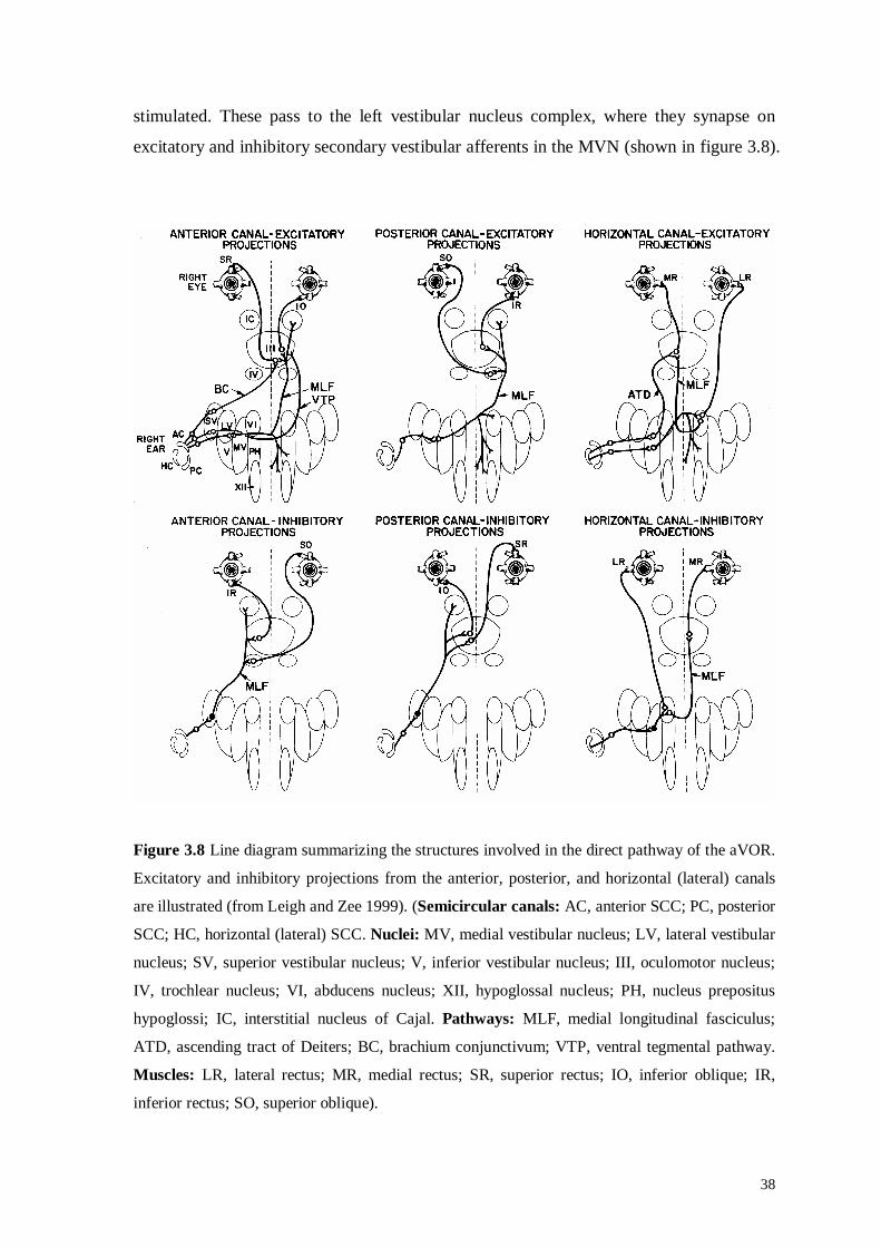
38
stimulated. These pass to the left vestibular nucleus complex, where they synapse on
excitatory and inhibitory secondary vestibular afferents in the MVN (shown in figure 3.8).
Figure 3.8 Line diagram summarizing the structures involved in the direct pathway of the aVOR.
Excitatory and inhibitory projections from the anterior, posterior, and horizontal (lateral) canals
are illustrated (from Leigh and Zee 1999). (Semicircular canals: AC, anterior SCC; PC, posterior
SCC; HC, horizontal (lateral) SCC. Nuclei: MV, medial vestibular nucleus; LV, lateral vestibular
nucleus; SV, superior vestibular nucleus; V, inferior vestibular nucleus; III, oculomotor nucleus;
IV, trochlear nucleus; VI, abducens nucleus; XII, hypoglossal nucleus; PH, nucleus prepositus
hypoglossi; IC, interstitial nucleus of Cajal. Pathways: MLF, medial longitudinal fasciculus;
ATD, ascending tract of Deiters; BC, brachium conjunctivum; VTP, ventral tegmental pathway.
Muscles: LR, lateral rectus; MR, medial rectus; SR, superior rectus; IO, inferior oblique; IR,
inferior rectus; SO, superior oblique).

39
The excitatory secondary afferents function to produce contraction of the left medial
rectus and right lateral rectus, resulting in a rightward eye movement. The signal for left
medial rectus contraction is carried by afferents passing directly to the medial rectus
subdivision of the oculomotor nucleus via the ascending tract of Deiters. Other signals
serving to produce the same muscle contraction pass indirectly to the ipsilateral
oculomotor nucleus, after synapsing on internuclear neurons in the contralateral abducens
nucleus, to ascend to the oculomotor nucleus via the ipsilateral medial longitudinal
fasciculus (MLF). To produce contraction of the right lateral rectus, secondary afferents
synapse on contralateral abducens motor neurons. The inhibitory pathways function to
relax antagonist muscles, in this case the left lateral rectus and right medial rectus
muscles. Most inhibitory secondary afferents pass to the ipsilateral abducens nucleus,
where they inhibit left abducens motor neurons, relaxing the left lateral rectus, and
internuclear neurons synapsing on right medial rectus motor neurons in the contralateral
oculomotor nucleus, relaxing the right medial rectus. Afferents from the right lateral canal,
which have been inhibited in this example, synapse on excitatory and inhibitory
secondary afferents in the right vestibular nucleus complex. The same pathways as
described above are utilized, producing the same end result, rightward rotation of the eyes.
When an anterior SCC is stimulated, the eye movement response includes both a
vertical and torsional component, with elevation/intorsion of the ipsilateral eye and
elevation/extorsion of the contralateral eye being observed. The pathways are shown in
figure 3.8. Excitatory secondary afferents pass from the vestibular nuclei to the
contralateral oculomotor nucleus via the brachium conjunctivum, where they excite motor
neurons of the ipsilateral superior rectus. Other afferents pass to the contralateral
oculomotor nucleus via the MLF and ventral tegmental pathway to excite motor neurons
of the contralateral inferior oblique. Inhibitory afferents pass to the ipsilateral trochlear
nucleus via the MLF to inhibit the contralateral superior oblique motor neurons. Other
inhibitory neurons pass to the ipsilateral oculomotor nucleus, to inhibit ipsilateral inferior
rectus motor neurons. Coordinated contraction and relaxation of the extra-ocular muscles
thereby produces the eye movement response to anterior canal stimulation.
When a posterior SCC is stimulated, similar pathways are utilized, producing
depression/intorsion of the ipsilateral eye and depression/extorsion of the contralateral
eye. There is contraction of the agonist muscles, the ipsilateral superior oblique and

40
contralateral inferior rectus, and relaxation of the antagonist muscles, the ipsilateral
inferior oblique and contralateral superior rectus. The excitatory and inhibitory pathways
involved are summarized in figure 3.8.
3.7b Indirect Pathways of the aVOR
The indirect pathways of the aVOR generate the eye position command to
produce normal vestibular-evoked eye movements and to help maintain fixation
following them. The indirect pathways are also utilized in generating the eye position
command for other types of eye movements. As previously discussed (see section 2.5c),
the neural substrate for the indirect pathway includes the MVN (Miles 1974; Cannon and
Robinson 1987), the NPH (Baker 1977; Lopez-Barneo et al. 1982; Cannon and Robinson
1987), the INC (King et al. 1980, 1981; Fukushima 1987, 1991; Crawford et al. 1991;
Crawford 1994), and the flocculus and paraflocculus (Takemori and Cohen 1974; Zee et
al. 1981; Waespe et al. 1983).
The mechanisms by which the indirect pathways produce a position command
from the eye velocity command have become controversial (Tweed and Vilis 1987;
Schnabolk and Raphan 1994; Tweed et al. 1994a; Raphan 1998; Smith and Crawford
1998). Recent theories and models of the indirect pathways are described and extensively
discussed in chapter 4.
3.8 Velocity Storage
The velocity storage mechanism improves the performance of the aVOR during
sustained head rotations (Raphan et al. 1979), by storing head velocity information from
the labyrinth and thereby lengthening the time constant of the aVOR (Cohen et al. 1981).
The role of the velocity storage mechanism is well illustrated by considering the
characteristics of the vestibular response to a constant velocity rotational stimulus (see
figure 3.9). In the case of a horizontal rotational stimulus, accelerating rapidly from rest,
the time constant of the mechanical response of the cupula in the lateral canal is thought
to be 4-6 seconds (Cohen et al. 1981; Dai et al. 1999; see section 3.4a). However, the

41
resulting nystagmus has a time constant of about 15 seconds, indicating that the raw
vestibular velocity signal has been prolonged by velocity storage (Raphan et al. 1979).
Figure 3.9 Horizontal eye position recording from a subject undergoing a constant velocity
rotation of 50°/s in the dark. The subject is accelerated rapidly from rest (at arrow), and constant
velocity is quickly achieved. The cupular response to the stimulus is thought to last for about 4-6s
after the onset of the stimulus (Cohen et al. 1981; Dai et al. 1999), however the nystagmus lasts
considerably longer due to velocity storage (adapted from Leigh and Zee 1999).
A number of structures are thought to be important in the regulation of velocity
storage, including the MVN and SVN (de Jong et al. 1980), the nodulus and ventral uvula
(Waespe et al. 1985; Wearne et al. 1998), and the vestibular commissures (Blair and
Gavin 1981; Katz et al. 1991; Wearne et al. 1997). Lesions of these structures have been
shown to result in velocity storage being abolished or enhanced.
3.9 Cerebellar Influences on the Angular Vestibulo-Ocular Reflex
The regions of the cerebellum that influence vestibular function, including the
flocculus, ventral paraflocculus, nodulus, and ventral uvula, together constitute the
vestibulo-cerebellum. Lesions of these structures produce characteristic abnormalities of
vestibular and ocular motor function (see section 8.1). Lesions of the flocculus and
ventral paraflocculus result in abnormalities of aVOR gain (Zee et al. 1981; Waespe et al.
1983) and a loss of the ability to adapt the aVOR (Ito et al. 1982; Lisberger et al. 1984;
see section 3.10). Ablation of these structures also produces a deficient velocity-position
integrator, with gaze-evoked nystagmus being observed clinically (Takemori and Cohen
1974; Zee et al. 1981; Waespe et al. 1983).
40 deg
R
L 5 s

42
Lesions of the nodulus and ventral uvula are well known to produce abnormalities
of velocity storage (Waespe et al. 1985; Wearne et al. 1998; see section 3.8). In addition,
periodic alternating nystagmus (PAN) may be seen with lesions of these structures. A rare
form of nystagmus, PAN is a spontaneous horizontal nystagmus that reverses direction
every 90-120s. Loss of the ability to suppress or cancel the aVOR, when the subject is
fixating on a target that is moving with the head, also results from damage to the
vestibulo-cerebellum. The concept of aVOR suppression is illustrated in figure 3.10.
Figure 3.10 Eye position traces recorded from a subject sitting in an oscillating chair, while the
subject is in total darkness (middle trace), and while fixating a target that is moving with the chair
(lower trace). Suppression, or cancellation, of the aVOR occurs when the subject can fixate a
target that is moving with the head during the oscillations. Lesions of the vestibulo-cerebellum
result in a loss of the ability to suppress the aVOR, producing eye rotations similar to those seen
in normal subjects being rotated in the dark (from Baloh and Honrubia 2001).
3.10 Vestibular Adaptation
The aVOR functions as a feed-forward open-loop control system, as no feedback
information is relayed to the receptors that provide the input for the reflex. When the
aVOR is not performing optimally, manifest by excessive retinal image slip, the aVOR
must be re-calibrated. The process of aVOR re-calibration is known as adaptation.
Gonshor and Melvill Jones (1976) illustrated the remarkable degree to which the aVOR
can be adapted. When human subjects wore “dove” prism goggles, which reverse the left-

43
right relations of the visual world, for a few weeks, they noted that the gain of the
horizontal aVOR was progressively reduced, and the eye movement responses even
reversed in polarity.
Destruction of the cerebellum results in a loss of the ability to adaptively modify
aVOR gain (Ito et al. 1974; Robinson 1976). The flocculus and ventral paraflocculus play
a critical role in aVOR adaptation (see section 3.9), with their destruction resulting in a
loss of the ability to adapt the aVOR (Zee et al. 1981; Ito et al. 1982; Lisberger et al.
1984), while the aVOR itself remains intact. Retinal slip information, thought to be the
stimulus for aVOR adaptation, is relayed to the flocculus via climbing fibres from the
inferior olive. Ito and Miyashita (1975) demonstrated that removal of visual input to the
inferior olive results in loss of the ability to adaptively modify aVOR gain. In addition,
local lesions in the dorsal cap of the inferior olive have been shown to abolish aVOR
adaptation (Haddad et al. 1980). Since both retinal slip information and vestibular inputs
are relayed to the flocculus, Ito (1982) proposed that the flocculus is the structure
responsible for making appropriate changes to the aVOR via its projection to the
vestibular nuclei. Recordings from neurons in the flocculus do not, however, show
modifications in their firing rate appropriate to learned increases or decreases in aVOR
gain (Miles et al. 1980). Therefore, it is possible that the flocculus acts as an input to
another structure where aVOR adaptation actually occurs.
As the aVOR only undergoes adaptation when head movements and image slip
occur simultaneously, Lisberger (1988) argued that the site of adaptation should receive
both visual and vestibular input. Experimental evidence implied that the flocculus was not
the site at which aVOR adaptation took place, so Lisberger suggested that the flocculus
might send an error correction signal to cells in the vestibular nuclei called floccular
target neurons (FTNs). The firing rate of FTNs has been observed to systematically
change depending on whether aVOR gain has been increased or decreased as a result of
adaptation (Lisberger and Pavelko 1988; see figure 3.11). Furthermore, FTNs receive
excitatory input from the primary and secondary vestibular afferents, and inhibitory input
from the Purkinje cells of the flocculus. Thus, FTNs receive both vestibular and visual
input, and since they display appropriate firing responses after changes in aVOR gain, it
is possible that they form the locus for aVOR adaptation.

44
Figure 3.11 aVOR responses from two monkeys who had undergone aVOR adaptation. Head and
eye velocity traces are plotted, as well as the firing rate of a representative FTN. A. The monkey
with the increased aVOR gain showed a high firing rate in response to the head movement. B.
The monkey with the decreased aVOR gain showed a lower firing rate in response to the same
head movement stimulus. The velocity calibration is 30°/s (from Lisberger and Pavelko 1988).
3.11 Summary
The receptors of the vestibular system are located in the peripheral vestibular
apparatus, in the inner ear. Angular accelerations of the head are detected by the three
semicircular canals in each ear, which contain hair cell receptors that have the ability to
respond to movements in both excitatory and inhibitory directions. The canals
mechanically integrate the head acceleration stimulus, so that the signal passed to the
brainstem by afferents in the vestibular nerve is proportional to head velocity.
The neural substrate for the aVOR is found in the brainstem and cerebellum. The
eye velocity and eye position signals required for the aVOR to function normally are
produced in the direct and indirect pathways, respectively. The signals are combined and
the resulting command is relayed to the eye muscles via the neurons in the ocular motor
nerves. The central vestibular system also contains mechanisms for enhancing the aVOR
response during constant velocity head rotations (velocity storage mechanism) and for
modifying the aVOR gain so that retinal image slip is minimized (aVOR adaptation).
The literature describing the 3-d kinematic properties of saccadic eye movements
and the aVOR is reviewed in chapter 4. In particular, eye position-dependent changes in
the eye movement kinematics will be discussed.