Embed Size (px)
Citation preview

CHAPTER VI I I
Synthesis, structure and spectral investigation of single stranded hydrogen bonded helical coordination polymer
with mixed ligand Cu (II) pyrimidone complex
8.1 Introduction
Hydrogen bonding [240] and metal coordination [241] are extensively
used in the fornnation of different crystal structures. Hydrogen bonding is an
efficient way of making monovalent linkages through hydrogen atoms. On the
other hand, metal coordination is multivalent and therefore it is a powerful tool
to make highly ordered networks. Moreover when metal-coordinated ligands
form hydrogen bonds with each other, highly ordered networks are often
constructed [242, 243]. Coordination polymers form an important class of
compounds with a potential for exhibiting unusual and desirable properties [244,
245]. The design of such compounds takes into account the coordination
characteristics (viz., geometrical and ligand atom preferences) of the metal ion
as well as the structural features (viz., charge, multifunctionality, and chelate
and bridge-forming ability) of the ligand.
Pyrimidines generally found in the deoxyribonucleic acid have attracted
immense interest among the biochemists [246-248].The role of metal ions in
probing the structure and biochemistry of nucleic acids have paved the way to
gain a wealth of information and understanding of nucleic acids in particular
[249-251]. Cytosine, one of the fundamental nucleic acids and its interaction
with Cu (II) center was studied using powder XRD and spectroscopic tools
165
[252]. Pyrimicline-2(1H)-one rearranges its hydrogen between nitrogen and
ketonic oxygen, as a result of which it exists both in keto (pyrimidone) and enol
(2-hydroxypyrimidine) forms. Cu (II) being a well recognized bio-element,
studies on the complexation and structural investigation of copper (II) with
nucleobases such as pyrimidine is considered to be very useful [253-255].
In the present study, a simple mixed ligand mononuclear Cu (II) complex
with pyrimidone possessing a simple nucleotide helical supramolecular
structure is reported. It also involves a thorough structural analysis and
spectroscopic investigation of the complex formed.
8.2. Experimental
8.2.1 Preparation of the complex
Cu(CI04)26H20 and 2-Hydroxypyrimidine hydrochloride were procured
from Aldrich & Co and were used as such Caution! Perchlorate salts can be
potential explosives during drying and grinding and hence proper care should
be exercised while handling such compounds. So far no problem has been
experienced during the preparation of these samples.
Synthesis: 2-Hydroxy pyrimidine hydrochloride (0.001 mmol, 25ml) was
dissolved in an ethanolic solution and mixed with copper (II) perchlorate
hexahydrate dissolved in ethanol (0.001 mmol, 25ml) and stirred for 30 minutes
with gentle heating. After the complete evaporation of the solvent the resultant
pasty material was dissolved in an excess of ethanol and allowed to crystallize
at room temperature. Glassy needle like crystals were obtained on slow
166

evaporation at room temperature after a period of tliree weeks. (1): ESI-IVIS,
[1]-H20 (m/z+Na*), Calc for [C4H4Cl2CuN20+Na1 253.8876. Found 254.
The chloride ion in the protonated ligand (N-H* CI") has formed a bond
with copper ion in copper perchlorate in addition to 2-pyrimidone and water
molecule forming a complex and perchloric acid. IR spectrum also justifies the
absence of perchlorate. The possible chemical reaction is given below.
2(C4N2H40.HCI)+2Cu(CI04)2.6H20—^2[Cu(C4N2H4O(CI)2(H2O)]+10H2O+2HCIO4
8.2.2 Physical measurements
X-ray diffraction data were collected using the Bruker AXS smart single
crystal diffractometer [72], using graphite monochromated MoKa
o
(A= 0.71073 A) radiation. The structure was solved by direct methods using SIR
2004 and refined with SHELX97 system of programmes [73]. Diagrams were
drawn using an ORTEP H [74] programs and mercury software. The refinement
was run in the normal way and all the non-hydrogen atoms were refined with
anisotropic displacement parameters. All the hydrogen atoms were found in the
difference Fourier map, positioned and refined well with greater accuracy. Mass
analysis was performed using electron spray ionization (ESI) technique on a
waters Q Tof-micro mass spectrometer. IR spectrum was recorded using KBr
pellets ( 1 % w/w) on a Perkin-Elmer Spectrum GX FTIR spectrophotometer.
Electronic spectrum was recorded on a Schimadzu UV 3101 PC
spectrophotometer. Thermogravimetric analysis (TGA) and differential thermal
analysis (DTA) were carried out simultaneously employing Perkin-Elmer
167

Differential thermal analyzer (Pyris Diamond TG/DTA). Single crystal Raman
spectrum was recorded using a BRUKER RFS 100/s FT-Raman
spectrophotometer and employing 1064 nm laser excitation and 2 cm"
resolution.
8.3 Results and discussion
8.3.1 Crystal structure analysis
Relevant crystallographic data and structural refinement details are given
in Table 8.1. The ORTEP diagram with atom numbering scheme for the
complex is shown in Fig. 8.1 and their corresponding bond length and angles
are given in Table 8.2. The complex crystallized in Pn space group, with two
molecules in the unit cell. The central metal ion Cu (II) is forming a square
planar geometry, coordinates with two chlorines (CM, CI2), nitrogens (N1)
of pyrimidine and oxygen of water molecule (02) sitting at the corner of
distorted square planar geometry. The water molecule coordinating strongly
with Cul with a distance Cu1-01 = 1.979A; whereas the chlorine and nitrogen
atoms possesses slightly longer distance from Cu [Cu l -N l = 2.033 A;
Cu1-CI1 = 2.239 A; CU1-CI2 = 2.246 A] with trans angle ZN1-Cu1-CI1 =
169.09°; Z02-Cu1-CI2 = 178.09° and cis angle: ZN1-Cu1-02 = 87.73°;
Z02-Cu1-CI1 = 88.02°; ZCI1-Cu1-CI2 = 93.36°; ZN1-Cu1-CI2 = 91.14°. Among
the four coordinating atoms with copper, the shortest bond distance observed in
water oxygen suggests that water was bound strongly with Cu (II) central metal
ion. The degree of distortion in the square planar complex can be easily
measured using the distortion parameter (x) suggested by Addison et al. [256].
168

According to them a perfect square pyramidal and trigonal bipyramidal
geometry should have a x value [(p-a/) 60° where p and a are the trans angles
i.e. ZCI1-Cu1-N1= 169.09° and CI2-Cu1-02 = 178.09°)] of zero and unity
respectively. The T value of 0.17, for the present complex suggests a significant
distortion in the geometry, which lies in an intermediate geometry between the
[Cu(dien)(2Melm)] (0104)* and [Cu(dien)(2MeBzlm)f the details of which have
been reported earlier [257]. The H-O-H angle in the present case is 112.57°,
which deviates from the H-O-H angle of free water molecule. This shows a
possible distortion in the water molecule arising due to its direct coordination
with the metal and the strong intermolecular hydrogen bonding prevailed in the
structure.
8.3.2 Weak exchange interaction and molecular association
Intermolecular H-bonding interaction and K-% interaction plays significant
roles in bringing the molecules together in solid state and forms a single
stranded helical assembly in the supramolecular assembly network. The
exocyclic ketonic oxygen (01) of the pyrazole ring of the pyrimidine is
interlinked through their H-bond contact with water molecule (02) of the next
neighboring molecule forming a chain. This extends as a one dimensional
helical chain that appears as P and M sense of helix as shown in Fig. 8.2. Thus
the supramolecular self assembled P and M sense of helix running through
O-H-O hydrogen bonding interaction formed between Cu1-02(w)"- 01(C=0)
with H21-01 = 1.853 A, 01 ••• 02= 2.644 A and angle Z02-H21-01 = 172.87°.
In addition to the O-H-O hydrogen bonding interaction, the molecules are
169

further involved in an additional weak n -n stacking interaction mediated
between the aromatic rings of the pyrimidine moiety within the chain. The plane
of the Cu (II) geometry (CI1CI2N102) and the plane of the phenyl ring
(N1C1N2C2C3C4) are twisted to each other with an angle 65.18°. The distance
between the crystallographically calculated centroid of the aromatic rings within
the chain suggests that they possess weak n- n stacking interaction and
possesses Cg1-••Cg1'= 3.857 A. Cg and Cg' represents the center of pyrimidine
rings of the adjacent molecules in the chain (Fig. 8.2). The other significant
short contacts are (1) between the chlorine atoms and the H atoms of the C2,
C4 and N1 of the ring (2) between the chlorine atom and H atom of water
molecule and (3) between 0(2) of water molecule and H atom of C3 ring. These
contribute to C-H"CI, N-H-Ci and 0-H-CI hydrogen bonds of bond lengths
2.752 A, 2.525 A and 2.722 A. One more hydrogen bond is established due to
the short contacts, is C-H-0 of bond length 2.638 A. The combined strong
hydrogen bonding interaction O-H-O, the strong C-H--CI, N-H-CI , 0-H-CI
hydrogen bonds and the weak n- u stacking interaction, brings the adjacent
Cu(ll) metal center into close proximity through its helical assembly within the
polymeric chain. The packing of the complex is shown in Fig. 8.3. A comparison
of the bond length of Cu-0, Cu-N and 0 - 0 hydrogen bonding of the present
complex with related complexes [131,174,258-260] are given in Table 8.3. It
justifies the strong bonding of water molecule with the metal and the strong
hydrogen bonding of the complex under study.
170

8.3.3 Thermogravimetric analysis
To understand the thermal stability of the coordinated water molecules in
the Cu (II) system, thermo gravimetric analysis at temperatures ranging from
30°C to 160°C were carried out. The 1®* derivative TGA curve (Fig. 8.4),
indicates a distinct weight loss at a temperature of 120°C. The endothermic
curve showing 7.12% (calc = 7.28) weight loss at 105°C -140°C corresponds to
one water molecule.
8.3.4 Factor group analysis
The crystal mono-aqua-dichloro copper (II) pyrimidone complex
crystallizes in the monoclinic system with the space group Pn (Cs). The primitive
cell contains two formulae units with Ci site group symmetry. The unit cell
contains 34 atoms giving rise to a total of 102 vibrational modes. These modes
were classified according to the irreducible representations of the point group
Ci. The site correlation method of factor group analysis [39] was applied to
classify the vibrational modes of the title crystal. The representation, f total, of all
the vibrations can be decomposed according to the irreducible representation of
the point group Cs as [51A+51A], among which are included the three
acoustic modes corresponding to the block translations of the crystal f vib,
acoustic = 2A +A. The remaining 99 vibrations are optic modes. Group
theoretical consideration shows that these 99 optical modes can be divided into
36 external modes (including rotational and translationai lattice modes) and 66
internal modes. Among the 66 internal modes, the irreducible representation of
171

pyrimidone molecule [C4N2H4O] is 27 A'+27A" and the remaining 12 [6 A '+6 A"]
shared by the water molecule and CuCb molecular group. The correlation
scheme of the internal vibrations of the molecular group 2- pyrimidone
[C4N2H4O] is given in Table 8.4. The internal vibrations of both water and CuCIa
molecular group each with six vibrations are given in Table 8.5. Total external
vibrations specifically translations and rotations for pyrimidone, water molecule
and CuClg molecular groups are given by 18 A' +18 A". Summary of the factor
group analysis is given in Table 8.6.
8.3.5 Spectral investigation
The FTIR spectrum obtained in the range 4000-400 cm'^ and the
FT Raman spectra in the range 10 - 3500 cm"\ are shown in Figs. 8.5 - 8.8
respectively. The detailed vibrational assignments are given in Table 8.7. The
IR spectrum shows medium bands at 3600, 3500, 3454 and 3402 cm'^ and
Raman spectrum shows weak bands at 3467, 3453 and 3417 cm' \ This
confirms the presence of a water molecule in the complex. The IR bands which
are medium intense may be due to the locking of water molecule strongly with
the metal in the lattice. Group of vibrational bands in the region between 3100-
2900 cm'^ in both IR and Raman are assigned to CH stretching vibrations.
However the medium strong band at 3088 cm' in IR and 3075 cm" in Raman
were assigned to NH stretching vibrations may be due to the shorter hydrogen
bonding distance of N-H--CI over C-H-CI and also based on earlier studies
[115,129].The weak band at 2300 cm' in IR has been assigned to OH
stretching mode and the combinational bands appear in these region (not
172

mentioned in the assignments) usually corresponds to the presence of
hydrogen bonds. The Raman bands in this high frequency region were found to
have lesser intensity. The weak band at 1733 cm" and strong band 1656 cm'^
in IR and medium bands at 1731 cm'^ and 1655 cm" in Raman can be ascribed
to C=0 stretching [261], in support of the short bond distance of CO (double
bond) found in our single crystal X-ray analysis. Further, the double bond of
carbonyl oxygen undergoes only small changes upon formation of the complex,
so that any strong bonding of 2-pyrimidone to the metal through C=0 oxygen
can be ruled out [262]. The band at 1606 cm" in IR and 1616 cm'^ in Raman
has been assigned to H-O-H bending [194]. CH and NH bending vibrations and
ring stretching vibrations were assigned to the region 1500 to 1000 cm" [263,
264]. The regions between 1000 and 450 cm" were assigned to asymmetric
bending vibrations of CH and NH and ring bending vibrations [238]. The band at
724 and 591 cm" in Raman has been assigned to rocking and wagging of water
molecule [265]. In the low wavenumber region, lattice water exhibits vibrational
modes due to restricted rotations and oscillations of the water molecule. The
other lower bands exclusively correspond to lattice vibrations. The very strong
band at 279 cm" has been assigned to Cu-N and Cu-CI stretching [266, 267] of
metal ligand covalent coordination.
8.3.6 UV studies
The solid state UV spectrum is shown in Fig. 8.9. The absorption band in
the 600 nm range predicts the distorted square planar nature of the complex,
confirming the results of single crystal X-ray analysis.
173

Usually Square planar complexes have absorption in the 500 - 600 nm
region in the UV-visible spectroscopy. Copper complexes have distorted
geometry due to John - Teller effect.
8.4 Conclusions
An important mixed ligand mono-aqua-dichloro copper (II) pyrimidone
complex is synthesized. X-ray diffraction of the single crystal brings out the
formation of a single stranded coordination polymer with left and right-handed
(P and M sense) helical superstructure. The extensive hydrogen bonding in the
crystal resulted in the formation of this structure. TGA confirms the presence of
a water molecule. Most of the internal modes of 2-pyrimidone and water
molecule of the crystal are identified and assigned using IR and Raman
analysis out of the estimated values found from factor group analysis. Fourteen
out of thirty six lattice modes are identified. The copper pyrimidone ligand
coordination through coordinate bond has also been confirmed from spectral
analyses. UV-Visible spectral studies are in support of the single crystal
analysis confirming the square planar structure.
Supporting information available
Crystallographic data for the structural analysis have been deposited
with the Cambridge Crystallographic Data Centre, CCDC No 634351. Copies of
this information may be obtained free of charge from The Director, CCDC 12
Union Road, Cambridge CB2 1EZ, UK (Fax: [D44-1223-336033; E-mail:
[email protected] or www: http: //www.ccdc. cam.ac.uk).
174

Table 8.1 Crystallographic data and structure refinement.
Identification code kalyal
Empirical formula C4H6N202Ci2CU
Formula weight 253.5286
Temperature 293(2) K
Wavelength 0.71073 A
Crystal system Monoclinic
Space group Pn
Unit cell dimensions a = 9.657(2) A a= 90.00.
b = 3.857(2) A p= 107.54(2)
c= 10.781(2) A Y= 90.00.
Volume 382.9(2) A 3
Z 2
Density (calculated) 3.081 Mg/m3
Absorption coefficient 3.234mm-''
F(OOO) 364
Crystal size 0.11x0.14x0.22 mm3
Theta range for data collection 2.49 to 29.14
Index ranges -13<=h<=12,-5<=k<=5, • .14<=|<=14
Reflections collected 4115
Independent reflections 1837 [R(int) = 0.0240]
Completeness to theta = 29.14 92.7%
Absorption correction none
Refinement method Full-matrix least-squares onF2
Data / restraints / parameters 1837/2/109
Goodness-of-fit on F^ 1.033
Final R indices [l>2sigma (1)] R1 = 0.0261, wR2 = 0.0658
R indices (all data) R1 = 0.0264, wR2 = 0.0660
Refine abs structure flack .055(11)
Largest diff. peak and hole 0.640 and -0.477 e.A-3
175

Table 8.2 Bond lengths [A ] and angles ["].
Cu1 02 1.979(3)
Cu1 N1 2.033(3)
Cu1 CM 2.2394(9)
Cu1 CI2 2.2462(9)
01 C1 1.232(4)
N1 C4 1.324(4)
N1 C1 1.379(4)
N2 02 1.338(4)
N2 01 1.379(4)
02 03 1.364(5)
03 04 1.390(4)
O2 0u1 N1 87.72(11)
02 Oul Oil 88.03(8)
N1 Oul Oil 169.08(8)
02 Oul 012 178.09(9)
N1 Ou1 012 91.14(8)
on Ou1 012 93.36(3)
04 N1 01 119.7(3)
04 N1 Oul 121.9(2)
01 N1 Oul 118.3(2)
02N2 01 123.8(3)
01 01 N1 123.7(3)
01 01 N2 119.8(3)
N1 01 N2 116.5(3)
N2 02 03 119.4(3)
02 03 04 117.0(3)
N1 04 03 123.5(3)
Symmetry transformations used to generate equivalent atoms:
176

Table 8.3
A comparison of the geometries of Cu—O interactions in some related
complexes
Compound Cu-0 O(1)---0(2) Cu-N Reference 0 0 0
A A A CU(C4N2H40(CI)2 (H2O) 1.979 2.644 2.033 This work
[Cu(2-pyrimidinone)4] 2+ 2.78, 2.95 2.90
[Cu2(PTMMC)2(MeCOO)2(H20)2]4EtOH 1.971, 2.74, 2.102 2.95
2.00, [131] 1.99
- - [258]
[Cu(N-salicylidene-N '-methylenediamine)(cytosine)] *
2.77 2.902, 2.761
2.01 [259]
Cu(glycylglycinato)(cytosine) 2.82 1.98 [260]
Cu (C4H5N30)4 (CI04)2.2H20 2.772, 2.741
2.027, 2.032
[174]
177

Table 8.4
Internal vibrations of 2-pyrimidone
Free ion
symmetry Cs
Site
symmetry Ci
Factor group
symmetry Cs
27 A'
27 A"
27 A'
27 A"
54
Table 8.5
Internal vibrations of Water and CuCb molecular group
Free ion Site Factor group
symmetry symmetry symmetry Cs
C2v Ci
2A i
As
^ ^ * cr ^ ^ _ _ ^ 3 A
2B i • - ^
2B2 ^ ^ 3A"
178
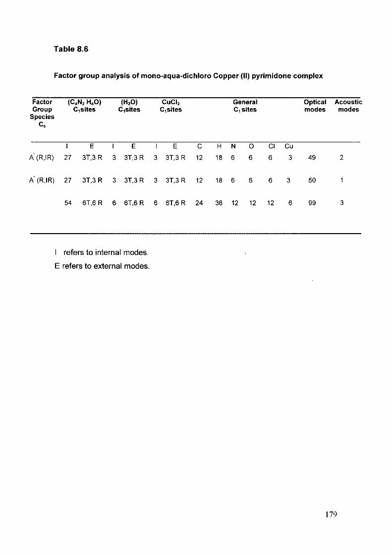
Table 8.6
Factor group analysis of mono-aqua-dichloro Copper (II) pyrimidone complex
Factor (C4N2 H4O) (H2O) CUCI2 General Group Cisites CiSites Cisites Ci sites
Species
Optical Acoustic modes modes
I E I E I E C H N O C I C u
A(R,IR) 27 3T,3R 3 3T,3 R 3 3T,3 R 12 18 6 6 6 3 49
A"(R,IR) 27 3T,3R 3 3T,3 R 3 3T,3 R 12 18 6 6 6 3 50
54 6T,6R 6 6T,6 R 6 6T,6 R 24 36 12 12 12 6 99
I refers to internal modes.
E refers to external modes.
179

Table 8.7
Assignment of various vibrational modes of mono-aqua-dichloro Copper
(II) pyrimidone complex
Mono-aqua-dichloro Copper (II) pyrimidone complex
Wave number (cm' ) Assignment
IR Raman
3600 m 3476 w v(OH) 3500 ms — v(OH)
3454 ms 3453 vw v(OH)
3402 m 3417 vw v(OH)
3397 vw v(NH) 3365 vw v(NH)
3303 w v(NH) 3241 w v(NH)
3212 w v(NH)
3189 w v(CH)
3133 w 3127 w v(CH) 3111 w v(CH)
3088 ms 3075 w v(NH)
3045 s 3058 vw v(CH) 3000 m 3000 vw v(CH)
2977 vw v(CH) 2924 m 2941 vw v(CH)
2905 vw v(CH)
2866 m 2866 vw v(CH)
2839 w v(CH)
2817 vw v(CH)
1792 w 5(OH)
1767 w 1750VW 5(0H)
1733 w — 5(0H)
1700 w 1671 w 6(0H)
1656 vs 1656 w v(C=0)
1606 ms 1602 m 6 H-O-H
1589 s 1563 m v(C-N),5(NH), V ring
1462 vw 1528 w v(C-N),5(NH), V ring
1424 vw 1444 w v(C-N),5(NH), V ring
1327 m 1336 ms v(C-N),8(CH), V ring
— 1266 w V ring, 8{NH)
180

1212 m 1212 m 5(CH)
1154W 1153W 6(CH) — 1099 s V ring
1077W 1079 m 5(CH) 1038 vw — 8(CH)
— 1010 w V ring, 5(CH)
— 971 w Y(NH)
— 934 w V ring, 6 ring
860 vw 872 s V ring, 5 ring
780 m 793 w Y(CH) — 724 w P (H2O)
600 w 650 s 8 ring — 591 w ©(HzO)
500 w 527 m Y(CO)
487 w 8 ring
458 w 8 ring
317w v(Cu-CI)
279 vs V(cu-pyrimidone) + v ( C U - C I )
189 w Lattice
171 m Lattice
155 m Lattice
148 w Lattice
126 w Lattice
106 m Lattice
vs-very strong ; s-strong ; ms-medium strong ; m-medium ; w-weak v:stretching
p: in- plane bending ; y : out- of- plane bending ; 8 : deformation ; co-wagging.
181

Fig. 8.1 ORTEP showing the atomic numbering scheme of cu(ll) pyrimidone complex.
IVI
Fig. 8.2 Packing diagram with n-n stacking between the aromatic rings and
the 0-H-O, H- bonding distance assembled in both P and M
sense of helical coordination polymeric adjacent chain.
182

Fig. 8.3 Packing of the complex in the unit cell.
Fig. 8.4 TGA Curve for compound 1 (inset DTA curve indicating two
endothermic curves)
183

40-3r
3000 2000 1500
Wavenumber (cftT')
1000 400
Fig. 8.5 FTIR spectrum of mono-aqua-dichloro Copper (II) pyrimidone complex.
500 -
100 — I — 150
1— 250 200 250 300
Wavenumber(cm"')
400
Fig. 8.6 Single crystal Raman spectrum of mono-aqua-dichloro Copper (II)
pyrimidone complex in the range 10-400 cm"
184

1 6 0 0
(0
*^ 'E 3
ji 1 2 0 0 ra (/> 1 0 0 0 -
c
c n E (0
3 0 0 -
6 0 0 -
Wl/ylUv^ v»Vv
4 0 0 1 —
6 0 0 8 0 0 1 0 0 0 1 2 0 0 1 4 0 0
W a v e n u m b e r ( c m " ' )
1 6 0 0 1 8 0 0
Fig. 8.7 Single crystal Raman spectrum of mono-aqua-dichloro Copper (II) pyrimidone complex in the range 400-1800 cm"
2800 2900 3000 3100 3200 3300 3400 3500
Wavenumber(cm"^)
Fig. 8.8 Single crystal Raman spectrum of mono-aqua-dichloro Copper (II) -1 pyrimidone complex in the range 2800-3500 cm .
185

T 1 1 ' 1 1 1 1 1 1 1 < 1 1 1 1 1 1 1 r-0 200 400 600 800 1000 1200 1400 1600 1800 2000 2200
Wavelength (nm)
Fig. 8.9 Optical transmission spectrum of mono-aqua-dichloro Copper (II)
pyrimidone complex.
186