Embed Size (px)
Citation preview
Kim et al. Crit Care (2021) 25:11 https://doi.org/10.1186/s13054-020-03421-4
RESEARCH
Characteristics and clinical outcomes of culture-negative and culture-positive septic shock: a single-center retrospective cohort studyJune‑sung Kim, Youn‑Jung Kim and Won Young Kim*
Abstract
Background: We evaluated the characteristics and outcomes of culture‑negative versus culture‑positive septic shock.
Methods: We performed a retrospective observational study of data from a prospective registry from 2014 to 2018. A total of 2,499 adult patients with septic shock were enrolled. The primary outcome was 90‑day mortality, and the secondary outcomes were the length of hospital stay, a requirement for mechanical ventilation or renal replacement therapy, and in‑hospital mortality.
Results: Of 1,718 patients with septic shock, 1,012 (58.9%) patients were culture‑positive (blood 803, urine 302, sputum 102, others 204) and the median pathogen detection time was 9.5 h (aerobic 10.2 h and anaerobic 9.0 h). The most common site of culture‑positive infection was the hepatobiliary tract (39.5%), while for the culture‑negative it was the lower respiratory tract (38.2%). The culture‑negative group had a lower mean body temperature (37.3 vs 37.7 ℃), lactate (2.5 vs. 3.2 mmol/L), C‑reactive protein (11.1 vs 11.9 mg/dL), and sequential organ failure assessment score (7.0 vs. 8.0) than that of the culture‑positive group. However, 90‑day mortality between the groups was not signifi‑cantly different (32.7 vs 32.2%, p = 0.83), and the other clinical outcomes also did not differ significantly. Moreover, a shorter culture detection time was correlated with a higher sequential organ failure assessment score but not with mortality.
Conclusion: Patients with septic shock are frequently culture‑negative, especially in cases where the infection focus is in the lower respiratory tract. Although culture‑negative was associated with a degree of organ dysfunction, it was not an independent predictor of death.
Keywords: Sepsis, Septic shock, Mortality, Culture, Time to blood culture positivity
© The Author(s) 2020. Open Access This article is licensed under a Creative Commons Attribution 4.0 International License, which permits use, sharing, adaptation, distribution and reproduction in any medium or format, as long as you give appropriate credit to the original author(s) and the source, provide a link to the Creative Commons licence, and indicate if changes were made. The images or other third party material in this article are included in the article’s Creative Commons licence, unless indicated otherwise in a credit line to the material. If material is not included in the article’s Creative Commons licence and your intended use is not permitted by statutory regulation or exceeds the permitted use, you will need to obtain permission directly from the copyright holder. To view a copy of this licence, visit http://creat iveco mmons .org/licen ses/by/4.0/. The Creative Commons Public Domain Dedication waiver (http://creat iveco mmons .org/publi cdoma in/zero/1.0/) applies to the data made available in this article, unless otherwise stated in a credit line to the data.
IntroductionThe incidence of sepsis and septic shock has been increasing worldwide over the past decade, and its mor-bidity and mortality are still unacceptably high [1]. Early
resuscitation for maintaining adequate tissue perfusion and the choice of proper antibiotics are mainstays to improve outcomes [2]. Although current guidelines rec-ommend obtaining cultures before prescribing empirical antibiotics, in many cases, isolation of specific organ-isms by culture remains challenging. Previous studies have reported the proportion of culture-negative cases was between 28 and 49% of all patients with sepsis [3–5];
Open Access
*Correspondence: [email protected] of Emergency Medicine, University of Ulsan College of Medicine, Asan Medical Center, 88, Olympic‑ro 43‑gil., Songpa‑gu, Seoul 05505, Korea
Page 2 of 9Kim et al. Crit Care (2021) 25:11
however, clinical outcomes between culture-negative and -positive patients have only been documented inconsist-ently and are controversial. Moreover, there is a remarka-ble paucity of investigations evaluating the characteristics and clinical outcomes of culture-negative patients with septic shock, especially among those visiting emergency departments.
Time-to-positivity (TPP) is defined as the duration of time from the start of incubation to the detection of bacterial growth by an automated culture system. It has been suggested as a prognostic marker of a fatal outcome. Numerous reports have revealed that a shorter TTP is associated with mortality among patients with each specific type of bacteremia [6, 7]. Septic shock is a het-erogeneous syndrome, involving a variety of microorgan-isms and affecting numerous vital organs. However, the relationship between TTP and the entire sepsis entity is rarely reported.
We hypothesized that culture negativity was associ-ated with worse outcomes than culture positivity because identifying the pathogens might help physicians choose the most appropriate antibiotics and determine the opti-mal duration of their use. Moreover, we presumed that an earlier detection time of culture could reflect the extent of bacterial loading and would be correlated with the severity of the illness. To prove this, we compared the clinical characteristics and outcomes of culture-negative septic shock (CNSS) cases versus culture-positive septic shock (CPSS) cases. We also evaluated the relationship between TTP and 90-day mortality in patients with sep-tic shock.
MethodsStudy designWe conducted a retrospective cohort study by analyzing data from a prospective septic shock registry at an urban tertiary emergency department with annual admissions of more than 120,000 patients from January 1, 2014, to December 31, 2018. This registry was designed to enhance internal quality improvement of septic shock patients visiting emergency departments and to manage multicenter cohort data of the Korean Shock Society for future analysis [8]. The Institutional Review Board of the study facility approved the study (no. 2016–0548) and waived the requirement for informed consent because of its retrospective characteristics.
Data collection and definition of variablesThis registry included all adult septic shock patients (≥ 18 years) consecutively diagnosed in the emergency department [9]. Infection was defined clinically by the emergency physicians on duty. In brief, they evalu-ated the systemic inflammatory response syndrome
and applied the quick sequential organ failure assess-ment (SOFA) criteria for every patient with suspected or confirmed infection [9]. In addition, we used a defi-nition of septic shock as refractory hypotension (mean arterial pressure ≤ 65 mmHg) requiring vasopressors despite adequate fluid infusion or a blood lactate level of at least 4 mmol/L, based on a previous definition [10]. This registry excluded those who were transferred from other hospitals after proper resuscitation, transferred to another hospital because of no room for admission, had a “do-not-resuscitate” order, or refused to accept treat-ment because of nonmedical issues, such as healthcare cost, fear of invasive procedures, or just not wanting to intensive care unit care (ICU) without definite reasons. We also excluded patients from this secondary analysis with microbiologically proven viral, fungal, and parasitic infections.
All enrolled patients were treated with protocol-driven resuscitation following the Surviving Sepsis Campaign guidelines [3]. In brief, aggressive fluids infusion and vasopressors were applied with blood pressure monitor-ing. Lactate levels were checked via venous or arterial blood gas analysis, and a central venous catheter was placed routinely for administering high-dose vasopres-sors and repetitive sampling [11, 12]. Blood cultures were obtained within 3 h of recognition from samples at two or more different anatomical sites according to the local practice [13]. When indwelling catheters were present, one blood sample was obtained through the catheter, and the remainder were taken from different periph-eral venous sites. Site-specific cultures, including urine, sputum, pleural, ascites, stool, and pus, were performed following the physicians’ decisions. Broad-spectrum empirical antibiotics were infused as soon as possible, and percutaneous or endoscopic drainages were aggres-sively conducted after image work-ups. The sites of infec-tion were grouped as the lower respiratory tract, urinary tract, gastrointestinal, and hepatobiliary systems. Minor sites, such as isolated blood stream infections, skin and soft tissue, and the central nervous system were catego-rized as others. The infectious sites were determined and recorded in the registry by the primary physicians on duty through the patients’ histories, physical examina-tions, and results of laboratory and imaging. We deemed fever without a definite focus as unknown.
Data regarding demographics, underlying diseases, ini-tial vital signs, sites of infection, and clinical outcomes, such as intensive care unit (ICU) admission, require-ments for a mechanical ventilator or renal replacement therapy, and duration of ICU stay and mechanical ven-tilation were obtained from the registry. The date of the patient’s death was obtained from the National Health Insurance Service in South Korea, and in-hospital
Page 3 of 9Kim et al. Crit Care (2021) 25:11
mortality and 90-day mortality were extracted. Subgroup analysis of in-hospital mortality according to the site of infection was also investigated to confirm the clinical dif-ferences based on the infection sites. Laboratory exami-nations, including white blood cell counts, hemoglobin, prothrombin time (international normalization ratio), lactate, and C-reactive protein, were also extracted. Source control measures, including all physical proce-dures to eliminate the sources of the pathogen, including draining abscesses, debriding infected soft tissues, and extracting contaminated devices or foreign bodies, were recorded [14]. Escalation of antibiotics occurred when-ever the initial antibiotics were changed to cover more extensive pathogens because of refractory shock or the identification of antibiotic-resistance pathogens. SOFA scores were calculated from the initial clinical and labo-ratory data on admission.
Moreover, microbiological culture results and the detection time of the enrolled patients were extracted from the electronic medical records and reviewed by three investigators (J.S.K., Y.J.K., and W.Y.K.). An expe-rienced infection specialist made a final decision when there was a disagreement about discriminating the exact pathogen. Detection time was defined as the time to pos-itive detection of the pathogens and was calculated by subtracting the time of receipt in the laboratory from the time required to detect a positive culture. When growth was detected in both aerobic and anaerobic bottles, each time was recorded [see Additional file 1].
The primary outcome was 90-day mortality, and the secondary outcomes were ICU admission, ICU length of stay, mechanical ventilator or renal replacement therapy requirement, mechanical ventilation duration, and in-hospital mortality.
Statistical analysisDescriptive statistics were stratified by culture results (i.e., culture-positive and -negative). Baseline demo-graphics, clinical characteristics, and outcomes are pre-sented as the frequency and percentage for categorical and median with interquartile range (IQR) for continu-ous variables. The Kolmogorov–Smirnov test was used to check the normality of the distribution. Categorical vari-ables were analyzed using Chi-squared or Fisher’s exact tests. Univariate logistic regression tests were performed with potential risk factors, which showed differences between two groups. Multivariate logistic regression was conducted with the variables that had significance in the univariate logistic regression analysis. The absence of multicollinearity was confirmed by using regression anal-ysis with calculating the variance inflation factor values. Kaplan–Meier survival curves with the log-rank test were stratified by the culture results. The correlations between
the severity of the septic shock and culture detection time were assessed by the Spearman rank correlation coefficient. We considered P values less than 0.05 as sta-tistically significant. Analyses were performed using SPSS Statistics for Windows, version 26 (IBM Corp., Armonk, NY, USA).
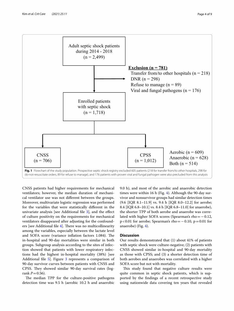
ResultsDuring the study period, 2,499 adult septic shock patients visited the emergency department, and 781 were not included in this study due to the exclusion criteria (218 transferred from/to other hospitals, 298 had do-not-resuscitate orders, 89 refused treatment, and 176 had known viral or fungal pathogens) (Fig. 1). Of the 1,718 cases, 706 (41.1%) were in the CNSS and 1,012 (58.9%) were in the CPSS group.
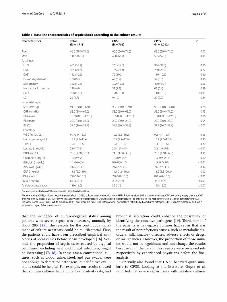
Table 1 presents the baseline characteristics of the study population according to the culture results. The CPSS group tended to be older and had a higher propor-tion of female patients. All underlying diseases, except chronic kidney diseases (10.9 vs. 18.1%, P < 0.01; CPSS vs. CNSS, respectively), were similar between the groups. Patients who were pathogen-positive showed slightly lesser tachypnea and a higher mean body temperature than those who were pathogen-negative. In the labora-tory results, initial lactate (3.2 vs. 2.5 mmol/L, P < 0.01) and total bilirubin (1.3 vs. 0.9 mg/dL, P < 0.01) were ele-vated more in the CPSS than in the CNSS patients. Fur-thermore, the initial SOFA score (8.0 vs. 7.0, P < 0.01) was higher, and source control (44.6 vs. 28.6%, P < 0.01) and the escalation of antibiotics (15.6 vs. 4.4%, P < 0.01) were more frequent in patients with CPSS.
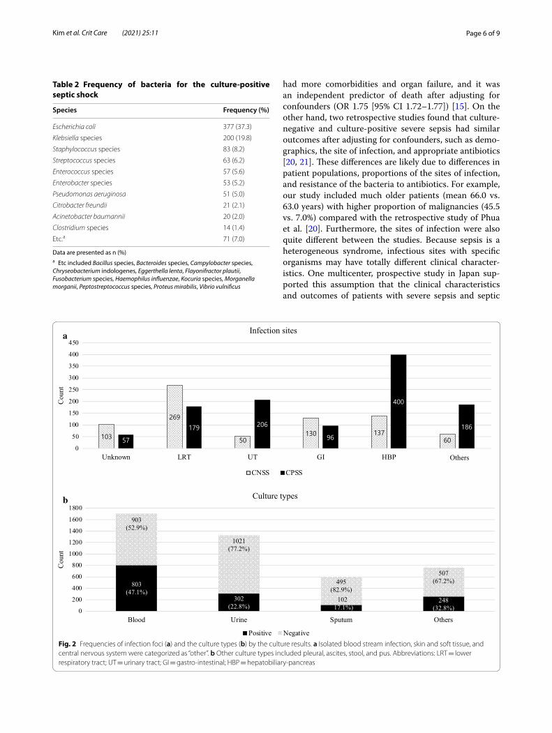
Escherichia coli was the most common causative organ-ism, followed by Klebsiella species (including Klebsiella pneumoniae, oxytoca, and ornithinolytica) (Table 2). Gram-positive bacteria, such as Staphylococcus and Streptococcus, were less frequent than Gram-negative bacteria. An additional table shows this in more detail [see Additional file 2].
Figure 2 shows the frequencies of infection focuses and culture types according to the culture results. Lower respiratory tract and gastrointestinal focuses were the most frequent sites in the negative culture cases; mean-while, the urinary tract and hepatobiliary pancreas were the most common sites among the positive culture cases. Blood cultures were predominantly performed and showed the highest positive rates (47.1%) followed by the others (32.8%), including pleural effusion, ascites, open pus, and closed pus obtained by endoscopic or percuta-neous drainage.
For the clinical outcomes, both groups had similar ICU admission rates, ICU length of stay, and require-ments for renal replacement therapy (Table 3). The
Page 4 of 9Kim et al. Crit Care (2021) 25:11
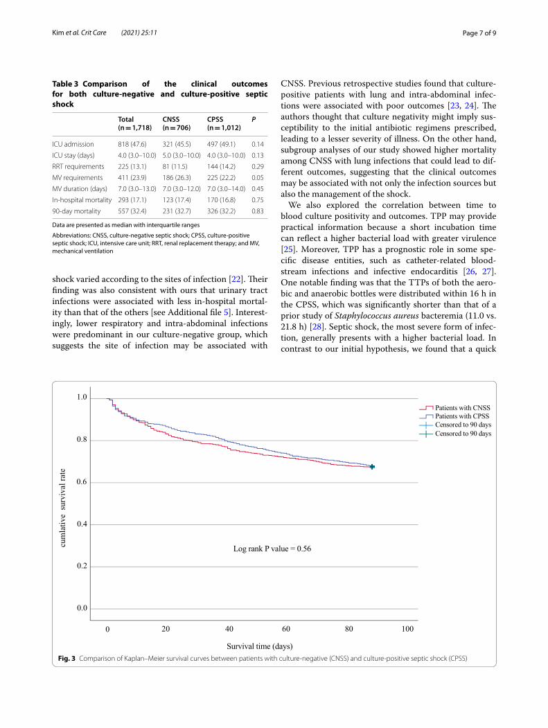
CNSS patients had higher requirements for mechanical ventilators; however, the median duration of mechani-cal ventilator use was not different between the groups. Moreover, multivariate logistic regression was performed for the variables that were statistically different in the univariate analysis [see Additional file 3], and the effect of culture positivity on the requirements for mechanical ventilators disappeared after adjusting for the confound-ers [see Additional file 4]. There was no multicollinearity among the variables, especially between the lactate level and SOFA score (variance inflation factors 1.084). The in-hospital and 90-day mortalities were similar in both groups. Subgroup analysis according to the sites of infec-tion showed that patients with lower respiratory infec-tions had the highest in-hospital mortality (38%) [see Additional file 5]. Figure 3 represents a comparison of 90-day survivor curves between patients with CNSS and CPSS. They showed similar 90-day survival rates (log-rank P = 0.56).
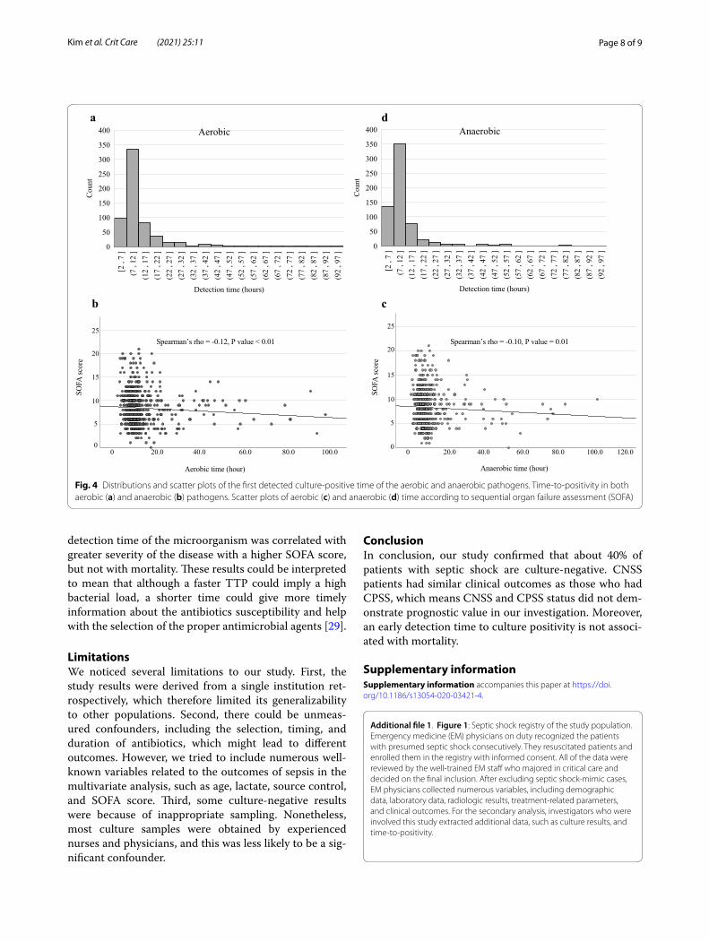
The median TPP for the culture-positive pathogens detection time was 9.5 h (aerobic 10.2 h and anaerobic
9.0 h), and most of the aerobic and anaerobic detection times were within 16 h (Fig. 4). Although the 90-day sur-vivor and nonsurvivor groups had similar detection times (9.6 [IQR 8.1–11.9] vs. 9.4 h [IQR 8.0–12.2] for aerobe; 8.4 [IQR 6.8–10.1] vs. 8.4 h [IQR 6.8–11.0] for anaerobe), the shorter TPP of both aerobe and anaerobe was corre-lated with higher SOFA scores (Spearman’s rho = − 0.12, p < 0.01 for aerobe; Spearman’s rho = − 0.10, p = 0.01 for anaerobe) (Fig. 4).
DiscussionOur results demonstrated that (1) about 41% of patients with septic shock were culture-negative; (2) patients with CNSS showed similar in-hospital and 90-day mortality as those with CPSS; and (3) a shorter detection time of both aerobes and anaerobes was correlated with a higher SOFA score but not with mortality.
This study found that negative culture results were quite common in septic shock patients, which is sup-ported by the findings of a recent retrospective study using nationwide data covering ten years that revealed
Enrolled patients with septic shock
(n = 1,718)
CNSS(n = 706)
Adult septic shock patientsduring 2014 - 2018
(n = 2,499)
Exclusion (n = 781)Transfer from/to other hospitals (n = 218)DNR (n = 298)Refuse to manage (n = 89)Viral and fungal pathogens (n = 176)
CPSS(n = 1,012)
Aerobic (n = 609)Anaerobic (n = 628)Both (n = 514)
Fig. 1 Flowchart of the study population. Prospective septic shock registry excluded 605 patients (218 for transfer from/to other hospitals, 298 for do‑not‑resuscitate orders, 89 for refuse to manage), and 176 patients with proven viral and fungal pathogen were also precluded from this analysis
Page 5 of 9Kim et al. Crit Care (2021) 25:11
that the incidence of culture-negative status among patients with severe sepsis was increasing annually by about 28% [15]. The reasons for the continuous incre-ment of culture negativity could be multifactorial. First, the patients could have been prescribed empirical anti-biotics at local clinics before sepsis developed [16]. Sec-ond, the proportion of sepsis cases caused by atypical pathogens, including viral and fungal infections, might be increasing [17, 18]. In these cases, conventional cul-tures, such as blood, urine, stool, and pus swabs, were not enough to detect the pathogens, but definitive evalu-ations could be helpful. For example, our results showed that sputum cultures had a quite low positivity rate, and
bronchial aspiration could enhance the possibility of identifying the causative pathogens [19]. Third, some of the patients with negative cultures had sepsis that was the result of noninfectious causes, such as metabolic dis-orders, inflammatory diseases, adverse effects of drugs, or malignancies. However, the proportion of those mim-ics would not be significant and not change the results because all of the data in this registry were reviewed ret-rospectively by experienced physicians before the final analysis.
Our study also found that CNSS behaved quite simi-larly to CPSS. Looking at the literature, Gupta et al. reported that severe sepsis cases with negative cultures
Table 1 Baseline characteristics of septic shock according to the culture results
Data are presented as n (%) or mean with standard deviation
Abbreviations: CNSS, culture negative septic shock; CPSS, culture positive septic shock; HTN, hypertension; DM, diabetes mellitus; CAD, coronary artery disease; CKD, chronic kidney disease; LC, liver cirrhosis; SBP, systolic blood pressure; DBP, diastolic blood pressure; PR, pulse rate; RR, respiratory rate; BT, body temperature; GCS, Glasgow Coma Scale; WBC, white blood cells; PT, prothrombin time; INR, international normalized ratio; BUN, blood urea nitrogen; CRP, C-reactive protein; and SOFA, sequential organ failure assessment
Characteristics Total(N = 1,718)
CNSS(N = 706)
CPSS(N = 1,012)
P
Age 66.0 (58.0–74.0) 66.0 (56.0–74.0) 68.0 (59.0–74.0) 0.02
Male 1,035 (60.2) 450 (63.7) 585 (57.8) 0.01
Past illness
HTN 605 (35.2) 261 (37.0) 344 (34.0) 0.20
DM 425 (24.7) 165 (23.4) 260 (25.7) 0.27
CAD 182 (10.6) 72 (10.2) 110 (10.9) 0.66
Pulmonary disease 108 (6.3) 49 (6.9) 59 (5.8) 0.36
Malignancy 782 (45.5) 302 (42.8) 480 (47.4) 0.06
Hematologic disorder 118 (6.9) 53 (7.5) 65 (6.4) 0.39
CKD 238 (13.9) 128 (18.1) 110 (10.9) < 0.01
LC 29 (1.7) 9 (1.3) 20 (2.0) 0.34
Initial vital signs
SBP (mmHg) 91.0 (80.0–112.0) 90.0 (80.0–109.0) 93.0 (80.0–113.0) 0.28
DBP (mmHg) 58.0 (50.0–69.0) 58.0 (50.0–68.0) 58.0 (50.0–71.0) 0.73
PR (/min) 107.0 (89.0–125.0) 105.0 (88.0–123.0) 108.0 (90.0–126.0) 0.06
RR (/min) 20.0 (20.0–24.0) 20.0 (20.0–24.0) 20.0 (20.0–22.0) 0.04
BT (℃) 37.6 (36.6–38.7) 37.2 (36.5–38.2) 37.7 (36.7–38.9) < 0.01
Laboratory
WBC (× 103/μL) 9.7 (5.0–15.9) 10.2 (5.5–16.2) 9.2 (4.7–15.7) 0.06
Hemoglobin (g/dL) 10.7 (9.1–12.4) 10.7 (9.2–12.6) 10.7 (9.0–12.3) 0.30
PT (INR) 1.3 (1.1–1.5) 1.3 (1.1–1.4) 1.3 (1.1–1.5) 0.20
Lactate (mmol/L) 2.9 (1.7–5.1) 2.5 (1.5–4.5) 3.2 (1.9–5.4) < 0.01
BUN (mg/dL) 25.0 (17.0–38.0) 24.0 (17.0–39.0) 25.0 (17.0–37.0) 0.95
Creatinine (mg/dL) 1.3 (0.9–2.1) 1.3 (0.9–2.2) 1.3 (0.9–2.1) 0.10
Bilirubin (mg/dL) 1.1 (0.6–2.4) 0.9 (0.5–1.7) 1.3 (0.7–3.0) < 0.01
Albumin (g/dL) 2.6 (2.2–3.1) 2.6 (2.2–3.1) 2.6 (2.2–3.1) 0.37
CRP (mg/dL) 11.6 (5.0–19.8) 11.1 (4.6–19.2) 11.9 (5.3–20.2) 0.02
SOFA score 7.0 (5.0–10.0) 7.0 (5.0–10.0) 8.0 (6.0–10.0) < 0.01
Source control 653 (38.0) 202 (28.6) 451 (44.6) < 0.01
Antibiotics escalation 189 (11.0) 31 (4.4) 158 (15.6) < 0.01
Page 6 of 9Kim et al. Crit Care (2021) 25:11
had more comorbidities and organ failure, and it was an independent predictor of death after adjusting for confounders (OR 1.75 [95% CI 1.72–1.77]) [15]. On the other hand, two retrospective studies found that culture-negative and culture-positive severe sepsis had similar outcomes after adjusting for confounders, such as demo-graphics, the site of infection, and appropriate antibiotics [20, 21]. These differences are likely due to differences in patient populations, proportions of the sites of infection, and resistance of the bacteria to antibiotics. For example, our study included much older patients (mean 66.0 vs. 63.0 years) with higher proportion of malignancies (45.5 vs. 7.0%) compared with the retrospective study of Phua et al. [20]. Furthermore, the sites of infection were also quite different between the studies. Because sepsis is a heterogeneous syndrome, infectious sites with specific organisms may have totally different clinical character-istics. One multicenter, prospective study in Japan sup-ported this assumption that the clinical characteristics and outcomes of patients with severe sepsis and septic
Table 2 Frequency of bacteria for the culture-positive septic shock
Data are presented as n (%)a Etc included Bacillus species, Bacteroides species, Campylobacter species, Chryseobacterium indologenes, Eggerthella lenta, Flayonifractor plautii, Fusobacterium species, Haemophilus influenzae, Kocuria species, Morganella morganii, Peptostreptococcus species, Proteus mirabilis, Vibrio vulnificus
Species Frequency (%)
Escherichia coli 377 (37.3)
Klebsiella species 200 (19.8)
Staphylococcus species 83 (8.2)
Streptococcus species 63 (6.2)
Enterococcus species 57 (5.6)
Enterobacter species 53 (5.2)
Pseudomonas aeruginosa 51 (5.0)
Citrobacter freundii 21 (2.1)
Acinetobacter baumannii 20 (2.0)
Clostridium species 14 (1.4)
Etc.a 71 (7.0)
a
b
Fig. 2 Frequencies of infection foci (a) and the culture types (b) by the culture results. a Isolated blood stream infection, skin and soft tissue, and central nervous system were categorized as “other”. b Other culture types included pleural, ascites, stool, and pus. Abbreviations: LRT = lower respiratory tract; UT = urinary tract; GI = gastro‑intestinal; HBP = hepatobiliary‑pancreas
Page 7 of 9Kim et al. Crit Care (2021) 25:11
shock varied according to the sites of infection [22]. Their finding was also consistent with ours that urinary tract infections were associated with less in-hospital mortal-ity than that of the others [see Additional file 5]. Interest-ingly, lower respiratory and intra-abdominal infections were predominant in our culture-negative group, which suggests the site of infection may be associated with
CNSS. Previous retrospective studies found that culture-positive patients with lung and intra-abdominal infec-tions were associated with poor outcomes [23, 24]. The authors thought that culture negativity might imply sus-ceptibility to the initial antibiotic regimens prescribed, leading to a lesser severity of illness. On the other hand, subgroup analyses of our study showed higher mortality among CNSS with lung infections that could lead to dif-ferent outcomes, suggesting that the clinical outcomes may be associated with not only the infection sources but also the management of the shock.
We also explored the correlation between time to blood culture positivity and outcomes. TPP may provide practical information because a short incubation time can reflect a higher bacterial load with greater virulence [25]. Moreover, TPP has a prognostic role in some spe-cific disease entities, such as catheter-related blood-stream infections and infective endocarditis [26, 27]. One notable finding was that the TTPs of both the aero-bic and anaerobic bottles were distributed within 16 h in the CPSS, which was significantly shorter than that of a prior study of Staphylococcus aureus bacteremia (11.0 vs. 21.8 h) [28]. Septic shock, the most severe form of infec-tion, generally presents with a higher bacterial load. In contrast to our initial hypothesis, we found that a quick
Table 3 Comparison of the clinical outcomes for both culture-negative and culture-positive septic shock
Data are presented as median with interquartile ranges
Abbreviations: CNSS, culture-negative septic shock; CPSS, culture-positive septic shock; ICU, intensive care unit; RRT, renal replacement therapy; and MV, mechanical ventilation
Total(n = 1,718)
CNSS(n = 706)
CPSS(n = 1,012)
P
ICU admission 818 (47.6) 321 (45.5) 497 (49.1) 0.14
ICU stay (days) 4.0 (3.0–10.0) 5.0 (3.0–10.0) 4.0 (3.0–10.0) 0.13
RRT requirements 225 (13.1) 81 (11.5) 144 (14.2) 0.29
MV requirements 411 (23.9) 186 (26.3) 225 (22.2) 0.05
MV duration (days) 7.0 (3.0–13.0) 7.0 (3.0–12.0) 7.0 (3.0–14.0) 0.45
In‑hospital mortality 293 (17.1) 123 (17.4) 170 (16.8) 0.75
90‑day mortality 557 (32.4) 231 (32.7) 326 (32.2) 0.83
Log rank P value = 0.56
Survival time (days)
Patients with CNSSPatients with CPSSCensored to 90 daysCensored to 90 days
cum
lativ
e su
rviv
al ra
te
0.4
0.0
0.2
0.6
0.8
1.0
100806040200
Fig. 3 Comparison of Kaplan–Meier survival curves between patients with culture‑negative (CNSS) and culture‑positive septic shock (CPSS)
Page 8 of 9Kim et al. Crit Care (2021) 25:11
detection time of the microorganism was correlated with greater severity of the disease with a higher SOFA score, but not with mortality. These results could be interpreted to mean that although a faster TTP could imply a high bacterial load, a shorter time could give more timely information about the antibiotics susceptibility and help with the selection of the proper antimicrobial agents [29].
LimitationsWe noticed several limitations to our study. First, the study results were derived from a single institution ret-rospectively, which therefore limited its generalizability to other populations. Second, there could be unmeas-ured confounders, including the selection, timing, and duration of antibiotics, which might lead to different outcomes. However, we tried to include numerous well-known variables related to the outcomes of sepsis in the multivariate analysis, such as age, lactate, source control, and SOFA score. Third, some culture-negative results were because of inappropriate sampling. Nonetheless, most culture samples were obtained by experienced nurses and physicians, and this was less likely to be a sig-nificant confounder.
ConclusionIn conclusion, our study confirmed that about 40% of patients with septic shock are culture-negative. CNSS patients had similar clinical outcomes as those who had CPSS, which means CNSS and CPSS status did not dem-onstrate prognostic value in our investigation. Moreover, an early detection time to culture positivity is not associ-ated with mortality.
Supplementary informationSupplementary information accompanies this paper at https ://doi.org/10.1186/s1305 4‑020‑03421 ‑4.
Additional file 1. Figure 1: Septic shock registry of the study population. Emergency medicine (EM) physicians on duty recognized the patients with presumed septic shock consecutively. They resuscitated patients and enrolled them in the registry with informed consent. All of the data were reviewed by the well‑trained EM staff who majored in critical care and decided on the final inclusion. After excluding septic shock‑mimic cases, EM physicians collected numerous variables, including demographic data, laboratory data, radiologic results, treatment‑related parameters, and clinical outcomes. For the secondary analysis, investigators who were involved this study extracted additional data, such as culture results, and time‑to‑positivity.
Detection time (hours)
[2 ,
7 ]
(7 ,
12 ]
(12
, 17
](1
7 , 2
2 ]
(22
, 27
](2
7 , 3
2 ]
(32
, 37
](3
7 , 4
2 ]
(42
, 47
](4
7 , 5
2 ]
(52
, 57
](5
7 , 6
2 ]
(62
, 67
](6
7 , 7
2 ]
(72
, 77
](7
7 , 8
2 ]
(82
, 87
](8
7 , 9
2 ]
(92
, 97
]
Cou
nt
0
50
100
150
200
250
300
350
400 Aerobic
Detection time (hours)
[2 ,
7 ]
(7 ,
12 ]
(12
, 17
](1
7 , 2
2 ]
(22
, 27
](2
7 , 3
2 ]
(32
, 37
](3
7 , 4
2 ]
(42
, 47
](4
7 , 5
2 ]
(52
, 57
](5
7 , 6
2 ]
(62
, 67
](6
7 , 7
2 ]
(72
, 77
](7
7 , 8
2 ]
(82
, 87
](8
7 , 9
2 ]
(92
, 97
]
Cou
nt
0
50
100
150
200
250
300
350
400 Anaerobic
Aerobic time (hour) Anaerobic time (hour)Aerobic time (hour) Anaerobic time (hour)
-0.12, P value < 0.01 -0.10, P value = 0.01
SOFA
scor
e
SOFA
scor
e
0
5
15
20
10
25
0 20.0 40.0 60.0 80.0 100.00
5
15
20
25
10
120.0100.080.060.040.020.00
a
b c
d
Fig. 4 Distributions and scatter plots of the first detected culture‑positive time of the aerobic and anaerobic pathogens. Time‑to‑positivity in both aerobic (a) and anaerobic (b) pathogens. Scatter plots of aerobic (c) and anaerobic (d) time according to sequential organ failure assessment (SOFA)
Page 9 of 9Kim et al. Crit Care (2021) 25:11
Additional file 2. Table 1: Frequency of the subspecies of the isolated bacteria.
Additional file 3. Table 2: Baseline characteristics of septic shock accord‑ing to the culture results.
Additional file 4. Table 3: Univariate and multivariate analysis for predict‑ing mechanical ventilator requirements.
Additional file 5. Table 4: In‑hospital mortality according to sites of infec‑tion among patients with culture‑negative septic shock.
AbbreviationsCNSS: Culture‑negative septic shock; CPSS: Culture‑positive septic shock; DNR: Do‑not‑resuscitate; ICU: Intensive care unit; IQR: Interquartile range; SOFA: Sequential organ failure assessment; TTP: Time‑to‑positivity.
AcknowledgementsNone.
Authors’ contributionsW.Y.K. designed and supervised the paper. J.S.K. contributed to the literature search, figures, study design, data collection, data analysis, interpretation and writing, and revisions. Y.J.K. contributed to design, revisions, and validation. All authors read and approved the final manuscript.
FundingNone.
Availability of data and materialsThe datasets generated and analyzed during the current study are available from the corresponding author on reasonable request.
Ethics approval and consent to participateThis study was reviewed and approved by Asan Medical Center IRB with waiver of written consent.
Consent for publicationNot applicable.
Competing interestsThe authors declare that they have no competing interests.
Received: 20 July 2020 Accepted: 2 December 2020
References 1. Rudd KE, Johnson SC, Agesa KM, Shackelford KA, Tsoi D, Kievlan DR, et al.
Global, regional, and national sepsis incidence and mortality, 1990–2017. Lancet. 2020;395:200–11.
2. De Backer D, Cecconi M, Lipman J, Machado F, Myatra SN, Ostermann M, et al. Challenges in the management of septic shock. Intensive Care Med. 2019;45:420–33.
3. Rhodes A, Evans LE, Alhazzani W, Levy MM, Antonelli M, Ferrer R, et al. Surviving Sepsis Campaign: international guidelines for management of sepsis and septic shock: 2016. Intensive Care Med. 2017;43:304–77.
4. Kumar A, Ellis P, Arabi Y, Roberts D, Light B, Parrillo JE, et al. Initiation of inappropriate antimicrobial therapy results in a fivefold reduction of survival in human septic shock. Chest. 2009;136:1237–48.
5. Rannikko J, Syrjänen J, Seiskari T, Aittoniemi J, Huttunen R. Sepsis‑related mortality in 497 cases with blood culture‑positive sepsis in an emergency department. Int J Infect Dis. 2017;58:52–7.
6. Palmer HR, Palavecino EL, Johnson JW, Ohl CA, Williamson JC. Clinical and microbiological implications of time‑to‑positivity of blood cultures in patients with Gram‑negative bacilli bacteremia. Eur J Clin Microbiol Infect Dis. 2013;32:955–9.
7. Tang P‑C, Lee C‑C, Li C‑W, Li M‑C, Ko W‑C, Lee N‑Y. Time‑to‑positivity of blood culture. J Microbiol Immunol Infect. 2017;50:486–93.
8. Shin TG, Hwang SY, Kang GH, Kim WY, Ryoo SM, Kim K, et al. Korean Shock Society septic shock registry: a preliminary report. Clin Exp Emerg Med. 2017;4:146–53.
9. Kim JS, Kim Y‑J, Ryoo S, Sohn C, Ahn S, Seo D, et al. Risk factors for same pathogen sepsis readmission following hospitalization for septic shock. J Clin Med. 2019;8:181–8.
10. Levy MM, Fink MP, Marshall JC, Abraham E, Angus D, Cook D, et al. 2001 SCCM/ESICM/ACCP/ATS/SIS international sepsis definitions conference. Crit Care Med. 2003;31:1250–6.
11. Ryoo SM, Lee J, Lee Y‑S, Lee JH, Lim KS, Huh JW, et al. Lactate level versus lactate clearance for predicting mortality in patients with septic shock defined by Sepsis‑3. Crit Care Med. 2018;46:e489–95.
12. Hernández G, Ospina‑Tascón GA, Damiani LP, Estenssoro E, Dubin A, Hurtado J, et al. Effect of a resuscitation strategy targeting peripheral per‑fusion status vs serum lactate levels on 28‑day mortality among patients with septic shock. JAMA. 2019;321:654–711.
13. Lee A, Mirrett S, Reller LB, Weinstein MP. Detection of bloodstream infections in adults: how many blood cultures are needed? J Clin Microbiol. 2007;45:3546–8.
14. Kim HJ, Chung SP, Choi S‑H, Kang GH, Shin TG, Kim KS, et al. Impact of timing to source control in patients with septic shock: a prospective multi‑center observational study. J Crit Care. 2019;53:176–82.
15. Gupta S, Sakhuja A, Kumar G, McGrath E, Nanchal RS, Kashani KB. Culture‑negative severe sepsis. Chest. 2016;150:1251–9.
16. Scheer CS, Fuchs C, Gründling M, Vollmer M, Bast J, Bohnert JA, et al. Impact of antibiotic administration on blood culture positivity at the beginning of sepsis: a prospective clinical cohort study. Clin Microbiol Infect. 2019;25:326–31.
17. Lin G‑L, McGinley JP, Drysdale SB, Pollard AJ. Epidemiology and immune pathogenesis of viral sepsis. Front Immunol Frontiers. 2018;9:801–21.
18. Delaloye J, Calandra T. Invasive candidiasis as a cause of sepsis in the criti‑cally ill patient. Virulence. 2013;5:161–9.
19. Koslow M, Shochet GE, Matveychuk A, Israeli‑Shani L, Guber A, Shitrit D. The role of bacterial culture by bronchoscopy in patients with lung cancer. J Thorac Dis. 2017;9:5300–5.
20. Phua J, Ngerng WJ, See KC, Kiong T, Lim HF, Chew MY, et al. Characteristics and outcomes of culture‑negative versus culture‑positive severe sepsis. Crit Care. 2013;17:R202.
21. Kethireddy S, Bilgili B, Sees A, Kirchner HL, Ofoma UR, Light RB, et al. Culture‑negative septic shock compared with culture‑positive septic shock. Crit Care Med. 2018;2018:1–7.
22. Abe T, Ogura H, Kushimoto S, Shiraishi A, Sugiyama T, Deshpande GA, et al. Variations in infection sites and mortality rates among patients in intensive care units with severe sepsis and septic shock in Japan. J Inten‑sive Care. 2019;3:28–36.
23. Labelle AJ, Arnold H, Reichley RM, Micek ST, Kollef MH. A comparison of culture‑positive and culture‑negative health‑care‑associated pneumonia. Chest. 2010;137:1130–7.
24. Karvellas CJ, Abraldes JG, Zepeda‑Gomez S, Moffat DC, Mirzanejad Y, Vazquez‑Grande G, et al. The impact of delayed biliary decompression and anti‑microbial therapy in 260 patients with cholangitis‑associated septic shock. Aliment Pharmacol Ther. 2016;44:755–66.
25. Janapatla RP, Yan J‑J, Chien M‑L, Chen H‑M, Wu H‑M, Wu J‑J. Effect of over‑night storage of blood culture bottles on bacterial detection time in the BAC‑TEC 9240 blood culture system. J Microbiol Immunol Infect. 2010;43:126–32.
26. Puerta‑Alcalde P, Cardozo C, Suárez‑Lledó M, Rodríguez‑Núñez O, Morata L, Fehér C, et al. Current time‑to‑positivity of blood cultures in febrile neutropenia: a tool to be used in stewardship de‑escalation strategies. Clin Microbiol Infect. 2019;25:447–53.
27. Siméon S, Le Moing V, Tubiana S, Duval X, Fournier D, Lavigne JP, et al. Time to blood culture positivity. Clin Microbiol Infect. 2019;25:481–8.
28. Li Y, Li Q, Zhang G, Ma H, Wu Y, Yi Q, et al. Time to positivity of blood culture is a risk factor for clinical outcomes in Staphylococcus aureus bacteremia children: a retrospective study. BMC Infect Dis. 2019;19:437.
29. Lambregts MMC, Bernards AT, van der Beek MT, Visser LG, de Boer MG. Time to positivity of blood cultures supports early re‑evaluation of empiric broad‑spectrum antimicrobial therapy. Gołębiewska J editor. PLoS ONE. 2019;14(1):e0208819‑e208912.
Publisher’s NoteSpringer Nature remains neutral with regard to jurisdictional claims in pub‑lished maps and institutional affiliations.