Embed Size (px)
Citation preview
J. exp. Biol. 117, 389-399 (1985) 3 3 9Printed in Great Britain © The Company of Biologists Limited 1985
CHARACTERISTICS OF MECHANORECEPTORS INTHE AIR-BREATHING ORGAN OF THE HOLOSTEAN
FISH, AMIA CALVA
BY WILLIAM K. MILSOM AND DAVID R. JONES
Department of Zoology, University of British Columbia, Vancouver,British Columbia, V6T2A9, Canada
Accepted 14 February 1985
SUMMARY
Single nerve fibre discharge was recorded from mechanoreceptorsassociated with the air-breathing organ in double-pithed specimens of thebowfin, Amia calva L. These receptors were innervated by the vagus nerveand although their exact location was difficult to determine, most appearedto be located along the anterio-ventral wall of the single lung. All receptorsincreased tonic discharge with step increases in lung volume, above athreshold level, and were slowly adapting. There was a dynamic, rate-sensitive burst of activity associated with lung inflation and a dynamic, rate-sensitive inhibition of discharge associated with deflation. These responseswere qualitatively similar to those of the tonic stretch receptors found in fishswimbladder and mammalian gut. All receptors were insensitive to changesin intrapulmonary partial pressures of oxygen and carbon dioxide. Theseobservations suggest that receptors capable of transducing the rate, as wellas the degree, of inflation and deflation are associated with primitive lungs,and may have arisen from tonic gut receptors.
INTRODUCTION
Air breathing has evolved many times in the lower vertebrates with a variety ofstructural adaptations giving rise to gas exchange organs. Those found among the air-breathing fishes are extremely diverse. Most species of air-breathing fishes rely on avariety of modifications of the buccal cavity and pharynx, on the walls of the stomachand intestine, on air sacs or on lungs for aerial exchange. The mechanisms used toventilate these gas exchange organs are equally diverse as, undoubtedly, are thecontrol mechanisms involved (see Gans, 1970 for review).
Various cardiovascular and respiratory reflexes have been described which areassociated with inflation and/or deflation of these organs (see Johansen, 1970, 1972;Wood & Lenfant, 1976 for reviews; Johansen, Hanson & Lenfant, 1970; Babiker,1979; Smatresk & Cameron, 1982; Pack, Galante & Fishman, 1984). Furthermore,changes in volume of gas exchange organs have been implicated in adjusting breathingfrequency to match metabolic rate (Johansen, 1970; Smatresk & Cameron, 1982) andbuoyancy state (Gee & Graham, 1978; Gee, 1981). Such correlations between
Key words: Lunged fish, pulmonary receptors, bowfin, slowly adapting receptors.

390 W. K. MILSOM AND D. R. JONES
changes in lung volume, metabolic rate, buoyancy state, breathing frequency andcardiac output resulting from both lung inflation and deflation led Johansen (1970)to speculate that, if these vagus-mediated reflexes are homologous with the Hering-Breuer reflex found in mammals, the situation found in air-breathing fishes mayexemplify a basic reflex that exerted a more important regulatory role early in itsphylogenetic history.
Such observations and speculation suggest the presence of receptors in the gasexchange organs of fishes which convey volume-related information via vagal afferentfibres. Recently, both slowly adapting and rapidly adapting mechanoreceptors weredescribed in the lungs of the dipnoan lungfish Protopterns andLeipidosiren (DeLaneyet al. 1983), confirming these predictions. These receptors possessed dischargecharacteristics similar to those described for frogs (Taglietti & Casella, 1966;McKean, 1969; Milsom & Jones, 1977) suggesting phylogenetic continuity of recep-tor types and transduction properties from lunged fishes to terrestrial vertebrates.Given the diverse and independent origins of air-breathing organs in different groupsof fish, the question then arises: how uniform are the discharge characteristics foundin receptors associated with these organs? Are pulmonary receptors of the typedescribed in the dipnoan lungfish and amphibians a natural correlate of the evolutionof sac-like gas-exchange organs or are they peculiar, among fishes, to the sarcoptery-gian line which gave rise to the terrestrial vertebrates? The present study was designedto answer these questions by examining the air-breathing organ of the primitiveholostean fish Amia calva, a descendant of the actinopterygian line of fishes whichgave rise to the teleosts, for the presence of lung receptors, and, if such receptors werepresent, to characterize their discharge patterns.
METHODS
Observations were made on 10 bowfin, Amia calva, obtained from Lake Erie bycommercial fishermen (Port Colburn, Ontario) and air-freighted to the University ofBritish Colombia. They were held outdoors in large fibreglass tanks supplied withrunning, dechlorinated water (5-8 °C) and kept on a natural photoperiod. Severaldays to 1 week before experiments were run, the fish were moved to indoor tanks andmaintained at room temperature (20— 23 °C) and a 12 h light: 12h dark photoperiod.Room temperature acclimation was found to be essential for successful recording.
Surgical procedures and recording techniques
All experiments were performed on double pithed animals which were placed ontheir backs on a holding platform and covered with cotton soaked in saline to keepthem moist. A small opening was made in the left-hand body wall on the ventralsurface just behind the cleithrum exposing the anterior portion of the body cavity.The oesophagus was opened by a midventral incision revealing the dorsal, glottalopening to the single lung. The lung was then intubated with a polyvinyl catheter thatfitted snugly through the glottal sphincter and was held in place by a purse stringsuture. A side arm of this catheter allowed tracheal pressure measurement (StathamP23Db pressure transducer and Gould transducer amplifier) and gas sampling (Beck-man OM-11 and LB-2 gas analysers). Because the body cavity was open to the

Pulmonary mechanoreceptors in the bowfin 391
atmosphere throughout these experiments, intrapulmonary pressure was equal totranspulmonary pressure.
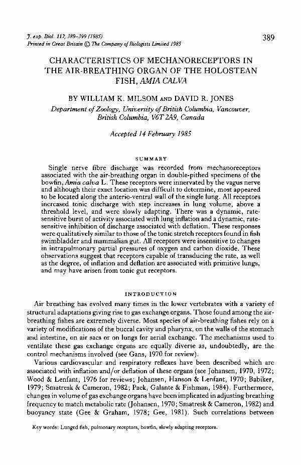
On leaving the cranial cavity, the vagus nerve on each side provides branches to theipsilateral gill arches as it courses along the top of the gill arches below the floor of thecardinal sinus. As it emerges into the body cavity, the vagus follows the intestinal veindown the anterior medial wall to the dorso-lateral side of the oesophagus. This is theramus intestinalis of the vagus which divides providing innervation to the lung andviscera (Fig. 1 A). In the present studies, the ramus intestinalis wa9 isolated on the leftside where it crosses from the posterior cardinal sinus to join the oesophagus (Fig.1A). The nerve was dissected free of surrounding tissue, placed on a dissectingplatform and desheathed. Small filaments were dissected from the distal end of a smallcut made in the nerve and single unit action potentials from afferent fibres wererecorded by conventional means using bipolar platinum electrodes. This activity wasamplified using a Framp (F. M. Smith, Vancouver, B. C , Canada) preamplifier(PRA-2) and amplifier (GPA-2), whose frequency bandwidth was set from 20 Hz to10 KHz. The amplifier output was further filtered with a 60 Hz notch filter. Both theunfiltered preamplifier output and filtered amplifier output were monitored with anoscilloscope and audio-amplifier. The filtered signal was also connected to a windowdiscriminator (Frederick Haer & Co.) and instantaneous rate meter (EKEG Electron-ics, Vancouver, B.C., Canada). The filtered and unfiltered signals and the windowdiscriminator output were stored on magnetic tape (Tandberg Series 115 Instru-mentation Tape Recorder) along with the intrapulmonary pressure signal. The out-put of the window discriminator, instantaneous rate meter and pressure transducerwere also recorded on a Beckman Type R Dynograph recorder.
Location of pulmonary receptors
Receptor discharge was attributed to pulmonary stretch receptors if the dischargewas modulated by artificial ventilation. No rapidly adapting receptors were observedin these experiments; all receptors were either slow or non-adapting during main-tained lung inflation. For further confirmation, following each experimental run, thelateral body wall of each fish was cut open, the lung exposed and the response of eachunit to punctate stimulation of the lung determined (Fig. 1C). All fibres which firedin synchrony with lung inflation responded to discrete local stimulation of the lung orglottal walls with a fine probe and were thus assumed to arise from pulmonary recep-tors.
Experimental protocol
While monitoring the discharge of each receptor, the bowfin were ventilated witha positive pressure respirator (Harvard Inc.) using mixtures of humidified air contain-ing 0, 5 or 10 % CO2, or 10 % CO2 in N2 at pump frequencies from 5 to 20 min"1 andtidal volumes of 10-25 ml. The pump was stopped to allow the lung to equilibrate toatmospheric pressure or hold inflation constant at various volumes. The lungs werealso inflated and deflated in a step-wise fashion using a syringe in place of the pump.The effect of changes in the rate of step inflation or deflation was also determinedusing a syringe.
A R
ecor
dmg
slte
R
-
2 cm
T
o gu
t and
vis
cera
O
ewph
agea
' op
enln
g
Fig
. 1.
(A) S
chem
atic
hag
ram
illu
stra
t~ng
the
cour
se of
the
vng
us n
erve
and
the
area
from
whi
ch r
ecor
d~ng
s wer
e m
ade.
(3) An
air-
drie
d lun
g of
Am
in in
crm
ae
ctio
n ill
ustr
atin
g in
tern
al a
epta
tion
and
regi
on o
f glo
ttal
ope
nlng
. (C
) D~s
char
ge of a
pul
mon
ary
stre
tch
rece
ptor
in r
espo
nse t
o pu
ncta
te st
imul
atio
n (m
m)
of t
he lu
ng.
Pulmonary mechanoreceptors in the bowfin 393
RESULTS
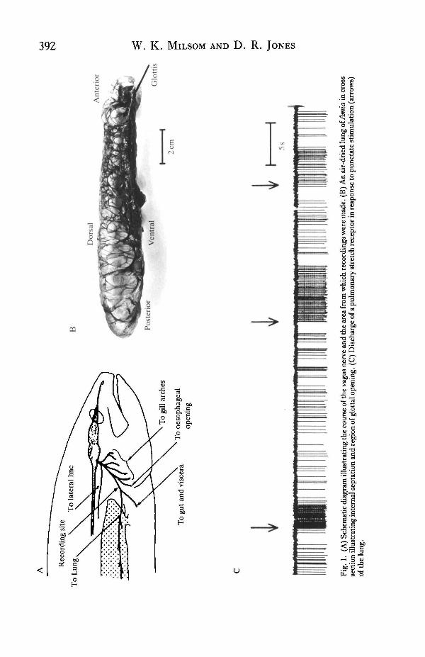
A total of 10 single-fibre preparations were obtained in this study from receptorslocated in the lung (Fig. IB). Their exact location, however, was difficult to deter-mine. One receptor was positively located in the glottal lips and another was locatedin the general area of the dorsal lung wall. The others were associated with the ventralwall of the lung along its anterior portion where it lies juxtaposed to the gut to whichit is joined by a connective tissue sheet. It was too difficult to dissect the lung free ofthe gut in this region without disturbing the preparation and therefore impossible todetermine whether the receptors lay in the outer lung wall or along one of the internalsepta (Fig. 1).
The volume threshold for discharge in these units was quite variable: several wereactive when the lungs were open to atmospheric pressure, others exhibited activityonly after the lungs had been inflated to varying degrees (5-25 ml). Consequently, theaverage discharge frequency at any given volume was also highly variable from fibreto fibre (25 to 300 impulsesmin"1 at 35 ml inflation volume, for instance). All fibres
Fibre 1 Fibre 2
25 ml
30 ml
35 ml
40 ml
45 ml
5s
Fig. 2. The effect of maintained lung inflation at various lung volumes on the discharge of two slowlyadapting pulmonary receptors.

394 W. K. MILSOM AND D. R. JONES
exhibited some adaptation to a step increase in volume over the range from 5 to 60 ml,but the rate of adaptation estimated from Index 1 of Davis, Fowler & Lambert (1956)was always less than 25 %. After adaptation had occurred, the steady discharge of thesefibres in response to maintained lung inflation at all volumes above threshold volume(or end-expiratory volume in those fibres active when the lung was open to theatmosphere) increased with increasing volume (Fig. 2). Neither the relationship be-tween receptor discharge and lung volume nor that between discharge and pulmonary
30
U
107
10 20 30 40
Volume (ml)
50
Ea.d.
50i B
40
30
20
10
50 i C
40
30
20
10
10 20 30 40
Volume (ml)
50 10 20
Pressure (cmH2O)
Fig. 3. The relationships between changes in transpulmonary pressure, inflation volume and pul-monary stretch receptor discharge during step inflation and deflation of the lung. (A) Plot of pulmon-ary pressure versus inflation volume. (B) Plot of discharge frequency of a single stretch receptorversus lung volume during the same inflation-deflation sequence, p. p.m., pulse per minute. (C) Plot ofdischarge frequency versus pulmonary pressure from the same receptor and during the same inflation-deflation sequence as in A and B. The time relationship of each plot is indicated by the arrows.
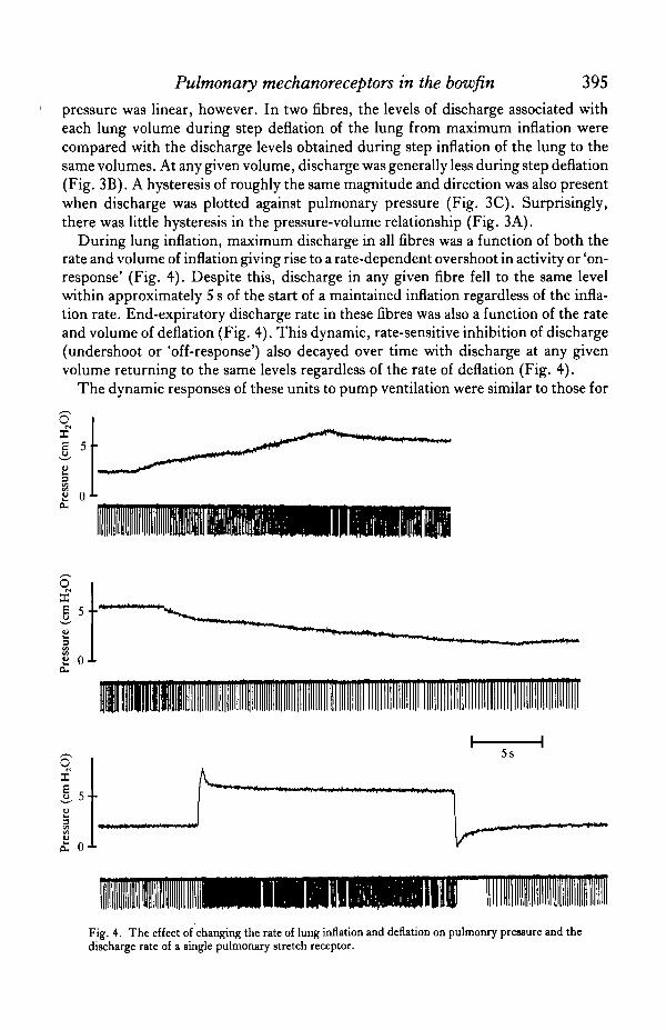
Pulmonary mechanoreceptors in the bowfin 395pressure was linear, however. In two fibres, the levels of discharge associated witheach lung volume during step deflation of the lung from maximum inflation werecompared with the discharge levels obtained during step inflation of the lung to thesame volumes. At any given volume, discharge was generally less during step deflation(Fig. 3B). A hysteresis of roughly the same magnitude and direction was also presentwhen discharge was plotted against pulmonary pressure (Fig. 3C). Surprisingly,there was little hysteresis in the pressure-volume relationship (Fig. 3A).
During lung inflation, maximum discharge in all fibres was a function of both therate and volume of inflation giving rise to a rate-dependent overshoot in activity or 'on-response' (Fig. 4). Despite this, discharge in any given fibre fell to the same levelwithin approximately 5 s of the start of a maintained inflation regardless of the infla-tion rate. End-expiratory discharge rate in these fibres was also a function of the rateand volume of deflation (Fig. 4). This dynamic, rate-sensitive inhibition of discharge(undershoot or 'off-response') also decayed over time with discharge at any givenvolume returning to the same levels regardless of the rate of deflation (Fig. 4).
The dynamic responses of these units to pump ventilation were similar to those for
oI
I ox
oX
5--
io- 0 •L
5s
Fig. 4. The effect of changing the rate of lung inflation and deflation on pulmonry pressure and thedischarge rate of a single pulmonary stretch receptor.
396 W. K. MILSOM AND D. R. JONES
oX 5 TE
15 ml 25 ml
o-1-
5s
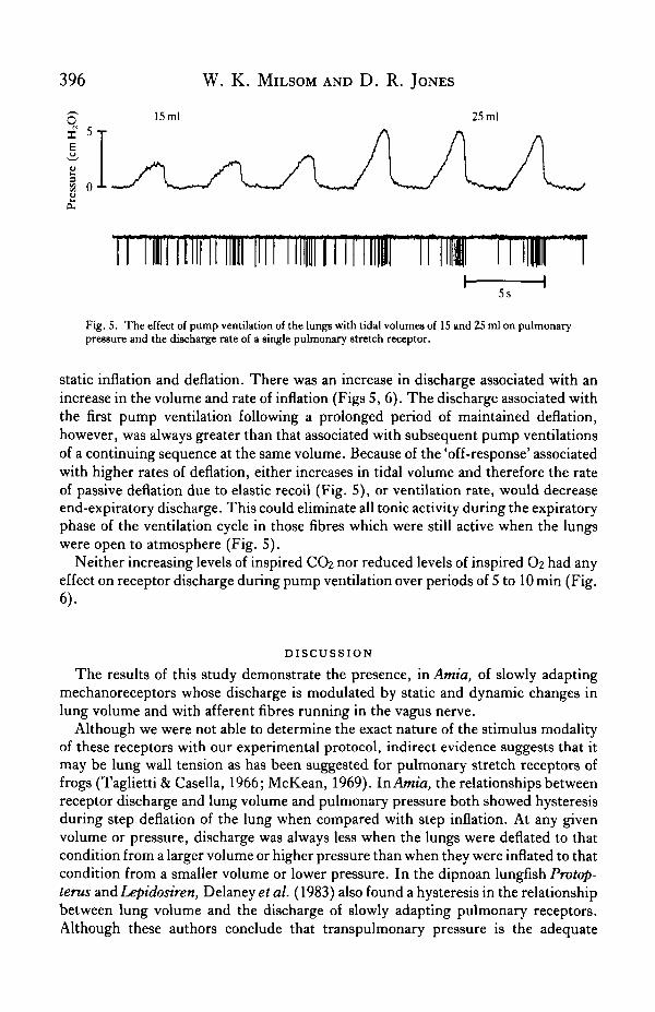
Fig. 5. The effect of pump ventilation of the lungs with tidal volumes of 15 and 25 ml on pulmonarypressure and the discharge rate of a single pulmonary stretch receptor.
static inflation and deflation. There was an increase in discharge associated with anincrease in the volume and rate of inflation (Figs 5,6). The discharge associated withthe first pump ventilation following a prolonged period of maintained deflation,however, was always greater than that associated with subsequent pump ventilationsof a continuing sequence at the same volume. Because of the 'off-response' associatedwith higher rates of deflation, either increases in tidal volume and therefore the rateof passive deflation due to elastic recoil (Fig. 5), or ventilation rate, would decreaseend-expiratory discharge. This could eliminate all tonic activity during the expiratoryphase of the ventilation cycle in those fibres which were still active when the lungswere open to atmosphere (Fig. 5).
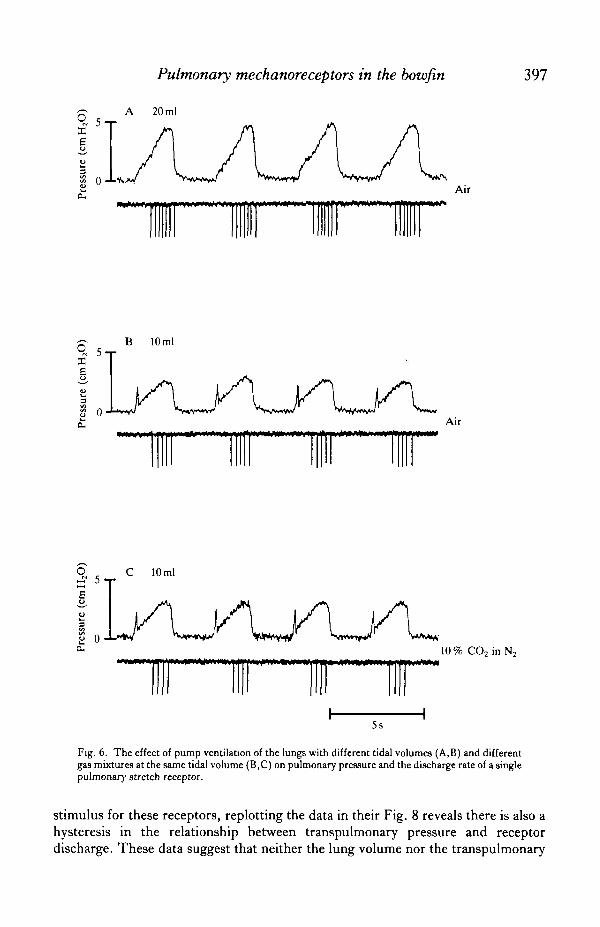
Neither increasing levels of inspired CO2 nor reduced levels of inspired O2 had anyeffect on receptor discharge during pump ventilation over periods of 5 to 10 min (Fig.6).
DISCUSSION
The results of this study demonstrate the presence, in Amia, of slowly adaptingmechanoreceptors whose discharge is modulated by static and dynamic changes inlung volume and with afferent fibres running in the vagus nerve.
Although we were not able to determine the exact nature of the stimulus modalityof these receptors with our experimental protocol, indirect evidence suggests that itmay be lung wall tension as has been suggested for pulmonary stretch receptors offrogs (Taglietti & Casella, 1966; McKean, 1969). In Amia, the relationships betweenreceptor discharge and lung volume and pulmonary pressure both showed hysteresisduring step deflation of the lung when compared with step inflation. At any givenvolume or pressure, discharge was always less when the lungs were deflated to thatcondition from a larger volume or higher pressure than when they were inflated to thatcondition from a smaller volume or lower pressure. In the dipnoan lungfish Protop-terus andLepidosiren, Delaney et al. (1983) also found a hysteresis in the relationshipbetween lung volume and the discharge of slowly adapting pulmonary receptors.Although these authors conclude that transpulmonary pressure is the adequate
Pulmonary mechanoreceptors in the bowfin 397
qxuu3
U
£
A 20 ml
Air
O c _(S J ""*•
B 10 ml
Air
5-r10 ml
I o -L-w ^*^u/ V***i*vV̂ w i >*̂ A-10% CO2in N2
5s
Fig. 6. The effect of pump ventilation of the lungs with different tidal volumes (A,B) and differentga9 mixtures at the same tidal volume (B,C) on pulmonary pressure and the discharge rate of a singlepulmonary stretch receptor.
stimulus for these receptors, replotting the data in their Fig. 8 reveals there is also ahysteresis in the relationship between transpulmonary pressure and receptordischarge. These data suggest that neither the lung volume nor the transpulmonary
398 W. K. MILSOM AND D. R. JONES
pressure can be the adequate stimulus for the slowly adapting pulmonary stretchreceptors in Amia or the dipnoan lungfish. It remains possible, therefore, that lungwall tension, which is a function of lung volume, pulmonary pressure and lung surfacearea (Taglietti & Casella, 1966; McKean, 1969), is the stimulus modality for pulmon-ary receptors in the holosteans and dipnoans as it is in the amphibia.
A major difference between the characteristics of the receptors in the Amia lung andthose in other lower vertebrate lungs is the apparent lack of any inhibitory effect ofCO2 on the discharge of receptors in the Amia lung. Although the effect of CO2 ondischarge of pulmonary receptors in lungfish, amphibia and reptiles was highly vari-able from fibre to fibre (see Jones & Milsom, 1982 for review) and the apparent lackof effect in Amia could simply stem from the small sample size rather than from anyreal functional difference, we do not feel this is the case. The consistent absence ofany effect in all fibres examined is strong evidence that CO2 does not have an inhibit-ory effect on these receptors. Another difference between the pulmonary stretchreceptors oiAmia and those of other vertebrates would appear to be the rate-sensitiveinhibition of discharge associated with lung deflation. This is a characteristic thesereceptors share in common with slowly adapting receptors in the swim bladder of thecyprinid fishes Leuciscus and Scardinius (Qutob, 1962) as well as with mostvertebrate tonic receptors such as those associated with photoreceptors, carotid sinusbaroreceptors and muscle and gut stretch receptors (Patton, 1965). Given the originsof the swimbladder and lung as outpocketings of the gut (Romer, 1970), this is nottoo surprising. The off-effect seen in tonic receptor discharge has a correlate in ahyperpolarizing off-effect in the stretch receptor generator potential. To what extentthis hyperpolarization reflects changes in the capacitance properties of the receptorending itself or changes in non-neural elements around the receptor ending, is unclear(Patton, 1965). Thus, whether the absence of such a response in the pulmonarystretch receptors of other vertebrates is a true difference in the receptors themselvesor simply a difference in the mechanical linkage between the receptors and the lungor tracheal walls remains an interesting question.
During pump ventilation of the Amia lung, the dynamic (phasic) characteristics ofreceptor discharge were predominant in establishing overall levels of receptor activity.Peak discharge rates during lung inflation were strongly influenced by the rate ofinflation and end-expiratory discharge rates were more a function of deflation ratethan lung volume. In spontaneously breathing Amia, however, lung ventilation isusually a solitary event beginning with an active expiration followed by inspirationand breath holding (Johansen et al. 1970). In this context, if these receptors arefunctionally equivalent to the pulmonary stretch receptors of higher vertebrates, it isquite possible that their dynamic discharge characteristics play an important role inestablishing the timing of the events associated with ventilation. It is more likely,however, that the cardio-respiratory and buoyancy-related reflexes associated withchanges in lung volume during the period of breath holding (Johansen, 1970, 1972;Johansen et al. 1970; Wood & Lenfant, 1976) are a consequence of changes in thetonic level of receptor discharge, although such speculation must remain highly con-jectural until a more direct link is established between these receptors and any func-tional events.

Pulmonary mechanoreceptors in the bowfin 399
We are grateful to Drs D. J. Randall, C. Daxboeck and G. Iwama for assistance inobtaining the Amia. This work was supported by operating grants from the NSERCof Canada.
R E F E R E N C E S
BABIKER, M. M. (1979). Respiratory behaviour, oxygen consumption and relative dependence on aerial respira-tion in the African lungfish (Pmtopterus annecteus, Owen) and an air-breathing teleost (Clarias lazera, C ) .Hydrobiologia 65, 177-187.
DAVIS, H. L., FOWLER, W. S. & LAMBERT, E. H. (1956). Effect of volume and rate of inflation and deflationon transpulmonary pressure and response of pulmonary stretch receptors. Am.J. Physiol. 187, 558-566.
DELANEY, R. G., LAURENT, P., GALANTE, R., PACK, A. I. & FISHMAN, A. P. (1983). Pulmonary
mechanoreceptors in the dipnoi lungfish Pmtopterus and Lepidosiren. Am. J. Physiol. 244, R418-R428.GANS, C. (1970). Strategy and sequence in the evolution of the external gas exchangers of ectothermal
vertebrates. Forma et Functio 3, 61-104.GEE, J. H. (1981). Coordination of respiratory and hydrostatic functions of the swimbladder in the central
mudminnow, Umbra limi. J. exp. Biol. 92, 37—52.GEE, J. H. & GRAHAM, J. B. (1978). Respiratory and hydrostatic functions of the intestine of the catfishes
Hopbsternum thoracatum and Brochis splendens, (Callichthyidae). J. exp. Biol. 74, 1-16.JOHANSEN, K. (1970). Air breathing in fishes. In Fish Physiology, Vol. IV, (edsW. S. Hoar&D. J. Randall),
pp. 361—411. New York: Academic Press.JOHANSEN, K. (1972). Heart and circulation in gill, skin and lung breathing. Respir, Physiol. 14, 193-210.JOHANSEN, K., HANSON, D. & LENFANT, C. (1970). Respiration in a primitive airbreather, Amia calva.
Respir. Physiol. 9, 162-174.JONES, D. R. & MILSOM, W. K. (1982). Peripheral receptors affecting breathing and cardiovascular function
in non-mammalian vertebrates. J. exp. Biol. 100, 59-91.MCKEAN, T. A. (1969). A linear approximation of the transfer function of pulmonary mechanoreceptors of the
frog. J. appl. Physiol. 27, 775-781.MILSOM, W. K. & JONES, D. R. (1977). Carbon dioxide sensitivity of pulmonary receptors in the frog.
Experiential, 1167-1168.PACK, A. I., GALANTE, R. J. & FISHMAN, A. P. (1984). Breuer-Hering reflexes in the African lungfish
(Pmtopterus annecteus). Fedn Proc. Fedn Am. Socs exp. Biol. 43, 433.PATTON, H. D. (1965). Receptor mechanism. In Neurophysiology, (eds T. C. Ruch, H. D. Patton, J. W.
Woodbury k A. L. Towe), pp. 95-112. London: W. B. Saunders Co.QUTOB, Z. (1962). The swimbladder of fishes as a pressure receptor. Arch, neerl. Zool. 15, 1-67.ROMER, A. S. (1970). The Vertebrate Body (4th ed.). London: W. B. Saunders Co.SMATRESK, N. J. & CAMERON , J. N. (1982). Respiration and acid-base physiology of the spotted gar, a bimodal
breather. I l l . Response to a transfer from fresh water to 50 % sea water, and control of breathing. J . exp. Biol.96, 295-306.
TAGUETTI , V. &CASELLA, C. (1966). Stretch receptor stimulation in frog lungs./y?«gerjj4rcA.gcj. Physiol. 292,297-308.
WOOD, S. C. & LENFANT, C. J. M. (1976). Physiology of fish lungs. In Respiration of Amphibious Vertebrates,(ed. G. M. Hughes), pp. 257-270. New York: Academic Press.