Embed Size (px)
Citation preview
CHARACTERIZATION OF MAJOR VIRULENCE REGULATORS
OF ERWINIA AMYLOVORA
BY
WENTING LI
THESIS
Submitted in partial fulfillment of the requirements
for the degree of Master of Science in Crop Sciences
in the Graduate College of the
University of Illinois at Urbana-Champaign, 2012
Urbana, Illinois
Adviser:
Associate Professor Frank (Youfu) Zhao
ii
ABSTRACT
Erwinia amylovora is the causal agent of the devastating fire blight disease which is a
major concern to the apple and pear industry. Fire blight costs millions of dollars of economic
losses all over the world. Exopolysaccharide (EPS) amylovoran and type III secretion system
(T3SS) are two major virulence factors in E. amylovora. However, how these virulence factors
are regulated is not completely understood. In bacteria, gene expression is mainly regulated at
the transcription initiation level and its core RNA polymerase (RNAP) requires sigma factors for
promoter recognition and initiation. In this study, we investigated the role of several sigma
factors in regulating virulence gene expression in E.amylovora. Early studies have shown that
hrp-type III secretion (T3SS) in E. amylovora is regulated by HrpS, a member of the σ54
enhancer binding proteins, and the master regulator HrpL, which belongs to the ECF subfamily
of σ factors. Other sigma factors characterized included RpoN, a nitrogen limitation σ54
factor,
and its modulation protein YhbH. Our results showed that mutations in hrpS, hrpL, rpoN and
yhbH resulted in nonpathogenic phenotype in host plant and no hypersensitive response in non-
host tobacco. Consistently, expression of T3SS genes including hrpL, dspE, hrpN and hrpA was
barely detected in hrpS, hrpL, rpoN and yhbH mutants. Amylovoran (EPS) production was
higher in these mutants than that of wild type (WT) strain, indicating sigma factors may also play
roles in regulating exopolysaccharide production. These results suggest that sigma factors in E.
amylovora are important virulence regulators and sigma factor cascade exists in its regulatory
networks.
Two-component signal transduction systems (TCSTs) in E. amylovora play a major role
in virulence and in regulating amylovoran production, including EnvZ/OmpR and GrrS/GrrA,
two widely distributed systems in gamma-proteobacteria. While both systems negatively control
amylovoran biosynthesis, deletion mutants of envZ/ompR and grrA/grrS have opposite swarming
motility phenotypes. In order to determine how the two systems interact, two triple mutants,
envZ/ompR/grrA (ERA) and envZ/ompR/grrS (ERS) were generated. Our results showed that both
triple mutants had slightly increased virulence on apple shoots as compared to that of wild type
(WT) as well as mutants deleting a single system. In an in vitro amylovoran assay, amylovoran
production was significantly increased in the two triple mutants, indicating the two systems
synergistically regulate amylovoran production. In consistent with amylovoran production, amsG

iii
gene expression was expressed significantly higher in the triple mutants in vitro than those in
WT as well as mutants deleting a single system. In contrast, exopolysaccharide levan was
significantly reduced in the triple mutants compared with that of WT and deletion of a single
system. In addition, the triple mutants showed reduced swarming motility on swarming plates
compared to that of grrA/grrS mutants and WT strain, but moved slightly faster than that of
envZ/ompR mutants, indicating that the two systems antagonistically regulate swarming motility
in E. amylovora. Furthermore, type III secretion (T3SS) genes were significantly upregulated in
the triple mutants as well as deletion of a single system than that of the WT strain. These results
indicate that EnvZ/OmpR and GrrS/GrrA systems play major roles in virulence and in regulating
virulence gene expression.

iv
ACKNOWLEDGMENTS
This project would not have been possible without the support of many people. First of
all, I wish to express my sincere gratitude to my advisor, Dr. Frank (Youfu) Zhao, who was
extremely helpful and offered invaluable guidance during the past two and half years as well as
his extensive editing of this thesis. His abundant knowledge and creativity are precious for my
career training. I would like to thank my committee members, Dr. Lila Vodkin and Dr. Schuyler
Korban, for their knowledge and helpful feedback on the project. I am also grateful to the
support I got from Dr. Jack Widholm and Dr. Schuyler Korban’s lab at the University of Illinois
at Urbana-Champaign for their support. Sincere thanks to all my colleagues, Dr. Dongping Wang,
Dr. Mingsheng Qi, Mr. Fan Yang, Dr. Veronica Anconca and Mr. Jae Hoon Lee for their
assistance and friendship. I would like to convey my thanks to Dr. Larry Pusey at USDA-ARS
for providing immature apple fruits. I am also grateful to my friends that we had a great time
during the last two and half years. Last but not the least, I own my appreciation to my dear
parents and their love across the Pacific. I could not finish my study without their support.
v
TABLE OF CONTENTS
LIST OF FIGURES ................................................................................ vii
LIST OF TABLES .................................................................................. ix
Chapter 1: Literature Review ................................................................ 1
1.1 Fire blight disease and symptoms ................................................................................. 1
1.2 Erwinia amylovora and virulence factors ..................................................................... 1
1.3 Exopolysaccharide .......................................................................................................... 3
1.4 Type III secretion system ............................................................................................... 4
1.5 Sigma factor .................................................................................................................... 8
1.6 Two-component signal transduction systems (TCSTs) ............................................ 11
1.7 EnvZ/OmpR and GrrS/GrrA system ......................................................................... 12
Chapter 2: Effect of Sigma Factor RpoN and Its Modulation
Protein on Erwinia amylovora Virulence ............................................. 17
2.1 Abstract ......................................................................................................................... 17
2.2 Introduction .................................................................................................................. 17
2.3 Materials and methods................................................................................................. 20
2.4 Results ........................................................................................................................... 28
2.5 Discussion ...................................................................................................................... 39
Chapter 3: Effect of EnvZ/OmpR and GrrS/GrrA Systems on
Erwinia amylovora Virulence………………………………………....42
3.1 Abstract ......................................................................................................................... 42
3.2 Introduction .................................................................................................................. 42
3.3 Materials and methods................................................................................................. 46

vi
3.4 Results ........................................................................................................................... 54
3.5 Discussion ...................................................................................................................... 65
References ............................................................................................... 67
vii
LIST OF FIGURES
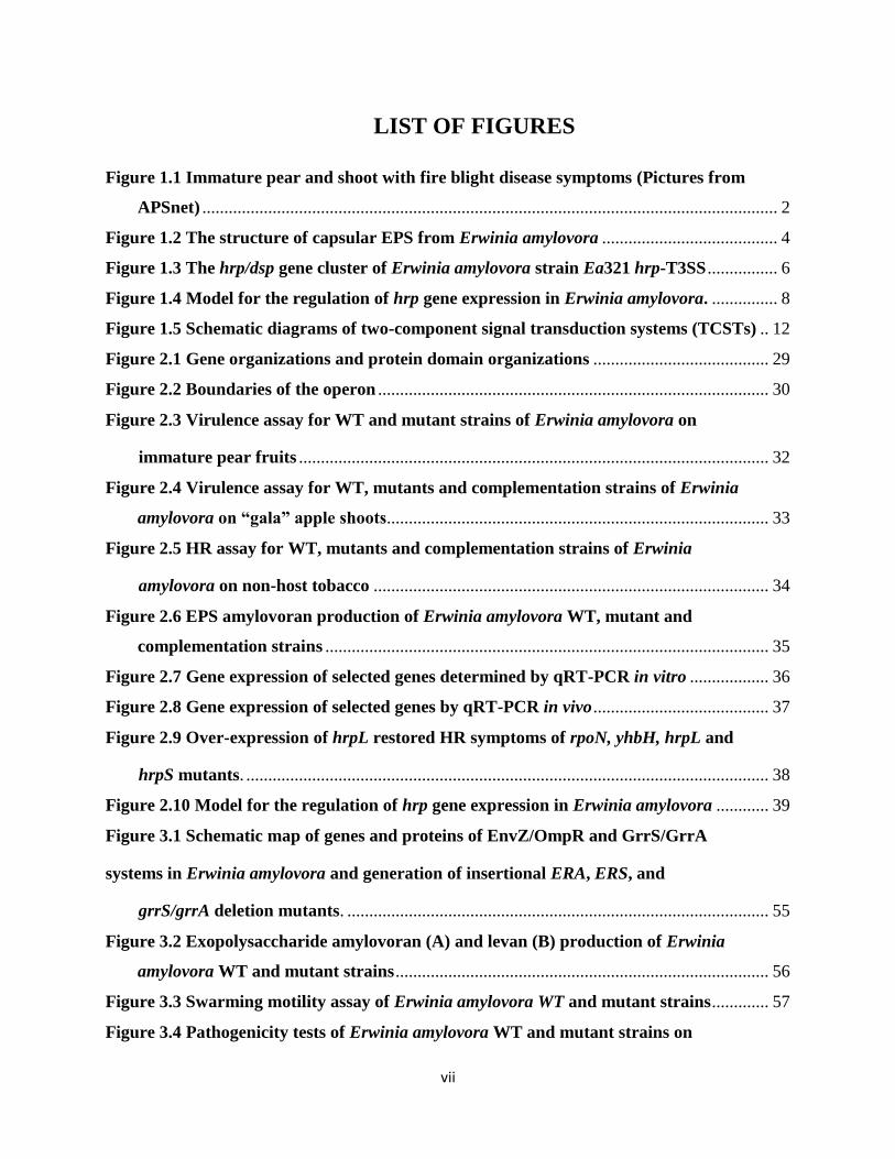
Figure 1.1 Immature pear and shoot with fire blight disease symptoms (Pictures from
APSnet) ................................................................................................................................... 2
Figure 1.2 The structure of capsular EPS from Erwinia amylovora ........................................ 4
Figure 1.3 The hrp/dsp gene cluster of Erwinia amylovora strain Ea321 hrp-T3SS ................ 6
Figure 1.4 Model for the regulation of hrp gene expression in Erwinia amylovora. ............... 8
Figure 1.5 Schematic diagrams of two-component signal transduction systems (TCSTs) .. 12
Figure 2.1 Gene organizations and protein domain organizations ........................................ 29
Figure 2.2 Boundaries of the operon ......................................................................................... 30
Figure 2.3 Virulence assay for WT and mutant strains of Erwinia amylovora on
immature pear fruits ........................................................................................................... 32
Figure 2.4 Virulence assay for WT, mutants and complementation strains of Erwinia
amylovora on “gala” apple shoots....................................................................................... 33
Figure 2.5 HR assay for WT, mutants and complementation strains of Erwinia
amylovora on non-host tobacco .......................................................................................... 34
Figure 2.6 EPS amylovoran production of Erwinia amylovora WT, mutant and
complementation strains ..................................................................................................... 35
Figure 2.7 Gene expression of selected genes determined by qRT-PCR in vitro .................. 36
Figure 2.8 Gene expression of selected genes by qRT-PCR in vivo ........................................ 37
Figure 2.9 Over-expression of hrpL restored HR symptoms of rpoN, yhbH, hrpL and
hrpS mutants. ....................................................................................................................... 38
Figure 2.10 Model for the regulation of hrp gene expression in Erwinia amylovora ............ 39
Figure 3.1 Schematic map of genes and proteins of EnvZ/OmpR and GrrS/GrrA
systems in Erwinia amylovora and generation of insertional ERA, ERS, and
grrS/grrA deletion mutants. ................................................................................................ 55
Figure 3.2 Exopolysaccharide amylovoran (A) and levan (B) production of Erwinia
amylovora WT and mutant strains ..................................................................................... 56
Figure 3.3 Swarming motility assay of Erwinia amylovora WT and mutant strains ............. 57
Figure 3.4 Pathogenicity tests of Erwinia amylovora WT and mutant strains on

viii
immature pears .................................................................................................................... 59
Figure 3.5 Virulence assay for Erwinia amylovora WT and mutant strains ......................... 60
Figure 3.6 Gene expression of selected genes determined by qRT-PCR in vitro .................. 63
Figure 3.7 Gene expression of selected genes determined by qRT-PCR in vivo ................... 65
ix
LIST OF TABLES
Table 2.1: Bacterial strains and plasmids used in this study .................................................. 21
Table 2.2: Primers used in this study ........................................................................................ 22
Table 3.1: Bacterial strains and plasmids used in this study .................................................. 47
Table 3.2: Primers used in this study ........................................................................................ 49
Table 3.3: Promoter activities of amsG, dspE, hrpL, hrpN, ysaE1, ysaE2, prgH1 and
prgH2 genes in Erwinia amylovora WT and mutant strains.. .......................................... 61
1
Chapter 1
Literature Review
1.1 Fire blight disease and symptoms
Fire blight is a devastating disease of Rosaceae family plants including many economic
important fruit trees, such as apple (Malus sylvestris) and pear (Pyrus communis L.). Fire blight
is also the first plant disease attributed to bacterium. The disease is native to North America and
the first report of fire blight as a disease of apple and pear occurred in 1780 in the Hudson Valley
of New York. Since then, it has spread into every region of the U.S.A. Continually, it was found
in some other countries, such as England and New Zealand. By 1990, fire blight was widespread
in North America, west Pacific region, Europe and in the Mediterranean area (Bonn & van Zwet,
2000).
The term "fire blight" describes the typical symptoms of this disease as the affected areas
turned into black, shrunken and cracked just like scorched by fire. The bacteria are dormant in
winter in the infected plant tissues. Open blossoms, tender new shoots, and leaves are the
primary infection sites in the spring. Injured tissues are also highly susceptible to infection,
including punctures and tears caused by plant-sucking or biting insects. Natural openings, like
open stamata, are the entrance sites to cause blackened necrosis, produce viscous exudates and
spread throughout the host via vascular system. In some cases, creamy white or yellow ooze
droplets containing bacteria and exopolysaccharide are formed at the infected site, which can
serve as the source of secondary infection distributed by rain, birds or insects. Abundant moist
and heat contribute to disease epidemic: under optimal conditions, this devastating disease can
destroy an entire orchard in a single growing season. Over-pruning and over-fertilization can
lead to water sprouts and other mid-summer growths that render the tree more susceptible to
disease.
1.2 Erwinia amylovora and virulence factors
Erwinia amylovora, a highly virulent, necrogenic, vascular pathogen, is the causal agent
of disease fire blight. It is a Gram-negative, 0.5-1.0 x 3.0 µm in size, facultative anaerobic, rod
shaped bacterium with peritrichous flagella. As a member of the Enterobacteriaceae, it is closely
2
related to Salmonella entica, E. coli and Yersinia pestis. Two pathogenicity (virulence) factors
are strictly required for E. amylovora to cause disease: the exopolysaccharide amylovoran and
the type III secretion system.
Figure 1.1 Immature pear and shoot with fire blight disease symptoms (Pictures from
APSnet)
Recent development of molecular techniques allows the comparison of genome
organization of different strains. Great homogeneity was found within Erwinia species. Genomic
comparison of two E. amylovora strains, CFBP1430 (isolated from Crataegus in France) and
ATCC 49946 (referred as Ea273, isolated from apple in New York), revealed a large-scale
chromosomal rearrangement, although they shared more than 99.99% identity at the nucleotide
level (Smits et al., 2010; Zhao and Qi, 2011).
Genome sequences of some closely related Erwinia species were recently reported and
comparative analysis was conducted with genome sequences of E. amylovora, Erwinia pyrifoliae
(isolated from shoot blight in South Korea and Japan), and Erwinia tasmaniensis (saprophyte,
isolated from trees in UK) (Smits et al., 2011). The presence of several virulence factors, such as
T3SS PAI-3, levansucrase, protease A and some effectors, may be responsible for their variance
in host range and virulence (Zhao and Qi, 2011). Genetic analysis also revealed that horizontal
gene transfer may account for these differential features between the three species (Smits et al.,
2011).
3
1.3 Exopolysaccharide
Exopolysaccharides (EPS) are high-molecular-weight polymers that are composed of
sugar residues and are secreted by microorganisms to attach to the cell wall, or secreted into
growth medium. EPS is important in biofilm formation and cell attachment to surfaces (Donlan
& Costerton, 2002) and is barely immunogenic, which allows pathogens to elude host
recognition and escape host defense.
1.3.1 Amylovoran
Like many plant-pathogenic bacteria, E. amylovora produces large amount of acidic
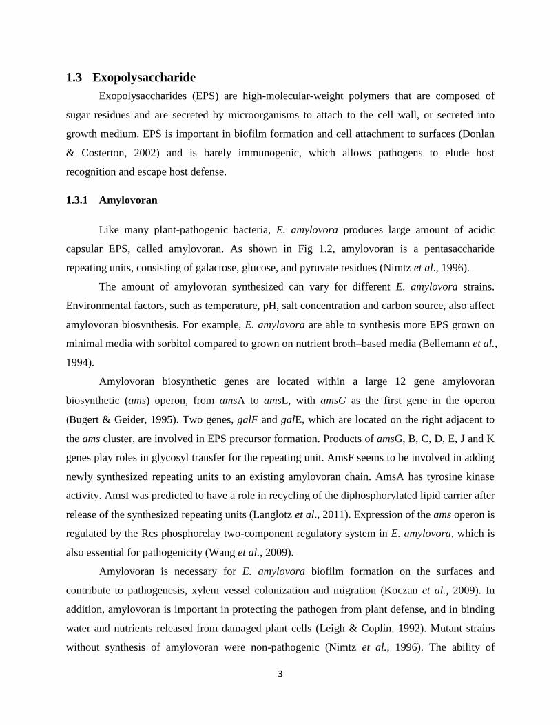
capsular EPS, called amylovoran. As shown in Fig 1.2, amylovoran is a pentasaccharide
repeating units, consisting of galactose, glucose, and pyruvate residues (Nimtz et al., 1996).
The amount of amylovoran synthesized can vary for different E. amylovora strains.
Environmental factors, such as temperature, pH, salt concentration and carbon source, also affect
amylovoran biosynthesis. For example, E. amylovora are able to synthesis more EPS grown on
minimal media with sorbitol compared to grown on nutrient broth–based media (Bellemann et al.,
1994).
Amylovoran biosynthetic genes are located within a large 12 gene amylovoran
biosynthetic (ams) operon, from amsA to amsL, with amsG as the first gene in the operon
(Bugert & Geider, 1995). Two genes, galF and galE, which are located on the right adjacent to
the ams cluster, are involved in EPS precursor formation. Products of amsG, B, C, D, E, J and K
genes play roles in glycosyl transfer for the repeating unit. AmsF seems to be involved in adding
newly synthesized repeating units to an existing amylovoran chain. AmsA has tyrosine kinase
activity. AmsI was predicted to have a role in recycling of the diphosphorylated lipid carrier after
release of the synthesized repeating units (Langlotz et al., 2011). Expression of the ams operon is
regulated by the Rcs phosphorelay two-component regulatory system in E. amylovora, which is
also essential for pathogenicity (Wang et al., 2009).
Amylovoran is necessary for E. amylovora biofilm formation on the surfaces and
contribute to pathogenesis, xylem vessel colonization and migration (Koczan et al., 2009). In
addition, amylovoran is important in protecting the pathogen from plant defense, and in binding
water and nutrients released from damaged plant cells (Leigh & Coplin, 1992). Mutant strains
without synthesis of amylovoran were non-pathogenic (Nimtz et al., 1996). The ability of
4
individual E. amylovora strains to produce amylovoran is positively correlated with the degree of
virulence (Ayers et al., 1979).
Figure 1.2 The structure of capsular EPS from Erwinia amylovora. Glycosyl residues and
their linkages in the repeating units of amylovoran are shown. The number 0.5 refers to the
glucose residue added to half of the repeating units (Langlotz et al., 2011).
1.3.2 Levan
E. amylovora also produces a minor EPS component, levan. Levan is a homo-polymer of
fructose residues, the major storage and transport carbohydrates in Rosaceae family (Chong &
Tapper, 1971). Levan production is controlled by the lsc gene, encoding the levansucrase
enzyme which is used by E. amylovora to cleave sucrose to fructose, then polymerized into levan
(Geier & Geider, 1993).
Levan plays a role in E. amylovora biofilm formation because a levansucrase-deficient
mutant was reduced in biofilm formation and in cell-to-cell aggregation in vitro (Koczan et al.,
2009). In addition, secretion of levansucrase is thought to contribute to colonization of sucrose-
containing tissue by E. amylovora (Geier & Geider, 1993) .
1.4 Type III secretion system
Type III secretion system (T3SS) is a protein appendage found in many Gram-negative
bacteria, such as Salmonella, Burkholderia, Yesinia, Pseudomonas, Erwinia, Ralstonia,
5
Rhizobium, Vibro and Xanthomonas. It is a dedicated mechanism used by bacteria to deliver
proteins to cytosol of host cells or apoplast (Galan & Wolf-Watz, 2006; He et al., 2004). In
pathogenic bacteria, this needle-like structure is utilized to detect the presence
of eukaryotic organisms and secret a variety of effectors across the plant cell wall and plasma
membrane in order to assist infection, cause disease symptoms in host plant or elicit
hypersensitive response (HR) in non-host plant.
T3SS is one of the most complex secretion systems, which composed of about 30
different proteins (Gophna et al., 2003). A high degree of sequence similarity is observed
between T3SS proteins and flagellar proteins (Blocker et al., 2003). The genome sequence
reveals three type III secretion systems in E. amylovora, including the pathogenicity island 1
(PAI-1) encoded hypersensitive response and pathogenicity T3SS (hrp-T3SS), and two inv/spa-
like non-flagellarT3SS islands (PAI-2 and PAI-3) (Zhao et al., 2009a). Hrp-T3SS has been
known for its role as a pathogenicity factor that functions to deliver effectors into eukaryotic host
(He et al., 2004). PAI-2 and PAI-3 are similar to SPI1 T3SS of Salmonella typhimurium LT-2
and inv/spa T3SS of the insect endosymbiont Sodalis glossidinius str. morsitans, respectively.
However, their functions are still unknown.
1.4.1 T3SS-mediated infection
T3SS is encoded by the hrp (hypersensitive response and pathogenicity) genes and hrc
(hrp-conserved) genes among plant pathogenic bacteria (Cornelis & Van, 2000). T3SS proteins
can be categorized into three groups, regulatory proteins (e.g. HrpL), secreting proteins
(structural components, e.g. HrpA) and secreted proteins (effectors, e.g. HrpN).
T3SS effectors enter into the base of T3SS apparatus and move inside the needle towards
the host cell. The detailed mechanism for the entrance of effectors into host cells is still not clear.
It is possible that translocators (a set of effectors) are secreted first and form a pore (translocon)
in the host cell membrane. Subsequently, other effectors enter into host cells through this
translocation pore (He et al., 2004). Mutation in translocator genes didn’t affect the secretion of
translocator proteins, however, suppressed their ability to deliver them into host cells.
Manipulation of host cells by T3SS effectors can be found in several ways: promoting uptake of
the bacterium by host cell, tampering with host's cell cycle, inducing apoptosis, or acting as
transcription activators.
6
In E. amylovora, hrc and hrp genes are located in a pathogenicity island (PAI-1) (Oh &
Beer, 2005), which also includes dsp (disease-specific) genes. Harpin-like proteins (a subset of
T3SS substrates) which have the unusual ability to elicit HR, have been reported in all genera of
phytopathogens (Ahmad et al., 2001; Alfano and Collmer, 1997). For example, E. amylovora
and Pseudomonas syringae were previously shown to produce two harpins, i.e. HrpN/HrpW and
HrpZ/HrpW, respectively (Kim & Beer, 1998).
1.4.2 Regulation of T3SS in Erwinia amylovora
In general, T3SS is required by plant pathogenic bacteria for the translocation of certain
bacterial proteins to the cytoplasm of plant cell, or secretion of some proteins to the apoplast
(Charkowski et al., 1998).
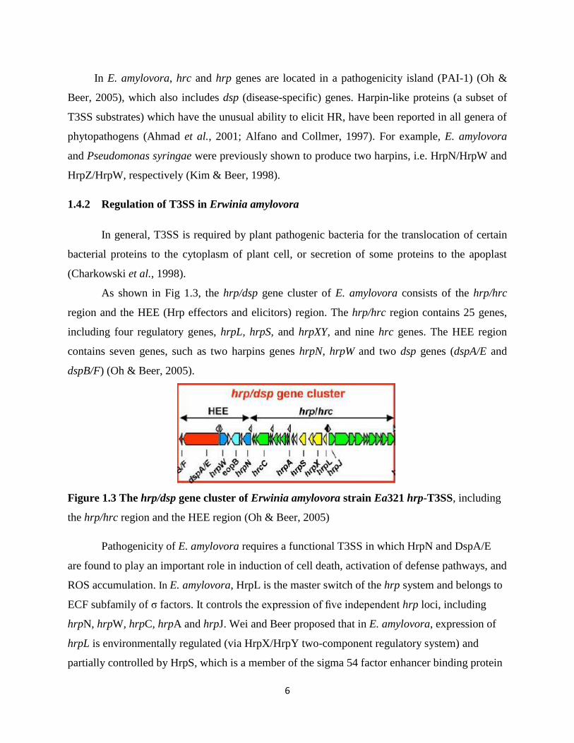
As shown in Fig 1.3, the hrp/dsp gene cluster of E. amylovora consists of the hrp/hrc
region and the HEE (Hrp effectors and elicitors) region. The hrp/hrc region contains 25 genes,
including four regulatory genes, hrpL, hrpS, and hrpXY, and nine hrc genes. The HEE region
contains seven genes, such as two harpins genes hrpN, hrpW and two dsp genes (dspA/E and
dspB/F) (Oh & Beer, 2005).
Figure 1.3 The hrp/dsp gene cluster of Erwinia amylovora strain Ea321 hrp-T3SS, including
the hrp/hrc region and the HEE region (Oh & Beer, 2005)
Pathogenicity of E. amylovora requires a functional T3SS in which HrpN and DspA/E
are found to play an important role in induction of cell death, activation of defense pathways, and
ROS accumulation. In E. amylovora, HrpL is the master switch of the hrp system and belongs to
ECF subfamily of σ factors. It controls the expression of five independent hrp loci, including
hrpN, hrpW, hrpC, hrpA and hrpJ. Wei and Beer proposed that in E. amylovora, expression of
hrpL is environmentally regulated (via HrpX/HrpY two-component regulatory system) and
partially controlled by HrpS, which is a member of the sigma 54 factor enhancer binding protein
7
(Wei & Beer, 1995). However, there is no report as how these factors are involved in regulation
of T3SS gene expression.
In general, hrp genes are activated in planta, but repressed in rich media. In order to
induce T3SS gene expression, minimal media that mimic apoplast conditions is required
(Lindgren, 1997), but the conditions can vary considerably between species. For example, P.
syringae hrp genes are induced at low pH in minimal media, but are nonspecifically repressed by
high salt concentrations (Rahme et al., 1992). In contrast, hrp genes in E. amylovora are
stimulated likewise by low pH and repressed by glucose, ammonium salts, asparagine, histidine,
and nicotinic acid, however, unaffected by osmolarity (Wei et al., 1992).

8
Figure 1.4 Model for the regulation of hrp gene expression in Erwinia amylovora. Thick
arrow lines indicate genes or operons, ovals and circles indicate proteins and arrowheads in
thinner lines indicate the directions of information flow. CM, cytoplasmic membrane; OM, outer
membrane; P, phosphate; E, RNA polymerase; closed half-circle, σ70
promoter; open triangle,
σ54
promoter; filled triangle, HrpL promoter (Wei & Beer, 1995).
1.5 Sigma factor
In bacteria, gene expression is mainly regulated at the transcription initiation level.
Transcription in bacteria is initiated by a PNA polymerase (RNAP) isomerization process in
which the promoter DNA is melted close to the transcription start site (Browning & Busby,
2004). Bacterial RNAP holoenzymes are composed of the α2ββ'ω core enzyme associated with
one of a range of sigma factors. Different sigma factors are activated in response to different
environmental conditions. These specialized sigma factors bind to the promoters of genes
appropriate to the environmental conditions, increasing the transcription of those genes. There
are seven sigma factors in E. coli, including the “housekeeping” sigma factor σ70
(RpoD), and
alternative sigma factors, such as σ54/N
(RpoN), σ38 (RpoS), σ32
(RpoH), etc. The primary sigma
factor σ70
transcribes most genes in growing cells which keeps essential genes and pathways
9
operating (Gruber & Gross, 2003). Since every molecule of RNA polymerase contains exactly
only one sigma factor subunit, there is a competition of RNAP between different sigma factors
(Malik et al., 1987).
1.5.1 Classification of sigma factors
Based on their mode of activation, sigma factors can be categorized into two families, sigma
70 family and sigma 54 family. σ70
and all alternative sigma factors, except for the homologs of
E. coli σ54
, belong to the extensive sigma 70 family, which directs the binding of RNAP to the
consensus −10 (TATAAT) and −35 (TTGACA) sequences to form an open complex to initiate
transcription. In contrast, sigma 54 family contains just a single member, σ54
, which directs the
binding of RNAP to conserved −12 (TGC) and −24 (GG) promoter elements and requires the
presence of a specialized activator (bacterial enhancer binding protein) to start transcription
(Bush & Dixon, 2012).
1.5.2 RpoN
RpoN (σ54/N
factor) is the nitrogen-limitation sigma factor that belongs to the sigma 54
family. The range of σN-dependent genes is still not clear, as the regulated genes described to
date control a wide diversity of processes, from flagella, pili to T3SS and EPS production. In
general, nitrogen metabolism is regulated by σ54/N
, but many other regulons of σ54/N
have been
identified in several organisms (Kazmierczak et al., 2005).
The σ54
has been found to contribute to virulence in a number of Gram-negative bacterial
pathogens. In P. aeruginosa, algD and algC, two important genes for biosynthesis of alginate (a
virulence factor), are controlled by σ54
(Peñaloza-Vázquez et al., 2004). Moreover, rpoN mutant
didn’t produce pilin or form pili and had dramatic reduced adhesion (Zielinski et al., 1992).
Besides, rpoN mutant didn’t produce flagellin subunit or form flagella, lost motility, and has
attenuated virulence. Similar to P. aeruginosa, Vibrio cholerae and Vibrio anguillarum rpoN
mutant lacked flagella and was completely nonmotile. A mutation lacking σ54
in fish pathogen V.
anguillarum severely impaired its ability to infect fish immersed in contaminated water (Damron
et al., 2012; Dong & Mekalanos, 2012; O’Toole et al., 1997).
In the case of phytopathogenic bacterial pathogens, RpoN has been implicated indirectly
as a regulator of the hrp gene cluster. For example, in P. syringae pv. maculicola, rpoN mutant
10
displayed nonmotile, defected in nitrogen utilization, as well as lost the ability of producing
coronatine, causing disease and inducing HR (Hendrickson et al., 2000). RpoN was known to
work in conjunction with members of EBPs. In Pectobacterium carotovora subsp. carotovora
strain 71, σ54
together with HrpS, one of the NtrC transcriptional activators, are required for
activating hrpLEcc transcription (Chatterjee et al., 2002). P. syringae RpoN controls hrp gene
expression and influences virulence via a short regulatory cascade, where HrpR/S activates hrpL,
and HrpL activates transcription of the remaining hrp genes (Grimm et al., 1995). Similar to P.
syringae, expression of hrpL in E. amylovora seems to response to various signals and depends
on both RpoN and HrpS to activate all hrp operons, hrpin and dsp/avr genes.
1.5.3 Bacterial enhancer binding protein (bEBP)
Bacterial enhancer binding proteins (bEBPs) are also called σ54
activators, which are
members of the AAA+ (ATPases associated with various cellular activities) family of proteins
that open transcriptional conformation by ATP hydrolysis. The function of AAA+ proteins is
converting the chemical energy stored in ATP into a mechanical force that can be used by a
series of cellular process (Bush & Dixon, 2012). In the case of bEBPs, they typically bind at -80
to -150 bp upstream of the promoter, which is referred as enhancer sites or upstream activator
sequences (UASs) to assist σ54
factor.
bEBPs in general consist of three domains, N-terminal regulatory domain, central AAA+
domain and C-terminal DNA binding domain. N-terminal regulatory domain senses signal and
modulates the activity of bEBPs; AAA+ domain activates σ54
-dependent transcription by
providing energy via ATP hydrolysis; C-terminal DNA binding domain contains a helix-turn-
helix (HTH) motif, which is responsible for specific UAS site recognition (Bush & Dixon, 2012).
The AAA+ domain is the most conserved among the three domains and contains seven
conserved regions, including the GAFTGA motif, which forms a loop on the surface of the
AAA+ domain to directly contacts σ54
during ATP hydrolysis (Bush & Dixon, 2012).
Molecular mechanism of bEBPs in initiating transcription is that at the beginning, six
monomers of bEBP form a homohexamer and bind to the UAS site, whereas σ54
–RNAP complex
binds to the promoter sequence at position −12 (TGC) and −24 (GG), which remains
transcriptionally silent. With the assistant of integration host factor (IHF) and DNA looping, σ54
directly contacts bEBPs at the conserved motif (GAFTGA). Using the energy provided by ATP
11
hydrolysis, σ54
-RNAP-promoter complex undertakes conformational change, from closed DNA
complex to an open one, and thus initiates transcription. Since transcription of a σ54
regulated
gene can be completely turned on by this mechanism, σ54
-dependent gene expression is often
responsible for creating swift and precise responses to environmental changes (Kazmierczak et
al., 2005).
1.6 Two-component signal transduction systems (TCSTs)
Two-component signal transduction systems are widely distributed in prokaryotes,
serving as a basic stimulus-response coupling mechanism to allow organisms to sense and
respond to different environmental conditions. In contrast, only a few TCSTs have been reported
in eukaryotic organisms.
In bacteria, most TCSTs consist of a membrane-bound sensor kinase (HK) that senses a
specific environmental stimulus, and a corresponding response regulator (RR) that mediates the
cellular response, mostly through differential expression of target genes. Normally, signal
transduction occurs through autophosphorylation reaction (Mascher et al., 2006). After detection
of a signal, e.g. a change iron concentration in the medium, two HK monomers dimerize (Stock
et al., 2000) and transfer phosphoryl groups from adenosine triphosphate (ATP) to a specific
histidine residue in HK. Subsequently, the phosphate groups are transferred to an aspartate
residue in the RR. This short phosphorylation cascade causes the conformational change of RR,
leading to gene expression (Fig 1.5). The phosphorylation level of the RR controls its regulatory
activity (West & Stock, 2001; Stock et al., 1989).
A minority of TCSTs are more sophisticated, which may include a “hybrid kinase”, such
as the Rcs phosphorelay system. The hybrid kinase consists of not only a kinase domain, but also
a receiver domain and an additional phosphorylatable histidine residue, rendering the system to
integrate signals into the phosphorelay signaling cascade, and thus can be better fine-tuned (Fig
1.5 bottom) (West & Stock, 2001).
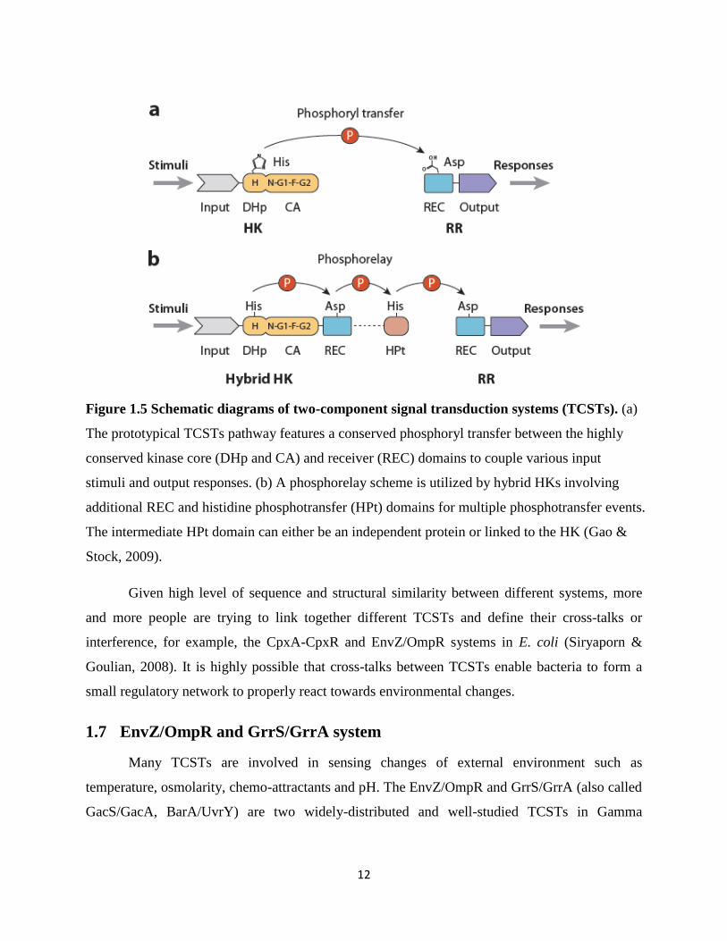
12
Figure 1.5 Schematic diagrams of two-component signal transduction systems (TCSTs). (a)
The prototypical TCSTs pathway features a conserved phosphoryl transfer between the highly
conserved kinase core (DHp and CA) and receiver (REC) domains to couple various input
stimuli and output responses. (b) A phosphorelay scheme is utilized by hybrid HKs involving
additional REC and histidine phosphotransfer (HPt) domains for multiple phosphotransfer events.
The intermediate HPt domain can either be an independent protein or linked to the HK (Gao &
Stock, 2009).
Given high level of sequence and structural similarity between different systems, more
and more people are trying to link together different TCSTs and define their cross-talks or
interference, for example, the CpxA-CpxR and EnvZ/OmpR systems in E. coli (Siryaporn &
Goulian, 2008). It is highly possible that cross-talks between TCSTs enable bacteria to form a
small regulatory network to properly react towards environmental changes.
1.7 EnvZ/OmpR and GrrS/GrrA system
Many TCSTs are involved in sensing changes of external environment such as
temperature, osmolarity, chemo-attractants and pH. The EnvZ/OmpR and GrrS/GrrA (also called
GacS/GacA, BarA/UvrY) are two widely-distributed and well-studied TCSTs in Gamma
13
proteobacteria. They represent paradigms of signal transduction systems, which have pleiotropic
effects, suggesting both systems are global regulators.
1.7.1 EnvZ/OmpR system and its function
EnvZ/OmpR is one of the well-studied TCSTs. It is originally reported to be responsible
for osmo-regulation in E. coli by governing the expression of ompC and ompF genes, which
encode two major outer-membrane porins, OmpC and OmpF, respectively (Cai & Inouye, 2002).
The sensory domain inside HK EnvZ recognizes the variations in membrane surface tension,
which triggers conformational changes in EnvZ. At high osmolyte concentration, EnvZ exhibits
higher kinase activity than phosphatase activity, resulting in phosphorylation of OmpR. As the
number of phosphorylated OmpR proteins increases, OmpR binds to both high affinity binding
sites and low affinity-binding repressor site upstream of the ompF promoter. OmpR also binds to
three low affinity sites upstream of the ompC promoter, leading to increased ompC expression
and thus OmpC becomes the major porin. In low osmolarity state, however, EnvZ exhibits
relatively low kinase activity (i.e., high phosphatase activity) towards OmpR, resulting in
relatively less phosphorylated OmpR. In this situation, OmpR binds to high-affinity OmpR-
binding sites within ompF promoter, resulting in OmpF porin production (Kato et al., 1989).
In addition to its role in porin osmoregulation, OmpR has been found to be involved in
regulating virulence and various cellular components as a dual regulator, such as EPS synthesis,
flagella gene expression, fatty acid transport (Brzostek et al., 2007).
EnvZ/OmpR has been reported to regulate genes associated with virulence in several
pathogenic bacteria. The Shigella flexneri ompB locus was found to modulate expression of the
vir genes, which are responsible for invasion of epithelial cells. Mutation of envZ gene reduced
its virulence (Bernardini et al., 1990). Meanwhile, a mutation in S. typhimurium ompR locus
resulted in highly attenuated strain (Dorman et al., 1989). OmpR also negatively regulates
expression of invasin, a protein that allows enteric bacteria to penetrate cultured mammalian
cells in Yersinia enterocolitica and T3SS in P. syringae (Brzostek et al., 2007). In Salmonella
spp., OmpR activates another TCST, SsrA/SsrB, which in turn regulates T3SS produced by
Salmonella pathogenicity island 2 (SPI-2) (Feng et al., 2003). On the contrary, although OmpR
was found to be involved in building resistance against phagocytosis or survival within
macrophages, mutation in ompR did not affect the virulence of Y. pestis (Gao et al., 2011).
14
In addition, EnvZ/OmpR system has been reported to regulate bacterial EPS production.
EnvZ/OmpR plays an important role in regulation of Vi polysaccharide synthesis in S. typhi and
one of the environmental signals for this regulation may be osmolarity (Pickard et al., 1994). The
S. typhi ompR mutant no longer agglutinates with Vi antiserum. Meanwhile, complementation of
the ompR mutant with the ompR and envZ genes of S. typhi restores its ability to agglutinate with
Vi antiserum. Furthermore, OmpR activates algD in E. coli, whose transcription activation is
essential for the EPS alginate synthesis and virulence factor expressed by Pseudomonas
aeruginosa in cystic fibrosis under high osmolarity conditions (Berry et al., 1989).
Swarming is a flagella-driven form of motility for movement across solid surfaces as a
group. Hyper-flagellated swarmer cells require EPS and surfactants for mass migration.
Inactivation of ompR promotes precocious swarming and flhDC expression in E. coli and
Xenorhabdus nematophila (Kim et al., 2003). In contrast, Y. enterocolitica ompR mutant showed
a decrease in flhDC expression and a non-motile phenotype, suggesting a positive effect of
OmpR in regulating flagella master regulator FlhDC (Raczkowska et al., 2011).
A microarray-based comparative transcriptome analysis of Y. pestis identified 224 genes
whose expression was altered by ompR mutation, indicating a global regulatory role in Y. pestis
(Gao et al., 2011). A similar global regulatory effect of OmpR in E. coli was observed (Oshima
et al., 2002).
1.7.2 GrrS/GrrA system
The hybrid HK GacS (initially called LemA) was first reported in the bean pathogen P.
syringae pv. syringae B728a. The corresponding RR GacA was described shortly thereafter in
the biological control bacterium P. fluorescence strain CHA0. Subsequently, GrrS and GrrA
homologs were identified in many enteric bacteria (E. coli, S. enterica, P. carotovora, P.
fluorescent, Vibrio and Azotobacter). The GrrSA system has since been reported to regulate an
array of phenotypes, including biofilm formation, alginate biosynthesis, production of toxins and
extracellular enzymes, proteases, siderophores, swarming motility and type III secretion system
(Zhao et al., 2009b). Two main properties of gacS/gacA mutants stand out: partial or complete
reduced biocontrol ability in a group of plant-beneficial Pseudomonads and significantly
attenuated virulence in plant- or animal-pathogenic bacteria (Altier et al., 2000; Gaffney et al.,

15
1994; Laville et al., 1992; Whistler et al., 1998; Zhang & Normark, 1996). Similar to
EnvZ/OmpR, GrrSA also functions as a dual regulator, i.e. positive or negative.
GacS/A controls virulence gene expression in a variety of host-pathogen systems,
including Pseudomonas, Vibrio, E. coli, Salmonella and Erwinia. In most cases, gacS/gacA
mutants showed reduced production of virulence factors and attenuated virulence. In plant
pathogen P. syringae, GacS/GacA was found to play a role in regulating hrpRS expression (Heeb
& Haas, 2001). In D. dadantii, GacA upregulated dspE, hrpA, and hrpN in vitro and in vivo
(Yang et al., 2008). The gacS and gacA mutants of P. syringae pv. syringae B728 were
completely nonpathogenic in foliar infiltration assays (Willis et al., 2001). Besides, gacS and
gacA mutants of animal pathogens such as Salmonella spp. (Johnston et al., 1996), V. cholera
and P. aeruginosa PAO1 (Parkins et al., 2001) were attenuated in colonizing infant mice,
suggesting an important role in regulating virulence factors. In APEC (avian pathogenic E. coli ),
GacS/A regulates a variety of virulence factors, such as its abilities to adhere, invade, persist
within tissues, survive within macrophages, as well as resistance to serum complement (Herren
et al., 2006). In S. enterica, GacA (SirA) has been found to contribute to the regulation of T3SS
through hilA (Ahmer et al., 1999).
Moreover, GacSA regulates EPS production (e.g. alginate) as well as extracellular
enzyme production in a variety of species (Cui et al., 2001). In P. fluorescences CHA0, GacS/A
system tightly controls the expression of antifungal secondary metabolites (e.g. hcnA) and
extracellular enzymes (e.g. aprA) (Heeb & Haas, 2001). The gacA gene product of P. carotovora
subsp. carotovora strain 71 regulates a number of extracellular enzymes (pel-1, a pectate lyase
gene; peh-1, polygalacturonase gene; and celV, a cellulase gene) (Cui et al., 2001).
As a negative regulator, GacSA was also found to down-regulate flagella gene expression
of P. fluorescens and E. coli. Mutation in both gacS and gacA genes in P. fluorescens strain
CHA0 affected its motility on swarming plate compared with wild-type strain (Kato et al., 1989).
D. dadantii gacA mutant showed reduced maceration and systemic invasion ability (Yang et al.,
2008). Both gacS and gacA mutants of P. syringae B728a showed reduced ability of swarming
(Kinscherf & Willis, 1999). In S. enterica serovar typhimurium, GacA was found to affect
flagella gene expression indirectly by binding to csrB promoter and activating its expression
(Teplitski et al., 2003).
16
In addition, GacSA system positively controls the expression of one to five small RNAs
(sRNAs), thus upregulates the production of proteins that are otherwise repressed by RNA
binding proteins, such as RsmA/CsrA (Cui et al., 2001). In E. coli, BarA/UvrY affects the
activity of RNA-binding protein CsrA by regulating the expression of csrB and csrC untranslated
regulatory RNA, which bind to CsrA protein and prevents it from binding to target genes
(Timmermans & Melderen, 2010).
Recent researches have provided much information that gene expression is mainly
regulated at the transcription initiation level in plant- and animal-pathogenic bacterium. E.
amylovora virulence is controlled by two virulence factors, EPS amylovoran and type III
secretion system. However, it is important to note that detailed mechanism of how virulence
regulators, such as sigma factors and two-component signal transduction systems are involved in
this process is still unclear. Early studies have shown that hrp-type III secretion (T3SS) in E.
amylovora is regulated by the master regulator HrpL, which belongs to the ECF subfamily of σ
factors; whereas two-component signal transduction systems (TCSTs) are important regulators of
amylovoran production. The main purpose of this project is to identify and characterize major
virulence regulators in regulating Erwinia amylovora virulence. The specific objectives are:
1. To understand the role of sigma factor 54 and its modulation proteins in Erwinia
amylovora virulence
2. To determine the interaction between EnvZ/OmpR and GrrS/GrrA systems in regulating
Erwinia amylovora virulence

17
Chapter 2
Effect of Sigma Factor RpoN and Its Modulation Protein on Erwinia
amylovora Virulence
2.1 Abstract
In bacteria, gene expression is mainly regulated at the transcription initiation level and its
core RNA polymerase (RNAP) requires sigma factors for promoter recognition and initiation. In
this study, we investigated the role of several sigma factors in regulating virulence gene
expression in Erwinia amylovora, a necrogenic enterobacterium causing fire blight of apples and
pears. Early studies have shown that hrp-type III secretion (T3SS) in E. amylovora is regulated
by HrpS, a member of the σ54
enhancer binding proteins, and the master regulator HrpL, which
belongs to the ECF subfamily of σ factors. Other sigma factors characterized included RpoN, a
nitrogen limitation σ factor, and its modulation protein YhbH. Our results showed that mutations
in hrpS, hrpL, rpoN and yhbH resulted in nonpathogenic phenotype in host plant and no
hypersensitive response in non-host tobacco. Consistently, expression of T3SS genes including
hrpL, dspE, hrpN and hrpA was barely detected in hrpS, hrpL, rpoN and yhbH mutants.
Amylovoran (EPS) production was higher in these mutants than that of WT strain, indicating
sigma factors may also play roles in regulating exopolysaccharide production. These results
suggest that sigma factors in E. amylovora are important virulence regulators and sigma factor
cascade exists in its regulatory networks.
2.2 Introduction
E. amylovora is the causal agent of fire blight of apples and pears. Its pathogenicity
depends on function of hypersensitive response and pathogenicity (hrp) type III protein secretion
system (T3SS) and production of exopolysaccharide amylovoran (Bellemann & Geider 1992).
The regulatory region of the hrp gene cluster in E. amylovora consists of three adjacent operons:
hrpXY encodes a two component regulatory system, consisting of histidine kinase (HK) HrpX
and response regulator (RR) HrpY; hrpS encodes an NtrC-like σ54
-dependent enhancer-binding
protein; and hrpL encodes an ECF (extra cytoplasmic functions) subfamily sigma factor, which
belongs to sigma 70 family. The promoter region of hrpL contains a putative σ54
promoter
consensus sequence (Wei & Beer, 1995).
18
Transcription in bacteria is initiated by a RNA polymerase (RNAP) isomerization process
in which the promoter DNA is melted close to the transcription start site (Browning & Busby,
2004). Bacterial RNAP holoenzyme is composed of the α2ββ'ω core enzyme associated with one
of a range of sigma factors. Every molecule of RNA polymerase contains exactly one sigma
factor subunit, so there is a competition of RNAP between different sigma factors. There are
mainly two families of sigma factors in bacteria, the sigma 70 family including σ 70
, σ 38
, σ32
, and
σ24
(ECF); and the sigma 54 family. Different sigma factors are activated in response to different
environmental conditions. σ70
(RpoD) is the housekeeping sigma factor that transcribes stringent
genes in growing cells and keeps essential genes and pathways operating (Gruber and Gross,
2003). All the other sigma factors are called alternative sigma factors, which competitively bind
the promoters of genes under certain environmental conditions. On the other hand, σ54/N
(RpoN)
is the nitrogen-limitation sigma factor whose function requires enhancer binding proteins (EBPs),
also are referred to as σ54
activators. Typically, EBPs have an N-terminal regulatory domain, a
central AAA+ (ATPases Associated with diverse cellular Activities) domain that directly
contacts σ54
and a C-terminal DNA binding domain (HTH). EBPs usually bind to regulatory
DNA sequences upstream from the σ54
promoters, from which interact with RNAP associated
with σ54
(σ54
-RNAP) at the promoter.
The mechanisms how sigma 70 and 54 factors work are different. In contrast to σ70
–like
family sigma factors, which characteristically bind to the -35 (TTGACA) and -10 (TATAAT)
positions from the transcription start, σ54
family sigma factors bind to specific promoter
sequences at positions -24 (GG) and -12 (TGC) and the σ54
–RNAP complex forms a closed loop
which is transcriptionally silent. EBPs open the transcriptional conformation by ATP hydrolysis
within the AAA+ domain, which provides the energy for the conformational change, and thus
transcription starts. Since the transcription of a σ54
regulated gene can be completely turned on by
this mechanism, σ54
-dependent gene expression is often counted for creating swift and precise
responses to environmental change (Schumacher et al., 2006).
The sigma 54 factors have been indirectly shown to contribute to virulence as a regulator
of the hrp gene cluster in a number of Gram-negative bacterial pathogens. In Pseudomonas
syringae pv. maculicola, the rpoN mutant lost its ability to cause disease and induce HR. In P.
syringae, expression of hrpL was strongly reduced in an rpoN mutant (Fellay et al., 1991) and
19
expression of Pantoea stewartii hrp genes was reduced in an E. coli rpoN mutant strain
(Frederick et al., 1993).
RpoN is also involved in regulating various cellular components, such EPS production
and flagella gene expression. In P. syringae pv. syringae and P. aeruginosa, transcription of
algD and algC, two important genes for biosynthesis of alginate (a virulence factor), are
regulated by AlgR and RpoN. Moreover, rpoN mutant didn’t produce pilin or form pili and had
dramatic reduced adhesion (Peñaloza-Vázquez et al., 2004; Zielinski et al., 1992). Besides, the
rpoN mutant didn’t produce flagellin subunit or form flagella, thus losing motility. Similar to P.
aeruginosa, Vibrio cholera and Vibrio anguillarum rpoN mutant lacked flagella and were
completely non-motile (Damron et al., 2012; Dong et al., 2012; O’Toole et al., 1997). A
mutation lacking σ54
in fish pathogen V. anguillarum severely impaired its ability to infect fish
immersed in contaminated water. In the case of P. syringae pv. maculicola, the rpoN mutant was
nonmotile and lost its ability to produce coronatine, a phytotoxin (Hendrickson et al., 2000).
In P. syringae, two EBPs, HrpR and HrpS are involved in regulating hrp T3SS. In this
hrp regulatory cascade, cooperative action of HrpR, HrpS and RpoN controls hrpL gene
expression, and HrpL activates transcription of the hrp genes (Grimm et al., 1995). In contrast,
there is only one EBP HrpS in E. amylovora which has been suggested to be required for
activating hrpL gene expression. HrpL then enables the recognition and transcriptional activation
of hrp promoters containing “Hrp boxes” in their –10/-35 regions (Fellay et al., 1991).
In the genome of E. amylovora, next to rpoN is yhbH gene (Fig 2.1 A). YhbH is
annotated as sigma 54 (RpoN) modulation protein in E. amylovora. YhbH, renamed recently as
hibernation promoting factor (HPF) in E. coli, is involved in ribosome stabilization and
preservation in stationary phase by binding specifically to 90S ribosome (a dimer of the 70S
ribosomes) to form 100S ribosome. The latter has no translational activity (Kato et al., 2010).
However, there is no direct evidence showing exactly how YhbH is involved in sigma 54
regulatory process. On the other hand, both rpoN and hrpS have not been characterized in E.
amylovora. The purpose of this study is to systematically characterize the role of RpoN, EBP
HrpS and its modulation protein YhbH in E. amylovora virulence.
20
2.3 Materials and methods
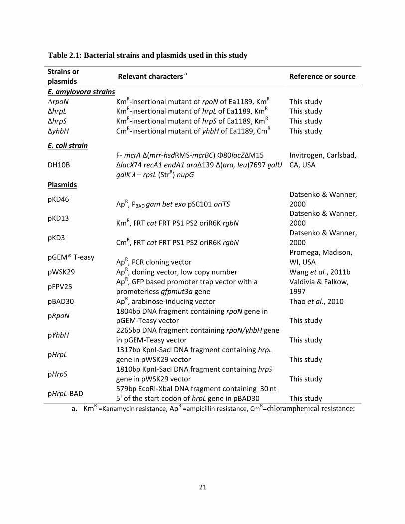
2.3.1 Bacterial strains and growth condition
The bacterial strains and plasmids used in this study are summarized in Table 2.1. LB
medium is used routinely for culture E. amylovora. When necessary, the following antibiotics
were added to the medium: 50 µg/ml kanamycin (Km), 100 µg/ml ampicillin (Ap) and 20 µg/ml
chloramphenical (Cm). Amylovoran production was determined by growing bacteria in MBMA
medium (3 g KH2PO4, 7 g K2HPO4, 1 g [NH4]2SO4, 2 ml glycerol, 0.5 g citric acid, 0.03 g
MgSO4) amended with 1% sorbitol (Zhao et al., 2009a). A specific hrp-inducing minimal
medium (HrpMM) containing 20 mmol galactose (1g [NH4]2SO4, 0.246 g MgCl2·6H2O, 0.099 g
NaCl, 8.708 g K2HPO4, 6.804 g KH2PO4) was used in vitro to mimic conditions of the plant
apoplast (Wei et al., 1992).
21
Table 2.1: Bacterial strains and plasmids used in this study
Strains or plasmids
Relevant characters a Reference or source
E. amylovora strains ∆rpoN KmR-insertional mutant of rpoN of Ea1189, KmR This study
∆hrpL KmR-insertional mutant of hrpL of Ea1189, KmR This study ∆hrpS KmR-insertional mutant of hrpS of Ea1189, KmR This study ∆yhbH CmR-insertional mutant of yhbH of Ea1189, CmR This study
E. coli strain
DH10B F- mcrA Δ(mrr-hsdRMS-mcrBC) Φ80lacZΔM15 ΔlacX74 recA1 endA1 araΔ139 Δ(ara, leu)7697 galU galK λ – rpsL (StrR) nupG
Invitrogen, Carlsbad, CA, USA
Plasmids
pKD46 ApR, PBAD gam bet exo pSC101 oriTS
Datsenko & Wanner, 2000
pKD13 KmR, FRT cat FRT PS1 PS2 oriR6K rgbN
Datsenko & Wanner, 2000
pKD3 CmR, FRT cat FRT PS1 PS2 oriR6K rgbN
Datsenko & Wanner, 2000
pGEM® T-easy ApR, PCR cloning vector
Promega, Madison, WI, USA
pWSK29 ApR, cloning vector, low copy number Wang et al., 2011b
pFPV25 ApR, GFP based promoter trap vector with a promoterless gfpmut3a gene
Valdivia & Falkow, 1997
pBAD30 ApR, arabinose-inducing vector Thao et al., 2010
pRpoN 1804bp DNA fragment containing rpoN gene in pGEM-Teasy vector This study
pYhbH 2265bp DNA fragment containing rpoN/yhbH gene in pGEM-Teasy vector This study
pHrpL 1317bp KpnI-SacI DNA fragment containing hrpL gene in pWSK29 vector This study
pHrpS 1810bp KpnI-SacI DNA fragment containing hrpS gene in pWSK29 vector This study
pHrpL-BAD 579bp EcoRI-XbaI DNA fragment containing 30 nt 5' of the start codon of hrpL gene in pBAD30 This study
a. KmR =Kanamycin resistance, ApR =ampicillin resistance, CmR=chloramphenical resistance;
22
Table 2.2: Primers used in this study
Primer Sequences (5’—3’) a Reference or source
rpoN F ATGAAGCAAGGTCTACAACTCAGGCTGAGCCAACAGCTTGCCATGACGCCGCGATTGTGTAGGCTGGAGCT This study
rpoN R TCAAACCAGCTGTTTACGCTGATTCGATGGCGGGATGGATAAAGACTCTC ATTCCGGGGATCCGTCGACC
This study
yhbH F GTTGCATCGTCGACCGACAGCAGGCTTTTTTTGAACAAGGTGAAGAGTTT GCGATTGTGTAGGCTGGAGCT
This study
yhbH R TAGTTTCACTTACTTATTCACTTCCGCAGGGCGCATGGCATTTTCCCAGG ATTCCGGGGATCCGTCGACC
This study
hrpL F ATGACAGAAATTCACCTGCAAACAACTGAATCAACATCGGTCAACGATGGGCGATTGTGTAGGCTGGAGCT This study
hrpL R TTAAGAAAATACTGACTGTTTCAGCGTGACGCGCGCACGCGACAGACGTGATTCCGGGGATCCGTCGACC
This study
hrpS F AGAGCACATCTCTTTGACAGAAGAACAACCCATCGATATCCACGACACATGCGATTGTGTAGGCTGGAGCT
This study
hrpS R GATATAGCGTACGCAAAGGAATACCCAACTCCTGCGCCGCATCATCAATGATTCCGGGGATCCGTCGACC This study
rpoN Cm1 GTAACAAACTCGCGCAATGG This study
rpoN Cm2 GCCGATGAACAAGTGAAGC This study
yhbH Cm1 GTGCCGCGGCTAAAGATTA This study
yhbH Cm2 TTGTGGCAGGTTAAGCTGTTT This study
hrpL Cm1 TGCAAATTTTGGCGGTTTA This study
hrpL Cm2 GCTGGGAAAATTGCATCTC This study
hrpS Cm1 TGTTCAGCATAAGACGATGG This study
hrpS Cm2 ATCCCGGCATAACCTTTGTA This study
Km1 CAGTCATAGCCGAATAGCCT Zhao et al., 2009a
Km2 CGGTGCCCTGAATGAACTGC Zhao et al., 2009a
Cm1 TTATACGCAAGGCGACAAGG Zhao et al., 2009a
Cm2 GATCTTCCGTCACAGGTAGG Zhao et al., 2009b
16S1 CCTCCAAGTCGACATCGTTT Wang et al., 2011b
16S2 TGTAGCGGTGAAATGCGTAG Wang et al., 2011b
amsG-rt1 CAAAGAGGTGCTGGAAGAGG Wang et al., 2011b
amsG-rt2 GTTCCATAGTTGCGGCAGTT Wang et al., 2011b
amsD-rt1 GATGCGTCTGTTCAAGCTGT Wang et al., 2011b
amsD-rt2 TCGCAACAAATCAGTCTGGA Wang et al., 2011b
rcsA-rt1
TTAAACCTGTCTGTGCGTCA Wang et al., 2011b
23
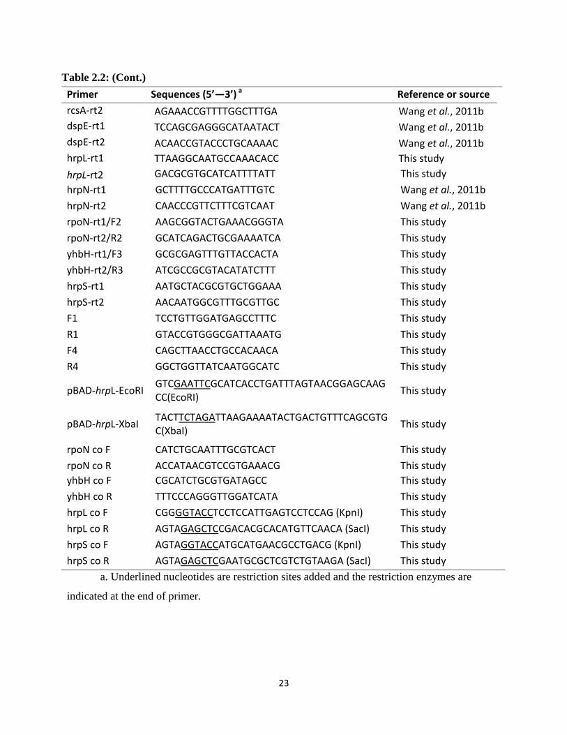
Table 2.2: (Cont.)
Primer Sequences (5’—3’) a Reference or source
rcsA-rt2 AGAAACCGTTTTGGCTTTGA Wang et al., 2011b
dspE-rt1 TCCAGCGAGGGCATAATACT Wang et al., 2011b
dspE-rt2 ACAACCGTACCCTGCAAAAC Wang et al., 2011b
hrpL-rt1 TTAAGGCAATGCCAAACACC This study
hrpL-rt2 GACGCGTGCATCATTTTATT This study
hrpN-rt1 GCTTTTGCCCATGATTTGTC Wang et al., 2011b
hrpN-rt2 CAACCCGTTCTTTCGTCAAT Wang et al., 2011b
rpoN-rt1/F2 AAGCGGTACTGAAACGGGTA This study
rpoN-rt2/R2 GCATCAGACTGCGAAAATCA This study
yhbH-rt1/F3 GCGCGAGTTTGTTACCACTA This study
yhbH-rt2/R3 ATCGCCGCGTACATATCTTT This study
hrpS-rt1 AATGCTACGCGTGCTGGAAA This study
hrpS-rt2 AACAATGGCGTTTGCGTTGC This study
F1 TCCTGTTGGATGAGCCTTTC This study
R1 GTACCGTGGGCGATTAAATG This study
F4 CAGCTTAACCTGCCACAACA This study
R4 GGCTGGTTATCAATGGCATC This study
pBAD-hrpL-EcoRI GTCGAATTCGCATCACCTGATTTAGTAACGGAGCAAGCC(EcoRI)
This study
pBAD-hrpL-XbaI TACTTCTAGATTAAGAAAATACTGACTGTTTCAGCGTGC(XbaI)
This study
rpoN co F CATCTGCAATTTGCGTCACT This study
rpoN co R ACCATAACGTCCGTGAAACG This study
yhbH co F CGCATCTGCGTGATAGCC This study
yhbH co R TTTCCCAGGGTTGGATCATA This study
hrpL co F CGGGGTACCTCCTCCATTGAGTCCTCCAG (KpnI) This study
hrpL co R AGTAGAGCTCCGACACGCACATGTTCAACA (SacI) This study
hrpS co F AGTAGGTACCATGCATGAACGCCTGACG (KpnI) This study
hrpS co R AGTAGAGCTCGAATGCGCTCGTCTGTAAGA (SacI) This study
a. Underlined nucleotides are restriction sites added and the restriction enzymes are
indicated at the end of primer.
24
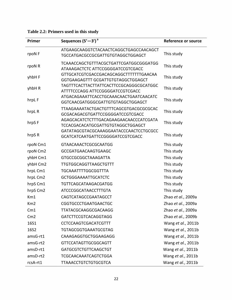
2.3.2 DNA manipulation and bioinformatics analysis
Plasmid DNA purification, PCR amplification of genes, isolation of fragments from
agarose gels, cloning, restriction enzyme digestion and T4 DNA ligation were performed using
standard molecular procedures (Sambrook & Russell, 1989). Protein domain organizations were
derived from the graphical output of the NCBI web interface.
2.3.3 Construction of mutants in Erwinia amylovora by Lambda-Red recombinase
E. amylovora stable mutants were generated by using the λ phage recombinases as
previously described (Zhao et al., 2009a). Briefly, E. amylovora Ea1189 was transformed with
plasmid pKD46 expressing recombinases red α, β, and γ. The transformant Ea1189 (pKD46)
were grown overnight at 28°C, reinoculated in LB broth containing 0.1% arabinose, and grown
to exponential phase OD600=0.8. Cells were collected, made electro-component, and stored at -
80°C. Recombination fragments consisting of a kanamycin (kan) or chloramphenical (cat) gene
with its own promoter, flanked by a 50-nucleotide (nt) homology arm, was generated by PCR
using plasmid pKD13 or pKD3 as a template. The primers that used for generating mutants are
listed in Table 2.2. Primers and internal primer pair Km1 and Km2 of the kan gene, Cm1 and
Cm2 of the cat gene, were used to confirm mutants by PCR. For the resulting mutants, the
majority of the coding region of each gene was replaced by the kan or cat gene, except for the
first and last 50 nt. The resulting mutants were designated and listed in Table 2.1.
2.3.4 Virulence assays on apple shoot and immature pear fruit
For E. amylovora WT and mutant strains, bacteria were grown overnight in LB broth,
harvested by centrifugation, and resuspended in ½*sterile phosphate buffered-saline (PBS) with
bacterial cells adjusted to OD600 = 0.001 in ½*PBS. Immature fruits of pear (Pyrus communis L.
cv Bartlett) were surface-sterilized, and pricked with a sterile needle as described previously
(Zhao et al., 2006). Two μl of cell suspensions was inoculated on the wounded tissue and
incubated the pears in a humidified chamber at 26°C. Symptoms were recorded at 4 and 8 days
post-inoculation. For each strain tested, fruits were assayed in triplicate, and each experiment
was performed at least three times.
25
Apple shoot virulence assay was performed on young annual shoots of ‘gala’ apple, 25 to
40 cm in length. After pricking the tip with a sterilized needle, five μl of pathogen suspension
with an initial OD600=0.1 was pipette onto the wounded tissue. For each bacterial strain, seven
shoots were inoculated. Plants were kept in a greenhouse at 25°C and a 16 hours light
photoperiod, and recorded disease development after 7 days following inoculation by measuring
length of the necrotic tissue. The experiment was performed at least three times.
2.3.5 CPC assay for determining amylovoran concentration
Amylovoran concentration in supernatants of bacterial cultures was quantitatively
determined by a turbidity assay with cetylpyrimidinium chloride (CPC) as described (Hildebrand
et al., 2006). For E. amylovora WT and mutants strains, bacterial suspensions was grown
overnight in LB broth w/o appropriate antibiotics, harvested by centrifugation and washed with
½*PBS for three times, then resuspended the bacterial pellet in 200 μl PBS and inoculated into
5ml MBMA+1% sorbitol medium with an initial OD600=0.2, inoculated for 24 hours at 28°C
with shaking. Following centrifugation, 50 μl CPC at 50 mg /ml were added to one ml
supernatant, incubated 10 min at room temperature, and determined amylovoran concentration
by measuring OD600 turbidity. The final concentration of amylovoran production was normalized
for a cell density of 1.0. For each strain tested, the experiment was repeated at least three times.
2.3.6 HR assay
E. amylovora Ea1189 and mutant strains were grown overnight at 28°C. Cells were
resuspended to OD600 = 0.2 in sterile half-phosphate buffered-saline (½*PBS). When necessary,
arabinose was added to bacterial suspension to a final concentration of 0.2%. The mixture was
infiltrated into tobacco (Nicotiana benthamiana) leaves by needle-less syringe. Infiltrated plants
were kept in a humid growth chamber, and HR symptoms were recorded at 24 hours post
infiltration. The experiment was repeated at least 3 times.
2.3.7 RNA isolation
For in vitro assay, WT and mutant bacterial strains were grown overnight in MBMA+1%
sorbitol medium for 24 hours or hrp-inducing medium at 18 ºC for 6 hours. For in vivo assay,
bacterial strains were collected from inoculated pear fruits 18 hour after inoculation as described
26
above. In both cases, four mL of RNA Protect Reagent (Qiagen) were added to two ml bacterial
cultures (at OD600 of about 0.5–0.8) to stabilize RNA, harvested the cells by centrifugation for 10
min at 4000 g and extracted RNA using Qiagen Bacterial RNA Mini Kit. Dnase (Qiagen, Hilden,
Germany) was used to eliminate residue genomic DNA by an on-column digestion method.
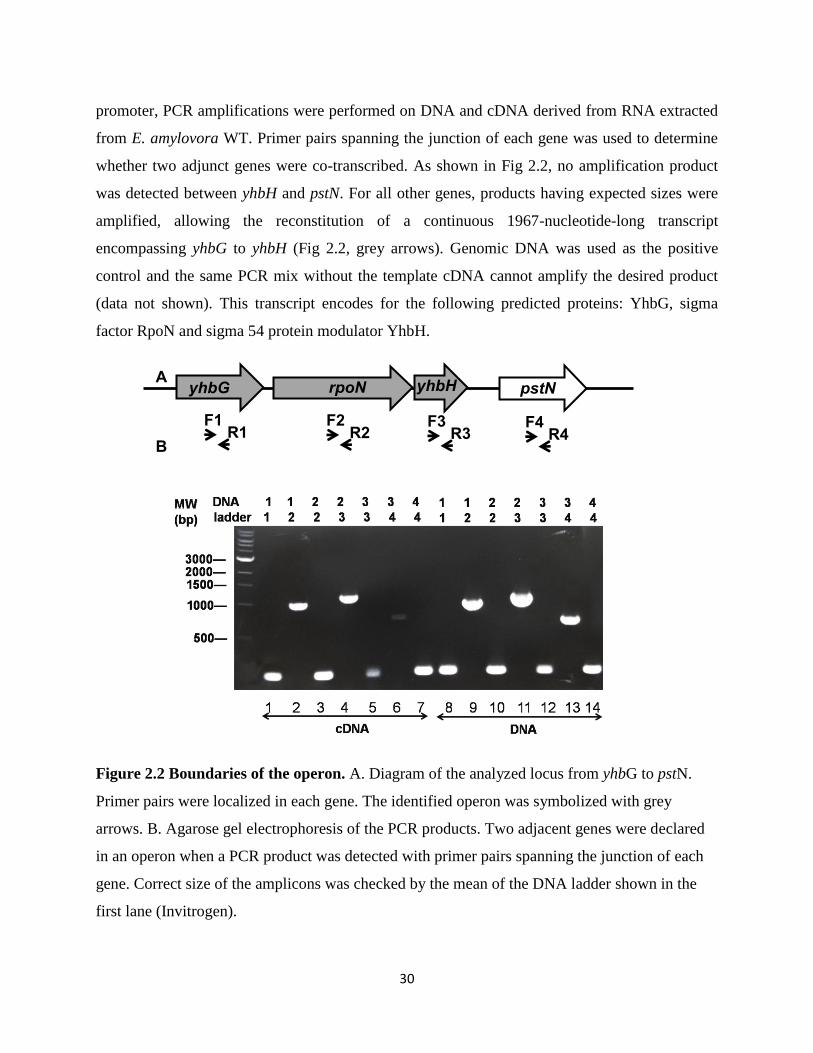
2.3.8 Operon determination
cDNA conversion was performed in a 50-μl reaction mixture by combining five μg of
total RNA and 100 ng of random hexamers using the Superscript™ First-Strand Synthesis Kit
(Invitrogen). Aliquots of diluted cDNA (2.5 μl, 1:10) were used as template for PCR experiments
using 0.3 μM of the required primers and 0.5 μl of Taq DNA Polymerase (Invitrogen). The
primer pairs F1 R1, F2 R2, F3 R3 and F4 R4 were used to test their abilities to generate a PCR
product for yhbG, rpoN, yhbH and pstN, respectively (Fig 2.1A). The following primer pairs
were used for the transcriptional analysis of the yhbG-yhbH operon structure: F1–R2 for the
junction between yhbG and rpoN, F2–R3 for the junction between rpoN and yhbH, F3–R4 the
junction between for yhbH and pstN. PCR products were analyzed onto 1% agarose gel
electrophoresis with ethidium bromide staining (Loisel et al., 2008).
2.3.9 Quantitative real-time PCR (qRT-PCR)
qRT-PCR was performed as previously described (Wang et al., 2011) to compare the
relative expression of target genes of E. amylovora rpoN, yhbH, hrpL and hrpS mutants with the
WT strain. One microgram of total RNA was reverse-transcribed in a 20 µl reaction using
SuperScript® III Reverse Transcriptase (Invitrogen, Carlsbad, CA, USA). For each sample,
negative reverse transcription reaction was done to verify the absence of genomic DNA
contamination in subsequent qPCR. Primers (Table 2.2) were designed using Primer3
(http://frodo.wi.mit.edu/primer3/). BLAST searches were performed to confirm gene specificity
and the absence of multi-locus matching at the primer site. ABI 7300 System (Applied
Biosystems) was used to perform the SYBRGreenq PCR reactions in 96-well optical reaction
plates. One µl of cDNA (2 ng/ reaction) or water (no-template control) was used as template for
qPCR reactions with Power SYBR Green PCR Master Mix (Applied Biosystems) with a final
primer concentration of 500 nmol. Primers in Table 2.2 were used to detect the expression of E.
amylovora amsG, rcsA, dspE, hrpL, hrpS, hrpN, hrpA, rpoN and yhbH gene, respectively. qPCR
27
amplifications were carried out with a cycle of 95°C for 10 min, followed by 40 cycles of 95°C
for 15 sec and 60°C for 1 min, and a final dissociation curve analysis step from 65°C to 95°C.
Technical replicate experiments were performed for each biological triplicate sample.
Amplification specificity for each qPCR reaction was confirmed by the dissociation curve
analysis. Determined Ct values were then exploited for further analysis.
Gene expression levels were analyzed using the relative quantification (∆∆Ct) method. A
16S rRNA rrsA gene was used as the housekeeping gene to normalize our samples (∆Ct =
Cttarget−CtrssA). A relative quantification (RQ) value was calculated as 2-(∆∆Ct = ∆Cttarget−∆Ctreference)
for each gene with the control group as a reference. A p-value was computed using a moderated
t-test to measure the significance associated with each RQ value. Variations were considered
statistically significant when the p-value was <0.05. RQ values for rpoN, yhbH, hrpL and hrpS
mutants were then normalized to those of WT (Wang et al., 2011).
2.3.10 Statistical analysis
One-way ANOVA and Student-Newman-Keuls test were used determine differences in
virulence progress, amylovoran production and gene expression data means within a = 0.05,
analyzed by SAS 9.2 program.
28
2.4 Results
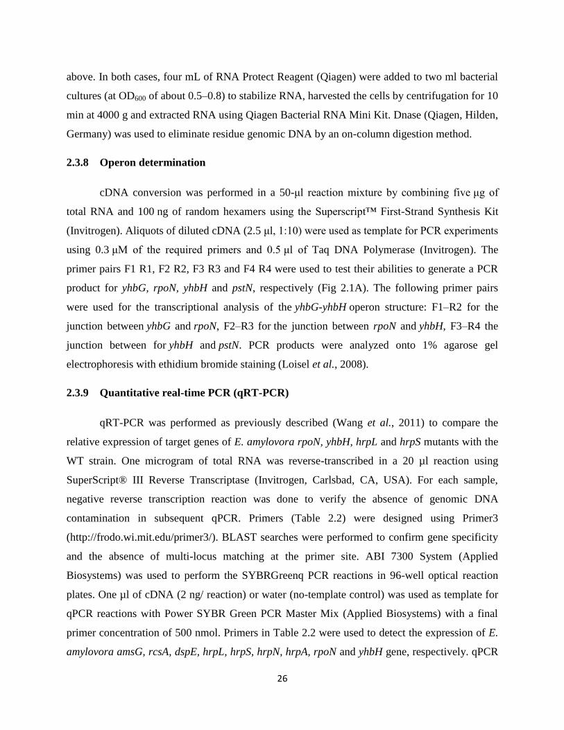
2.4.1 Domain organization of HrpL, HrpS, YhbH, and RpoN proteins.
The ECF sigma factor HrpL contains two functional regions (Fig. 2.1B). Sigma70 region
2 is the most conserved part of the protein, which contains both the -10 promoter recognition
helix and the primary core RNA polymerase binding site. The other region, sigma70 region 4 is
involved in binding to the -35 promoter element via a helix-turn-helix (HTH) motif (Campbell et
al., 2002).
Meanwhile, HrpS contains two functional regions, the AAA+ domain that directly
contacts σ54
and a C-terminal DNA binding HTH domain that binds upstream DNA activator
sequences; but lacks the regular cis-acting regulatory domain in the N-terminal (Fig. 2.1B).
Walker A and Walker B motifs are the most conserved sequences among all the AAA+ members
(Schumacher et al., 2006) and GAFTGA is the conserved motif that directly interacts with RpoN.
The sigma 54 protein RpoN has three functional regions (Fig. 2.1B). Activator interaction
domain (AID) contacts directly with EBPs, thus required for coupling ATP hydrolysis with
isomerization of the σ54
-RNAP holoenzyme from transcription silent to active states. DNA
binding domain (DBD) includes a segment recognizing the -12 promoter region, and a helix-
turn-helix motif that specifically interacts with the -24 promoter region. Core RNAP-binding
domain (CBD) is a linker between AID and DBD (Hong et al., 2009).
YhbH contains seventeen 30S subunit ribosome binding sites, indicating its ability to
attach to 30S subunit of the 90S ribosome in order to form translational silent 100S ribosome.
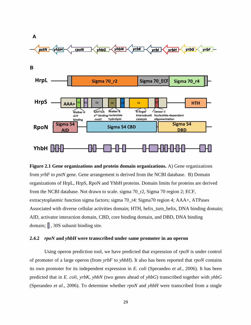
29
Figure 2.1 Gene organizations and protein domain organizations. A) Gene organizations
from yrbF to pstN gene. Gene arrangement is derived from the NCBI database. B) Domain
organizations of HrpL, HrpS, RpoN and YhbH proteins. Domain limits for proteins are derived
from the NCBI database. Not drawn to scale. sigma 70_r2, Sigma 70 region 2; ECF,
extracytoplasmic function sigma factors; sigma 70_r4: Sigma70 region 4; AAA+, ATPases
Associated with diverse cellular activities domain; HTH, helix_turn_helix, DNA binding domain;
AID, activator interaction domain, CBD, core binding domain, and DBD, DNA binding
domain; , 30S subunit binding site.
2.4.2 rpoN and yhbH were transcribed under same promoter in an operon
Using operon prediction tool, we have predicted that expression of rpoN is under control
of promoter of a large operon (from yrbF to yhbH). It also has been reported that rpoN contains
its own promoter for its independent expression in E. coli (Sperandeo et al., 2006). It has been
predicted that in E. coli, yrbK, yhbN (two genes ahead of yhbG) transcribed together with yhbG
(Sperandeo et al., 2006). To determine whether rpoN and yhbH were transcribed from a single
A
B
30
promoter, PCR amplifications were performed on DNA and cDNA derived from RNA extracted
from E. amylovora WT. Primer pairs spanning the junction of each gene was used to determine
whether two adjunct genes were co-transcribed. As shown in Fig 2.2, no amplification product
was detected between yhbH and pstN. For all other genes, products having expected sizes were
amplified, allowing the reconstitution of a continuous 1967-nucleotide-long transcript
encompassing yhbG to yhbH (Fig 2.2, grey arrows). Genomic DNA was used as the positive
control and the same PCR mix without the template cDNA cannot amplify the desired product
(data not shown). This transcript encodes for the following predicted proteins: YhbG, sigma
factor RpoN and sigma 54 protein modulator YhbH.
Figure 2.2 Boundaries of the operon. A. Diagram of the analyzed locus from yhbG to pstN.
Primer pairs were localized in each gene. The identified operon was symbolized with grey
arrows. B. Agarose gel electrophoresis of the PCR products. Two adjacent genes were declared
in an operon when a PCR product was detected with primer pairs spanning the junction of each
gene. Correct size of the amplicons was checked by the mean of the DNA ladder shown in the
first lane (Invitrogen).
yhbG yhbH rpoN
pstN
F1 R1
F2 R2
F3 R3
F4 R4
B
A
31
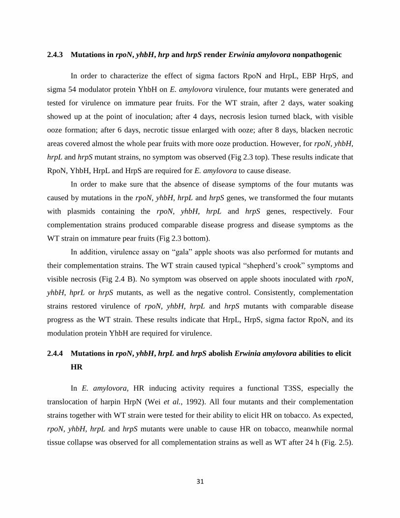
2.4.3 Mutations in rpoN, yhbH, hrp and hrpS render Erwinia amylovora nonpathogenic
In order to characterize the effect of sigma factors RpoN and HrpL, EBP HrpS, and
sigma 54 modulator protein YhbH on E. amylovora virulence, four mutants were generated and
tested for virulence on immature pear fruits. For the WT strain, after 2 days, water soaking
showed up at the point of inoculation; after 4 days, necrosis lesion turned black, with visible
ooze formation; after 6 days, necrotic tissue enlarged with ooze; after 8 days, blacken necrotic
areas covered almost the whole pear fruits with more ooze production. However, for rpoN, yhbH,
hrpL and hrpS mutant strains, no symptom was observed (Fig 2.3 top). These results indicate that
RpoN, YhbH, HrpL and HrpS are required for E. amylovora to cause disease.
In order to make sure that the absence of disease symptoms of the four mutants was
caused by mutations in the rpoN, yhbH, hrpL and hrpS genes, we transformed the four mutants
with plasmids containing the rpoN, yhbH, hrpL and hrpS genes, respectively. Four
complementation strains produced comparable disease progress and disease symptoms as the
WT strain on immature pear fruits (Fig 2.3 bottom).
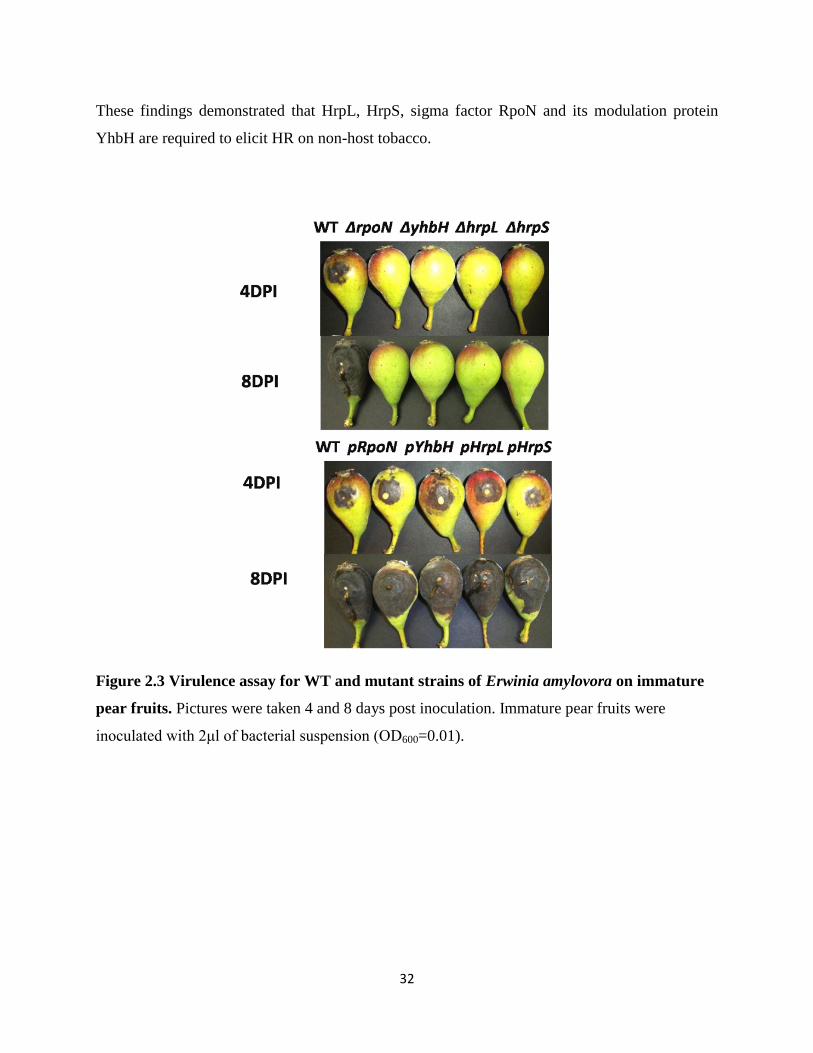
In addition, virulence assay on “gala” apple shoots was also performed for mutants and
their complementation strains. The WT strain caused typical “shepherd’s crook” symptoms and
visible necrosis (Fig 2.4 B). No symptom was observed on apple shoots inoculated with rpoN,
yhbH, hprL or hrpS mutants, as well as the negative control. Consistently, complementation
strains restored virulence of rpoN, yhbH, hrpL and hrpS mutants with comparable disease
progress as the WT strain. These results indicate that HrpL, HrpS, sigma factor RpoN, and its
modulation protein YhbH are required for virulence.
2.4.4 Mutations in rpoN, yhbH, hrpL and hrpS abolish Erwinia amylovora abilities to elicit
HR
In E. amylovora, HR inducing activity requires a functional T3SS, especially the
translocation of harpin HrpN (Wei et al., 1992). All four mutants and their complementation
strains together with WT strain were tested for their ability to elicit HR on tobacco. As expected,
rpoN, yhbH, hrpL and hrpS mutants were unable to cause HR on tobacco, meanwhile normal
tissue collapse was observed for all complementation strains as well as WT after 24 h (Fig. 2.5).
32
These findings demonstrated that HrpL, HrpS, sigma factor RpoN and its modulation protein
YhbH are required to elicit HR on non-host tobacco.
Figure 2.3 Virulence assay for WT and mutant strains of Erwinia amylovora on immature
pear fruits. Pictures were taken 4 and 8 days post inoculation. Immature pear fruits were
inoculated with 2μl of bacterial suspension (OD600=0.01).
33
Figure 2.4 Virulence assay for WT, mutants and complementation strains of Erwinia
amylovora on “gala” apple shoots. “Gala” apple shoots were inoculated with 5μl of bacterial
suspension (OD600=0.1). Picture was taken at 7 days post inoculation. For all the strains tested in
virulence assay, experiment was repeated at least three times.

34
Figure 2.5 HR assay for WT, mutants and complementation strains of Erwinia amylovora
on non-host tobacco. Pictures were taken 24h post inoculation. Tobacco leaves were infiltrated
with bacteria suspension (OD600=0.2) using syringe and incubated at 28°C growth chamber for
24 hours.
2.4.5 RpoN, YhbH, HrpL and HrpS suppress amylovoran production in vitro
To determine whether sigma factors and its modulation proteins of E. amylovora affect
amylovoran biosynthesis, bacterial cells were grown in MBMA+1% sorbitol medium and
quantified by CPC turbidity assay (Bellemann et al., 1994). rpoN, yhbH, hrpL and hrpS mutants
produced higher amount of amylovoran than that of WT at 24 hours post inoculation
(OD600=0.08), indicating a negative effect on EPS production (Fig 2.6). Introduction of original
copy of rpoN and yhbH gene into the rpoN and yhbH mutant strains can partially restore the
amylovoran synthesis, producing approximately 0.3 and 0.5 times of amylovoran as the mutants,
respectively. These results suggest that RpoN and YhbH of E. amylovora are negative regulators
of amylovoran production in vitro.
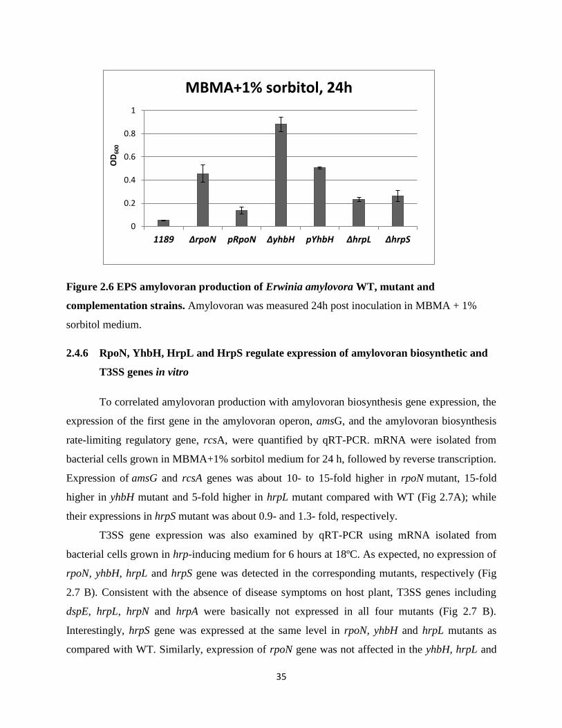
35
Figure 2.6 EPS amylovoran production of Erwinia amylovora WT, mutant and
complementation strains. Amylovoran was measured 24h post inoculation in MBMA + 1%
sorbitol medium.
2.4.6 RpoN, YhbH, HrpL and HrpS regulate expression of amylovoran biosynthetic and
T3SS genes in vitro
To correlated amylovoran production with amylovoran biosynthesis gene expression, the
expression of the first gene in the amylovoran operon, amsG, and the amylovoran biosynthesis
rate-limiting regulatory gene, rcsA, were quantified by qRT-PCR. mRNA were isolated from
bacterial cells grown in MBMA+1% sorbitol medium for 24 h, followed by reverse transcription.
Expression of amsG and rcsA genes was about 10- to 15-fold higher in rpoN mutant, 15-fold
higher in yhbH mutant and 5-fold higher in hrpL mutant compared with WT (Fig 2.7A); while
their expressions in hrpS mutant was about 0.9- and 1.3- fold, respectively.
T3SS gene expression was also examined by qRT-PCR using mRNA isolated from
bacterial cells grown in hrp-inducing medium for 6 hours at 18ºC. As expected, no expression of
rpoN, yhbH, hrpL and hrpS gene was detected in the corresponding mutants, respectively (Fig
2.7 B). Consistent with the absence of disease symptoms on host plant, T3SS genes including
dspE, hrpL, hrpN and hrpA were basically not expressed in all four mutants (Fig 2.7 B).
Interestingly, hrpS gene was expressed at the same level in rpoN, yhbH and hrpL mutants as
compared with WT. Similarly, expression of rpoN gene was not affected in the yhbH, hrpL and
0
0.2
0.4
0.6
0.8
1
1189 ΔrpoN pRpoN ΔyhbH pYhbH ΔhrpL ΔhrpS
OD
60
0
MBMA+1% sorbitol, 24h
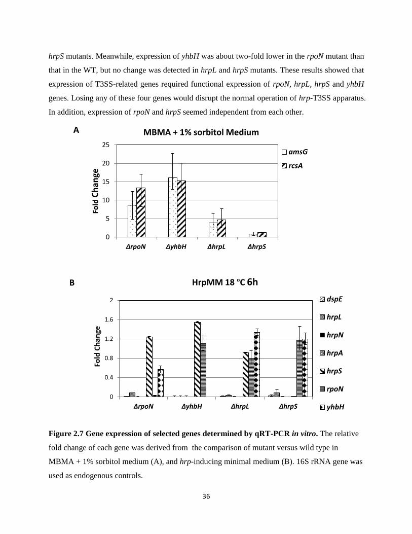
36
hrpS mutants. Meanwhile, expression of yhbH was about two-fold lower in the rpoN mutant than
that in the WT, but no change was detected in hrpL and hrpS mutants. These results showed that
expression of T3SS-related genes required functional expression of rpoN, hrpL, hrpS and yhbH
genes. Losing any of these four genes would disrupt the normal operation of hrp-T3SS apparatus.
In addition, expression of rpoN and hrpS seemed independent from each other.
Figure 2.7 Gene expression of selected genes determined by qRT-PCR in vitro. The relative
fold change of each gene was derived from the comparison of mutant versus wild type in
MBMA + 1% sorbitol medium (A), and hrp-inducing minimal medium (B). 16S rRNA gene was
used as endogenous controls.
0
5
10
15
20
25
ΔrpoN ΔyhbH ΔhrpL ΔhrpS
Fold
Ch
ange
MBMA + 1% sorbitol Medium
amsG
rcsA
0
0.4
0.8
1.2
1.6
2
ΔrpoN ΔyhbH ΔhrpL ΔhrpS
Fold
Ch
ange
HrpMM 18 ºC 6h
dspE
hrpL
hrpN
hrpA
hrpS
rpoN
yhbH
A
B
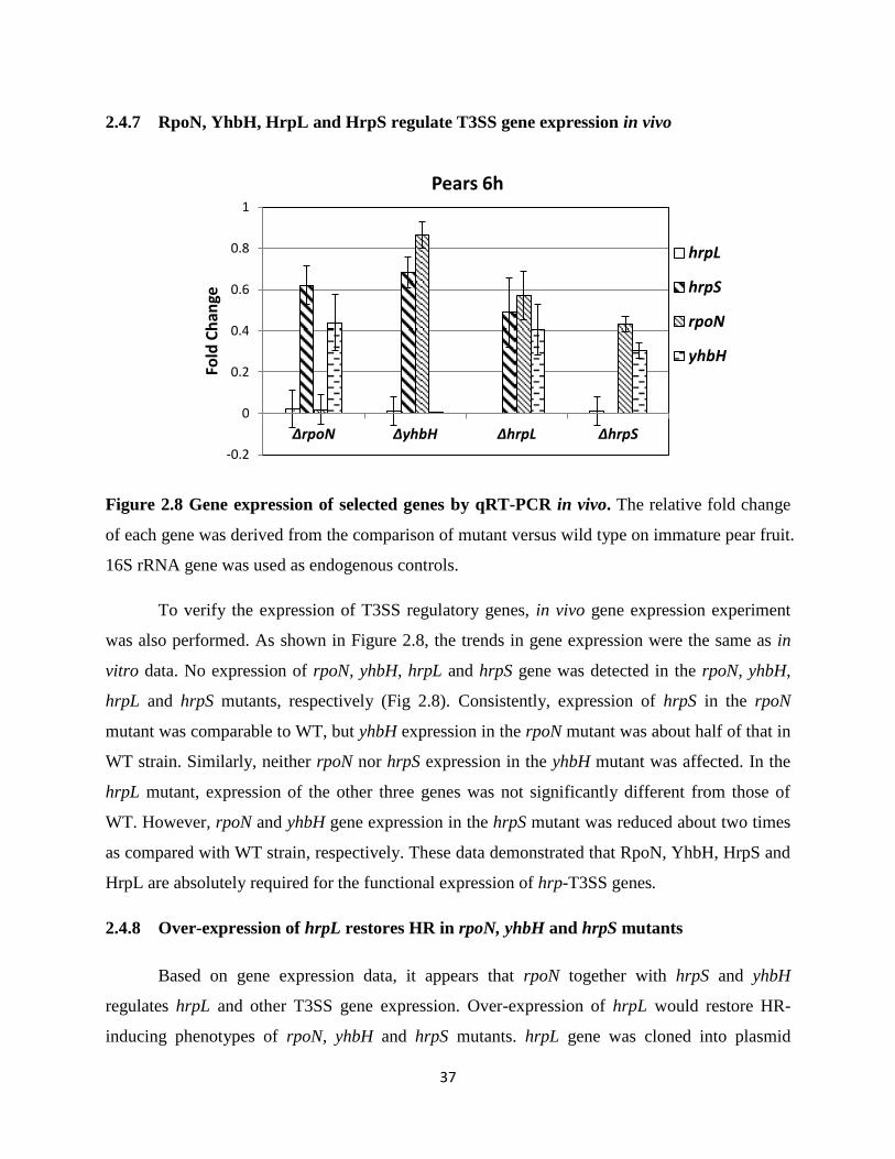
37
2.4.7 RpoN, YhbH, HrpL and HrpS regulate T3SS gene expression in vivo
Figure 2.8 Gene expression of selected genes by qRT-PCR in vivo. The relative fold change
of each gene was derived from the comparison of mutant versus wild type on immature pear fruit.
16S rRNA gene was used as endogenous controls.
To verify the expression of T3SS regulatory genes, in vivo gene expression experiment
was also performed. As shown in Figure 2.8, the trends in gene expression were the same as in
vitro data. No expression of rpoN, yhbH, hrpL and hrpS gene was detected in the rpoN, yhbH,
hrpL and hrpS mutants, respectively (Fig 2.8). Consistently, expression of hrpS in the rpoN
mutant was comparable to WT, but yhbH expression in the rpoN mutant was about half of that in
WT strain. Similarly, neither rpoN nor hrpS expression in the yhbH mutant was affected. In the
hrpL mutant, expression of the other three genes was not significantly different from those of
WT. However, rpoN and yhbH gene expression in the hrpS mutant was reduced about two times
as compared with WT strain, respectively. These data demonstrated that RpoN, YhbH, HrpS and
HrpL are absolutely required for the functional expression of hrp-T3SS genes.
2.4.8 Over-expression of hrpL restores HR in rpoN, yhbH and hrpS mutants
Based on gene expression data, it appears that rpoN together with hrpS and yhbH
regulates hrpL and other T3SS gene expression. Over-expression of hrpL would restore HR-
inducing phenotypes of rpoN, yhbH and hrpS mutants. hrpL gene was cloned into plasmid
-0.2
0
0.2
0.4
0.6
0.8
1
∆rpoN ∆yhbH ∆hrpL ∆hrpS
Fold
Ch
ange
Pears 6h
hrpL
hrpS
rpoN
yhbH
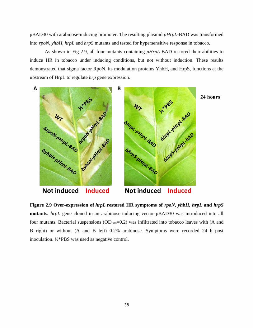
38
pBAD30 with arabinose-inducing promoter. The resulting plasmid pHrpL-BAD was transformed
into rpoN, yhbH, hrpL and hrpS mutants and tested for hypersensitive response in tobacco.
As shown in Fig 2.9, all four mutants containing pHrpL-BAD restored their abilities to
induce HR in tobacco under inducing conditions, but not without induction. These results
demonstrated that sigma factor RpoN, its modulation proteins YhbH, and HrpS, functions at the
upstream of HrpL to regulate hrp gene expression.
Figure 2.9 Over-expression of hrpL restored HR symptoms of rpoN, yhbH, hrpL and hrpS
mutants. hrpL gene cloned in an arabinose-inducing vector pBAD30 was introduced into all
four mutants. Bacterial suspensions (OD600=0.2) was infiltrated into tobacco leaves with (A and
B right) or without (A and B left) 0.2% arabinose. Symptoms were recorded 24 h post
inoculation. ½*PBS was used as negative control.
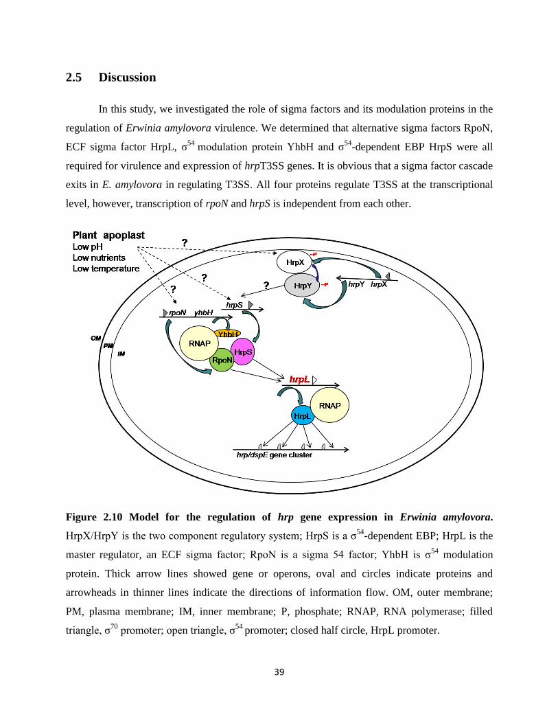
39
2.5 Discussion
In this study, we investigated the role of sigma factors and its modulation proteins in the
regulation of Erwinia amylovora virulence. We determined that alternative sigma factors RpoN,
ECF sigma factor HrpL, σ54
modulation protein YhbH and σ54
-dependent EBP HrpS were all
required for virulence and expression of hrpT3SS genes. It is obvious that a sigma factor cascade
exits in E. amylovora in regulating T3SS. All four proteins regulate T3SS at the transcriptional
level, however, transcription of rpoN and hrpS is independent from each other.
Figure 2.10 Model for the regulation of hrp gene expression in Erwinia amylovora.
HrpX/HrpY is the two component regulatory system; HrpS is a σ54
-dependent EBP; HrpL is the
master regulator, an ECF sigma factor; RpoN is a sigma 54 factor; YhbH is σ54
modulation
protein. Thick arrow lines showed gene or operons, oval and circles indicate proteins and
arrowheads in thinner lines indicate the directions of information flow. OM, outer membrane;
PM, plasma membrane; IM, inner membrane; P, phosphate; RNAP, RNA polymerase; filled
triangle, σ70
promoter; open triangle, σ54
promoter; closed half circle, HrpL promoter.
40
Sigma factors play important roles in the virulence of E. amylovora. We have
demonstrated, for the first time, that mutation in rpoN, hrpL, yhbH, and hrpS gene render E.
amylovora nonpathogenic. The lack of disease symptoms of all four mutants on both immature
pear fruits and apple shoots clearly indicated that sigma factors (RpoN and HrpL), σ54
-dependent
EBP HrpS, as well as sigma 54 factor modulator (YhbH) are required to cause disease symptoms
on host plant. HR assay also illustrated that loss of rpoN, hrpL, yhbH, and hrpS gene would lead
to the loss of HR-eliciting ability of E. amylovora as a result of lack of T3SS gene expression.
Gene expression assay further indicated that rpoN, yhbH and hrpS were all necessary to
activate hrpL transcription, which in turn activates the expression of other hrp-T3SS genes.
Mutation in rpoN, hrpL, yhbH and hrpS gene disrupted the formation of transcription machine,
thus paralyzed transcription initiation of hrpL. Interestingly, mutation in rpoN gene had no effect
on hrpS expression, and vice versa. It seemed that transcription of rpoN and hrpS was
independent from each other. It also appeared that sigma factors and related modulation proteins
are required for amylovoran production. RpoN, yhbH, hrpL and hrpS mutant disrupted the ability
of E. amylovora to produce normal amount of amylovoran. Gene expression assay verified that
these phenotypes were correlated with amsG and rcsA gene expression. It is possible that there is
an interaction between T3SS and amylovoran biosynthesis.
Overexpression of hrpL via an arabinose-inducing vector in rpoN, hrpS and yhbH mutant
bypassed these genes and rescued E. amylovora with HR symptoms on non-host plant tobacco,
further indicating that rpoN, hrpS and yhbH genes function at upstream of hrpL.
Based on our results, we proposed the following model of regulation of T3SS gene
expression in E. amylovora (Fig. 2.10). The bacteria may sense environmental stimulus through
HrpX/HrpY two-component regulatory system and somehow transmit the signal to HrpS, which
needs further study. HrpS is a σ54
-dependent EBP, which binds to upstream DNA activator
sequences (UAS), located −80 to −150 bp from HrpL transcriptional start site, and forms a
hexamer. Meanwhile, RpoN together with core RNAP forms σ54
-RNAP complex and binds to −
24 (GG) and −12 (TGC) position from HrpL transcription start, and remains transcriptionally
silent. With the assistance of integration host factor (IHF), hexametric EBPs contact the σ54
-
RNAP-promoter complex via the consensus GAFTGA motif and DNA looping. The energy
provided by ATP hydrolysis of the EBPs AAA+ domain triggers the formation of σ54-RNAP-
promoter complex open, with hrpL DNA melting. HrpL is the master switch of hrp system and
41
recognizes a conserved “hrp box” at the promoter regions of HrpL-dependent operons or genes
and regulates the other hrp gene expression. Our results indicate that both RpoN and HrpS are
required to active hrpL transcription and YhbH is also involved in this process. On the other
hand, expression of rpoN and hrpS is independent from each other.
However, the detailed function of YhbH is still unknown. In E. coli, level of σ38
protein
was observed to be significantly increased when cell growth stops and enters into the stationary
phase. Besides, σ38
factor assists the function of ribosome modulation factor (RMF), another
ribosome-associated protein, in cell viability (Apirakaramwong et al., 1999). It is possible that
YhbH aids RpoN in the competition with σ70 since it was found to be involved in 100S
ribosome stabilization and preservation (Kato et al., 2010). Studies to determine the molecular
mechanism of YhbH function are currently underway.
42
Chapter 3
Effect of EnvZ/OmpR and GrrS/GrrA Systems on Erwinia
amylovora Virulence
3.1 Abstract
Two-component signal transduction systems (TCSTs) in Erwinia amylovora play a major
role in virulence and in regulating amylovoran production, including EnvZ/OmpR and
GrrS/GrrA, two widely distributed systems in gamma-proteobacteria. While both systems
negatively control amylovoran biosynthesis, deletion mutants of envZ/ompR and grrA/grrS have
opposite swarming motility phenotypes. In order to determine how the two systems interact, two
triple mutants, envZ/ompR/grrA (ERA) and envZ/ompR/grrS (ERS) were generated. Our results
showed that both triple mutants had slightly increased virulence on apple shoots as compared to
that of wild type (WT) as well as mutants deleting a single system. In an in vitro amylovoran
assay, amylovoran production was significantly increased in the two triple mutants, indicating
the two systems synergistically regulate amylovoran production. In consistent with amylovoran
production, amsG gene expression was expressed significantly higher in the triple mutants in
vitro than those in WT as well as mutants deleting a single system. In contrast,
exopolysaccharide levan was significantly reduced in the triple mutants compared with that of
WT and deletion of a single system. In addition, the triple mutants showed reduced swarming
motility on swarming plates compared to that of grrA/grrS mutants and WT strain, but moved
slightly faster than that of envZ/ompR mutants, indicating that the two systems antagonistically
regulate swarming motility in E. amylovora. Furthermore, type III secretion (T3SS) genes were
significantly unregulated in the triple mutants as well as deletion of a single system than that of
the WT strain. These results indicate that EnvZ/OmpR and GrrS/GrrA systems play major roles
in virulence and in regulating virulence gene expression.
3.2 Introduction
Erwinia amylovora is the causal agent of fire blight disease on Rosaceae plants,
especially pears and apples. Fire blight was the first disease attributed to bacterium and E.
amylovora was the first plant pathogenic bacterium shown to be vectored by insects. Symptoms
43
of fire blight can be observed on all above ground tissues including blossoms, fruits, shoots,
branches and rootstock. After infection, tips of shoots may wilt rapidly to form a typical
“shepherd's crook” appearance. Viscous exudates “ooze” containing bacteria and
exopolysaccharide (EPS) amylovoran would come out after a while and serve as the source for
secondary infection.
E. amylovora is a Gram-negative, rod shaped bacterium with peritrichous flagella. It
belongs to the Enterobacteriaceae family and is closely related to Salmonella, E. coli and
Yersinia pestis. Two major pathogenicity factors in E. amylovora have been identified, i.e. EPS
amylovoran and type III secretion system (T3SS).
Amylovoran is a complex EPS made up of a large number of repeating units.
Amylovoran contributes to E. amylovora pathogenesis by protecting the pathogen from plant
defense, and binding water and nutrients released from damaged plant cells (Leigh & Coplin,
1992). The ability of individual E. amylovora strains to produce amylovoran is positively
correlated with the degree of virulence (Ayers et al., 1979). Mutant strains without amylovoran
were non-pathogenic (Nimtz et al., 1996). The ams (amylovoran biosynthetic) operon, consisting
12 genes (from amsA to amsL), is responsible for amylovoran biosynthesis. Expression of the
ams operon is regulated by the Rcs phosphorelay system in E. amylovora, which is also essential
for pathogenicity (Wang et al., 2009).
In addition to amylovoran, E. amylovora also produces another homopolymer EPS, levan,
which is composed of β-2, 6-linked fructose molecules. Levan synthesis is mediated by
levansucrase (Lsc) (Bereswill & Geider, 1997). In contrast, levan-deficient strains only showed
attenuated virulence (Geier & Geider, 1993).
The genome sequence of E. amylovora strains revealed three type III secretion systems in
E. amylovora, including the pathogenicity island1 (PAI-1) containing the hypersensitive
response and pathogenicity (hrp) T3SS, and two inv/spa-like T3SS islands (PAI-2 and PAI-3)
(Zhao et al., 2009a). hrp-T3SS has been known for its role as a pathogenicity factor that
functions to deliver effectors into the eukaryotic host (He et al., 2004). PAI-2 and PAI-3 are
similar to the SPI1 T3SS of Salmonella typhimurium and the inv/spa T3SS of the insect
endosymbiont Sodalis glossidinius, respectively. However, the role of these T3SSs is still
unknown. The pathogenicity of E. amylovora requires a functional hrp-T3SS in which HrpN and
DspA/E were found to play an important role in induction of cell death, activation of defense
44
pathways, and ROS accumulation. In E. amylovora, HrpL is the master switch of the hrp system
and belongs to a subfamily of bacterial sigma factors. It activates all secretory hrp operons,
harpin genes and dsp/avr genes. In E. amylovora, expression of hrpL is environmentally
regulated and seems partially controlled by HrpS as well as HrpX/Y, a two-component
regulatory system.
Two-component signal transduction systems (TCSTs) in E. amylovora have been
recently found to play a major role in regulating virulence, amylovoran production and swarming
motility. EnvZ/OmpR and GrrS/GrrA systems are two widely distributed systems in gamma-
proteobacteria (Zhao et al., 2009a). TCSTs normally consists of a membrane-bound histidine
kinase (HK) that senses a specific environmental stimulus, and a corresponding response
regulator (RR) that mediates the cellular response mostly through differential expression of
target genes.
EnvZ/OmpR, as a global and dual regulator, has been found to be involved in regulation
of a series of cellar components, virulence and global gene expression. First of all, EnvZ/OmpR
is essential for bacterial EPS production in Salmonella typhi and Pseudomonas aeruginosa
(Pickard et al., 1994; Berry et al., 1989). Secondly, OmpR has been suggested to have a positive
regulatory effect on flagella master regulator, FlhDC, in Y. enterocolitica (Raczkowska et al.,
2011). However, in E. coli and Xenorhabdus nematophila, OmpR was reported as a negative
regulator of swarming motility (Kim et al., 2003). Furthermore, EnvZ/OmpR has also been
reported to regulate genes associated with virulence in several pathogenic bacteria, including
Shigella flexneri, Salmonella typhimurium, Salmonella spp., Yersinia enterocolitica and
Pseudomonas syringae (Bernardini et al., 1990; Feng et al., 2003; Dorman et al., 1989; Brzostek
et al., 2007). Besides, microarray-based comparative transcriptome analysis of Yersinia pestis
identified a set of 224 genes whose expressions were affected by ompR mutation, indicating a
global regulatory role in Y. pestis (Gao et al. 2011).
Similarly, GrrSA system (also called GacSA, BarA/UvrY) has also been reported to
regulate an array of phenotypes (Zhao et al., 2009b). Two main properties of gacS/gacA mutants
stand out: partial or complete reduced biocontrol ability in a group of plant-beneficial
Pseudomonads and significantly attenuated virulence in plant- or animal-pathogenic bacteria.
GacS/A TCSTs regulate EPS production as well as extracellular enzyme production in a variety
species, such as Pseudomonas fluorescences CHA0 and Pectobacterium carotovora subsp.
45
carotovora (Cui et al., 2001). As a negative regulator, GacSA was found to down-regulate
flagella gene expression in P. fluorescens, E. coli and S. typhimurium (Teplitski et al., 2003).
Mutation in gacA gene of P. fluorescens, P. syringae B728a and Dickeya dadantii showed
reduced motile ability (Kato et al., 1989; Kinscherf & Willis, 1999). Moreover, GacS/A controls
virulence gene expression in a variety of host-pathogen systems, such as P. syringae, P.
aeruginosa, V. cholera, E. coli, S. enterica, and D. dadantii (Willis et al., 2001; Johnston et al.,
1996; Parkins, et al., 2001). In addition, GrrSA in many γ-proteobacteria have been identified to
function through the RNA binding protein RsmA and sRNA rsmB regulatory system, including
E. coli, P. syringae and P. carotovora (Lapouge et al., 2008). GacSA system positively controls
expression of one to five small RNAs (sRNAs), thus upregulating the production of proteins that
are otherwise repressed by small RNAs, such as RsmA/CsrA (Cui et al., 2001).
In the last few years, a more complex view of TCSTs came into sight. Given the high
level of sequence and structural similarity between different systems, more and more people are
trying to link together different TCSTs and define their cross-talks or interference, such as the
interaction between CpxA-CpxR and EnvZ/OmpR systems in E. coli (Siryaporn & Goulian,
2008). It is possible that cross-talks and interactions between TCSTs enable bacteria to form a
regulatory network to properly react towards environmental changes. We have previously
reported that both EnvZ/OmpR and GrrSA systems negatively control amylovoran biosynthesis,
deletion mutants of envZ/ompR and grrA/grrS have opposite swarming motility phenotypes
(Zhao et al., 2009b). However, we do not know how these two systems interact with each other
in E. amylovora. The purpose of this study is to determine the interaction of EnvZ/OmpR and
GrrS/GrrA on E. amylovora amylovoran production, swarming motility and virulence.
46
3.3 Materials and methods
3.3.1 Bacterial strains and growth condition
The bacterial strains and plasmids used in this study are summarized in Table 3.1. LB
medium is used routinely for culture E. amylovora. When necessary, the following antibiotics
were added to the medium: 50 µg/ml kanamycin (Km), 100 µg/ml ampicillin (Ap) and 20 µg/ml
chloramphenical (Cm). Amylovoran production was determined by growing bacteria in MBMA
medium (3 g KH2PO4, 7 g K2HPO4, 1 g [NH4]2SO4, 2 ml glycerol, 0.5 g citric acid, 0.03 g
MgSO4) amended with 1% sorbitol (Zhao et al., 2009a). A specific hrp-inducing minimal
medium (HrpMM) (1g [NH4]2SO4, 0.246 g MgCl2·6H2O, 0.099 g NaCl, 8.708 g K2HPO4, 6.804
g KH2PO4) containing 20 mmol galactose was used in vitro to mimic conditions of the plant
apoplast (Wei et al., 1992).
3.3.2 DNA manipulation and bioinformatics analysis
Standard molecular procedures were followed in the experiment of plasmid DNA
purification, PCR amplification of genes, isolation of fragments from agarose gels, cloning,
restriction enzyme digestion and T4 DNA ligation (Sambrook & Russell 1989). Protein domain
organizations of the HKs and RRs were identified by searching the SMART program
http://smart.embl-heidelberg.de/website (Zhao et al., 2009a). Domain limits for proteins were
also be derived from the graphical output of the SMART web interface.

47
Table 3.1: Bacterial strains and plasmids used in this study
Strains or plasmids
Relevant characters a Reference or source
E. amylovora strain
Ea1189 Wild type; isolated from apple Zhao et al., 2009b
Z2946 ΔflhD flhD::Km; KmR-insertional mutant of flhD of Ea1189, KmR
Zhao et al., 2009b
Z0271 ΔompR KmR-insertional mutant of ompR of Ea1189, KmR Zhao et al., 2009b
Z0270 ΔenvZ KmR-insertional mutant of envZ of Ea1189, KmR Zhao et al., 2009b Z0270–71 ΔenvZ/ompR
KmR-insertional mutant of envZ-ompR operon of Ea1189, KmR
Zhao et al., 2009b
Z2198 ΔgrrA KmR-insertional mutant of grrA of Ea1189, KmR Zhao et al., 2009b
Z3742 ΔgrrS CmR-insertional mutant of grrS of Ea1189, KmR Zhao et al., 2009b ΔenvZ/ompR/grrA (ERA)
CmR-insertional mutant of envZ/ompR of Ea1189 grrA mutant, KmR, CmR
This study
ΔenvZ/ompR/grrS (ERS)
CmR-insertional mutant of envZ/ompR of Ea1189 grrS mutant, KmR, CmR
This study
ΔgrrS/grrA CmR-insertional mutant of grrS of Ea1189 grrA mutant, KmR, CmR
This study
E. coli strain
DH10B DH10B F- mcrA Δ(mrr-hsdRMS-mcrBC) Φ80lacZΔM15 ΔlacX74 recA1 endA1 araΔ139 Δ(ara, leu)7697 galU galK λ – rpsL (StrR) nupG
Invitrogen, Carlsbad, CA, USA
Plasmids
pKD46 ApR, PBAD gam bet exo pSC101 oriTS Datsenko & Wanner, 2000
pKD13 KmR, FRT cat FRT PS1 PS2 oriR6K rgbN Datsenko & Wanner, 2000
pKD3 CmR, FRT cat FRT PS1 PS2 oriR6K rgbN Datsenko & Wanner, 2000
pGEM® T-easy ApR, PCR cloning vector Promega, Madison, WI, USA
pWSK29 ApR, cloning vector, low copy number Wang & Kushner, 1991
pFPV25 ApR, GFP based promoter trap vector with a promoterless gfpmut3a gene
Valdivia & Falkow, 1997
pAmsG-GFP A 721 bp KpnI-XbaI DNA fragment containing promoter sequence of amsG gene in pFPV25
Zhao et al., 2009
pDspE-GFP A 570bp SmaI DNA fragment containing promoter sequence of dspE gene in pFPV25
Zhao et al., 2009
pHrpN-GFP A 608 bp EcoRI-BamHI DNA fragment containing promoter sequence of hrpN gene in pFPV25
Fan, unpublished
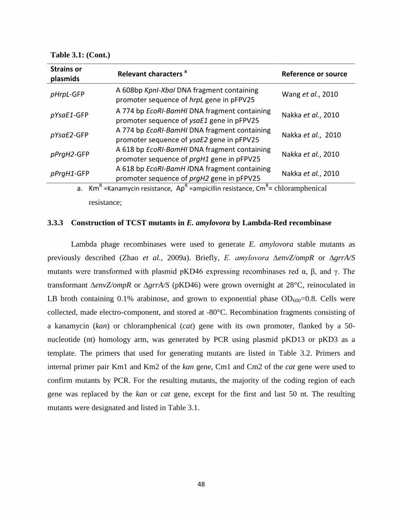
48
Table 3.1: (Cont.)
Strains or plasmids
Relevant characters a Reference or source
pHrpL-GFP A 608bp KpnI-XbaI DNA fragment containing promoter sequence of hrpL gene in pFPV25
Wang et al., 2010
pYsaE1-GFP A 774 bp EcoRI-BamHI DNA fragment containing promoter sequence of ysaE1 gene in pFPV25
Nakka et al., 2010
pYsaE2-GFP A 774 bp EcoRI-BamHI DNA fragment containing promoter sequence of ysaE2 gene in pFPV25
Nakka et al., 2010
pPrgH2-GFP A 618 bp EcoRI-BamHI DNA fragment containing promoter sequence of prgH1 gene in pFPV25
Nakka et al., 2010
pPrgH1-GFP A 618 bp EcoRI-BamH IDNA fragment containing promoter sequence of prgH2 gene in pFPV25
Nakka et al., 2010
a. KmR =Kanamycin resistance, ApR =ampicillin resistance, CmR= chloramphenical
resistance;
3.3.3 Construction of TCST mutants in E. amylovora by Lambda-Red recombinase
Lambda phage recombinases were used to generate E. amylovora stable mutants as
previously described (Zhao et al., 2009a). Briefly, E. amylovora ∆envZ/ompR or ∆grrA/S
mutants were transformed with plasmid pKD46 expressing recombinases red α, β, and γ. The
transformant ∆envZ/ompR or ∆grrA/S (pKD46) were grown overnight at 28°C, reinoculated in
LB broth containing 0.1% arabinose, and grown to exponential phase OD600=0.8. Cells were
collected, made electro-component, and stored at -80°C. Recombination fragments consisting of
a kanamycin (kan) or chloramphenical (cat) gene with its own promoter, flanked by a 50-
nucleotide (nt) homology arm, was generated by PCR using plasmid pKD13 or pKD3 as a
template. The primers that used for generating mutants are listed in Table 3.2. Primers and
internal primer pair Km1 and Km2 of the kan gene, Cm1 and Cm2 of the cat gene were used to
confirm mutants by PCR. For the resulting mutants, the majority of the coding region of each
gene was replaced by the kan or cat gene, except for the first and last 50 nt. The resulting
mutants were designated and listed in Table 3.1.
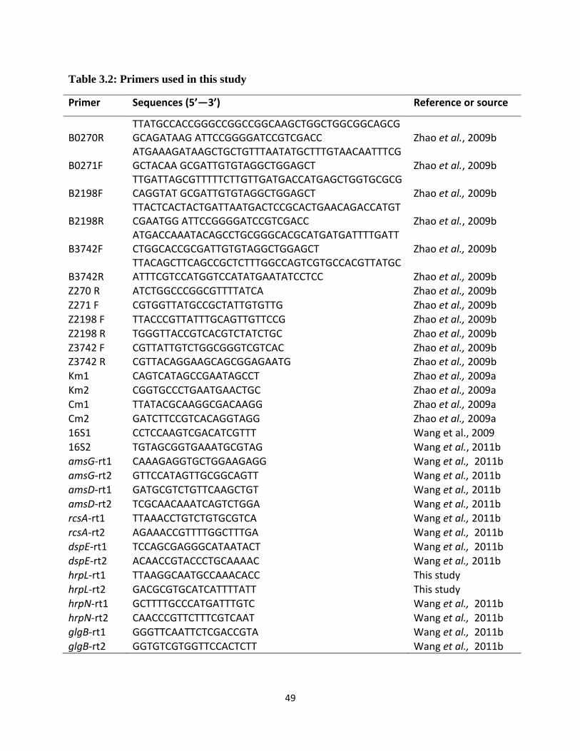
49
Table 3.2: Primers used in this study
Primer Sequences (5’—3’) Reference or source
B0270R TTATGCCACCGGGCCGGCCGGCAAGCTGGCTGGCGGCAGCGGCAGATAAG ATTCCGGGGATCCGTCGACC Zhao et al., 2009b
B0271F ATGAAAGATAAGCTGCTGTTTAATATGCTTTGTAACAATTTCGGCTACAA GCGATTGTGTAGGCTGGAGCT Zhao et al., 2009b
B2198F TTGATTAGCGTTTTTCTTGTTGATGACCATGAGCTGGTGCGCGCAGGTAT GCGATTGTGTAGGCTGGAGCT Zhao et al., 2009b
B2198R TTACTCACTACTGATTAATGACTCCGCACTGAACAGACCATGTCGAATGG ATTCCGGGGATCCGTCGACC Zhao et al., 2009b
B3742F ATGACCAAATACAGCCTGCGGGCACGCATGATGATTTTGATTCTGGCACCGCGATTGTGTAGGCTGGAGCT Zhao et al., 2009b
B3742R TTACAGCTTCAGCCGCTCTTTGGCCAGTCGTGCCACGTTATGCATTTCGTCCATGGTCCATATGAATATCCTCC Zhao et al., 2009b
Z270 R ATCTGGCCCGGCGTTTTATCA Zhao et al., 2009b Z271 F CGTGGTTATGCCGCTATTGTGTTG Zhao et al., 2009b Z2198 F TTACCCGTTATTTGCAGTTGTTCCG Zhao et al., 2009b Z2198 R TGGGTTACCGTCACGTCTATCTGC Zhao et al., 2009b Z3742 F CGTTATTGTCTGGCGGGTCGTCAC Zhao et al., 2009b Z3742 R CGTTACAGGAAGCAGCGGAGAATG Zhao et al., 2009b Km1 CAGTCATAGCCGAATAGCCT Zhao et al., 2009a Km2 CGGTGCCCTGAATGAACTGC Zhao et al., 2009a Cm1 TTATACGCAAGGCGACAAGG Zhao et al., 2009a
Cm2 GATCTTCCGTCACAGGTAGG Zhao et al., 2009a 16S1 CCTCCAAGTCGACATCGTTT Wang et al., 2009 16S2 TGTAGCGGTGAAATGCGTAG Wang et al., 2011b amsG-rt1 CAAAGAGGTGCTGGAAGAGG Wang et al., 2011b amsG-rt2 GTTCCATAGTTGCGGCAGTT Wang et al., 2011b
amsD-rt1 GATGCGTCTGTTCAAGCTGT Wang et al., 2011b amsD-rt2 TCGCAACAAATCAGTCTGGA Wang et al., 2011b rcsA-rt1 TTAAACCTGTCTGTGCGTCA Wang et al., 2011b rcsA-rt2 AGAAACCGTTTTGGCTTTGA Wang et al., 2011b dspE-rt1 TCCAGCGAGGGCATAATACT Wang et al., 2011b dspE-rt2 ACAACCGTACCCTGCAAAAC Wang et al., 2011b
hrpL-rt1 TTAAGGCAATGCCAAACACC This study hrpL-rt2 GACGCGTGCATCATTTTATT This study hrpN-rt1 GCTTTTGCCCATGATTTGTC Wang et al., 2011b hrpN-rt2 CAACCCGTTCTTTCGTCAAT Wang et al., 2011b glgB-rt1 GGGTTCAATTCTCGACCGTA Wang et al., 2011b glgB-rt2 GGTGTCGTGGTTCCACTCTT Wang et al., 2011b
50
3.3.4 Virulence assays on apple plants and immature pear fruit
In brief, E. amylovora WT and mutant strains were grown overnight in LB broth,
harvested by centrifugation, and resuspended in ½*sterile phosphate buffered-saline (PBS) with
bacterial cells adjusted to OD600 = 0.001 in ½*PBS. Immature fruits of pear (Pyruscommunis L.
cv Bartlett) were surface-sterilized with 10% bleach, dried in a flow hood and pricked with a
sterile needle followed by the standard procedure (Zhao et al., 2005). Two μl of cell suspensions
was inoculated on the wounded tissue and incubated pears in a humidified chamber at 26°C.
Symptoms were recorded at 4 and 8 days post-inoculation. For each strain tested, fruits were
assayed in triplicate, and each experiment was performed at least three times.
Apple shoot virulence assays were performed on young annual shoots of “gala” apple, 22
to 25 cm in length. After pricking the tip with a sterilized needle, five μl of pathogen suspension
with an initial OD600=0.1 was pipetted onto the wounded tissue. For each bacterial strain, seven
shoots were inoculated. Plants were kept in a greenhouse at 25°C and a 16 hours light
photoperiod, and periodically evaluated for disease development for up to 7 days following
inoculation by measuring length of the necrotic tissue. The experiment was performed at least
three times.
3.3.5 CPC assay for determining amylovoran concentration
The amylovoran concentration in supernatants of bacterial cultures was quantitatively
determined by a turbidity assay with cetylpyrimidinium chloride (CPC) as described (Hildebrand
et al., 2006). Briefly, E. amylovora WT and mutant strains were grown overnight in LB broth
and washed with ½*PBS three times. After the final wash, the bacteria pellet was resuspended in
200 μl PBS and inoculated into 5ml MBMA+1% sorbitol medium with an initial OD600=0.2. One
ml of bacterial cells was pelleted 24 hours after inoculation at 28°C with shaking. Following
centrifugation, 50 μl CPC at 50 mg /ml was added to one ml supernatant, incubated 10 min at
room temperature, and determined amylovoran concentration by measuring OD600 turbidity. The
final concentration of amylovoran production was normalized for a cell density of 1.0. For each
strain tested, the experiment was repeated at least three times.
51
3.3.6 Levan production
Levansucrase activity was carried out as previously described (Bellemann et al., 1994).
For E. amylovora WT and mutants, bacteria were grown overnight in LB broth, harvested by
centrifugation. One ml of cell-free supernatant was added to 1 ml of LS buffer (50 mM Na3PO4,
2 M sucrose, and 0.05% NaN3). After 24 h of incubation at 37°C, absorbance was determined at
OD590. Each experiment included four biological replicates per strain, and the experiment was
performed three times.
3.3.7 Swarming motility assay
For E. amylovora WT and mutant strains, bacteria were grown overnight in LB broth,
harvested by centrifugation, washed with PBS once, and resuspended in 200 μl PBS. Then,
bacterial suspensions were diluted 100×water, and five μl of the diluted bacterial suspension was
plated onto the center of swarming agar plates (10 g tryptone, 5 g NaCl, 3 g agar per l Liter) as
previously described (Zhao et al., 2009b;Skerker et al., 2005). Swarming diameters were
determined following incubation at 28°C for up to three days. The experiment was repeated at
least three times.
3.3.8 GFP reporter gene assay by flow cytometry
The BD FAC SCanto flow cytometer was used to monitor GFP intensity of WT and
mutant strains containing the corresponding promoter-GFP construct (Zhao et al., 2009b). For in
vitro amsG gene expression, WT and mutants containing the amsG promoter-GFP fusion
plasmid were grown in LB overnight, harvested, and resuspended in ½*PBS. Bacterial
suspensions were reinoculated in MBMA broth with 1% sorbitol and grown at 28°C with
shaking for up to two days. Bacterial cultures were then harvested by centrifugation, washed
once with ½*PBS, and resuspended in PBS for flow cytometry assay. For in vitro dspE, hrpL,
and hrpN gene expression, WT and mutants containing the dspE, hrpL and hrpN promoter-GFP
fusion plasmid were grown in LB overnight, harvested, and resuspended and washed twice with
hrp-inducing minimal medium and then resuspended in ½*PBS, respectively. Bacterial
suspensions then were reinoculated in hrp-inducing minimal medium and grown at 18°C with
shaking for 18 hours for flow cytometry assay. For in vitro ysaE1, ysaE2, prgH1 and prgH2 gene
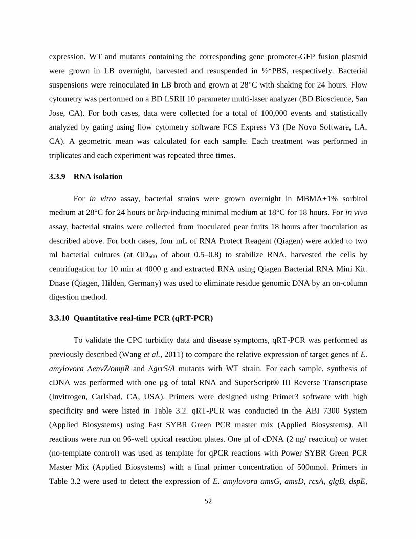
52
expression, WT and mutants containing the corresponding gene promoter-GFP fusion plasmid
were grown in LB overnight, harvested and resuspended in ½*PBS, respectively. Bacterial
suspensions were reinoculated in LB broth and grown at 28°C with shaking for 24 hours. Flow
cytometry was performed on a BD LSRII 10 parameter multi-laser analyzer (BD Bioscience, San
Jose, CA). For both cases, data were collected for a total of 100,000 events and statistically
analyzed by gating using flow cytometry software FCS Express V3 (De Novo Software, LA,
CA). A geometric mean was calculated for each sample. Each treatment was performed in
triplicates and each experiment was repeated three times.
3.3.9 RNA isolation
For in vitro assay, bacterial strains were grown overnight in MBMA+1% sorbitol
medium at 28°C for 24 hours or hrp-inducing minimal medium at 18°C for 18 hours. For in vivo
assay, bacterial strains were collected from inoculated pear fruits 18 hours after inoculation as
described above. For both cases, four mL of RNA Protect Reagent (Qiagen) were added to two
ml bacterial cultures (at OD600 of about 0.5–0.8) to stabilize RNA, harvested the cells by
centrifugation for 10 min at 4000 g and extracted RNA using Qiagen Bacterial RNA Mini Kit.
Dnase (Qiagen, Hilden, Germany) was used to eliminate residue genomic DNA by an on-column
digestion method.
3.3.10 Quantitative real-time PCR (qRT-PCR)
To validate the CPC turbidity data and disease symptoms, qRT-PCR was performed as
previously described (Wang et al., 2011) to compare the relative expression of target genes of E.
amylovora ∆envZ/ompR and ∆grrS/A mutants with WT strain. For each sample, synthesis of
cDNA was performed with one µg of total RNA and SuperScript® III Reverse Transcriptase
(Invitrogen, Carlsbad, CA, USA). Primers were designed using Primer3 software with high
specificity and were listed in Table 3.2. qRT-PCR was conducted in the ABI 7300 System
(Applied Biosystems) using Fast SYBR Green PCR master mix (Applied Biosystems). All
reactions were run on 96-well optical reaction plates. One µl of cDNA (2 ng/ reaction) or water
(no-template control) was used as template for qPCR reactions with Power SYBR Green PCR
Master Mix (Applied Biosystems) with a final primer concentration of 500nmol. Primers in
Table 3.2 were used to detect the expression of E. amylovora amsG, amsD, rcsA, glgB, dspE,
53
hrpL, and hrpN gene, respectively. Technical replicate experiments were performed for each
biological triplicate sample. The thermal cycling conditions included a step of 10 min at 95°C,
followed by 40 cycles of 95°C for 15 sec and 60°C for 1 min. The relative quantification (∆∆Ct)
method was used to determine expression level of selected genes and 16S rRNA (rrsA) gene was
used as an endogenous control to normalize our samples (∆Ct = Cttarget−CtrssA). A relative
quantification (RQ) value was calculated as 2-(∆∆Ct = ∆Cttarget−∆Ctreference)
for each gene with the
control group as a reference. A p-value was computed using a moderated t-test to measure the
significance associated with each RQ value. Variations were considered statistically significant
when the p-value was <0.05. RQ values for envZ/ompR and grrS/A single, double and triple
mutants were then normalized to those of WT (Wang et al., 2011).
3.3.11 Statistical analysis
One-way ANOVA and Student-Newman-Keuls test were used to determine differences
in virulence progress, amylovoran production, gene expression and swarming motility data
means within a = 0.05, analyzed by SAS 9.2 program.
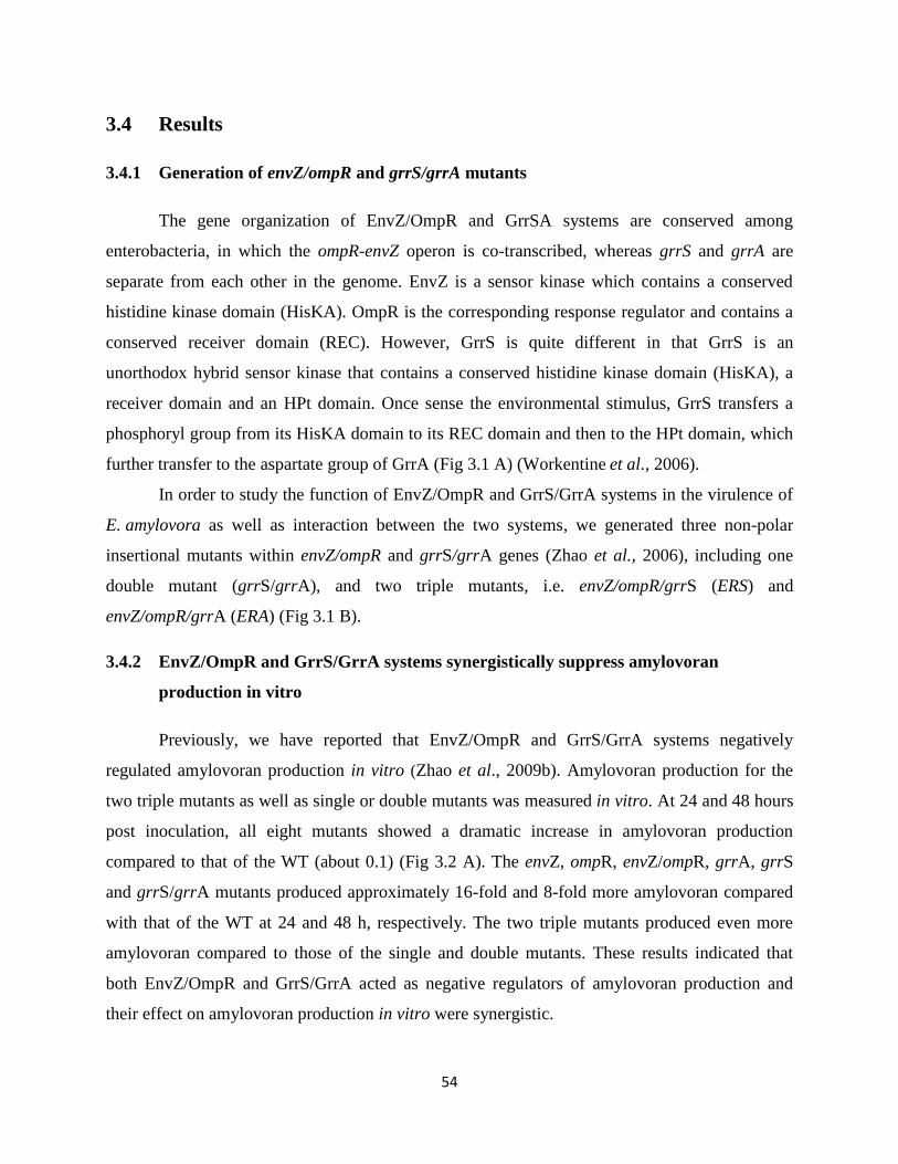
54
3.4 Results
3.4.1 Generation of envZ/ompR and grrS/grrA mutants
The gene organization of EnvZ/OmpR and GrrSA systems are conserved among
enterobacteria, in which the ompR-envZ operon is co-transcribed, whereas grrS and grrA are
separate from each other in the genome. EnvZ is a sensor kinase which contains a conserved
histidine kinase domain (HisKA). OmpR is the corresponding response regulator and contains a
conserved receiver domain (REC). However, GrrS is quite different in that GrrS is an
unorthodox hybrid sensor kinase that contains a conserved histidine kinase domain (HisKA), a
receiver domain and an HPt domain. Once sense the environmental stimulus, GrrS transfers a
phosphoryl group from its HisKA domain to its REC domain and then to the HPt domain, which
further transfer to the aspartate group of GrrA (Fig 3.1 A) (Workentine et al., 2006).
In order to study the function of EnvZ/OmpR and GrrS/GrrA systems in the virulence of
E. amylovora as well as interaction between the two systems, we generated three non-polar
insertional mutants within envZ/ompR and grrS/grrA genes (Zhao et al., 2006), including one
double mutant (grrS/grrA), and two triple mutants, i.e. envZ/ompR/grrS (ERS) and
envZ/ompR/grrA (ERA) (Fig 3.1 B).
3.4.2 EnvZ/OmpR and GrrS/GrrA systems synergistically suppress amylovoran
production in vitro
Previously, we have reported that EnvZ/OmpR and GrrS/GrrA systems negatively
regulated amylovoran production in vitro (Zhao et al., 2009b). Amylovoran production for the
two triple mutants as well as single or double mutants was measured in vitro. At 24 and 48 hours
post inoculation, all eight mutants showed a dramatic increase in amylovoran production
compared to that of the WT (about 0.1) (Fig 3.2 A). The envZ, ompR, envZ/ompR, grrA, grrS
and grrS/grrA mutants produced approximately 16-fold and 8-fold more amylovoran compared
with that of the WT at 24 and 48 h, respectively. The two triple mutants produced even more
amylovoran compared to those of the single and double mutants. These results indicated that
both EnvZ/OmpR and GrrS/GrrA acted as negative regulators of amylovoran production and
their effect on amylovoran production in vitro were synergistic.
55
Figure 3.1 Schematic map of genes and proteins of EnvZ/OmpR and GrrS/GrrA systems in
Erwinia amylovora and generation of insertional ERA, ERS and grrS/grrA deletion mutants.
A) Domain organization of EnvZ, OmpR, GrrS and GrrA proteins. Domain limits for proteins
are derived from the SMART program. Not drawn to scale. HisKA, Histidine Kinase A domain;
REC, Receiver domain; HATPase_c, Histidine kinase-like ATPases; HPT: Histidine
Phosphotransfer domain; HTH_LUXR domain, helix_turn_helix, Lux regulon. B) Organizations
of envZ, ompR, grrS and grrA genes and generation of insertional ERA, ERS and grrS/grrA
deletion mutants. Arrowheads show orientations. kan, kanamycin resistant gene; cat,
chloramphenicol resistant gene.
A
B
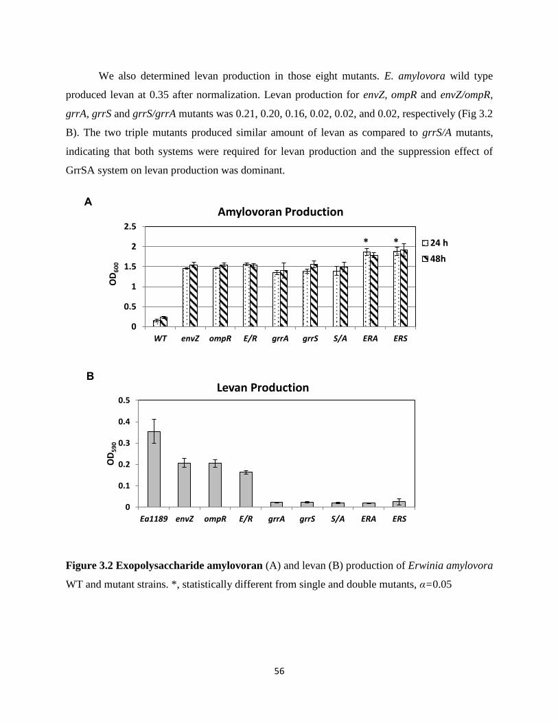
56
We also determined levan production in those eight mutants. E. amylovora wild type
produced levan at 0.35 after normalization. Levan production for envZ, ompR and envZ/ompR,
grrA, grrS and grrS/grrA mutants was 0.21, 0.20, 0.16, 0.02, 0.02, and 0.02, respectively (Fig 3.2
B). The two triple mutants produced similar amount of levan as compared to grrS/A mutants,
indicating that both systems were required for levan production and the suppression effect of
GrrSA system on levan production was dominant.
Figure 3.2 Exopolysaccharide amylovoran (A) and levan (B) production of Erwinia amylovora
WT and mutant strains. *, statistically different from single and double mutants, α=0.05
0
0.5
1
1.5
2
2.5
WT envZ ompR E/R grrA grrS S/A ERA ERS
OD
60
0
Amylovoran Production
24 h
48h
* *
0
0.1
0.2
0.3
0.4
0.5
Ea1189 envZ ompR E/R grrA grrS S/A ERA ERS
OD
59
0
Levan Production
A
B
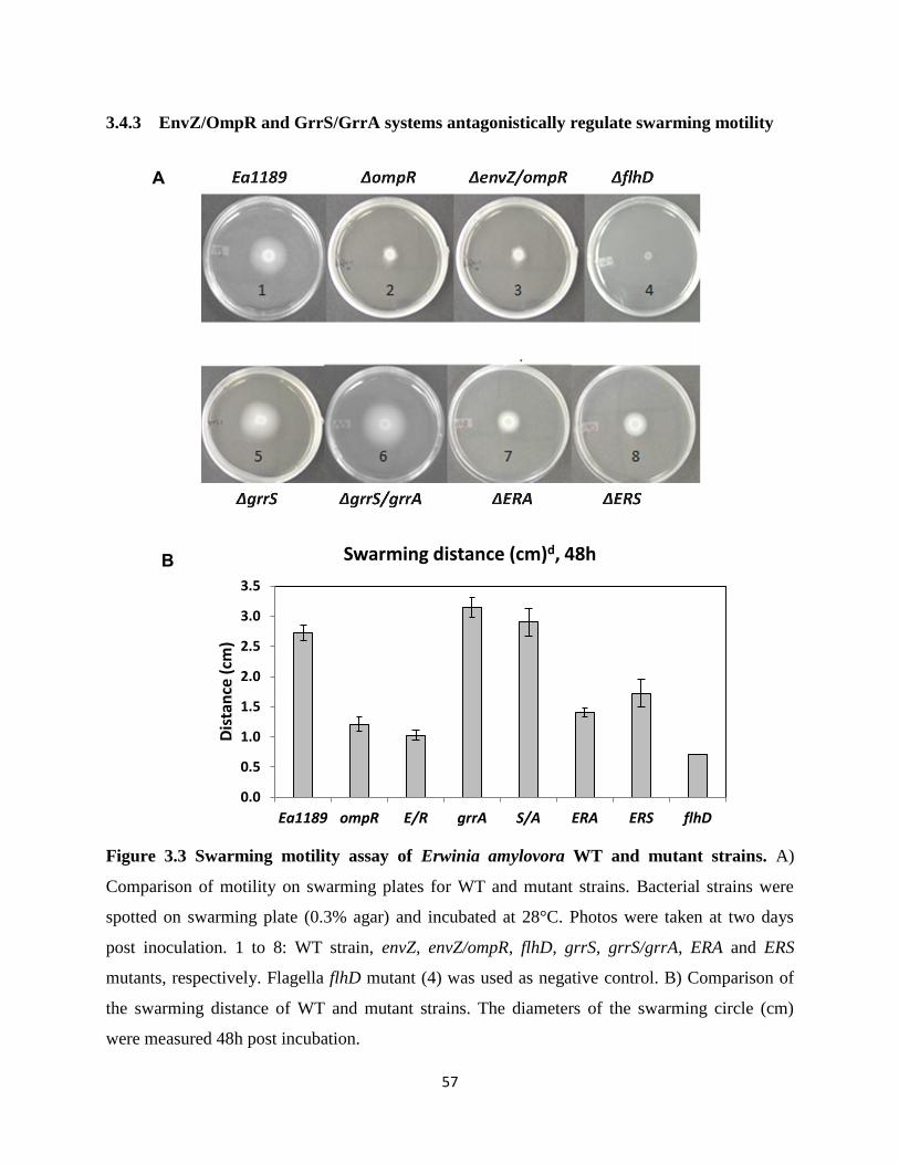
57
3.4.3 EnvZ/OmpR and GrrS/GrrA systems antagonistically regulate swarming motility
Figure 3.3 Swarming motility assay of Erwinia amylovora WT and mutant strains. A)
Comparison of motility on swarming plates for WT and mutant strains. Bacterial strains were
spotted on swarming plate (0.3% agar) and incubated at 28°C. Photos were taken at two days
post inoculation. 1 to 8: WT strain, envZ, envZ/ompR, flhD, grrS, grrS/grrA, ERA and ERS
mutants, respectively. Flagella flhD mutant (4) was used as negative control. B) Comparison of
the swarming distance of WT and mutant strains. The diameters of the swarming circle (cm)
were measured 48h post incubation.
0.0
0.5
1.0
1.5
2.0
2.5
3.0
3.5
Ea1189 ompR E/R grrA S/A ERA ERS flhD
Dis
tan
ce (
cm)
Swarming distance (cm)d, 48h
A
B

58
In our previous study, we have reported that EnvZ/OmpR system acts as a positive
regulator and GrrS/A system as a negative regulator of swarming motility (Zhao et al., 2009b).
Motility of the two triple mutants was evaluated using a swarming plate assay. As expected,
grrS/grrA double mutant strains showed precocious swarming and moved faster than the WT
strain as grrA single mutants, with a diameter of 3.0 cm within 48 hours compared to 2.7 cm for
the WT strain, from an original spot of 0.6 cm in diameter (Fig 3.3A). Two triple mutants
showed intermediate circular and swarming phenotype (Fig 3.3 B), i.e. the swarming distances
were 1.5 and 1.8 cm at 48 hours. The data clearly indicate that the regulatory role of
EnvZ/OmpR and GrrS/A on E. amylovora swarming motility and further suggested that their
effect were antagonistic.
3.4.4 Mutations in envZ/ompR and grrS/grrA render Erwinia amylovora slightly more
pathogenic
To determine the role of EnvZ/OmpR and GrrS/GrrA systems in E. amylovora virulence,
we conducted virulence assays for envZ/ompR, grrS/grrA, ERS and ERA mutants on both
immature pear fruits and apple shoots (Zhao et al., 2005; Zhao et al., 2006). At 2 days following
inoculation, E. amylovora WT strain Ea1189 produced water soaking symptoms on pears with
visible bacterial ooze. Four days after inoculation, Ea1189-inoculated immature pears showed
necrotic lesions and bacterial ooze formation. After 8 days, the necrosis turned black with more
ooze production at the inoculation site. Similar disease progresses were observed on immature
pear fruits inoculated with envZ/ompR and grrS/grrA double mutants as WT strain (Fig 3.2).
Meanwhile, similar disease symptom and ooze formation was observed on immature pear fruits
inoculated with the two triple mutants.
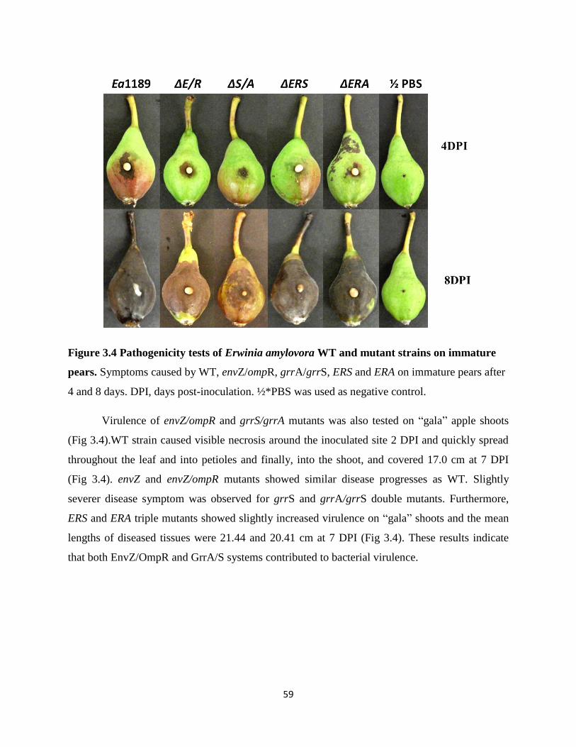
59
Figure 3.4 Pathogenicity tests of Erwinia amylovora WT and mutant strains on immature
pears. Symptoms caused by WT, envZ/ompR, grrA/grrS, ERS and ERA on immature pears after
4 and 8 days. DPI, days post-inoculation. ½*PBS was used as negative control.
Virulence of envZ/ompR and grrS/grrA mutants was also tested on “gala” apple shoots
(Fig 3.4).WT strain caused visible necrosis around the inoculated site 2 DPI and quickly spread
throughout the leaf and into petioles and finally, into the shoot, and covered 17.0 cm at 7 DPI
(Fig 3.4). envZ and envZ/ompR mutants showed similar disease progresses as WT. Slightly
severer disease symptom was observed for grrS and grrA/grrS double mutants. Furthermore,
ERS and ERA triple mutants showed slightly increased virulence on “gala” shoots and the mean
lengths of diseased tissues were 21.44 and 20.41 cm at 7 DPI (Fig 3.4). These results indicate
that both EnvZ/OmpR and GrrA/S systems contributed to bacterial virulence.
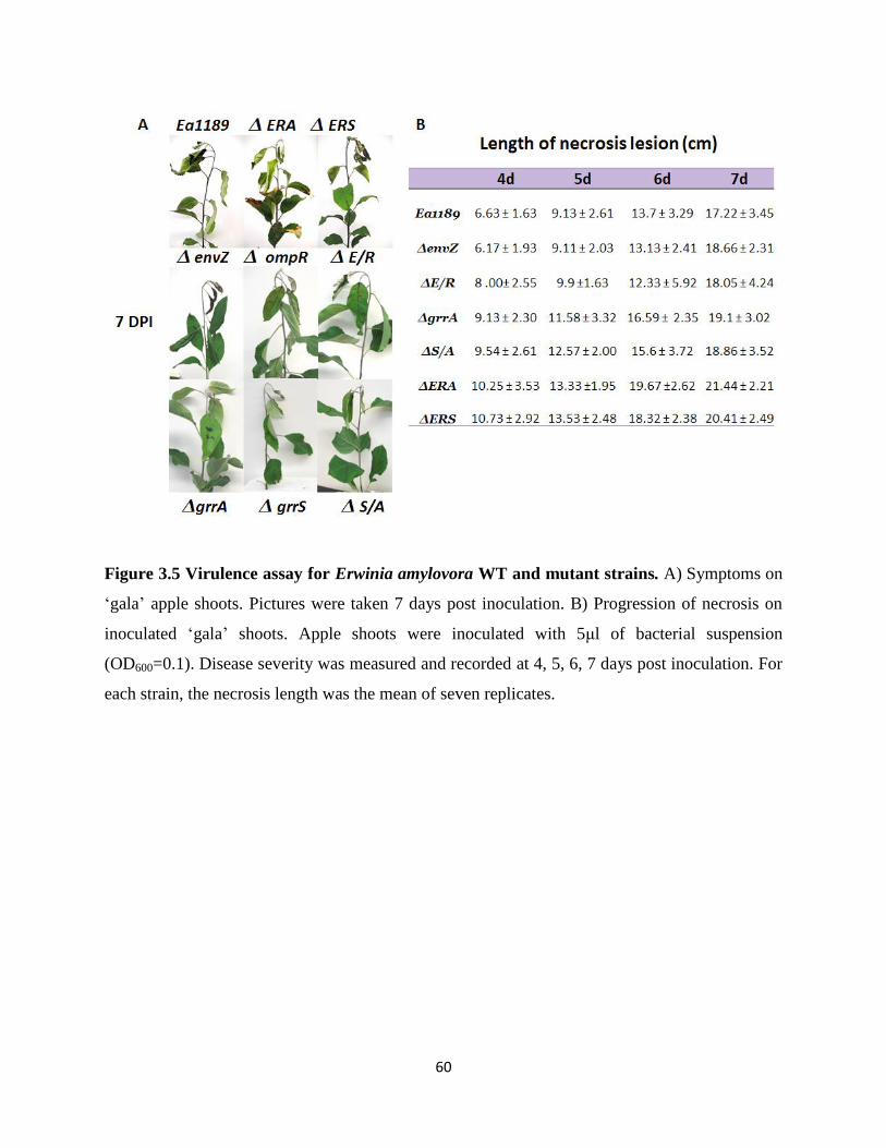
60
Figure 3.5 Virulence assay for Erwinia amylovora WT and mutant strains. A) Symptoms on
‘gala’ apple shoots. Pictures were taken 7 days post inoculation. B) Progression of necrosis on
inoculated ‘gala’ shoots. Apple shoots were inoculated with 5μl of bacterial suspension
(OD600=0.1). Disease severity was measured and recorded at 4, 5, 6, 7 days post inoculation. For
each strain, the necrosis length was the mean of seven replicates.
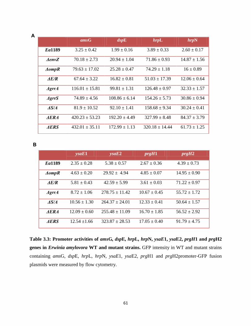
61
ysaE1 ysaE2 prgH1 prgH2
Ea1189 2.35 ± 0.28 5.38 ± 0.57 2.67 ± 0.36 4.39 ± 0.73
ΔompR 4.63 ± 0.20 29.92 ± 4.94 4.85 ± 0.07 14.95 ± 0.90
ΔE/R 5.81 ± 0.43 42.59 ± 5.99 3.61 ± 0.03 71.22 ± 0.97
ΔgrrA 8.72 ± 1.06 278.75 ± 11.42 10.67 ± 0.45 55.72 ± 1.72
ΔS/A 10.56 ± 1.30 264.37 ± 24.01 12.33 ± 0.41 50.64 ± 1.57
ΔERA 12.09 ± 0.60 255.48 ± 11.09 16.70 ± 1.85 56.52 ± 2.92
ΔERS 12.54 ±1.66 323.87 ± 28.53 17.05 ± 0.40 91.79 ± 4.75
Table 3.3: Promoter activities of amsG, dspE, hrpL, hrpN, ysaE1, ysaE2, prgH1 and prgH2
genes in Erwinia amylovora WT and mutant strains. GFP intensity in WT and mutant strains
containing amsG, dspE, hrpL, hrpN, ysaE1, ysaE2, prgH1 and prgH2promoter-GFP fusion
plasmids were measured by flow cytometry.
amsG dspE hrpL hrpN
Ea1189 3.25 ± 0.42 1.99 ± 0.16 3.89 ± 0.33 2.60 ± 0.17
ΔenvZ 70.18 ± 2.73 20.94 ± 1.04 71.86 ± 0.93 14.87 ± 1.56
ΔompR 79.63 ± 17.02 25.28 ± 0.47 74.29 ± 1.18 16 ± 0.89
ΔE/R 67.64 ± 3.22 16.82 ± 0.81 51.03 ± 17.39 12.06 ± 0.64
ΔgrrA 116.01 ± 15.81 99.81 ± 1.31 126.48 ± 0.97 32.33 ± 1.57
ΔgrrS 74.89 ± 4.56 108.86 ± 6.14 154.26 ± 5.73 30.86 ± 0.94
ΔS/A 81.9 ± 10.52 92.10 ± 1.41 158.68 ± 9.34 30.24 ± 0.41
ΔERA 420.23 ± 53.23 192.20 ± 4.49 327.99 ± 8.48 84.37 ± 3.79
ΔERS 432.01 ± 35.11 172.99 ± 1.13 320.18 ± 14.44 61.73 ± 1.25
B
A

62
3.4.5 EnvZ/OmpR and GrrS/GrrA systems synergistically regulate expression of
amylovoran biosynthetic and T3SS genes in vitro
To correlate amylovoran production with amylovoran biosynthesis gene expression,
promoter activity of amsG gene was measured using GFP as a reporter in WT and eight mutants
using flow cytometry (Zhao et al., 2009b). The amsG gene was expressed at a basal level in WT
strain, with a GFP intensity value of 3.25 (geometric mean), compared to a geometric mean
value of 1.5 for the vector control. The intensity of the amsG promoter was 70.18, 79.63, 67.64,
116.01, 74.89 and 81.9 for envZ, ompR, envZ/ompR, grrS, grrA and grrS/grrA mutants,
respectively. A significant increase in amsG promoter activity of two triple mutants (ERS, ERA)
was observed, with a geometric mean value of 432.01 and 420.23, respectively (Table 3.3).
These results indicated that there is a synergistic effect of EnvZ/OmpR and GrrA/S systems on
amylovoran biosynthetic gene expression in E. amylovora.
Next, we determined the promoter activities for three T3SS genes in WT and mutant
strains. T3SS genes were expressed at a basal level in WT strain, with a GFP intensity value of
1.99, 3.89 and 2.60 (geometric mean) for dspE, hrpL and hrpN, respectively (Table 3.3 A). The
geometric mean values of the GFP intensity for dspE, hrpL and hrpN promoters were all
increased in envZ, ompR, envZ/ompR, grrS, grrA and grrS/grrA mutants. For the two triple
mutants, geometric mean values of the GFP intensity for dspE, hrpL and hrpN promoters
reached highest at192.20, 327.99, 84.37 in ERS mutant, and 172.99, 320.18 and 61.73 in ERA
mutant, respectively (Table 3.3A). These data indicated that EnvZ/OmpR and GrrA/S systems
negatively regulate T3SS gene expression in vitro in a synergistic way.
The promoter activities of two other PAIs (PAI-2 and PAI-3) T3SS genes, including
ysaE1, ysaE2, prgH1 and prgH2 were also measured by GFP-promoter fusion reporter in LB
medium. These genes were expressed at a basal level in WT strain, with a GFP intensity value of
2.35, 5.38, 2.67 and 4.39 (geometric mean) for ysaE1, ysaE2, prgH1 and prgH2, respectively
(Table 3.3 B). Interestingly, all six (2 single, 2 double and 2 triple) mutants showed increased
GFP intensity value for all four genes, especially in the triple mutants. These results suggest that
PAI-2 and PAI-3 genes were all negatively regulated by EnvZ/OmpR and GrrSA systems in
vitro in similar way as other hrp-T3SS genes.

63
.
Figure 3.6 Gene expression of selected genes determined by qRT-PCR in vitro. The relative
fold change of each gene was derived from the comparison of each mutant versus wild type in
MBMA +1% sorbitol medium (A), and Hrp inducing minimal medium (B). 16S rRNA gene was
used as endogenous controls.
qRT-PCR was also performed to confirm the in vitro promoter activity results. Total
mRNA of WT and mutant strains extracted from MBMA+1% sorbitol medium was used to test
the expression of amylovoran biosynthetic genes (amsG and amsD), amylovoran regulatory gene
0
2
4
6
8
10
12
14
amsG amsD glgB rcsA
Fold
Ch
ange
MBMA+1%sorbitol Medium OmpR
E/R
grrA
S/A
ERA
ERS
0
5
10
15
20
25
dspE hrpL hrpN
Fold
Ch
ange
HrpMM, 18h
OmpR
E/R
grrA
S/A
ERA
ERS
A
B
64
(rcsA) and glycogen branching enzyme gene (glgB). As expected, all genes were upregulated in
six mutants more than two folds. It also revealed that amsG was up-regulated by 5.3- and 11.7-
fold in ERA and ERS mutants, respectively. Similarly, amsD was up-regulated 4.9- and 5.1- fold,
respectively in the two triple mutants. Expression of glgB gene showed similar increase as amsD
gene with more than two folds in the six mutants (Fig 3.6A). These results suggested that
EnvZ/OmpR and GrrSA synergistically affect amylovoran gene expression.
Consistently, T3SS genes (dspE, hrpL and hrpN) showed up-regulated expression in
ompR, envZ/ompR, grrA, grrS/grrA, ERA and ERS mutants as compared with WT. Expression of
dspE was up-regulated about 4.2-, 3.4-, 5.8-, 10.0-, 17.4- and 11.7-fold higher in the six mutants
than wild type, respectively. Similarly, expression of hrpL was up-regulated by 2.0-, 3.0-, 3.7-,
3.1-, 9.1- and 10- fold in ompR, envZ/ompR, grrA, grrS/grrA, ERA and ERS mutants,
respectively. Furthermore, expression of hrpN was significantly higher in the ERA and ERS
mutants, up15.1- and 12.0-fold, as compared to 2.6- to 7.4-fold increases in ompR, envZ/ompR,
grrA and grrS/grrA mutants (Fig 3.6 B). These data verified the synergistic effect of
EnvZ/OmpR and GrrSA on T3SS gene expression.
3.4.6 EnvZ/OmpR and GrrS/GrrA systems synergistically regulate expression of T3SS
genes in vivo
As shown in Fig 3.7, all genes, except for the amsG and hrpL, was slightly upregulated in
the six mutants. For example, expression of amsD was 1.8-, 1.9-, 2.1-, 1.7-, 2.4- and 3.0- fold
higher in the ompR, envZ/ompR, grrA, grrS/grrA, ERA and ERS mutants as compared to the WT,
respectively, while amsG expressed only slightly higher than the WT strain. Meanwhile, glgB
gene expressed more than two folds higher in the six mutants than in WT. Similarly, expression
of dspE and hrpN was up-regulated in ompR, envZ/ompR, grrA, grrS/grrA, ERA and ERS
mutants. However, expression hrpL didn’t appear to be significant different between the six
mutants and WT at 18 h (Fig 3.7).
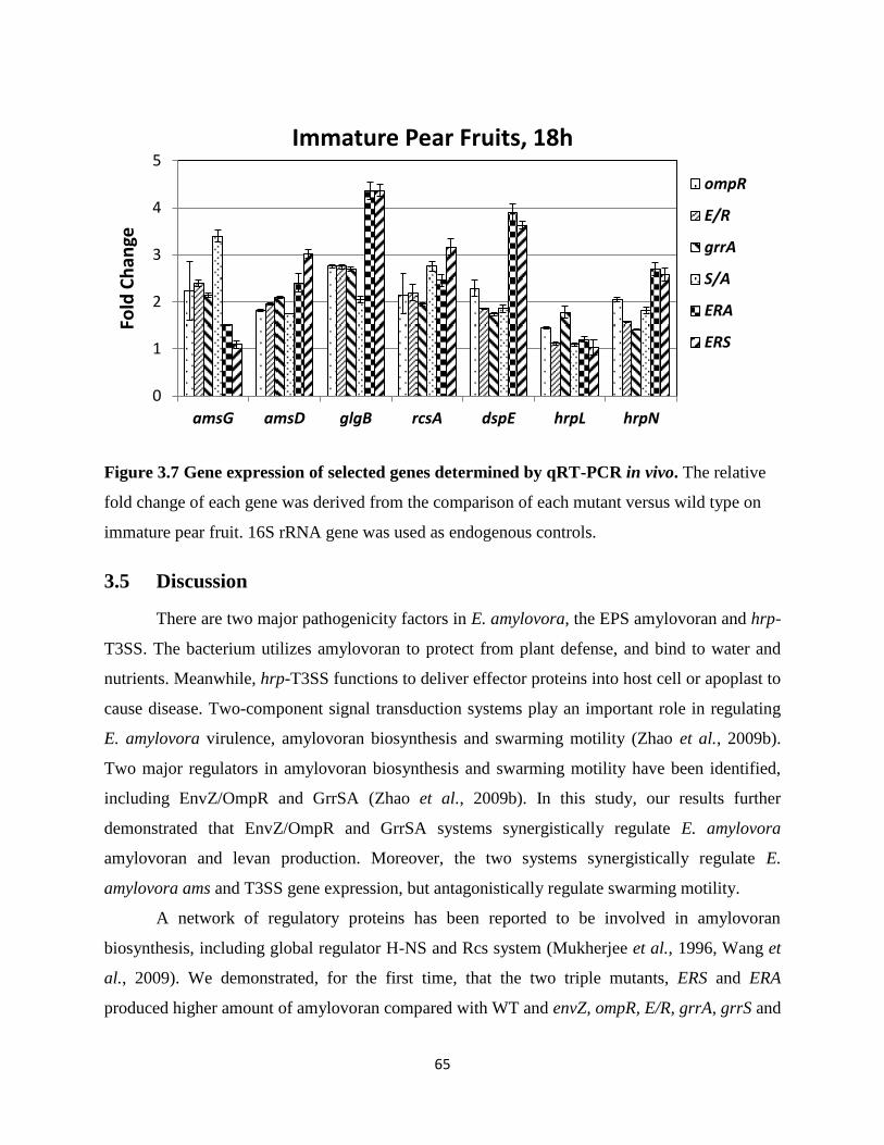
65
Figure 3.7 Gene expression of selected genes determined by qRT-PCR in vivo. The relative
fold change of each gene was derived from the comparison of each mutant versus wild type on
immature pear fruit. 16S rRNA gene was used as endogenous controls.
3.5 Discussion
There are two major pathogenicity factors in E. amylovora, the EPS amylovoran and hrp-
T3SS. The bacterium utilizes amylovoran to protect from plant defense, and bind to water and
nutrients. Meanwhile, hrp-T3SS functions to deliver effector proteins into host cell or apoplast to
cause disease. Two-component signal transduction systems play an important role in regulating
E. amylovora virulence, amylovoran biosynthesis and swarming motility (Zhao et al., 2009b).
Two major regulators in amylovoran biosynthesis and swarming motility have been identified,
including EnvZ/OmpR and GrrSA (Zhao et al., 2009b). In this study, our results further
demonstrated that EnvZ/OmpR and GrrSA systems synergistically regulate E. amylovora
amylovoran and levan production. Moreover, the two systems synergistically regulate E.
amylovora ams and T3SS gene expression, but antagonistically regulate swarming motility.
A network of regulatory proteins has been reported to be involved in amylovoran
biosynthesis, including global regulator H-NS and Rcs system (Mukherjee et al., 1996, Wang et
al., 2009). We demonstrated, for the first time, that the two triple mutants, ERS and ERA
produced higher amount of amylovoran compared with WT and envZ, ompR, E/R, grrA, grrS and
0
1
2
3
4
5
amsG amsD glgB rcsA dspE hrpL hrpN
Fold
Ch
ange
Immature Pear Fruits, 18h
ompR
E/R
grrA
S/A
ERA
ERS
66
grrS/A single and double mutants. These data was verified by in vitro promoter activity assay
and in vitro qRT-PCR.
Meanwhile, both EnvZ/OmpR and GrrSA TCSTs positively regulate levan production.
Mutation in either system could partially reduce levan biosynthesis, and it appeared that GrrSA
system has a dominant effect on levan production as compared with EnvZ/OmpR system since
levan production of the two triple mutants was similar to those of grrSA single and double
mutants.
On the other hand, EnvZ/OmpR and GrrSA systems antagonistically regulate E.
amylovora swarming motility. In earlier report, we have identified that GrrSA and EnvZ/OmpR
systems are negative and positive regulators of swarming motility, respectively. In this study, we
further demonstrated that these two systems function oppositely in controlling the swarming
ability of E. amylovora. The triple mutant ERS and ERA showed intermediate swarming ability.
Moreover, EnvZ/OmpR and GrrSA systems are involved in T3SS regulation. Our in vitro
promoter activity and both in vitro and in vivo qRT-PCR results indicated that both systems
negatively regulate hrp-T3SS genes expression. Similarly, the other two T3SS, PAI-2 and PAI-3,
also seemed to be negatively regulated by EnvZ/OmpR and GrrSA systems. In addition, there is
a synergistic effect between EnvZ/OmpR and GrrSA systems on T3SS gene expression.
However, when qRT-PCR assay using mRNA isolated from immature pear fruits was performed,
expression of hrpL gene was not changed. It is highly possible that hrpL mRNA level decreased
after 18 hours in vivo.
Further study is needed to investigate the mechanism of EnvZ/OmpR and GrrSA in
regulating amylovoran, swarming motility and T3SS gene expression. One possibility is through
the RNA-binding protein RsmA & small regulatory RNA rsmB system. Systematic studies of the
Rsm system in γ-proteobacteria have revealed that this system play an important role in
controlling a large variety of physiological processes including central carbon metabolism,
motility, virulence and biofilm formation, by binding to conserved sequences in its target gene
mRNAs and altering their translation and/or turnover (Romeo et al., 2012). It is likely that
GrrSA system in E. amylovora may regulate EPS production and T3SS through the RsmA/rsmB
system.
67
References
Ahmer, B. M., van Reeuwijk, J., Watson, P. R., Wallis, T. S., & Heffron, F. (1999). Salmonella
SirA is a global regulator of genes mediating enteropathogenesis. Molecular Microbiology,
31(3), 971-982.
Altier, C., Suyemoto, M., & Lawhon, S. D. (2000). Regulation of Salmonella enterica Serovar
typhimurium invasion genes by CsrA. Infection and Immunity, 68(12), 6790-6797.
Altier, C., Suyemoto, M., Ruiz, A. I., Burnham, K. D., & Maurer, R. (2000). Characterization of
two novel regulatory genes affecting Salmonella invasion gene expression. Molecular
Microbiology, 35(3), 635-646.
Apirakaramwong, A., Kashiwagi, K., Raj, V. S., Sakata, K., Kakinuma, Y., Ishihama, A.,
&Igarashi, K. (1999). Involvement of ppGpp, ribosome modulation factor, and stationary
phase-specific sigma factor sigma(S) in the decrease in cell viability caused by spermidine.
Biochemical and Biophysical Research Communications, 264(3), 643-647.
Ayers, A. R., Ayers, S. B., & Goodman, R. N. (1979). Extracellular polysaccharide of Erwinia
amylovora: A correlation with virulence. Applied and Environmental Microbiology, 38(4),
659-666.
Berry, A., DeVault, J. D., & Chakrabarty, A. M. (1989). High osmolarity is a signal for enhanced
algD transcription in mucoid and nonmucoid Pseudomonas aeruginosa strains. Journal of
Bacteriology, 171(5), 2312-2317.
Blocker, A., Komoriya, K., & Aizawa, S. (2003). Type III secretion systems and bacterial
flagella: Insights into their function from structural similarities. Proceedings of the National
Academy of Sciences of the United States of America, 100(6), 3027-3030.
Bonn, W. G., & van der Zwet, T. (2000). Distribution and economic importance of fire blight. In
J. Vanneste (Ed.), Fire blight, the disease and its causative agent Erwinia amylovora (pp.
9). UK.: CABI Wallingford.
Brzostek, K., Brzostkowska, M., Bukowska, I., Karwicka, E., & Raczkowska, A. (2007). OmpR
negatively regulates expression of invasin in Yersinia enterocolitica. Microbiology
(Reading, England), 153(Pt 8), 2416-2425.
Buban, T., Orosz-Kova´cs, Z., & Farkas, A. (2003). The nectary as the primary site of infection
by Erwinia amylovora. Plant Syst. Evol, (238), 183-194.
68
Bugert, P., & Geider, K. (1995). Molecular analysis of the ams operon required for
exopolysaccharide synthesis of Erwinia amylovora. Molecular Microbiology, 15(5), 917-
933.
Bush, M., & Dixon, R. (2012). The role of bacterial enhancer binding proteins as specialized
activators of sigma 54-dependent transcription. Microbiology and Molecular Biology
Reviews: MMBR, 76(3), 497-529.
Cai, S. J., & Inouye, M. (2002). EnvZ-OmpR interaction and osmoregulation in Escherichia coli.
The Journal of Biological Chemistry, 277(27), 24155-24161.
Campbell, E. A., Muzzin, O., Chlenov, M., Sun, J. L., Olson, C. A., Weinman, O., Trester-
Zedlitz, M. L., Darst, S.A. (2002). Structure of the bacterial RNA polymerase promoter
specificity sigma subunit. Molecular Cell, 9(3), 527-539.
Charkowski, A. O., Alfano, J. R., Preston, G., Yuan, J., He, S. Y., & Collmer, A. (1998). The
Pseudomonas syringae pv. tomato HrpW protein has domains similar to harpins and pectate
lyases and can elicit the plant hypersensitive response and bind to pectate. Journal of
Bacteriology, 180(19), 5211-5217.
Chatterjee, A., Cui, Y., & Chatterjee, A. K. (2002). Regulation of Erwinia carotovora HrpL(Ecc)
(sigma-L(Ecc)), which encodes an extracytoplasmic function subfamily of sigma factor
required for expression of the Hrp regulon. Molecular Plant-Microbe Interactions: MPMI,
15(9), 971-980.
Chong, C., & Tapper, C.D. (1971). Daily variation of sorbitol and related carbohydrates
in Malus leaves. Canadian Journal of Botany, 49, 173–177.
Cui, Y., Chatterjee, A., & Chatterjee, A. K. (2001). Effects of the two-component system
comprising GacA and GacS of Erwinia carotovora subsp. carotovora on the production of
global regulatory rsmB RNA, extracellular enzymes, and HarpinEcc. Molecular Plant-
Microbe Interactions: MPMI, 14(4), 516-526.
Damron, F. H., Owings, J. P., Okkotsu, Y., Varga, J. J., Schurr, J. R., Goldberg, J. B., Schurr, M.
J., Yu, H. D. (2012). Analysis of the Pseudomonas aeruginosa regulon controlled by the
sensor kinase KinB and sigma factor RpoN. Journal of Bacteriology, 194(6), 1317-1330.
Datsenko, K. A., & Wanner, B. L. (2000). One-step inactivation of chromosomal genes in
Escherichia coli K-12 using PCR products. Proceedings of the National Academy of
Sciences of the United States of America, 97(12), 6640-6645.

69
Dong, T. G., & Mekalanos, J. J. (2012). Characterization of the RpoN regulon reveals
differential regulation of T6SS and new flagellar operons in Vibrio cholerae O37 strain
V52. Nucleic Acids Research, 40(16), 7766-7775.
Donlan, R. M., & Costerton, J. W. (2002). Biofilms: Survival mechanisms of clinically relevant
microorganisms. Clinical Microbiology Reviews, 15(2), 167-193.
Fellay, R., Rahme, L. G., Mindrinos, M. N., Frederick, R. D., Pisi, A., & Panopoulos, N. J.
(1991). Genes and signals controlling the Pseudomonas syringae pv. phaseolocola-plant
interaction. In H. Henneke, & D. P. S. Verma (Eds.), Advances in molecular genetics of
plant-microbe interactions (pp. 45-52). Dordrecht, Netherlands: Kluwer Academic
Publishers.
Feng, X., Oropeza, R., & Kenney, L. J. (2003). Dual regulation by phospho-OmpR of ssrA/B
gene expression in Salmonella pathogenicity island 2. Molecular Microbiology, 48(4),
1131-1143.
Feng, X., Walthers, D., Oropeza, R., & Kenney, L. J. (2004). The response regulator SsrB
activates transcription and binds to a region overlapping OmpR binding sites at Salmonella
pathogenicity island 2. Molecular Microbiology, 54(3), 823-835.
Frederick, R. D., Majerczak, D. R., & Coplin, D. L. (1993). Erwinia stewartii WtsA, a positive
regulator of pathogenicity gene expression, is similar to Pseudomonas syringae pv.
phaseolicola HrpS. Molecular Microbiology, 9(3), 477-485.
Gaffney, T. D., Lam, S. T., Ligon, J., Gates, K., Frazelle, A., Di Maio, J., Hill, S., Goodwin, S.,
Torkewitz, N., & Allshouse, A.M.(1994). Global regulation of expression of antifungal
factors by a Pseudomonas fluorescens biological control strain. Molecular Plant-Microbe
Interactions: MPMI, 7(4), 455-463.
Galan, J. E., & Wolf-Watz, H. (2006). Protein delivery into eukaryotic cells by type III secretion
machines. Nature, 444(7119), 567-573.
Gao, H., Zhang, Y., Han, Y., Yang, L., Liu, X., Guo, Z., Tan, Y., Huang, X., Zhou, D., & Yang,
R. (2011). Phenotypic and transcriptional analysis of the osmotic regulator OmpR in
Yersinia pestis. BMC Microbiology, 11, 39.
Gao, R., & Stock, A. M. (2009). Biological insights from structures of two-component proteins.
Annual Review of Microbiology, 63, 133-154.
70
Geier, G., & Geider, K. (1993). Characterization and influence on virulence of the levansucrase
gene from the fire blight pathogen Erwinia amylovora. Physiological and Molecular Plant
Pathology, 42(6), 387-404.
Gophna, U., Ron, E. Z., & Graur, D. (2003). Bacterial type III secretion systems are ancient and
evolved by multiple horizontal-transfer events. Gene, 312, 151-163.
Grimm, C., Aufsatz, W., & Panopoulos, N. J. (1995). The hrpRS locus of Pseudomonas syringae
pv. phaseolicola constitutes a complex regulatory unit. Molecular Microbiology, 15(1), 155-
165.
Gruber, T. M., & Gross, C. A. (2003). Multiple sigma subunits and the partitioning of bacterial
transcription space. Annual Review of Microbiology, 57, 441-466.
He, S. Y., Nomura, K., & Whittam, T. S. (2004). Type III protein secretion mechanism in
mammalian and plant pathogens. Biochimica Et Biophysica Acta, 1694(1-3), 181-206.
Heeb, S., & Haas, D. (2001). Regulatory roles of the GacS/GacA two-component system in
plant-associated and other gram-negative bacteria. Molecular Plant-Microbe Interactions:
MPMI, 14(12), 1351-1363.
Hendrickson, E. L., Guevera, P., Penaloza-Vazquez, A., Shao, J., Bender, C., & Ausubel, F. M.
(2000). Virulence of the phytopathogen Pseudomonas syringae pv. maculicola is rpoN
dependent. Journal of Bacteriology, 182(12), 3498-3507.
Herren, C. D., Mitra, A., Palaniyandi, S. K., Coleman, A., Elankumaran, S., & Mukhopadhyay,
S. (2006). The BarA-UvrY two-component system regulates virulence in avian pathogenic
Escherichia coli O78:K80:H9.Infection and Immunity, 74(8), 4900-4909.
Hildebrand, M., Aldridge, P., & Geider, K. (2006). Characterization of hns genes from Erwinia
amylovora. Molecular Genetics and Genomics: MGG, 275(3), 310-319.
Hong, E., Doucleff, M., & Wemmer, D. E. (2009). Structure of the RNA polymerase core-
binding domain of sigma (54) reveals a likely conformational fracture point. Journal of
Molecular Biology, 390(1), 70-82.
Johnston, C., Pegues, D. A., Hueck, C. J., Lee, A., & Miller, S. I. (1996). Transcriptional
activation of Salmonella typhimurium invasion genes by a member of the phosphorylated
response-regulator superfamily. Molecular Microbiology, 22(4), 715-727.
71
Kazmierczak, M. J., Wiedmann, M., & Boor, K. J. (2005). Alternative sigma factors and their
roles in bacterial virulence. Microbiology and Molecular Biology Reviews: MMBR, 69(4),
527-543.
Kato, M., Aiba, H., & Mizuno, T. (1989). Molecular analysis by deletion and site-directed
mutagenesis of the cis-acting upstream sequence involved in activation of the ompF
promoter in Escherichia coli. Journal of Biochemistry, 105(3), 341-347.
Kato, T., Yoshida, H., Miyata, T., Maki, Y., Wada, A., & Namba, K. (2010). Structure of the
100S ribosome in the hibernation stage revealed by electron cryomicroscopy. Structure
(London, England: 1993), 18(6), 719-724.
Kim, D. J., Boylan, B., George, N., & Forst, S. (2003). Inactivation of ompR promotes
precocious swarming and flhDC expression in Xenorhabdus nematophila. Journal of
Bacteriology, 185(17), 5290-5294.
Kinscherf, T. G., & Willis, D. K. (1999). Swarming by Pseudomonas syringae B728a requires
gacS (lemA) and gacA but not the acyl-homoserine lactone biosynthetic gene ahlI. Journal
of Bacteriology, 181(13), 4133-4136.
Koczan, J. M., McGrath, M. J., Zhao, Y., & Sundin, G. W. (2009). Contribution of Erwinia
amylovora exopolysaccharides amylovoran and levan to biofilm formation: Implications in
pathogenicity. Phytopathology, 99(11), 1237-1244.
Langlotz, C., Schollmeyer, M., Coplin, D. L., Nimtz, M., & Geider, K. (2011). Biosynthesis of
the repeating units of the exopolysaccharides amylovoran from Erwinia amylovora and
stewartan from Pantoea stewartii. Physiological and Molecular Plant Pathology, 75(2011),
163-169.
Laville, J., Voisard, C., Keel, C., Maurhofer, M., Defago, G., & Haas, D. (1992). Global control
in Pseudomonas fluorescens mediating antibiotic synthesis and suppression of black root rot
of tobacco. Proceedings of the National Academy of Sciences of the United States of
America, 89(5), 1562-1566.
Leigh, J. A., & Coplin, D. L. (1992). Exopolysaccharides in plant-bacterial interactions. Annual
Review of Microbiology, 46, 307-346.
Lindgren, P. B. (1997). The role of hrp genes during plant-bacterial interactions. Annual Review
of Phytopathology, 35, 129-152.
72
Loisel, E., Jacquamet, L., Serre, L., Bauvois, C., Ferrer, J. L., Vernet, T. Di., Guilmi, A.M.,
Durmort, C. (2008). AdcAII, a new pneumococcal Zn-binding protein homologous with
ABC transporters: Biochemical and structural analysis. Journal of Molecular Biology,
381(3), 594-606.
Malik, S., Zalenskaya, K., & and Goldfarb, A. (1987). Competition between sigma factors for
core RNA polymerase. Nucl. Acids Res., 15(20), 8521-8530.
Mascher, T., Helmann, J. D., & Unden, G. (2006). Stimulus perception in bacterial signal-
transducing histidine kinases. Microbiology and Molecular Biology Reviews: MMBR, 70(4),
910-938.
Mukherjee, A., Cui, Y., Liu, Y., Dumenyo, C. K., & Chatterjee, A. K. (1996). Global regulation
in Erwinia species by Erwinia carotovora RsmA, a homologue of Escherichia coli CsrA:
Repression of secondary metabolites, pathogenicity and hypersensitive reaction.
Microbiology (Reading, England), 142(Pt 2), 427-434.
Nakka, S., Qi, M., & Zhao, Y. (2010). The Erwinia amylovora PhoPQ system is involved in
resistance to antimicrobial peptide and suppresses gene expression of two novel type III
secretion systems. Microbiological Research, 165(8), 665-673.
Nimtz, M., Mort, A., Domke, T., Wray, V., Zhang, Y., Qiu, F., Coplin, D., & Geider, K. (1996).
Structure of amylovoran, the capsular exopolysaccharide from the fire blight pathogen
Erwinia amylovora. Carbohydrate Research, 287(1), 59-76.
Oh, C. S., & Beer, S. V. (2005). Molecular genetics of Erwinia amylovora involved in the
development of fire blight. FEMS Microbiology Letters, 253(2), 185-192.
Oshima, T., Aiba, H., Masuda, Y., Kanaya, S., Sugiura, M., Wanner, B. L., Mori, H., & Mizuno,
T.(2002). Transcriptome analysis of all two-component regulatory system mutants of
Escherichia coli K-12. Molecular Microbiology, 46(1), 281-291.
O'Toole, R., Milton, D. L., Horstedt, P., & Wolf-Watz, H. (1997). RpoN of the fish pathogen
Vibrio (Listonella) anguillarum is essential for flagellum production and virulence by the
water-borne but not intraperitoneal route of inoculation. Microbiology (Reading, England),
143(Pt 12), 3849-3859.
Parkins, M. D., Ceri, H., & Storey, D. G. (2001). Pseudomonas aeruginosa GacA, a factor in
multihost virulence, is also essential for biofilm formation. Molecular Microbiology, 40(5),
1215-1226.
73
Penaloza-Vazquez, A., Fakhr, M. K., Bailey, A. M., & Bender, C. L. (2004). AlgR functions in
algC expression and virulence in Pseudomonas syringae pv. syringae. Microbiology
(Reading, England), 150(Pt 8), 2727-2737.
Pickard, D., Li, J., Roberts, M., Maskell, D., Hone, D., Levine, M., Dougan, G., & Chatfield,
S.(1994). Characterization of defined ompR mutants of Salmonella typhi: OmpR is involved
in the regulation of Vi polysaccharide expression. Infection and Immunity, 62(9), 3984-
3993.
Raczkowska, A., Skorek, K., Bielecki, J., & Brzostek, K. (2011). OmpR controls Yersinia
enterocolitica motility by positive regulation of flhDC expression. Antonie Van
Leeuwenhoek, 99(2), 381-394.
Rahme, L. G., Mindrinos, M. N., & Panopoulos, N. J. (1992). Plant and environmental sensory
signals control the expression of hrp genes in Pseudomonas syringae pv. phaseolicola.
Journal of Bacteriology, 174(11), 3499-3507.
Romeo, T., Vakulskas, C. A. & Babitzke, P. (2012). Post-transcriptional regulation on a global
scale: form and function of Csr/Rsm systems. Environmental Microbiology. doi:
10.1111/j.1462-2920.2012.02794.x
Sambrook, J. & Russel, D.W. (1989). Molecular cloning: A laboratory manual. 3nd
ed. Cold
Spring Harbor Laboratory Press. 213 p. ISBN 0-87-969577-3.
Schumacher, J., Joly, N., Rappas, M., Zhang, X., & Buck, M. (2006). Structures and
organization of AAA+ enhancer binding proteins in transcriptional activation. Journal of
Structural Biology, 156(1), 190-199.
Siryaporn, A., & Goulian, M. (2008). Cross-talk suppression between the CpxA-CpxR and
EnvZ-OmpR two-component systems in E. coli. Molecular Microbiology, 70(2), 494-506.
Skerker, J. M., Prasol, M. S., Perchuk, B. S., Biondi, E. G., & Laub, M. T. (2005). Two-
component signal transduction pathways regulating growth and cell cycle progression in a
bacterium: A system-level analysis. PLoS Biology, 3(10), e334.
Smits, T. H., Rezzonico, F., Kamber, T., Blom, J., Goesmann, A., Frey, J. E., & Duffy, B.
(2010). Complete genome sequence of the fire blight pathogen Erwinia amylovora CFBP
1430 and comparison to other Erwinia spp. Molecular Plant-Microbe Interactions: MPMI,
23(4), 384-393.
74
Smits, T. H., Rezzonico, F., & Duffy, B. (2011). Evolutionary insights from Erwinia amylovora
genomics. Journal of Biotechnology, 155(1), 34-39.
Sperandeo, P., Pozzi, C., Deho, G., & Polissi, A. (2006). Non-essential KDO biosynthesis and
new essential cell envelope biogenesis genes in the Escherichia coli yrbG-yhbG locus.
Research in Microbiology, 157(6), 547-558.
Stock, A. M., Robinson, V. L., & Goudreau, P. N. (2000). Two-component signal transduction.
Annual Review of Biochemistry, 69, 183-215.
Stock, J. B., Ninfa, A. J., & Stock, A. M. (1989).Protein phosphorylation and regulation of
adaptive responses in bacteria. Microbiological Reviews, 53(4), 450-490.
Teplitski, M., Goodier, R. I., & Ahmer, B. M. (2003). Pathways leading from BarA/SirA to
motility and virulence gene expression in Salmonella. Journal of Bacteriology, 185(24),
7257-7265.
Timmermans, J., & Van Melderen, L. (2010). Post-transcriptional global regulation by CsrA in
bacteria. Cellular and Molecular Life Sciences: CMLS, 67(17), 2897-2908.
Valdivia, R. H., & Falkow, S. (1997). Fluorescence-based isolation of bacterial genes expressed
within host cells. Science (New York, N.Y.), 277(5334), 2007-2011.
Wang, D., Calla, B., Vimolmangkang, S., Wu, X., Korban, S. S., Huber, S. C., Clough, S. J., &
Zhao, Y. (2011). The orphan gene ybjN conveys pleiotropic effects on multicellular
behavior and survival of Escherichia coli. PloS One, 6(9), e25293.
Wang, D., Korban, S. S., Pusey, P. L., & Zhao, Y. (2011). Characterization of the RcsC sensor
kinase from Erwinia amylovora and other enterobacteria. Phytopathology, 101(6), 710-717.
Wang, D., Korban, S. S., & Zhao, Y. (2009). The rcs phosphorelay system is essential for
pathogenicity in Erwinia amylovora. Molecular Plant Pathology, 10(2), 277-290.
Wang, R. F., & Kushner, S. R. (1991). Construction of versatile low-copy-number vectors for
cloning, sequencing and gene expression in Escherichia coli. Gene, 100, 195-199.
Wei, Z. M., & Beer, S. V. (1995). HrpL activates Erwinia amylovora hrp gene transcription and
is a member of the ECF subfamily of sigma factors. Journal of Bacteriology, 177(21), 6201-
6210.
Wei, Z. M., Sneath, B. J., & Beer, S. V. (1992). Expression of Erwinia amylovora hrp genes in
response to environmental stimuli. Journal of Bacteriology, 174(6), 1875-1882.
75
West, A. H., & Stock, A. M. (2001). Histidine kinases and response regulator proteins in two-
component signaling systems. Trends in Biochemical Sciences, 26(6), 369-376.
Whistler, C. A., Corbell, N. A., Sarniguet, A., Ream, W., & Loper, J. E. (1998). The two-
component regulators GacS and GacA influence accumulation of the stationary-phase sigma
factor sigma S and the stress response in Pseudomonas fluorescens pf-5. Journal of
Bacteriology, 180(24), 6635-6641.
Willis, D. K., Holmstadt, J. J., & Kinscherf, T. G. (2001). Genetic evidence that loss of virulence
associated with gacS or gacA mutations in Pseudomonas syringae B728a does not result
from effects on alginate production. Applied and Environmental Microbiology, 67(3), 1400-
1403.
Workentine, M. L., Chang, L., Ceri, H., & Turner, R. J. (2009). The GacS-GacA two-component
regulatory system of Pseudomonas fluorescens: A bacterial two-hybrid analysis. FEMS
Microbiology Letters, 292(1), 50-56.
Yang, S., Peng, Q., Zhang, Q., Yi, X., Choi, C. J., Reedy, R. M., Charkowski, A. O., &Yang, C.
H. (2008). Dynamic regulation of GacA in type III secretion, pectinase gene expression,
pellicle formation, and pathogenicity of Dickeya dadantii (Erwinia chrysanthemum 3937).
Molecular Plant-Microbe Interactions: MPMI, 21(1), 133-142.
Zhang, J. P., & Normark, S. (1996). Induction of gene expression in Escherichia coli after pilus-
mediated adherence. Science (New York, N.Y.), 273(5279), 1234-1236.
Zhao, Y. F., & Qi, M. S. (2011). Comparative genomics of Erwinia amylovora and related
Erwinia species—what do we learn? Genes, 2 (Special Issue [Genes and Genomes of Plant
Pathogenic Bacteria]), 627-639.
Zhao, Y., Blumer, S. E., & Sundin, G. W. (2005). Identification of Erwinia amylovora genes
induced during infection of immature pear tissue. Journal of Bacteriology, 187(23), 8088-
8103.
Zhao, Y., He, S. Y., & Sundin, G. W. (2006). The Erwinia amylovora avrRpt2EA gene
contributes to virulence on pear and AvrRpt2EA is recognized by Arabidopsis RPS2 when
expressed in Pseudomonas syringae. Molecular Plant-Microbe Interactions: MPMI, 19(6),
644-654.
Zhao, Y., Sundin, G. W., & Wang, D. (2009a). Construction and analysis of pathogenicity island
deletion mutants of Erwinia amylovora. Canadian Journal of Microbiology, 55(4), 457-464.
76
Zhao, Y., Wang, D., Nakka, S., Sundin, G. W., & Korban, S. S. (2009b). Systems level analysis
of two-component signal transduction systems in Erwinia amylovora: Role in virulence,
regulation of amylovoran biosynthesis and swarming motility. BMC Genomics, 10, 245.
Zielinski, N. A., Maharaj, R., Roychoudhury, S., Danganan, C. E., Hendrickson, W., &
Chakrabarty, A. M. (1992). Alginate synthesis in Pseudomonas aeruginosa: Environmental
regulation of the algC promoter. Journal of Bacteriology, 174(23), 7680-7688.