Embed Size (px)
Citation preview
Characterization of MicA interactions suggests apotential novel means of gene regulation by smallnon-coding RNAsCharlotte A. Henderson, Helen A. Vincent, Carlanne M. Stone, Jack O. Phillips,
Peter D. Cary, Darren M. Gowers and Anastasia J. Callaghan*
Biophysics Laboratories, School of Biological Sciences, Institute of Biomedical and Biomolecular Sciences,University of Portsmouth, Portsmouth PO1 2DT, UK
Received January 18, 2012; Revised December 18, 2012; Accepted December 21, 2012
ABSTRACT
MicA is a small non-coding RNA that regulatesompA mRNA translation in Escherichia coli. MicAhas an inhibitory function, base pairing to the trans-lation initiation region of target mRNAs throughshort sequences of complementarity, blockingtheir ribosome-binding sites. The MicA structurecontains two stem loops, which impede its inter-action with target mRNAs, and it is thought thatthe RNA chaperone protein Hfq, known to beinvolved in MicA regulation of ompA, may structur-ally remodel MicA to reveal the ompA-binding sitefor cognate pairing. To further characterize theseinteractions, we undertook biochemical and bio-physical studies using native MicA and a ‘stabilized’version, modified to mimic the conformational stateof MicA where the ompA-binding site is exposed.Our data corroborate two proposed roles for Hfq:first, to bring both MicA and ompA into close prox-imity, and second, to restructure MicA to allowexposure of the ompA-binding site for pairing,thereby demonstrating the RNA chaperonefunction of Hfq. Additionally, at accumulated MicAlevels, we identified a Mg2+-dependent self-association that occludes the ompA-recognitionregion. We discuss the potential contribution of anMg2+-mediated conformational switch of MicA forthe regulation of MicA function.
INTRODUCTION
In Escherichia coli, cellular stresses that compromise theintegrity of the bacterial envelope are mediated through
the sE pathway. This sigma factor activates the expressionof genes that respond to stresses like ethanol exposure,heat shock, hyperosmotic pressure and the accumulationof mis-folded proteins. In normal conditions, sE is seques-tered in the cytoplasmic membrane by an anti-s factor,RseA (1,2). On exposure to extracytoplasmic stress,however, DegS and RseP proteases cleave the RseA,releasing sE (2,3). Consequently, >80 genes that partici-pate in the homeostasis, synthesis and assembly of outermembrane proteins are induced (4,5).One of the induced genes, affected in the manner
outlined earlier in the text, encodes the small non-codingRNA (sRNA) MicA, which interacts with the 50-untrans-lated region of specific messenger RNAs through shortsequences of complementarity (6). For MicA to accommo-date interaction with several mRNA targets, base pairingis often limited to an imperfect region of 10–20 nt, typic-ally located near the ribosome-binding site of the mRNA.Interestingly, some sRNAs must be structurally remo-delled to present their mRNA-interaction site, as is thecase for MicA binding to its target, ompA (6), whichencodes a protein involved in maintaining the structuralintegrity of the cell’s outer membrane (7,8). In this way,MicA can interact with ompA to cause a negative regula-tory effect on its translation. This results from the pairingof MicA to the translation initiation region within ompA,preventing its translation and making it vulnerable to deg-radation by RNases (6,9).A crucial factor for efficient regulation of many
mRNAs via an sRNA-mediated pathway is the Sm-likeprotein Hfq. This protein is highly abundant in Gram-negative bacteria and was first discovered in the 1960s asa host factor for the RNA bacteriophage Qb, in whichHfq melted the 30-end of the genomic RNA to improvethe replication efficiency (10,11). Hfq has since beenshown to aid the regulation of many sRNA-mediated
*To whom correspondence should be addressed. Tel: +44 023 9284 2055; Fax: +44 023 9284 2070; Email: [email protected] address:Helen A. Vincent, School of Chemistry and Manchester Institute of Biotechnology, University of Manchester, 131 Princess Street, Manchester, M17DN, UK.
Nucleic Acids Research, 2013, 1–12doi:10.1093/nar/gkt008
� The Author(s) 2013. Published by Oxford University Press.This is an Open Access article distributed under the terms of the Creative Commons Attribution Non-Commercial License (http://creativecommons.org/licenses/by-nc/3.0/), which permits unrestricted non-commercial use, distribution, and reproduction in any medium, provided the original work is properly cited.
Nucleic Acids Research Advance Access published January 29, 2013
pathways, although the mechanism by which this occurs islargely unknown. One proposed mechanism for regulationis that Hfq improves the likelihood of pairing by simul-taneously binding both the sRNA and mRNA, therebyincreasing their intermolecular proximity. The distinctionof two RNA binding faces on Hfq (distal and proximal)may enable this proposed mode of action (12–14). Ternarycomplexes have also been visualized in vitro for anincreasing number of sRNA–mRNA pairs, supportingthe proposed mechanism (15,16). Alternatively, Hfq mayalter the structure of the sRNAs, to make accessible thecomplementary region to its target mRNA. Restructuringof RNAs by Hfq has been described recently for themRNAs, sodB and rpoS, and the sRNA, OxyS (17–19).Moreover, it is unknown whether Hfq provides the samerole to all sRNA–mRNA pairs.We undertook biochemical and biophysical character-
ization of the MicA:ompA interaction using native MicA,and a ‘stabilized’ MicA version (MicAstab) modified torepresent the structure of MicA in the ompA-binding siteexposed conformation. Our data show that Hfq plays amultifaceted role in facilitating the MicA:ompA inter-action. It causes both a change in structure in MicA andserves as a platform for both RNAs to bind. We furtherpresent data that at accumulated MicA levels, an Mg2+-dependent self-association occurs. We discuss the poten-tial impact of this Mg2+-mediated effect.
MATERIALS AND METHODS
Hfq protein expression and purification
E. coli BL21 (DE3) cells containing the plasmidpEH-10(hfq), which encodes Hfq, were a kind gift fromDr I. Moll (Max F. Perutz Laboratories, University ofVienna, Austria). The cells were grown at 37�C in LuriaBroth (LB) medium supplemented with 100mg/ml of ampi-cillin, to an OD600 of 0.6. Protein expression was inducedwith 1mM isopropyl b-D-1-thiogalactopyranoside(IPTG), and the cells were left to incubate for 3 h beforeharvesting by centrifugation (5000g, 20 min, 4�C). Hfqwas purified as described by Vassilieva et al., (20),except that after the hydrophobic interaction chromatog-raphy column, Hfq protein was concentrated withSartorius VivaSpin 2 centrifugal concentrators (10 kDamolecular weight cut-off) and loaded onto a Superdex200 10/300 size-exclusion column equilibrated in 20 mMTris, pH 8, 500mM NaCl, 0.5mM ethylenediaminete-traacetic acid (EDTA) and 10% glycerol. Peak fractionswere collected and concentrated as described earlier in thetext to �10mg/ml before storing at �80�C. All Hfq con-centrations relate to the protein in its hexameric form.
Preparation of RNAs
DNA templates encoding MicA, MicAstab, DsrA sRNAsand the ompA leader (encoding �132 to +33) weregenerated through the extension of overlapping primers(21), with KOD hot start polymerase (Novagen). ForrpoS, the plasmid rpoS-Blunt II TOPO (encoding �576to+10 of rpoS) was used as template DNA. To generatethis, rpoS (�576 to +10) was amplified from genomic
DNA and ligated into a linearized pCR-Blunt II TOPOvector using the Zero Blunt� TOPO� polymerase chainreaction cloning kit (Invitrogen), (see SupplementaryTable S1 for all primer sequences). Each sequence wasdesigned to contain a T7 promoter sequence (50-TAATACGACTCACTATA) and up to three guanines at the 50-end to enhance the yield from transcription. Analysis byMfold indicated that these additional guanines would notbe expected to interfere with the RNA structures formed.RNAs were transcribed in vitro by T7 RNA polymerase(Ambion Megascript kit) over 4 h. Template DNA wasremoved with TurboDNase, and the remaining RNAwas purified (Ambion MegaClear kit). Some RNAs wereradiolabelled at the 30-end with [32P]pCp (cytidinebis-phosphate) using T4 RNA ligase. Other RNAs wereCy labelled through the incorporation of 0.05mM Cy3/5uridine triphosphate (UTP) into the transcriptionreaction. Mfold was used to model RNA secondary struc-tures (22).
Thermal melting of RNAs
Melting titrations of RNAs were carried out using 400 nMMicA or MicAstab in 10 mM Tris, pH 8.0, 50mM NaCland 50mM KCl. Samples were measured in 0.5-cm pathlength quartz cuvettes with a lambda 35 spectrophotom-eter (Perkin Elmer). The absorbance was monitored at260 nm from 20�C–95�C using a Peltier system, controlledby timedrive software (UVWINLAB).
Electrophoretic mobility shift assays (EMSAs)
RNAs were heated to 80�C for 2 min in 10mM Tris, pH8.0, 50mM NaCl, 50mM KCl, 0.5mM EDTA and 10%v/v glycerol. For dimerization assays, 10mM MgCl2 wasused in replacement of the EDTA. RNAs were thencooled for 5 min at room temperature (RT) to allowthem to fold. All binding assays were carried out in10mM Tris, pH 8.0, 50mM NaCl, 50mM KCl and10% v/v glycerol in 10 ml volumes. Reaction productswere separated on 6% (w/v) native polyacrylamide gels(29:1 acrylamide:bis-acrylamide), run in 90mM Tris,90mM borate, 2mM EDTA (TBE) at 100V for 1.5 h at4�C. Cy5- and Cy3-labelled samples were analysed at 632and 473 nm, respectively, and imaged with a Fujifilmimager (FLA-5000). Gels containing radiolabelledsamples were imaged with a Fujifilm phosphoimager(FLA-5000) and analysed using MultiGuage software.Gels stained with SYBR Gold (Invitrogen) were visualizedwith a transilluminator.
Determination of ligand binding affinities
For RNA–RNA and Hfq–RNA interactions, the fractionof 32P-labelled RNA in each RNA complex was calculatedas a proportion of the total counts in each lane.MicA:ompA, MicAstab:ompA and MicAstab:Hfq werefitted to a single binding isotherm, whereas MicA:Hfqwas fitted to a partition function for co-operativebinding of Hfq to two independent sites, as described inLease and Woodson (23) using Grafit 5 (ErithacusSoftware). The Hill coefficient, n, was 2. The concentra-tions of Hfq used in these experiments ranged from 0 to
2 Nucleic Acids Research, 2013
100 nM (based on hexamer size; see Hfq protein expres-sion and purification). At these concentrations, recentwork by Panja and Woodson (24) suggests that Hfqwould be monomeric [as their findings suggest that166 nM Hfq (hexamer) or 1 mM Hfq (monomer) (24) isrequired for hexamer formation]. However, under the con-ditions used here, we observe the same mobility shift forMicA binding to Hfq at concentrations (based on hexamersize) above and below 166 nM (Supplementary Figure S1).Based on this, we conclude that the Hfq used in theseexperiments is in the hexameric form. The discrepancyof the lower stability of Hfq hexamer observed by Panjaand Woodson (24) may potentially be explained by theirintroduction of a Cy label at Ser65, a location that hasotherwise been implicated in impacting Hfq hexamer sta-bility (25).
Size-exclusion chromatography and analyticalultracentrifugation
RNAs were heated to 80�C for 2 min and cooled at RT for5 min in annealing buffer (10mM Tris, pH 8.0, 50mMKCl, 50mM NaCl±10mM MgCl2). A volume (100ml)of 1 mM of RNA was then applied to a Superdex 200 10/300 size-exclusion column equilibrated in the respectiveannealing buffer. Purified peaks were concentrated andwere adjusted to three different concentrations (162, 362and 638 nM for MicA; 167, 337 and 672 nM for DsrA).These were loaded into a six-channel centrepiece analyt-ical ultracentrifugation (AUC) equilibrium cell, with ref-erence buffer in the remaining three channels. Sampleswere sedimented with an Optima XL-A AUC (AnTi-50eight hole rotor, Beckman Coulter), at 10 000 g for 24 hwith a constant temperature of 10�C. Radial absorbancescans were measured at 265 nm after 18, 21 and 24 h.ProteomelabTM software was used to program the centri-fuge and to record the data. Data were analysed usingOrigin software (Microcal Software Inc., developed byBeckman Coulter), whereby molecular weights werecalculated using a partial specific volume for RNA of0.53 ml/g.
Small angle X-ray scattering
Small angle X-ray scattering (SAXS) experiments wereperformed on the ID14-3 bioSAXs beamline at theEuropean Synchrotron Radiation Facility (ESRF,Grenoble, France) with a wavelength of 0.931 A and acamera length of 2.42m, covering a Q range of 0.005–0.5 A�1 [where Q is the scattering vector (4psiny/�)].BsxCuBE software was used to acquire, record andprocess the raw data into 1D files. MicA/MicAstab andDsrA RNAs were buffer exchanged in to 10mM Tris,pH 8.0, 50mM NaCl and 50mM KCl (±10mM MgCl2)with Amicon Ultra 0.5ml 10 k centrifugal concentrators.RNA samples were heated at 80�C for 2 min followed by 5min at RT to allow them to fold. RNAs were centrifugedat 17 000g for 15 min before loading to ensure removal ofany particulates. Data were collected at 25�C for threeconcentrations of each sample to overcome inter-particleeffects and noise levels: MicAstab (1.19, 0.6 and 0.28mg/ml), MicA dimer (1.16, 0.26 and 0.06mg/ml), DsrA
monomer (0.975, 0.6 and 0.28mg/ml) and DsrA dimer(1.17, 0.76 and 0.28mg/ml). The 10� 10 s frames wereacquired under a constant flow rate to avoid the effectsof radiation damage. tRNA was used as a control to cal-culate molecular weight [MW of sRNA=I(0) sRNA/I(0)tRNA�MW of tRNA]. Scattering curves werebuffer-subtracted and merged using Primus software(26). At low angles, the radius of gyration (Rg) wasfound using the Guinier approximation, I(Q)= I(0) exp1/3 R
2
g Q2. Transformation of the scattering curve by theGNOM program (27) generated a distribution of particledistances allowing the maximum dimension (Dmax) to bedetermined. Confirmation of correct dimensions wasachieved when the Rg from GNOM matched thatobtained from the Guinier approximation. Dammif wasused to make low resolution ab inito models (28). Twentymodels were generated, averaged by Damaver and filteredwith Damfilt to make a compact model that representedthe most probable conformation (29).
Circular dichroism
Circular dichroism (CD) measurements were carried outon an Applied Photophysics p*� 180 spectrometer at20�C. RNAs were heated for 2 min in 10mM Tris, pH8.0, supplemented with 0.5mM EDTA for monomericRNA formation or 10mM Tris, pH 8.0, and 10mMMgCl2 for dimeric formation, before cooling at RT for 5min. RNAs were buffer exchanged into 10mM Tris, pH8.0, 100mM NaCl and Hfq into 20mM Tris, pH 8.0,500mM NaCl with Micro BioSpin columns (Biorad). Inall, 800 nM of RNA was measured with Hfq (hexamer)additions of 0, 800 nM and 1.6mM. Measurements werethen taken in a 0.4-mm path length over a wavelengthrange spanning 200–350 nm in 1-nm step sizes. Theprotein contribution was subtracted, and —four to sixscans were averaged, baseline subtracted and smoothedusing the Savitsky–Golay routine to reduce noise. Thespectra were converted into molar ellipticity units(deg cm2dmol�1).
RESULTS
MicAstab as a tool to characterize MicA regulation
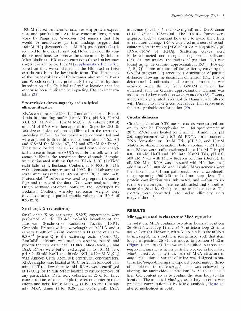
In isolation, MicA contains two stem loops at positions26–46 nt (stem loop 1) and 54–71 nt (stem loop 2) in itsnative form (6). However, when MicA binds to the mRNAtarget, ompA, the structure is reorganized, such that stemloop 1 at position 26–46 nt is moved to position 34–52 nt(Figure 1a and b) (6). This switch is required to expose theompA-binding site, which is partially blocked in the nativeMicA structure. To test the role of MicA structure inompA regulation, a variant of MicA was designed to sta-bilize the ‘ompA-binding site exposed’ conformation (here-after referred to as MicAstab). This was achieved byaltering the nucleotides at positions 34–52 to include ahigh GC content so as to confine the stem loop to thislocation. The modified MicAstab secondary structure waspredicted computationally by Mfold analysis (Figure 1c;altered nucleotides in bold).
Nucleic Acids Research, 2013 3
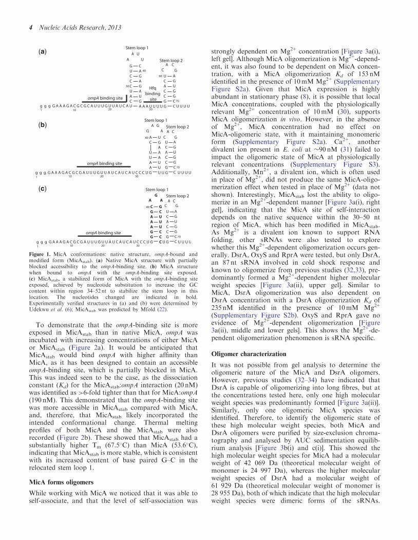
To demonstrate that the ompA-binding site is moreexposed in MicAstab than in native MicA, ompA wasincubated with increasing concentrations of either MicAor MicAstab (Figure 2a). It would be anticipated thatMicAstab would bind ompA with higher affinity thanMicA, as it has been designed to contain an accessibleompA-binding site, which is partially blocked in MicA.This was indeed seen to be the case, as the dissociationconstant (Kd) for the MicAstab:ompA interaction (20 nM)was identified as >6-fold tighter than that for MicA:ompA(190 nM). This demonstrated that the ompA-binding sitewas more accessible in MicAstab compared with MicA,and, therefore, that MicAstab likely incorporated theintended conformational change. Thermal meltingprofiles of both MicA and the MicAstab were alsorecorded (Figure 2b). These showed that MicAstab had asubstantially higher Tm (67.5�C) than MicA (53.6�C),indicating that MicAstab is more stable, which is consistentwith its increased content of base paired G–C in therelocated stem loop 1.
MicA forms oligomers
While working with MicA we noticed that it was able toself-associate, and that the level of self-association was
strongly dependent on Mg2+ concentration [Figure 3a(i),left gel]. Although MicA oligomerization is Mg2+-depend-ent, it was also found to be dependent on MicA concen-tration, with a MicA oligomerization Kd of 153 nMidentified in the presence of 10mM Mg2+ (SupplementaryFigure S2a). Given that MicA expression is highlyabundant in stationary phase (8), it is possible that localMicA concentrations, coupled with the physiologicallyrelevant Mg2+ concentration of 10mM (30), supportsMicA oligomerization in vivo. However, in the absenceof Mg2+, MicA concentration had no effect onMicA-oligomeric state, with it maintaining monomericform (Supplementary Figure S2a). Ca2+, anotherdivalent ion present in E. coli at �90 nM (31) failed toimpact the oligomeric state of MicA at physiologicallyrelevant concentrations (Supplementary Figure S3).Additionally, Mn2+, a divalent ion, which is often usedin place of Mg2+, did not produce the same MicA-oligo-merization effect when tested in place of Mg2+ (data notshown). Interestingly, MicAstab lost the ability to oligo-merize in an Mg2+-dependent manner [Figure 3a(i), rightgel], indicating that the MicA site of self-interactiondepends on the native sequence within the 30–50 ntregion of MicA, which has been modified in MicAstab.As Mg2+ is a divalent ion known to support RNAfolding, other sRNAs were also tested to explorewhether this Mg2+-dependent oligomerization occurs gen-erally. DsrA, OxyS and RprA were tested, but only DsrA,an 87 nt sRNA involved in cold shock response andknown to oligomerize from previous studies (32,33), pre-dominantly formed a Mg2+-dependent higher molecularweight species [Figure 3a(ii), upper gel]. Similar toMicA, DsrA oligomerization was also dependent onDsrA concentration with a DsrA oligomerization Kd of235 nM identified in the presence of 10mM Mg2+
(Supplementary Figure S2b). OxyS and RprA gave noevidence of Mg2+-dependent oligomerization [Figure3a(ii), middle and lower gels]. This shows the Mg2+-de-pendent oligomerization phenomenon is sRNA specific.
Oligomer characterization
It was not possible from gel analysis to determine theoligomeric nature of the MicA and DsrA oligomers.However, previous studies (32–34) have indicated thatDsrA is capable of oligomerizing into long fibres, but atthe concentrations tested here, only one high molecularweight species was predominantly formed [Figure 3a(ii)].Similarly, only one oligomeric MicA species wasidentified. Therefore, to identify the oligomeric state ofthese high molecular weight species, both MicA andDsrA oligomers were purified by size-exclusion chroma-tography and analysed by AUC sedimentation equilib-rium analysis [Figure 3b(i) and c(i)]. This showed thehigh molecular weight species for MicA had a molecularweight of 42 069 Da (theoretical molecular weight ofmonomer is 24 997 Da), whereas the higher molecularweight species of DsrA had a molecular weight of61 929 Da (theoretical molecular weight of monomer is28 955 Da), both of which indicate that the high molecularweight species were dimeric forms of the sRNAs.
(a)
(b)
(c)
Figure 1. MicA conformations: native structure, ompA-bound andmodified form (MicAstab). (a) Native MicA structure with partiallyblocked accessibility to the ompA-binding site. (b) MicA structurewhen bound to ompA with the ompA-binding site exposed.(c) MicAstab, a stabilized form of MicA with the ompA-binding siteexposed, achieved by nucleotide substitution to increase the GCcontent within region 34–52 nt to stabilize the stem loop in thislocation. The nucleotides changed are indicated in bold.Experimentally verified structures in (a) and (b) were determined byUdekwu et al. (6); MicAstab was predicted by Mfold (22).
4 Nucleic Acids Research, 2013
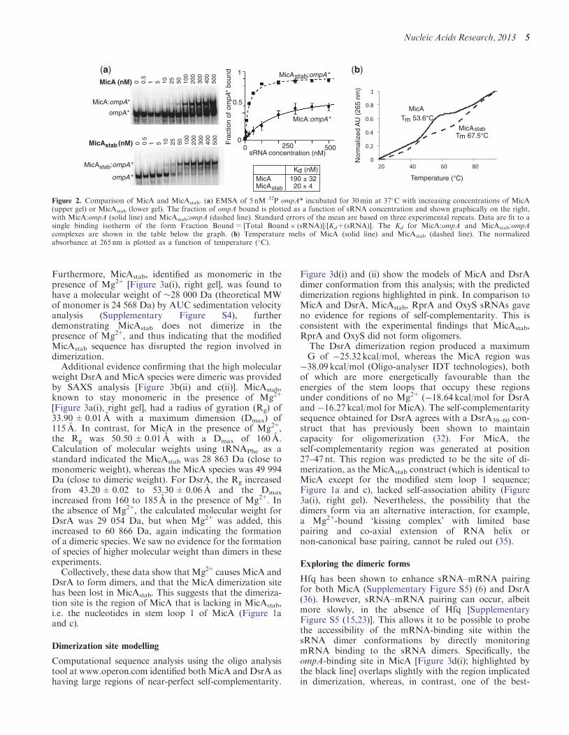
Furthermore, MicAstab, identified as monomeric in thepresence of Mg2+ [Figure 3a(i), right gel], was found tohave a molecular weight of �28 000 Da (theoretical MWof monomer is 24 568 Da) by AUC sedimentation velocityanalysis (Supplementary Figure S4), furtherdemonstrating MicAstab does not dimerize in thepresence of Mg2+, and thus indicating that the modifiedMicAstab sequence has disrupted the region involved indimerization.
Additional evidence confirming that the high molecularweight DsrA and MicA species were dimeric was providedby SAXS analysis [Figure 3b(ii) and c(ii)]. MicAstab,known to stay monomeric in the presence of Mg2+
[Figure 3a(i), right gel], had a radius of gyration (Rg) of33.90±0.01 A with a maximum dimension (Dmax) of115 A. In contrast, for MicA in the presence of Mg2+,the Rg was 50.50±0.01 A with a Dmax of 160 A.Calculation of molecular weights using tRNAPhe as astandard indicated the MicAstab was 28 863 Da (close tomonomeric weight), whereas the MicA species was 49 994Da (close to dimeric weight). For DsrA, the Rg increasedfrom 43.20±0.02 to 53.30±0.06 A and the Dmax
increased from 160 to 185 A in the presence of Mg2+. Inthe absence of Mg2+, the calculated molecular weight forDsrA was 29 054 Da, but when Mg2+ was added, thisincreased to 60 866 Da, again indicating the formationof a dimeric species. We saw no evidence for the formationof species of higher molecular weight than dimers in theseexperiments.
Collectively, these data show that Mg2+causes MicA andDsrA to form dimers, and that the MicA dimerization sitehas been lost in MicAstab. This suggests that the dimeriza-tion site is the region of MicA that is lacking in MicAstab,i.e. the nucleotides in stem loop 1 of MicA (Figure 1aand c).
Dimerization site modelling
Computational sequence analysis using the oligo analysistool at www.operon.com identified bothMicA and DsrA ashaving large regions of near-perfect self-complementarity.
Figure 3d(i) and (ii) show the models of MicA and DsrAdimer conformation from this analysis; with the predicteddimerization regions highlighted in pink. In comparison toMicA and DsrA, MicAstab, RprA and OxyS sRNAs gaveno evidence for regions of self-complementarity. This isconsistent with the experimental findings that MicAstab,RprA and OxyS did not form oligomers.The DsrA dimerization region produced a maximum
�G of �25.32 kcal/mol, whereas the MicA region was�38.09 kcal/mol (Oligo-analyser IDT technologies), bothof which are more energetically favourable than theenergies of the stem loops that occupy these regionsunder conditions of no Mg2+ (�18.64 kcal/mol for DsrAand �16.27 kcal/mol for MicA). The self-complementaritysequence obtained for DsrA agrees with a DsrA39–60 con-struct that has previously been shown to maintaincapacity for oligomerization (32). For MicA, theself-complementarity region was generated at position27–47 nt. This region was predicted to be the site of di-merization, as the MicAstab construct (which is identical toMicA except for the modified stem loop 1 sequence;Figure 1a and c), lacked self-association ability (Figure3a(i), right gel). Nevertheless, the possibility that thedimers form via an alternative interaction, for example,a Mg2+-bound ‘kissing complex’ with limited basepairing and co-axial extension of RNA helix ornon-canonical base pairing, cannot be ruled out (35).
Exploring the dimeric forms
Hfq has been shown to enhance sRNA–mRNA pairingfor both MicA (Supplementary Figure S5) (6) and DsrA(36). However, sRNA–mRNA pairing can occur, albeitmore slowly, in the absence of Hfq [SupplementaryFigure S5 (15,23)]. This allows it to be possible to probethe accessibility of the mRNA-binding site within thesRNA dimer conformations by directly monitoringmRNA binding to the sRNA dimers. Specifically, theompA-binding site in MicA [Figure 3d(i); highlighted bythe black line] overlaps slightly with the region implicatedin dimerization, whereas, in contrast, one of the best-
0 0.5
1 5 10
25
50
100
200
300
400
500
0 0.5
1 5 10
25
50
100
200
300
400
500
Nor
mal
ized
AU
(26
5 nm
)
Temperature (°C)
MicA (nM)
MicA:ompA*
MicA :ompA*
MicA (nM)
T 53.6°C
T 67.5°Cstab
stab
m
mMicA
MicA
stab
ompA*
ompA*
stab
K (nM)dMicA 190 ± 32MicA 20 ± 4
0
1
0.5
Fra
ctio
n of
om
pA*
boun
d
2500 500sRNA concentration (nM)
MicA:ompA*
MicA :ompA*stab(a) (b)
Figure 2. Comparison of MicA and MicAstab. (a) EMSA of 5 nM 32P ompA* incubated for 30min at 37�C with increasing concentrations of MicA(upper gel) or MicAstab (lower gel). The fraction of ompA bound is plotted as a function of sRNA concentration and shown graphically on the right,with MicA:ompA (solid line) and MicAstab:ompA (dashed line). Standard errors of the mean are based on three experimental repeats. Data are fit to asingle binding isotherm of the form Fraction Bound= [Total Bound� (sRNA)]/[Kd+(sRNA)]. The Kd for MicA:ompA and MicAstab:ompAcomplexes are shown in the table below the graph. (b) Temperature melts of MicA (solid line) and MicAstab (dashed line). The normalizedabsorbance at 265 nm is plotted as a function of temperature (�C).
Nucleic Acids Research, 2013 5
[a(i)]
[b(i)] [c(i)]
[c(ii)]
[d(i)] [d(ii)]
[b(ii)]
[a(ii)]
Figure 3. sRNA oligomerization. [a(i)] Native gel analysis of sRNAs in the presence of increasing concentrations of Mg2+. 2 mM MicA (left gel) andMicAstab (right gel) were heated for 2min at 80�C in 10mM Tris, pH 8.0, 50mM NaCl, 50mM KCl, 10% glycerol with 0–20mM Mg2+. Twopicomoles of each sample were analysed by 6% native polyacrylamide gel electrophoresis and stained with SYBR Gold. [a(ii)] As for [a(i)], but withsRNAs DsrA (top gel), OxyS (middle gel) and RprA (bottom gel). [b(i)] Size-exclusion chromatography profile monitoring RNA elution at 260 nmabsorbance over time shows MicA without MgCl2 (red) and with 10mM MgCl2 (orange). Inset shows AUC equilibrium analysis of the MicA specieswith 10mM MgCl2 (orange), which gave a molecular weight of 42 069 Da for the sample. [b(ii)] P(r) plots with corresponding ab initio models ofMicAstab (red) and MicA (orange) with 10mM MgCl2. The molecular weights for each sample, calculated from the scattering data with tRNAPhe as astandard, are shown. [c(i) and c(ii)] As for b(i) and b(ii) respectively, but using DsrA without MgCl2 (green) and with 10mM MgCl2 (blue). [d(i)]Model of MicA in dimer conformation. The pink lines and pink nucleotides highlight the predicted dimerization region. The complementary regionfor ompA-binding is highlighted by the black lines. [d(ii)] Model for DsrA in dimer conformation. Complementary regions for rpoS binding arehighlighted by the black lines, whilst the pink lines and nucleotides highlight the region involved in DsrA dimerization.
6 Nucleic Acids Research, 2013
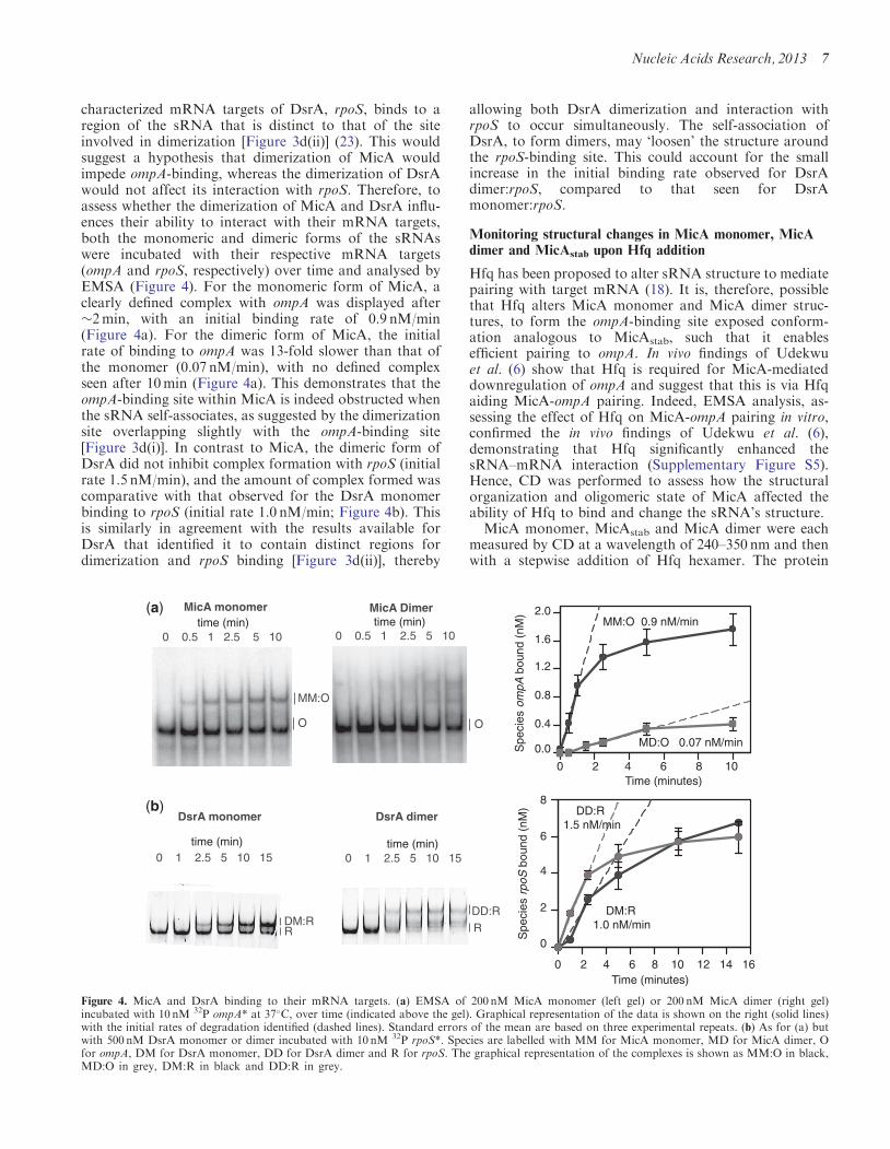
characterized mRNA targets of DsrA, rpoS, binds to aregion of the sRNA that is distinct to that of the siteinvolved in dimerization [Figure 3d(ii)] (23). This wouldsuggest a hypothesis that dimerization of MicA wouldimpede ompA-binding, whereas the dimerization of DsrAwould not affect its interaction with rpoS. Therefore, toassess whether the dimerization of MicA and DsrA influ-ences their ability to interact with their mRNA targets,both the monomeric and dimeric forms of the sRNAswere incubated with their respective mRNA targets(ompA and rpoS, respectively) over time and analysed byEMSA (Figure 4). For the monomeric form of MicA, aclearly defined complex with ompA was displayed after�2min, with an initial binding rate of 0.9 nM/min(Figure 4a). For the dimeric form of MicA, the initialrate of binding to ompA was 13-fold slower than that ofthe monomer (0.07 nM/min), with no defined complexseen after 10min (Figure 4a). This demonstrates that theompA-binding site within MicA is indeed obstructed whenthe sRNA self-associates, as suggested by the dimerizationsite overlapping slightly with the ompA-binding site[Figure 3d(i)]. In contrast to MicA, the dimeric form ofDsrA did not inhibit complex formation with rpoS (initialrate 1.5 nM/min), and the amount of complex formed wascomparative with that observed for the DsrA monomerbinding to rpoS (initial rate 1.0 nM/min; Figure 4b). Thisis similarly in agreement with the results available forDsrA that identified it to contain distinct regions fordimerization and rpoS binding [Figure 3d(ii)], thereby
allowing both DsrA dimerization and interaction withrpoS to occur simultaneously. The self-association ofDsrA, to form dimers, may ‘loosen’ the structure aroundthe rpoS-binding site. This could account for the smallincrease in the initial binding rate observed for DsrAdimer:rpoS, compared to that seen for DsrAmonomer:rpoS.
Monitoring structural changes in MicA monomer, MicAdimer and MicAstab upon Hfq addition
Hfq has been proposed to alter sRNA structure to mediatepairing with target mRNA (18). It is, therefore, possiblethat Hfq alters MicA monomer and MicA dimer struc-tures, to form the ompA-binding site exposed conform-ation analogous to MicAstab, such that it enablesefficient pairing to ompA. In vivo findings of Udekwuet al. (6) show that Hfq is required for MicA-mediateddownregulation of ompA and suggest that this is via Hfqaiding MicA-ompA pairing. Indeed, EMSA analysis, as-sessing the effect of Hfq on MicA-ompA pairing in vitro,confirmed the in vivo findings of Udekwu et al. (6),demonstrating that Hfq significantly enhanced thesRNA–mRNA interaction (Supplementary Figure S5).Hence, CD was performed to assess how the structuralorganization and oligomeric state of MicA affected theability of Hfq to bind and change the sRNA’s structure.MicA monomer, MicAstab and MicA dimer were each
measured by CD at a wavelength of 240–350 nm and thenwith a stepwise addition of Hfq hexamer. The protein
time (min) time (min)0 0.5 1 2.5 5 10 0 0.5 1 2.5 5 10
MicA monomer MicA Dimer
MM:O
O O
0 2 4 6 8 10 Time (minutes)
2.0
1.6
1.2
0.8
0.4
0.0 MD:O 0.07 nM/min
time (min) 0 1 2.5 5 10 15
time (min)0 1 2.5 5 10 15
DsrA monomer DsrA dimer
DD:RDM:RR R
Spe
cies
om
pA b
ound
(nM
)
MM:O 0.9 nM/min
Spe
cies
rpo
S b
ound
(nM
)
8
6
4
2
0
0 2 4 6 8 10 12 14 16 Time (minutes)
DM:R 1.0 nM/min
DD:R 1.5 nM/min
(a)
(b)
Figure 4. MicA and DsrA binding to their mRNA targets. (a) EMSA of 200 nM MicA monomer (left gel) or 200 nM MicA dimer (right gel)incubated with 10 nM 32P ompA* at 37�C, over time (indicated above the gel). Graphical representation of the data is shown on the right (solid lines)with the initial rates of degradation identified (dashed lines). Standard errors of the mean are based on three experimental repeats. (b) As for (a) butwith 500 nM DsrA monomer or dimer incubated with 10 nM 32P rpoS*. Species are labelled with MM for MicA monomer, MD for MicA dimer, Ofor ompA, DM for DsrA monomer, DD for DsrA dimer and R for rpoS. The graphical representation of the complexes is shown as MM:O in black,MD:O in grey, DM:R in black and DD:R in grey.
Nucleic Acids Research, 2013 7
contribution within the 240–350 nm region of the CDspectrum was subtracted, but because of the smallnumber of aromatic residues within Hfq, this contributionwas negligible. This means that the data at 240–350 nmshowed only the contribution from the RNA. The ellipti-city of the RNAs in the absence of Hfq and subsequentlyin the presence of increasing amounts Hfq was monitoreduntil no further change in ellipticity was observed. Anyellipticity changes observed upon Hfq addition could beinterpreted as indicating RNA conformational changes(37–39).For MicA monomer, the maximal ellipticity change
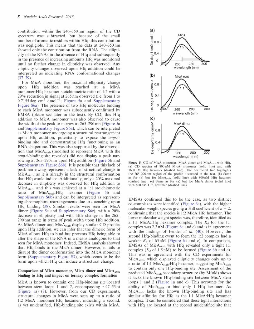
upon Hfq addition was reached at a MicAmonomer:Hfq hexamer stoichiometric ratio of 1:2 with a29% reduction in signal at 265 nm observed (i.e. from 1 to0.7155 deg cm2 dmol�1; Figure 5a and SupplementaryFigure S6a). The presence of two Hfq molecules bindingto each MicA monomer was subsequently confirmed byEMSA (please see later in the text). By CD, this Hfqaddition to MicA monomer was also observed to causethe width of the peak to narrow at 265–290 nm (Figure 5aand Supplementary Figure S6a), which can be interpretedas MicA monomer undergoing a structural rearrangementupon Hfq addition, potentially to expose the ompA-binding site and demonstrating Hfq functioning as anRNA chaperone. This was also supported by the observa-tion that MicAstab (modified to represent MicA with theompA-binding site revealed) did not display a peak nar-rowing at 265–290 nm upon Hfq addition (Figure 5b andSupplementary Figure S6b). It is possible that this lack ofpeak narrowing represents a lack of structural change inMicAstab, as it is already in the structural conformationthat Hfq would induce. Additionally, only a 20% maximaldecrease in ellipticity was observed for Hfq addition toMicAstab, and this was achieved at a 1:1 stoichiometricratio of MicAstab:Hfq hexamer (Figure 5b andSupplementary S6b) and can be interpreted as represent-ing chromophore rearrangements due to quenching uponHfq binding (38). Similar results were seen for MicAdimer (Figure 5c and Supplementary S6c), with a 20%decrease in ellipticity and with little change in the 265–290 nm range in terms of peak width upon Hfq addition.As MicA dimer and MicAstab display similar CD profilesupon Hfq addition, we can infer that the dimeric form ofMicA allows Hfq to bind but prevents Hfq being able toalter the shape of the RNA in a means analogous to thatseen for MicA monomer. Indeed, EMSA analysis showedthat Hfq binds to the MicA dimer. However, it fails todisrupt the dimer conformation into the MicA monomerform (Supplementary Figure S7), which seems to be theform upon which Hfq can induce a structural change.
Comparison of MicA monomer, MicA dimer and MicAstab
binding to Hfq and impact on ternary complex formation
MicA is known to contain one Hfq-binding site locatedbetween stem loops 1 and 2, encompassing �47–53 nt(Figure 1a) (8). However, from our CD experiments,structural changes in MicA were seen up to a ratio of1:2 MicA monomer:Hfq hexamer, indicating a second,as yet unidentified, Hfq-binding site exists within MicA.
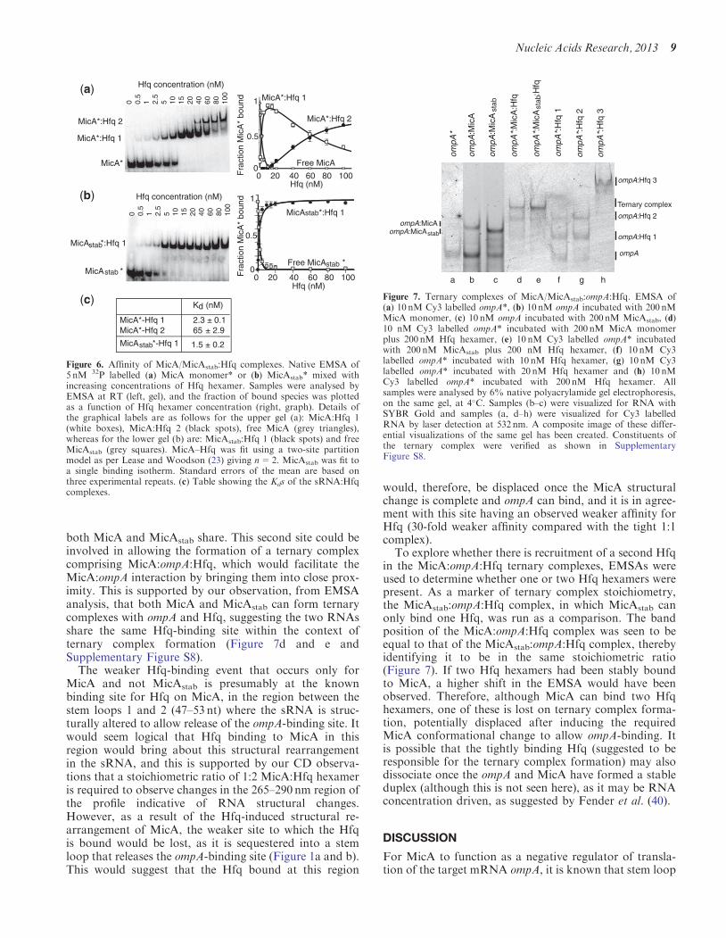
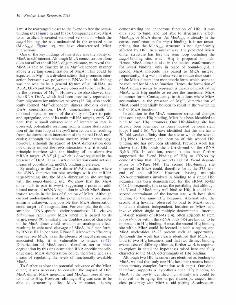
EMSAs confirmed this to be the case, as two distinctco-complexes were identified (Figure 6a), with the highermolecular weight species giving a Hill coefficient of n=2,confirming that the species is 1:2 MicA:Hfq hexamer. Thelower molecular weight species was, therefore, identified asa 1:1 MicA:Hfq hexamer complex. The Kd for the 1:1complex was 2.3 nM (Figure 6a and c) and is in agreementwith the findings of Fender et al. (40). However, thesecond Hfq-binding event to form the 1:2 complex had aweaker Kd of 65 nM (Figure 6a and c). In comparison,EMSAs of MicAstab with Hfq revealed only a tight 1:1complex (Kd of 1.5 nM) to be formed (Figure 6b and c).This was in agreement with the CD experiments forMicAstab, which displayed ellipticity changes only up toa ratio of 1:1 MicAstab:Hfq hexamer, suggesting MicAstab
to contain only one Hfq-binding site. Assessment of thepredicted MicAstab secondary structure (by Mfold) showsit lacks the known Hfq-binding site between MicA stemloops 1 and 2 (Figure 1a and c). This accounts for theability of MicAstab to bind only 1 Hfq hexamer. AsMicAstab lacks the known Hfq-binding site and hassimilar affinities for Hfq as the 1:1 MicA:Hfq hexamercomplex, it can be considered that these tight interactionswith Hfq are located at the second unidentified site that
0.8
0.4
0
De
deg-
1 cm
2 dm
ol-1
240 260 280 300wavelength (nm)
1:2
MicA
1:1
MicA stab
0.8
0.4
0
De
deg-
1 cm
2 dm
ol-1
240 260 280 300wavelength (nm)
0.8
0.4
0D
e de
g-1
cm2
dmol
-1260 280 300
wavelength (nm)
1:1
MicA dimer
(a)
(b)
(c)
Figure 5. CD of MicA monomer, MicA dimer and MicAstab with Hfq.(a) CD spectra of 800 nM MicA monomer (solid line) and with1600 nM Hfq hexamer (dashed line). The horizontal line highlightsthe 265–290 nm region of the profile discussed in the text. (b) Sameas for (a) but for MicAstab (solid line) with 800 nM Hfq hexamer(dashed line). (c) Same as for (a) but for MicA dimer (solid line)with 800 nM Hfq hexamer (dashed line).
8 Nucleic Acids Research, 2013
both MicA and MicAstab share. This second site could beinvolved in allowing the formation of a ternary complexcomprising MicA:ompA:Hfq, which would facilitate theMicA:ompA interaction by bringing them into close prox-imity. This is supported by our observation, from EMSAanalysis, that both MicA and MicAstab can form ternarycomplexes with ompA and Hfq, suggesting the two RNAsshare the same Hfq-binding site within the context ofternary complex formation (Figure 7d and e andSupplementary Figure S8).
The weaker Hfq-binding event that occurs only forMicA and not MicAstab is presumably at the knownbinding site for Hfq on MicA, in the region between thestem loops 1 and 2 (47–53 nt) where the sRNA is struc-turally altered to allow release of the ompA-binding site. Itwould seem logical that Hfq binding to MicA in thisregion would bring about this structural rearrangementin the sRNA, and this is supported by our CD observa-tions that a stoichiometric ratio of 1:2 MicA:Hfq hexameris required to observe changes in the 265–290 nm region ofthe profile indicative of RNA structural changes.However, as a result of the Hfq-induced structural re-arrangement of MicA, the weaker site to which the Hfqis bound would be lost, as it is sequestered into a stemloop that releases the ompA-binding site (Figure 1a and b).This would suggest that the Hfq bound at this region
would, therefore, be displaced once the MicA structuralchange is complete and ompA can bind, and it is in agree-ment with this site having an observed weaker affinity forHfq (30-fold weaker affinity compared with the tight 1:1complex).To explore whether there is recruitment of a second Hfq
in the MicA:ompA:Hfq ternary complexes, EMSAs wereused to determine whether one or two Hfq hexamers werepresent. As a marker of ternary complex stoichiometry,the MicAstab:ompA:Hfq complex, in which MicAstab canonly bind one Hfq, was run as a comparison. The bandposition of the MicA:ompA:Hfq complex was seen to beequal to that of the MicAstab:ompA:Hfq complex, therebyidentifying it to be in the same stoichiometric ratio(Figure 7). If two Hfq hexamers had been stably boundto MicA, a higher shift in the EMSA would have beenobserved. Therefore, although MicA can bind two Hfqhexamers, one of these is lost on ternary complex forma-tion, potentially displaced after inducing the requiredMicA conformational change to allow ompA-binding. Itis possible that the tightly binding Hfq (suggested to beresponsible for the ternary complex formation) may alsodissociate once the ompA and MicA have formed a stableduplex (although this is not seen here), as it may be RNAconcentration driven, as suggested by Fender et al. (40).
DISCUSSION
For MicA to function as a negative regulator of transla-tion of the target mRNA ompA, it is known that stem loop
MicA*
MicA *
MicA *:Hfq 1
MicA*:Hfq 1
MicA*:Hfq 2
Hfq concentration (nM)
Hfq concentration (nM)
Free MicA *
MicA *:Hfq 1
MicA*:Hfq 1
MicA*:Hfq 2
Free MicA
0 20 40 60 80 100
0 20 40 60 80 100Hfq (nM)
Fra
ctio
n M
icA
* bo
und 1
0.5
0
Hfq (nM)
Fra
ctio
n M
icA
* bo
und 1
0.5
0
stab
stabstab
stab
0 0.5
1 2.5
5 10 15 20 40 60 80 100
0 0.5
1 2.5
5 10 15 20 40 60 80 100
MicA *-Hfq 1stab
K (nM)d
MicA*-Hfq 1 2.3 ± 0.1MicA*-Hfq 2 65 ± 2.9
1.5 ± 0.2
(a)
(b)
(c)
Figure 6. Affinity of MicA/MicAstab:Hfq complexes. Native EMSA of5 nM 32P labelled (a) MicA monomer* or (b) MicAstab* mixed withincreasing concentrations of Hfq hexamer. Samples were analysed byEMSA at RT (left, gel), and the fraction of bound species was plottedas a function of Hfq hexamer concentration (right, graph). Details ofthe graphical labels are as follows for the upper gel (a): MicA:Hfq 1(white boxes), MicA:Hfq 2 (black spots), free MicA (grey triangles),whereas for the lower gel (b) are: MicAstab:Hfq 1 (black spots) and freeMicAstab (grey squares). MicA–Hfq was fit using a two-site partitionmodel as per Lease and Woodson (23) giving n=2. MicAstab was fit toa single binding isotherm. Standard errors of the mean are based onthree experimental repeats. (c) Table showing the Kds of the sRNA:Hfqcomplexes.
ompA
*
ompA
:Mic
A
ompA
*:M
icA
:Hfq
ompA
* :H
fq 1
ompA
*:H
fq 2
ompA
*:M
icA
:Hfq
stab stab
ompA
:Mic
A
Ternary complex
ompA
*:H
fq 3
ompA
ompA:Hfq 1
ompA:MicA
ompA:Hfq 3
ompA:Hfq 2
ompA:MicAstab
a b c d e f g h
Figure 7. Ternary complexes of MicA/MicAstab:ompA:Hfq. EMSA of(a) 10 nM Cy3 labelled ompA*, (b) 10 nM ompA incubated with 200 nMMicA monomer, (c) 10 nM ompA incubated with 200 nM MicAstab, (d)10 nM Cy3 labelled ompA* incubated with 200 nM MicA monomerplus 200 nM Hfq hexamer, (e) 10 nM Cy3 labelled ompA* incubatedwith 200 nM MicAstab plus 200 nM Hfq hexamer, (f) 10 nM Cy3labelled ompA* incubated with 10 nM Hfq hexamer, (g) 10 nM Cy3labelled ompA* incubated with 20 nM Hfq hexamer and (h) 10 nMCy3 labelled ompA* incubated with 200 nM Hfq hexamer. Allsamples were analysed by 6% native polyacrylamide gel electrophoresis,on the same gel, at 4�C. Samples (b–c) were visualized for RNA withSYBR Gold and samples (a, d–h) were visualized for Cy3 labelledRNA by laser detection at 532 nm. A composite image of these differ-ential visualizations of the same gel has been created. Constituents ofthe ternary complex were verified as shown in SupplementaryFigure S8.
Nucleic Acids Research, 2013 9
1 must be rearranged closer to the 30-end to free the ompA-binding site (Figure 1a and b) (6). Comparing native MicAto an artificially created stabilized version, in which theompA-binding site was maintained in the exposed state(MicAstab; Figure 1c), we have characterized MicAinteractions.One of the key findings of this study was the ability of
MicA to self-interact. Although MicA concentration alonedoes not affect the sRNA’s oligomeric state, we reveal thatMicA is able to dimerize in an Mg2+-dependent mannerabove a certain concentration of MicA. This could beexpected as Mg2+ is a divalent cation that promotes inter-action between two polyanionic RNAs, but this findingwas not seen to be a general feature of all sRNAs, asRprA, OxyS and MicAstab were observed to be unaffectedby the presence of Mg2+. However, we also showed thatthe sRNA DsrA, which had been previously reported toform oligomers for unknown reasons (32–34), also specif-ically formed Mg2+-dependent dimers above a certainDsrA concentration. DsrA dimerization results inminimal impact in terms of the ability of DsrA to pair,and upregulate, one of its main mRNA targets, rpoS. Wenote that a small enhancement of interaction rate isobserved, potentially resulting from a minor destabiliza-tion of the stem loop at the rpoS interaction site, resultingfrom the downstream interaction of the paired DsrA mol-ecules; although this remains unclear. More interestingly,however, although the region of DsrA dimerization doesnot directly impact the rpoS interaction site, it would inprinciple interfere with the binding of an alternativemRNA target, H-NS (41), which is downregulated in thepresence of DsrA. Thus, DsrA dimerization could act as ameans of coordinating mRNA binding preference.By analogy to the DsrA dimer:H-NS situation, where
the sRNA dimerization site overlaps with the mRNAtarget-binding site, the MicA dimerization site overlapswith the ompA-binding site. We show that the MicAdimer fails to pair to ompA, suggesting a potential add-itional means of mRNA regulation in which MicA dimer-ization results in the loss of function of MicA. Althoughcurrent understanding of this potential regulatory mech-anism is unknown, it is possible that MicA dimerizationcould target it for degradation. For example, the double-stranded RNA-specific endoribonuclease III cleavesSalmonella typhimurum MicA when it is paired to itstarget, ompA (9). Similarly, the double-stranded characterof the MicA dimer could result in an analogous effect,resulting in enhanced cleavage of MicA, in dimer form,by RNase III. In contrast, RNase E is known to efficientlydegrade free MicA, as in the absence of ompA pairing, orassociated Hfq, it is vulnerable to attack (9,42).Dimerization of MicA could, therefore, act to blockdegradation by this single-stranded RNA-specific endorib-onuclease. MicA dimerization could, therefore, act as ameans of regulating the levels of functionally availableMicA.To further explore the potential relevance of the MicA
dimer, it was necessary to consider the impact of Hfq.MicA dimer, MicA monomer and MicAstab were all seento bind to Hfq. However, although Hfq was seen to beable to structurally affect MicA monomer, thereby
demonstrating the chaperone function of Hfq, it wasonly able to bind, and not able to structurally affect,MicAstab or MicA dimer. As MicAstab is already in thecorrect conformation for ompA binding, it is not unsur-prising that the MicAstab structure is not significantlyaffected by Hfq. In a similar way, the predicted MicAdimer structure has lost the stem loop occluding theompA-binding site, which Hfq is proposed to melt.Hence, MicA dimer is also in the ‘active’ conformationfor ompA binding, only in place of bound-ompA, asecond MicA molecule has paired to MicA instead.Importantly, Hfq was not observed to induce dissociationof the MicA dimers into monomeric form, which seems tobe required for MicA to function. Hence, the formation ofMicA dimers seems to represent a means of inactivatingMicA, with Hfq unable to restore the functional MicAmonomer form. Consequently, in situations where MicAaccumulates in the presence of Mg2+, dimerization ofMicA could potentially be seen to result in the ‘switchingoff’ of MicA function.
In addition to the MicA monomer structural changesthat occur upon Hfq binding, MicA has been identified tobind to two Hfq hexamers. One Hfq-binding site hasalready been identified as being located between stemloops 1 and 2 (6). We have identified that this site has a30-fold weaker affinity than the site at which the secondHfq binds. However, the location of this second Hfqbinding site has not been identified. Previous work hasshown that Hfq binds the 30U-rich–end of the sRNARybB (43). In addition, recent studies have furthersupported the 30-end binding of Hfq to sRNAs bydemonstrating that Hfq protects against 30-end degrad-ation by PNPase (44). The second Hfq-binding siteobserved for MicA could, therefore, be at the 30U-rich–end of the sRNA. However, having multipleRNA-determinants involved in binding to a single Hfqhexamer has been demonstrated for the sRNA RybB(45). Consequently, this raises the possibility that althoughthe 30-end of MicA may well bind to Hfq, it could be asecond determinant of the known site, with both sitesbinding to the same Hfq hexamer. Alternatively, thesecond Hfq hexamer observed to bind to MicA, couldbind at a distinct, independent, location on MicA, andinvolve either single or multiple determinants. InternalU/A-rich regions of sRNAs (14), often adjacent to stemloops (46), or within the sRNA body (45) are known to beimportant in Hfq binding. Hence, the second Hfq-bindingsite within MicA could be located in such a region, andMicA nucleotides 15–23 present such an opportunity.Although this work has clearly identified that MicA canbind to two Hfq hexamers, and that two distinct bindingevents exist of differing affinities, further work is requiredto explore in detail the hypotheses raised here and fullycharacterize the MicA determinants of Hfq binding.
Although two Hfq hexamers are identified as binding toMicA, we find that only one Hfq hexamer remains boundupon ternary complex formation with ompA. Our data,therefore, supports a hypothesis that Hfq binding toMicA at the newly identified high affinity site could beinvolved in bringing the target transcript, ompA, intoclose proximity with MicA to aid pairing. A subsequent
10 Nucleic Acids Research, 2013
Hfq–MicA binding event at the lower affinity site,proposed to be at the known binding site between steploops 1 and 2, is necessary to allow restructuring ofMicA, such that the ompA-binding site becomesexposed. After MicA restructuring, the lower affinityHfq-binding site is lost and the Hfq involved then dissoci-ates. Upon exposure of the ompA-binding site withinMicA, MicA–ompA pairing occurs. It would beanticipated that the tightly bound Hfq would be lostonce a stable MicA–ompA duplex had formed, althoughthis may be driven by RNA concentration (40) and wasnot seen in our experiments.
In summary, our studies to characterize the interactionsof MicA have expanded our understanding of the sRNA’sfunction. We have demonstrated Hfq’s role as an RNAchaperone, impacting the structural conformation ofMicA. We have seen that MicA is capable of binding totwo Hfq hexamers and discuss a potential mechanismof MicA action which explains our observation of oneHfq hexamer within the context of a ternary complexwith ompA. In addition, we have identified that a poten-tially inactive Mg2+-dependent MicA dimer can form ataccumulated MicA levels. Although it is well establishedthat dimerization of proteins acts as a regulatory mechan-ism in signal transduction pathways (47) and RNA dimer-ization is an essential process in the retroviral replicationcycle (48), dimerization has never before been identified asa potential means of regulating sRNA function. Thecapacity of sRNA dimerization to prevent interactionswith specific mRNA targets, although continuing toallow interactions with others, provides a possible mech-anism of sRNA-dimer driven mRNA-target preferences.This could potentially provide part of the explanation asto how one sRNA can specifically act on multiple mRNAtargets. The ability of certain sRNAs to sense their envir-onment, specifically in terms of ion levels, could suggestthat a hitherto unknown level of regulation exists.
SUPPLEMENTARY DATA
Supplementary Data are available at NAR Online:Supplementary Table 1 and Supplementary Figures 1–8.
ACKNOWLEDGEMENTS
The authors thank Dr Isabella Moll (Max F. PerutzLaboratories, University of Vienna, Austria) for thekind gift of the E. coli Hfq expression strain. Theythank Dave Whitley (University of Portsmouth) for helpwith binding equations. They thank Petra Pernot(European Synchrotron Radiation Facility, Grenoble,France) for SAXS technical support and Marc Malfois(Diamond Light source) for useful SAXS analysis discus-sions. They also thank Ben Luisi (University ofCambridge, UK) for critical reading of the manuscript.
FUNDING
BBSRC research grant [BB/F013140/1 to A.J.C.]; MarieCurie Reintegration Grant from the European
Commission, FP7 [249154 to H.A.V.]; Institute ofBiomedical and Biomolecular Science (IBBS), Universityof Portsmouth bursary (to C.A.H); EuropeanSynchrotron Radiation Facility beamtime access grants[MX-1226 and MX-1102 to A.J.C.]. Funding for openaccess charge: BBSRC research grant.
Conflict of interest statement. None declared.
REFERENCES
1. De Las Penas,A., Connolly,L. and Gross,C.A. (1997) ThesigmaE-mediated response to extracytoplasmic stress inEscherichia coli is transduced by RseA and RseB, two negativeregulators of sigmaE. Mol. Microbiol., 24, 373–385.
2. Ades,S.E., Connolly,L.E., Alba,B.M. and Gross,C.A. (1999)The Escherichia coli sigma(E)-dependent extracytoplasmic stressresponse is controlled by the regulated proteolysis of ananti-sigma factor. Genes Dev., 13, 2449–2461.
3. Alba,B.M., Leeds,J.A., Onufryk,C., Lu,C.Z. and Gross,C.A.(2002) DegS and YaeL participate sequentially in the cleavage ofRseA to activate the sigma(E)-dependent extracytoplasmic stressresponse. Genes Dev., 16, 2156–2168.
4. Kabir,M.S., Yamashita,D., Koyama,S., Oshima,T., Kurokawa,K.,Maeda,M., Tsunedomi,R., Murata,M., Wada,C., Mori,H. et al.(2005) Cell lysis directed by sigmaE in early stationary phase andeffect of induction of the rpoE gene on global gene expression inEscherichia coli. Microbiology, 151, 2721–2735.
5. Rhodius,V.A., Suh,W.C., Nonaka,G., West,J. and Gross,C.A.(2006) Conserved and variable functions of the sigmaE stressresponse in related genomes. PLoS Biol., 4, e2.
6. Udekwu,K.I., Darfeuille,F., Vogel,J., Reimegard,J., Holmqvist,E.and Wagner,E.G. (2005) Hfq-dependent regulation of OmpAsynthesis is mediated by an antisense RNA. Genes Dev., 19,2355–2366.
7. Johansen,J., Eriksen,M., Kallipolitis,B. and Valentin-Hansen,P.(2008) Down-regulation of outer membrane proteins bynoncoding RNAs: unraveling the cAMP-CRP- and sigmaE-dependent CyaR-ompX regulatory case. J. Mol. Biol., 383, 1–9.
8. Rasmussen,A.A., Eriksen,M., Gilany,K., Udesen,C., Franch,T.,Petersen,C. and Valentin-Hansen,P. (2005) Regulation of ompAmRNA stability: the role of a small regulatory RNA in growthphase-dependent control. Mol. Microbiol., 58, 1421–1429.
9. Viegas,S.C., Silva,I.J., Saramago,M., Domingues,S. andArraiano,C.M. (2011) Regulation of the small regulatory RNAMicA by ribonuclease III: a target-dependent pathway. NucleicAcids Res., 39, 2918–2930.
10. Schuppli,D., Georgijevic,J. and Weber,H. (2000) Synergism ofmutations in bacteriophage Q beta RNA affecting host factordependence of Q beta replicase. J. Mol. Biol., 295, 149–154.
11. Franze de Fernandez,M.T., Eoyang,L. and August,J.T. (1968)Factor fraction required for the synthesis of bacteriophageQbeta-RNA. Nature, 219, 588–590.
12. Link,T.M., Valentin-Hansen,P. and Brennan,R.G. (2009)Structure of Escherichia coli Hfq bound to polyriboadenylateRNA. Proc. Natl Acad. Sci. USA, 106, 19292–19297.
13. Sauter,C., Basquin,J. and Suck,D. (2003) Sm-like proteins inEubacteria: the crystal structure of the Hfq protein fromEscherichia coli. Nucleic Acids Res., 31, 4091–4098.
14. Schumacher,M.A., Pearson,R.F., Moller,T., Valentin-Hansen,P.and Brennan,R.G. (2002) Structures of the pleiotropictranslational regulator Hfq and an Hfq-RNA complex: a bacterialSm-like protein. EMBO J., 21, 3546–3556.
15. Soper,T.J. and Woodson,S.A. (2008) The rpoS mRNA leaderrecruits Hfq to facilitate annealing with DsrA sRNA. RNA, 14,1907–1917.
16. Updegrove,T., Wilf,N., Sun,X. and Wartell,R.M. (2008) Effect ofHfq on RprA-rpoS mRNA pairing: Hfq-RNA binding and theinfluence of the 50 rpoS mRNA leader region. Biochemistry, 47,11184–11195.
Nucleic Acids Research, 2013 11
17. Geissmann,T.A. and Touati,D. (2004) Hfq, a new chaperoningrole: binding to messenger RNA determines access for smallRNA regulator. EMBO J., 23, 396–405.
18. Soper,T.J., Doxzen,K. and Woodson,S.A. (2011) Major role formRNA binding and restructuring in sRNA recruitment by Hfq.RNA, 17, 1544–1550.
19. Zhang,A., Wassarman,K.M., Ortega,J., Steven,A.C. and Storz,G.(2002) The Sm-like Hfq protein increases OxyS RNA interactionwith target mRNAs. Mol. Cell, 9, 11–22.
20. Vassilieva,I.M., Rouzanov,M.V., Zelinskaya,N.V., Moll,I.,Blasi,U. and Garber,M.B. (2002) Cloning, purification, andcrystallization of a bacterial gene expression regulator—Hfqprotein from Escherichia coli. Biochemistry (Mosc.), 67,1293–1297.
21. Gao,X., Yo,P., Keith,A., Ragan,T.J. and Harris,T.K. (2003)Thermodynamically balanced inside-out (TBIO) PCR-based genesynthesis: a novel method of primer design for high-fidelityassembly of longer gene sequences. Nucleic Acids Res., 31, e143.
22. Zuker,M. (2003) Mfold web server for nucleic acid folding andhybridization prediction. Nucleic Acids Res., 31, 3406–3415.
23. Lease,R.A. and Woodson,S.A. (2004) Cycling of the Sm-likeprotein Hfq on the DsrA small regulatory RNA. J. Mol. Biol.,344, 1211–1223.
24. Panja,S. and Woodson,S.A. (2012) Hexamer to monomerequilibrium of E. coli Hfq in solution and its impact on RNAannealing. J. Mol. Biol., 417, 406–412.
25. Vincent,H.A., Henderson,C.A., Ragan,T.J., Garza-Garcia,A.,Cary,P.D., Gowers,D.M., Malfois,M., Driscoll,P.C., Sobott,F.and Callaghan,A.J. (2012) Characterization of Vibrio cholerae Hfqprovides novel insights into the role of the Hfq C-terminalregion. J. Mol. Biol., 420, 56–69.
26. Konarev,P.V., Volkov,V.V., Sokolova,A.V., Koch,M.H.J. andSvergun,D.I. (2003) PRIMUS: a windows PC-based system forsmall-angle scattering data analysis. J. Appl. Cryst., 36,1277–1282.
27. Svergun,D.I. (1992) Determination of the regularization parameterin indirect-transform methods using perceptual criteria. J. Appl.Cryst., 25, 495–503.
28. Franke,D. and Svergun,D.I. (2009) DAMMIF, a program forrapid ab-initio shape determination in small-angle scattering.J. Appl. Cryst., 42, 342–346.
29. Volkov,V.V. and Svergun,D.I. (2003) Uniqueness of ab initioshape determination in small-angle scattering. J. Appl. Cryst., 36,860–864.
30. Hurwitz,C. and Rosano,C.L. (1967) The intracellularconcentration of bound and unbound magnesium ions inEscherichia coli. J. Biol. Chem., 242, 3719–3722.
31. Gangola,P. and Rosen,B.P. (1987) Maintenance of intracellularcalcium in Escherichia coli. J. Biol. Chem., 262, 12570–12574.
32. Cayrol,B., Geinguenaud,F., Lacoste,J., Busi,F., Le Derout,J.,Pietrement,O., Le Cam,E., Regnier,P., Lavelle,C. and Arluison,V.(2009) Auto-assembly of E. coli DsrA small noncoding RNA:molecular characteristics and functional consequences. RNA Biol.,6, 434–445.
33. Cayrol,B., Nogues,C., Dawid,A., Sagi,I., Silberzan,P. andIsambert,H. (2009) A nanostructure made of a bacterialnoncoding RNA. J. Am. Chem. Soc., 131, 17270–17276.
34. Busi,F., Cayrol,B., Lavelle,C., LeDerout,J., Pietrement,O., LeCam,E., Geinguenaud,F., Lacoste,J., Regnier,P. and Arluison,V.(2009) Auto-assembly as a new regulatory mechanism ofnoncoding RNA. Cell Cycle, 8, 952–954.
35. Ennifar,E., Walter,P., Ehresmann,B., Ehresmann,C. and Dumas,P.(2001) Crystal structures of coaxially stacked kissing complexes ofthe HIV-1 RNA dimerization initiation site. Nat. Struct. Biol., 8,1064–1068.
36. Sledjeski,D.D., Whitman,C. and Zhang,A. (2001) Hfq is necessaryfor regulation by the untranslated RNA DsrA. J. Bacteriol., 183,1997–2005.
37. Tan,R. and Frankel,A.D. (1992) Circular dichroism studiessuggest that TAR RNA changes conformation upon specificbinding of arginine or guanidine. Biochemistry, 31, 10288–10294.
38. Aparicio,F., Vilar,M., Perez-Paya,E. and Pallas,V. (2003) Thecoat protein of prunus necrotic ringspot virus specifically binds toand regulates the conformation of its genomic RNA. Virology,313, 213–223.
39. Daly,T.J., Rusche,J.R., Maione,T.E. and Frankel,A.D. (1990)Circular dichroism studies of the HIV-1 Rev protein and itsspecific RNA binding site. Biochemistry, 29, 9791–9795.
40. Fender,A., Elf,J., Hampel,K., Zimmermann,B. and Wagner,E.G.(2010) RNAs actively cycle on the Sm-like protein Hfq. GenesDev., 24, 2621–2626.
41. Lease,R.A. and Belfort,M. (2000) A trans-acting RNA as acontrol switch in Escherichia coli: DsrA modulates function byforming alternative structures. Proc. Natl Acad. Sci. USA, 97,9919–9924.
42. Bandyra,K.J., Said,N., Pfeiffer,V., Gorna,M.W., Vogel,J. andLuisi,B.F. (2012) The seed region of a small RNA drives thecontrolled destruction of the target mRNA by theendoribonuclease RNase E. Mol. Cell, 47, 943–953.
43. Sauer,E. and Weichenrieder,O. (2011) Structural basis for RNA30-end recognition by Hfq. Proc. Natl Acad. Sci. USA, 108,13065–13070.
44. Andrade,J.M., Pobre,V., Matos,A.M. and Arraiano,C.M. (2012)The crucial role of PNPase in the degradation of small RNAsthat are not associated with Hfq. RNA, 18, 844–855.
45. Sauer,E., Schmidt,S. and Weichenrieder,O. (2012) Small RNAbinding to the lateral surface of Hfq hexamers and structuralrearrangements upon mRNA target recognition. Proc. Natl Acad.Sci. USA, 109, 9396–9401.
46. Moll,I., Afonyushkin,T., Vytvytska,O., Kaberdin,V.R. andBlasi,U. (2003) Coincident Hfq binding and RNase Ecleavage sites on mRNA and small regulatory RNAs. RNA, 9,1308–1314.
47. Klemm,J.D., Schreiber,S.L. and Crabtree,G.R. (1998)Dimerization as a regulatory mechanism in signal transduction.Annu. Rev. Immunol., 16, 569–592.
48. Baig,T.T., Lanchy,J.M. and Lodmell,J.S. (2007) HIV-2 RNAdimerization is regulated by intramolecular interactions in vitro.RNA, 13, 1341–1354.
12 Nucleic Acids Research, 2013