Embed Size (px)
Citation preview

JOURNAL OF BACTERIOLOGY, Nov. 1967, p. 1451-1458Copyright © 1967 American Society for Microbiology
Vol. 94, No. 5Printed in U.S.A.
Characterization of Mycoplasma Strains from CatsB. C. COLE, L. GOLIGHTLY, AND J. R. WARD
Department of Internal Medicine, Division of Arthritis, University oj Utah Medical Center,Salt Lake City, Utah 84112
Received for publication 10 August 1967
Mycoplasma strains (Bi, B2, CS, and S1A) were isolated from the saliva of normalcats. These were compared with a strain (CO) isolated from the eye of a cat withsevere conjunctivitis. On the basis of morphology, biochemical reactions, andantigenic composition, two distinct species were recognizable. Strains CO, Bi, andB2 were antigenically unrelated to the other species tested; strains CS and SlApossessed antigenic components in common with Mycoplasma arthritidis, M. sali-varium, M. hominis, type 1, and M. orale, types 1 and 2. It was tentatively suggestedthat the two cat species be called M. felis and M. gateae, respectively.
The isolation of potentially pathogenic myco-plasma strains from laboratory and domesticanimals has been reported by many workers. Al-though Mycoplasma arthritidis is a well-knownpathogen for rats, it may be harbored by ap-parently healthy animals (13). Similarly, M.neurolyticum, M. pulmonis, and M. hyorhinis, allknown to be pathogenic species, may occur innormal animals (12, 21, 22, 25). Recent evidencesuggests that M. hominis, type 1, a common hu-man commensal, is pathogenic under certain cir-cumstances (19).
Switzer, in 1954, isolated a mycoplasma fromthe pneumonic lung of a 6-week-old kitten, butcould not induce disease experimentally (24). Thisstudy describes the isolation and characterizationof mycoplasma from cats, and discusses their rolein cat conjunctivitis.
MATERIAL,s AND METHODS
Strains. The original cat strain, designated CO,was isolated by J. K. Williams, Department ofPharmacology, University of Utah, from the eye of acat with severe conjunctivitis. Strains B1, B2, CS, andSIA were isolated from cat salivas by the authors. M.arthritidis, strain 14124, and M. hominis, type 1,strain 14027, were obtained from the American TypeCulture Collection (Rockville, Md.). M. orale, types 1and 2, M. salivariwn, strain 156, and M. hyorhinis.strain DGL, were kindly supplied by M. F. Barile(Bureau of Biologic Standards, National Institutes ofHealth, Bethesda, Md.). M. arthritidis, strain PN,was isolated by the authors from a spontaneous ratabscess.
Biochemical tests. Unless otherwise stated, themethods used were as described previously (4). PPLOAgar or Broth (Difco), supplemented with 0.25%(w/v) yeast extract (BBL), 0.2 ,ug of deoxyribonucleicacid per ml (calf thymus, MannResearchLaboratories,
New York, N.Y.), and 10% (v/v) horse serum wereused as the basal media throughout the study.
Fermentation of the following carbohydrates wastested: glucose, maltose, mannose, starch, fructose,and dextrin.
Hemolytic activity towards sheep, guinea pig,chicken, and duck erythrocytes was tested by growingthe organisms on base agar supplemented with 4%(v/v) of the various bloods. The plates were incubatedat 37 C for 48 hr and were then stored at 4 C foranother 24 hr before recording the results.
Peroxide production was tested by a modificationof the method of Kraus et al. (15). Base serum agarwas supplemented with 4% (v/v) sheep blood and0.01% (w/v) benzidine [added from a 1% (w/v)sterile aqueous solution]. The plates were inoculatedand examined 5 to 7 days later for the development ofa brown coloration to the growth. Earlier studies(B. C. Cole, Ph.D. Thesis, University of Birmingham,England, 1964) indicated the value of this test in thedifferentiation of mycoplasma.The reduction of methylene blue was performed by
use of concentrated suspensions of the strains. Cul-tures were centrifuged at 27,000 X g for 15 min,concentrated 100-fold in serum broth, and stored at-20 C in duplicate. Viable counts (18) were carriedout on one tube of each of the strains. Final suspen-sions were adjusted to contain 109 and 0l colony-forming units (CFU) per ml. The suspensions, in0.1-ml amounts, were mixed with 0.1 ml of sterile0.01% (w/v) aqueous methylene blue in precipitintubes. The tests were done in duplicate; one set oftubes was incubated anaerobically, and one set wascovered with sterile mineral oil and incubated aerobi-cally. Decolorization was recorded after 24 and 48 hrof incubation at 37 C.
Antibiotic sensitivites were performed by seedingagar plates from broth cultures of the strains to betested, and overlaying these with Multi-discs (Con-solidated Laboratories, Chicago Heights, Ill.). Anti-biotics and the amounts used were: erythromycin,2,g; tetracycline, 5 ,ug; neomycin, 5 jug; novobiocin,
1451
Dow
nloa
ded
from
http
s://j
ourn
als.
asm
.org
/jour
nal/j
b on
08
Dec
embe
r 20
21 b
y 14
.32.
99.1
86.

COLE, GOLIGHTLY, AND WARD
5 ,ug; lincomycin, 2 Mg; chloramphenicol, 5 ,ug; dihy-drostreptomycin, 2 /Ag; and penicillin G, 2 units.
Antigenic composition. Antigens for immunizationand subsequent testing were cloned three times andgrown in 750-ml amounts of rabbit infusion broth(27), supplemented with 10% (v/v) sterile rabbitserum. The cultures were incubated in 2-liter low-form flasks on a reciprocating shaker. The cells wereharvested by centrifugation, washed three times insaline, and finally resuspended in 5 ml of saline con-taining 0.2% (v/v) Formalin. Total nitrogen estima-tions of the suspensions were made by use of a Cole-man Nitrogen Analyzer, model 29. Antisera wereproduced in rabbits by foot-pad injection (H. E.Morton and R. J. Roberts, Bacteriol. Proc., p. 139,1966) and subcutaneous injection (20) for each strain.By use of these procedures, strains CO and Bi wouldnot induce antibody formation as detected by agargel double diffusion, although low titers of 1:160 weredetected by complement fixation. An improved im-munological response to these strains was obtained byreinjecting the same animals, but reversing the twoprocedures.
All antigens were subjected to 1 min of cavitationby use of a Branson Sonifier, model S75, prior to theserological determinations. Agar gel double diffusiontests were set up to determine individual antigeniccomponents. The sterile media used for PPLO growthwere centrifuged and all sera were tested against theresuspended deposits for possible false reactions.Precipitin bands were recorded after 4 to 5 days ofincubation at room temperature.Growth inhibition tests were carried out by the
method of Clyde (3). Complement-fixation tests werecarried out by the method of Kolmer et al. (14).Antigens of two different strains were adjusted to thesame optical density by use of a Beckman DB Spec-trophotometer at 530 mMA. The dilution of antigengiving maximal complement fixation against thehomologous sera was determined. As the antigen titerwas identical in these two cases, all other antigenswere adjusted to the same optical density. Both anti-gens and antisera were heated at 56 C for 30 minprior to use in the complement-fixation tests. Titerswere recorded as the reciprocal of the dilution of anti-serum which gave 100% fixation of complement.
RESULTS
Isolation and growth characteristics. The origi-nal cat strain, CO, was isolated in pure culture onColumbia Agar Base (BBL), supplemented with5% (v/v) horse blood, from the eye of a cat withsevere conjunctivitis. Minute a-hemolytic coloniesdeveloped after 24 hr of incubation in a candlejar. Microscopic examination by use of Dienesstain (6) revealed the typical morphology ofmycoplasma growth. The conjunctivitis re-sponded to tetracycline therapy (J. K. Williams,personal communication).
All other isolations were carried out on PPLOAgar supplemented with 10% (v/v) horse serumand 1,000 units of penicillin G per ml. Swabs
from the eyes and mouth of six normal cats wereinoculated in duplicate onto the base agar, andwere incubated aerobically and anaerobically at37 C. All eye cultures were negative for myco-plasma. Three out of six of the oral cultures con-tained mycoplasma after both aerobic and anaer-obic incubation. The isolates were designated Bi,B2, and SlA, respectively. Two kittens to be usedin pathogenicity experiments were similarly cul-tured. One contained mycoplasma, designated CS,in its saliva.On initial isolation, all strains grew well, pro-
ducing visible colonies after 24 hr of either aerobicor anaerobic incubation. No growth occurred onserum-free agar. Strains CO, Bi, and B2 did notsurvive refrigeration at 4 C for more than 1 week.The colonies of CO, Bi, and B2 were somewhatrough in appearance on initial isolation, but pos-sessed well-defined central regions of growth intothe agar (Fig. 1). In contrast, the colonies of SlAand CS did not possess well-defined regions ofagar growth (Fig. 2). For comparative purposes,the colonies of M. hominis, type 1, are recordedin Fig. 3. Colony impression preparations werefixed with Bouin's fixative and were stained for15 to 30 min in dilute carbol fuchsin. Colonies ofstrains CS and SIA were characterized by thepresence of numerous large bodies in variousstages of differentiation into minute elementarybodies (Fig. 2a). Such large bodies were almostcompletely absent from strains CO, Bi, and B2(Fig. la). Large bodies were infrequent in stainedpreparations of M. hominis, type 1.
Pathogenicity. Drops of a broth culture ofstrain CO, each containing approximately 107CFU, were implanted in the eyes of two kittensevery 48 hr. The mycoplasma could be reisolatedfrom the eyes up to 48 hr after implantation, butnot later. After 2 weeks of such treatment, one ofthe kittens developed a conjunctivitis with a pro-nounced mucous discharge. Inoculation ceased atthis point. During the course of the infection,which lasted 2 weeks, mycoplasma could beisolated from the eyes of the infected kitten, butnot from the normal kitten. The reisolated strainwas designated CO-R. When the apparent infec-tion had subsided, mycoplasma could no longerbe isolated.The reisolated mycoplasma was tested bio-
chemically and serologically and was found to beidentical with the strain implanted. The saliva ofboth animals was cultured for mycoplasma at thecompletion of the experiment. Both containedmycoplasma which produced colonies resemblingstrain CO, and one produced, in addition,colonies resembling strain CS, which had beenpreviously isolated at the beginning of the experi-ment.
1452 J. BACTERIOL.
Dow
nloa
ded
from
http
s://j
ourn
als.
asm
.org
/jour
nal/j
b on
08
Dec
embe
r 20
21 b
y 14
.32.
99.1
86.

MYCOPLASMA IN CATS
One week after the infection had subsided, bothanimals were bled, and the sera were tested bycomplement fixation against antigens of strainsCO and CS. No titers were detected.
Concentrated suspensions (100-fold) of strainsSIA and CS were injected subcutaneously andintravenously into groups of six rats. Neitherabscesses nor arthritis developed.
2
FIG. 1. Colonial morphology of cat strain CO after 2 days of aerobic growth. X 30.FIG. 2. Colonial morphology ofcat strain SIA after 2 days of aerobic growth. X 30.FIG. la and 2a. Impression preparations stained with carbol fuchsin, showing peripheral regions of colonies.
X 1,125.FIG. 3. Colonial morphology ofMycoplasma hominis, type 1, after 3 days ofaerobic growth. X 30.FIG. 4. Gel difusion plate showing relationship between cat strains CO, CO-R, BI, and SIA. Antigen ofCO-R
is in the center well; antisera are in the outer wells.
1453VOL. 94, 1967
A*
Dow
nloa
ded
from
http
s://j
ourn
als.
asm
.org
/jour
nal/j
b on
08
Dec
embe
r 20
21 b
y 14
.32.
99.1
86.

COLE, GOLIGHTLY, AND WARD
Biochemical properties. The results are sum-marized in Table 1. None of the strains attackedcarbohydrates, and peroxide production was notdetected. On egg yolk-agar, strains CO, Bi, andB2 produced a "film and spots" (7) reaction and aclearing of the emulsion around the growth; nolipase activity was detected on horse serum-agar.Strains SlA and CS produced ammonia fromarginine, but the others did not. Tetrazolium blueand triphenyltetrazolium chloride were weaklyreduced by all strains anaerobically, but notaerobically.
Strains CO, BI, and B2 reduced methyleneblue anaerobically in 24 hr when 5 X 10⁸ CFUper ml of reaction mixture was used. Partial reduc-tion occurred after 24 hr of aerobic incubationwhen the reaction mixture was covered with oil.With 5 x 10⁷ CFU per ml, and 48 hr of anaerobicincubation, only CO and B2 reduced the dye.SlA and CS were negative in all of the methyleneblue tests, even after 48 hr of incubation.
Strains SlA and CS produced weak 13-hemolysisof sheep and guinea pig blood after 24 hr ofrefrigeration of the 2-day aerobic cultures.Chicken and duck cells were not lysed. The otherstrains showed greater hemolytic activity, pro-ducing marked a-hemolysis of sheep and chickenbloods and strong a-hemolysis of duck cells.Strains CO, Bi, and B2 produced ,3-hemolysis ofguinea pig cells, even without refrigeration.
All strains were resistant to erythromycin anddihydrostreptomycin, and were inhibited by tetra-cycline, chloramphenicol, and lincomycin. StrainsSlA and CS were resistant to neomycin and novo-biocin, to which the others were sensitive.
Serology. Complement fixation. The results arerecorded in Table 2. On the basis of complementfixation, antigens of strains SlA and CS reactedidentically with SIA antiserum. Similarly, anti-gens of Bi, B2, and CO produced titers com-parable with the homologous systems by use ofBi and CO antisera. The other species, M. arth-ritidis, M. salivarium, and M. hominis, type 1,produced the highest titers against their own sera,although there was some evidence of cross-reac-tion between them and also with strains SlA andCS. For example, M. hominis, type 1, serum cross-reacted significantly with SlA and CS antigens.
Growth inhibition. The results (Table 3) con-firmed those obtained by complement fixation.Serum prepared against SIA inhibited the growthof strains SlA and CS. Antisera against CO andBi both inhibited the growth of strains CO, Bl,and B2. The other species tested were inhibitedonly by the homologous antisera.Agar gel double diffusion. The number of pre-
cipitin bands observed between the various anti-gens and sera are recorded in Table 4. By absorp-tion of sera with antigens and vice versa, and byalternating one serum or antigen with those con-
TABLE 1. Biochemical reactions of mycoplasma strains isolated from cats
Mycoplasma strainsaBiochemical tests
CO Bi B2 SIA CS
Ammonia from arginine................... + +Egg yolk reaction......................... + + + _ -Methylene blue reductionb................. + + + _Carbohydrate fermentation................Tetrazolium blue reductionAerobic.................................Anaerobic............. + + + + +
Triphenyltetrazolium chloride reductionAerobic.................................Anaerobic.... ...................................ss s s s
Neomycin................................. + + +Novobiocin............................... + + + _HemolysiscSheep ..+ a+ ae+ '3s s3SGuinea pig.............................. 3A+ A + A + O3s O,sChicken ................................ a+ a+ at+Duck ................................ a++ a++ a++ _
Reactions graded as: ++, strongly positive; +, positive; s, slight; -, no reaction. For neomycin andnovobiocin, + denotes inhibition of growth.
b Methylene blue reduction, recorded after 48 hr of anaerobic incubation, 5 X 10⁸ CFU/ml of reactionmixture.
- Hemolysis recorded after 24 hr of refrigeration at 4 C.
1454 J. BACTERIOL.
Dow
nloa
ded
from
http
s://j
ourn
als.
asm
.org
/jour
nal/j
b on
08
Dec
embe
r 20
21 b
y 14
.32.
99.1
86.

MYCOPLASMA IN CATS
TABLE 2. Comparison of mycoplasma strains as measured by complement fixationthrough use of specific antisera
Reciprocal of complement-fixing antibody titera against
Antigen Cat strains Others
CO BI SIA M. arthritidis M. salivarium M. hominis type 1
CO ....................... 1,280 320 20 20 40Bi .................... 1,280 320 <20 20 40 40B2.................... 640 320 40 40 40SIA .................... 20 20 320 80 40 160CS.40 20 320 80 40 160Cs........................ 40230 016M. arthritidis 14124 ........ 20 <20 40 1,280 40 160M. salivarium 156. 80 40 80 80 640 80M. hominis, type 1, 14027. 40 20 40 80 40 640
a Titer read as the highest dilution of serum showing complete fixation of complement.
TABLE 3. Comparison of mycoplasma strains byinhibition ofgrowth with specific antisera
TABLE 4. Number ofprecipitin bands observedbetween mycoplasma strains in agargel
double diffusion studies
Strain
Cs........ .....
CS.......M. arthritidis,
14124........M. salivarium,
156..........M. hominis,
type 1,14027........
Antisera5
CO
+++
BI
+
SlA M. ar- M. sal-S thritidis ivarium
++
+
++
M. hom-ipis,
type 1
++
a Symbols: +, denotes zone of growth inhibition2 mm or less; ++, denotes zone of growth inhibi-tion greater than 2 mm.
taining common components, it was possible toanalyze the antigenic structure of each organism.The results of these analyses are recorded in Table5. A complete analysis of strains other than thoseof cat origin was not undertaken, and only theantigens which were shared with the latter arerecorded. It is clear, however, from Table 5 thatthe other species all contained, in addition, theirown specific components.
Strains CO, B1, B2, and CO-R were closelyrelated to each other and distinct from all of theother strains tested (Fig. 4). The seven antigeniccomponents identified were designated a throughg.
Strains SIA and CS were closely related, pro-
Antigens
CO-R..........
SlA............B2.......
CS ............M. arthritidis,
14124........M. salivarium,
156..........M. hominis,
type 1,14027........
M. arthritidis,PN..........
M. orale,type 1.
M. orale,type 2.
M. pulmonis,Ti...........
M. hyorhinis,DGL........
Antiseraagainst cat
strains
CO B1 |SIA
575500
0
0
0
0
0
0
0
565500
0
0
0
0
0
0
0
0 10
000046
1
1
3
1
2
2
0
0
Antisera againstother species
M. ar-thritidis
000022
4
0
1
3
1
0
0
M. sal-ivarium
000011
1
5
2
1
1
1
0
0
M. homi-nis, type 1
000033
2
2
4
2
2
2
0
0
ducing at least four precipitin bands, respectively,when tested against SlA serum. The latter alsoproduced: one precipitin band with antigens ofM. arthritidis and M. salivarium; two bands withM. orale, types 1 and 2; and three bands withM. hominis, type 1 (Fig. 5). By alternating SlAserum with sera of the other species around M.
1455VOL. 94, 1967
Dow
nloa
ded
from
http
s://j
ourn
als.
asm
.org
/jour
nal/j
b on
08
Dec
embe
r 20
21 b
y 14
.32.
99.1
86.

COLE, GOLIGHTLY, AND WARD
TABLE 5. Antigenic components of mycoplasmastrains as identified by gel diffusion
Strains Components
Cat strainsBi...........a, b,c, d, e,gB2...a, b,c, d, gCO..................a, b,c,d, , f,gCO-R..................a, b,c, d, e, f,gS1A..................h,i, j, k, ICS ............... i, j, k,l
Other speciesM. arthritidis, 14124...... h, i, (j)aM. salivarium, 156........ h, (i)M. hominis, type 1,
14027.................. h,i,jM. orale, type I .. h, i,(j)M. orale, type 2.......... h, i, (j)
a Antigens in parentheses demonstrable by ab-sorption only.
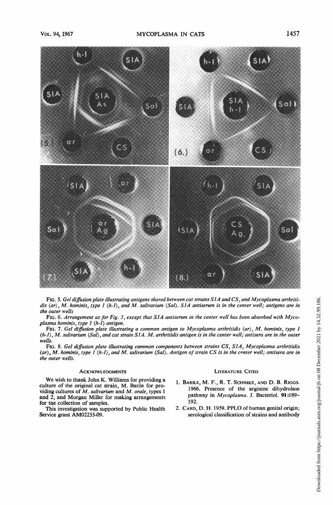
arthritidis antigen, a continuous band was pro-duced (Fig. 7). A similar continuous band wasalso produced by use of M. hominis, type 1, anti-gen. This component was designated h. The twoother components which SlA and CS shared withM. hominis, type 1, were designated i and j, re-spectively. Absorption of SlA serum with M.hominis, type 1, antigen removed components h,i, and j, but SlA serum still produced two bandswith its own antigen and that of CS (Fig. 5 and6). These components were designated k and 1.Absorption of SlA serum with M. arthritidis alsoremoved components h, i, and j, indicating thatsmall amounts of components i and j alsooccurred in M. arthritidis. Absorption with M.salivarium antigen removed only components hand i. Absorption with M. orale, types 1 and 2,removed all three components.
Similar reactions occurred in the opposite direc-tion by use of antigens of CS and SlA with seraagainst M. arthritidis, M. salivarium, and M.hominis, type 1 (Table 4 and Fig. 8). In thisinstance, two precipitin bands were producedwith M. arthritidis serum.None of the sera tested reacted with antigens
of M. pulmonis or M. hyorhinis.
DISCUSSIONSwitzer (24) reported the isolation of myco-
plasma from the pneumonic lung of a 6-week-oldkitten. The disease could not be produced experi-mentally, however, and the isolate was not charac-terized.The cat strains isolated in the present study
could be placed in two distinct groups on thebasis of physiology, morphology, and antigeniccomposition. Group 1, comprising strains CO,BI, and B2, was characterized by the reduction ofmethylene blue, lipase activity towards egg yolkemulsion, sensitivity to neomycin and novobiocin,marked hemolysis of various animal erythrocytes,and lack of ammonia production from arginine.Colonies produced well-defined regions of agargrowth.Group 2, strains SlA and CS, did not reduce
methylene blue, possessed no lipase activity to-wards egg yolk emulsion, were resistant to neo-mycin and novobiocin, produced only weakhemolysis of sheep and guinea pig erythrocytes,but produced ammonia from arginine. The col-onies were vacuolated without well-defined centra!regions. Groups 1 and 2 were serologically un-related, as tested by complement fixation, geldiffusion, and growth inhibition. The presence ofcommon antigens between M. arthritidis, M.salivarium, M. hominis, type 1, and M. orale iswell established (2, 5, 16, 17, 20, 26, 27). By useof agar gel double diffusion, it was shown thatthree of these antigens were also common to theGroup 2 cat strains. However, on the basis ofcomplement fixation, growth inhibition, and thepresence of two antigens specific to the Group 2strains, it was concluded that the latter formed adistinct group. This was also confirmed by thecharacteristic colonial morphology. On the basisof previous reports, the cat strains could also bedifferentiated from M. gallinarum, M. galli-septicum, M. canis, M. maculosum, M. mycoides,M.fermentans, M. laidlawii, M. neurolyticum, andM. pneumoniae (1, 8-10, 23). The physiologicalcharacteristics of most of these species have beenconfirmed by the present authors (unpublishedobservations).The original isolation and apparent induction
of conjunctivitis with strain CO suggests myco-plasma as one of the possible etiological agents ofcat conjunctivitis. The common occurrence ofidentical mycoplasma strains in the saliva ofhealthy cats suggests this as the potential sourceof the infection. This is supported by the findingthat strain CO survived in the saliva of bothkittens, even after they could no longer be iso-lated from the eyes. Further studies are required,however, to confirm the pathogenicity of thesestrains. It is interesting to note that mycoplasmahave also been isolated from cases of humanconjunctivitis (11).As a result of these studies, it is tentatively
suggested that Groups 1 and 2 be called M.1elis and M. gateae, respectively.
1456 J. BACTERIOL.
Dow
nloa
ded
from
http
s://j
ourn
als.
asm
.org
/jour
nal/j
b on
08
Dec
embe
r 20
21 b
y 14
.32.
99.1
86.
MYCOPLASMA IN CATS
FIG. 5. Gel diffusion plate illustrating antigens shared between cat strains SIA and CS, and Mycoplasma arthriti-dis (ar), M. hominis, type I (h-i), and M. salivarium (Sal). SIA antiserum is in the center well; antigens are inthe outer wells
FIG. 6. Arrangement as for Fig. 5, except that SIA antiserum in the center well has been absorbed with Myco-plasma hominis, type I (h-i) antigen.
FIG. 7. Gel diffusion plate illustrating a common antigen to Mycoplasma arthritidis (ar), M. hominis, type I(h-i), M. salivarium (Sal), and cat strain SIA. M. arthritidis antigen is in the center well; antisera are in the outerwells.
FIG. 8. Gel diffusion plate illustrating common components between strains CS, SIA, Mycoplasma arthiritidis(ar), M. hominis, type I (h-i), and M. salivarium (Sal). Antigen ofstrain CS is in the c"nter well; antisera are inthe outer wells.
ACKNOWLEDGMENTSWe wish to thank John K. Williams for providing a
culture of the original cat strain, M. Barile for pro-viding cultures of M. salivarium and M. orale, types 1and 2, and Morgan Miller for making arrangementsfor the collection of samples.
This investigation was supported by Public HealthService grant AM02255-09.
LITERATURE CITED
1. BARILE, M. F., R. T. SCHIMKE, AND D. B. RIGGS.1966. Presence of the arginine dihydrolasepathway in Mycoplasma. J. Bacteriol. 91:189-192.
2. CARD, D. H. 1959. PPLO of human genital origin;serological classification of strains and antibody
1457VOL. 94, 1967
Dow
nloa
ded
from
http
s://j
ourn
als.
asm
.org
/jour
nal/j
b on
08
Dec
embe
r 20
21 b
y 14
.32.
99.1
86.

COLE, GOLIGHTLY, AND WARD
distribution in man. Brit. J. Venereal Diseases35:27-34.
3. CLYDE, W. A., JR. 1964. Mycoplasma speciesidentification based upon growth inhibition byspecific antisera. J. Immunol. 92:958-965.
4. COLE, B. C., M. L. MILLER, AND J. R. WARD.1967. A comparative study on the virulence ofMycoplasma arthritidis and Mycoplasma homi-nis, type 2 strains in rats. Proc. Soc. Exptl. Biol.Med. 124:103-106.
5. CORELL, L. L. D. P. FABRIZIO, AND S. R. WILSON.1960. Comparison of pleuropneumonia-likeorganism strains from tissue culture by com-plement fixation. Ann. N.Y. Acad. Sci. 79:574-580.
6. DENES, L. 1939. "L" organism of Klienebergerand Streptobacillus moniliformis. J. Infect.Diseases 65:24-42.
7. EDWARD, D. G. FF. 1950. An investigation ofpleuropneumonia-like organisms isolated fromthe bovine genital tract. J. Gen. Microbiol.4:4-15.
8. EDWARD, D. G. FF. 1954. The pleuropneumoniagroup of organisms: a review together withsome new observations. J. Gen. Microbiol. 10:27-64.
9. EDWARD, D. G. FF., AND A. D. KANAREK. 1959.Organisms of the pleuropneumonia group ofavian origin: their classification into species.Ann. N.Y. Acad. Sci. 79:696-702.
10. FREUNDT, E. A. 1958. The Mycoplasmataceae.(The pleuropneumonia group of organisms).Munksgaard, Copenhagen.
11. HOLLAND, M. C. 1960. Uveitis and pleuropneu-monia-like organisms. Ann. N.Y. Acad. Sci.79:646-649.
12. KLENEBERGER, E., AmD D. B. STEABBEN. 1960.On the association of the pleuropneumonia-like organism L3 with bronchiectatic lesions inrats. J. Hyg. 40:223-227.
13. KLIENEBERGER-NOBEL, E. 1960. Pathogenicity andimmunology of organisms of the pleuropneu-monia group. Ann. N.Y. Acad. Sci. 79:615-625.
14. KOLMER, J. A., E. H. SPAULDING, AND H. W.ROBINSON. 1951. Approved laboratory technic,5th ed. Appleton-Century-Crofts, Inc., NewYork.
15. KRAUS, F. W., J. F. NICKERSON, W. I. PERRY,AND A. P. WALKER. 1957. Peroxide and peroxi-
dogenic bacteria in human saliva. J. Bacteriol.73:727-735.
16. LEMCKE, R. M. 1964. The serological differentia-tion of mycoplasma strains (pleuropneumonia-like organisms) from various sources. J. Hyg.62:199-129.
17. LEMCKE, R. M. 1965. A serological comparison ofvarious species of Mycoplasma by an agar geldouble diffusion technique. J. Gen. Microbiol.38:91-100.
18. MILES, A. A., AND S. S. MIsRA. 1938. The estima-tion of the bactericidal power of the blood. J.Hyg. 38:732.
19. MUFSON, M. A., W. M. LuDWIG, R. H. PURCELL,T. R. CATE, D. TAYLOR-ROBINSON, AND R. M.CHANOCK. 1965. Exudative pharyngitis follow-ing experimental Mycoplasma hominis, type 1injection. J. Am. Med. Assoc. 192:1146-1152.
20. PEASE, P. E. 1965. The antigenic structure ofPPLO (Mycoplasma hominis) and relatedbacteria. J. Gen. Microbiol. 41:299-308.
21. SABIN, A. B. 1939. Mice as carriers of pathogenicpleuropneumonia-like organisms. Science 90:18-19.
22. SABIN, A. B., AND B. JOHNSON. 1940. Pathogenicpleuropneumonia-like organisms in tissues ofnormal mice and isolation of new immuno-logical types. Proc. Soc. Exptl. Biol. Med. 44:569-571.
23. SOMERSON, N. L., D. TAYLOR-ROBINSON, ANDR. M. CHANOCK. 1963. Hemolysin productionas an aid in the identification and quantitationof Eaton agent (Mycoplasma pnewnoniae). Am.J. Hyg. 77:122-128.
24. SWITZER, W. P. 1967. The genus Mycoplasma, p.531-548. In I. A. Merchant and R. A. Packer[ed.], Veterinary bacteriology and virology, 7thed. Iowa State University Press, Ames.
25. SWITZER, W. P. 1964. Mycoplasmosis, p. 498-510.In H. W. Dunne [ed.], Diseases of swine, 2nded. Iowa State University Press, Ames.
26. TAYLOR-ROBINSON, D., J. CANCHOLA, H. Fox,AND R. M. CHANOCK. 1964. A newly identifiedoral mycoplasma (M. orale) and its relationshipto other human mycoplasmas. Am. J. Hyg.80:135-148.
27. TAYLOR-ROBINSON, D., N. L. SOMERSON, H. C.TURNER, AND R. M. CHANOCK. 1963. Sero-logical relationships among human myco-plasmas as shown by complement-fixation andgel diffusion. J. Bacteriol. 85:1261-1273.
1458 J. BACTERIOL.
Dow
nloa
ded
from
http
s://j
ourn
als.
asm
.org
/jour
nal/j
b on
08
Dec
embe
r 20
21 b
y 14
.32.
99.1
86.