Embed Size (px)
Citation preview
University of North DakotaUND Scholarly Commons
Theses and Dissertations Theses, Dissertations, and Senior Projects
January 2017
Characterization Of The Immune StimulatingProperties Of Type III Secretion System NeedleProtein Bscf From Bordetella Pertussis: TowardsThe Development Of A New Acellular PertussisVaccineTravis Douglas Alvine
Follow this and additional works at: https://commons.und.edu/theses
This Dissertation is brought to you for free and open access by the Theses, Dissertations, and Senior Projects at UND Scholarly Commons. It has beenaccepted for inclusion in Theses and Dissertations by an authorized administrator of UND Scholarly Commons. For more information, please [email protected].
Recommended CitationAlvine, Travis Douglas, "Characterization Of The Immune Stimulating Properties Of Type III Secretion System Needle Protein BscfFrom Bordetella Pertussis: Towards The Development Of A New Acellular Pertussis Vaccine" (2017). Theses and Dissertations. 2158.https://commons.und.edu/theses/2158
CHARACTERIZATION OF THE IMMUNE STIMULATING PROPERTIES OF TYPE III SECRETION SYSTEM NEEDLE PROTEIN BSCF FROM BORDETELLA PERTUSSIS: TOWARDS THE
DEVELOPMENT OF A NEW ACELLULAR PERTUSSIS VACCINE
by
Travis Douglas Alvine Bachelor of Science, University of North Dakota, 2005
Master of Science, University of North Texas, 2011
A Dissertation Submitted to the Graduate Faculty
of the
University of North Dakota
in partial fulfillment of the requirements
for the degree of
Doctor of Philosophy
Grand Forks, North Dakota
December 2017

ii
iii
PERMISSION
Title Characterization of the Immune Stimulating Properties of Type III
Secretion System Needle Protein BscF from Bordetella pertussis: Towards the Development of a New Acellular Pertussis Vaccine
Department Biomedical Sciences Degree Doctor of Philosophy In presenting this dissertation in partial fulfillment of the requirements for a graduate degree from the University of North Dakota, I agree that the library of this University shall make it freely available for inspection. I further agree that permission from extensive copying for scholarly purposes may be granted by the professor who supervised my dissertation work or, in his absence, by the Chairperson of the department or the dean of the School of Graduate Studies. It is understood that any copying or publication or other use of this dissertation or part thereof for financial gain shall not be allowed without my written permission. It is also understood that due recognition shall be given to me and to the University of North Dakota in any scholarly use which may be made of any material in my dissertation. Travis Alvine December 3, 2017
iv
TABLE OF CONTENTS
LIST OF FIGURES..................................................................................................................v ACKNOWLEDGMENTS.......................................................................................................vii ABSTRACT.........................................................................................................................viii CHAPTERS
I. INTRODUCTION.......................................................................................................1
II. CHARACTERIZATION OF THE IMMUNE RESPONSE INDUCED BY BSCF, A PURIFIED TYPE III SECRETION SYSTEM NEEDLE PROTEIN FROM BORDETELLA PERTUSSIS........................................................................................21
III. PURIFIED TYPE III SECRETION SYSTEM NEEDLE PROTEINS INDUCE
CLATHRIN-DEPENDENT NF-𝜅B/AP-1 SIGNALING FROM ENDOSOMAL COMPARTMENTS............................................................................54
IV. BSCF AS A VACCINE CANDIDATE FOR A NEXT GENERATION BORDETELLA PERTUSSIS ACELLULAR VACCINE...........................................................................73
V. DISCUSSION..........................................................................................................97 REFERENCES....................................................................................................................103
v
LIST OF FIGURES
Figure Page 1. Characterization of BscF protein preparation.............................................................44
2. BscF activates NF-𝜅B/AP-1 signaling in THP1-XBlue and HEK293 cells in a TLR2 and TLR4 dependent mechanism................................................................45
3. BscF stimulation induces robust inflammatory cytokine release from murine and human innate cells..................................................................................46
4. BscF induction of inflammatory cytokines is largely dependent upon TLR4 activation............................................................................................................47
5. BscF stimulates IL-1𝛽 release in an Nlrp3 caspase-1 dependent mechanism............48
6. Pre-treatment with cytochalasin D disrupts IL-1𝛽 release and results in an increased accumulation of cytosolic pro-IL-1𝛽......................................................49
7. BscF immunization induces a robust humoral response and B. pertussis opsonizing antibodies.................................................................................................50
8. BscF enhances activation of Th1 and Th17 cells, but not Th2 cells............................51
9. BscF vaccination acts as a protective antigen against B. pertussis.............................52
10. Proposed model of T3S system needle protein-induced IL-1𝛽 secretion...................53
11. Endocytosis inhibitors reduce NF-𝜅B/AP-1 signaling induced by T3S system needle proteins in HEK-Blue TLR2 and HEK-Blue TLR4 cells.......................................67
12. Endocytosis inhibitors reduce TNF-𝛼 production in T3S system needle protein human THP-1 cells..........................................................................................68
13. siRNA gene knockdown of heavy chain clathrin reduced NF-𝜅B/AP-1 signaling induced by T3S system needle proteins in HEK-Blue TLR2 cells..................69
vi
14. CD14 mediates NF-𝜅B/AP-1 signaling induced by T3S system needle proteins
in HEK-Blue TLR2 and HEK-Blue TLR4 cells..................................................................70
15. Direct cell cytotoxicity of endocytosis inhibition in HEK-Blue TLR2 and HEK-Blue TLR4 cells.....................................................................................................71
16. Proposed model of T3S system needle protein endosomal activation of NF-𝜅B/AP-1 and pro-inflammatory release................................................................72
17. BscF promotes murine DC maturation and inflammatory cytokine release...............92
18. BscF acts as an adjuvant to enhance aP vaccine specific antibody responses............93
19. BscF indirectly enhances IFN-𝛾 and IL-17 production from ex vivo stimulated splenocytes.................................................................................................................94
20. The addition of BscF to the laboratory aP vaccine enhanced a central memory T cell phenotype...........................................................................................95
21. The addition of BscF to the laboratory aP vaccine enhanced protective immunity against a sub lethal B. pertussis challenge..................................................96

vii
ACKNOWLEDGMENTS I would first like to thank Dr. David S. Bradley for giving me the opportunity to grow not
only as a scientist, but also as a person during my time in his laboratory. Your willingness
to allow me to develop my research skills, often times through trial and error, is greatly
appreciated. I would also like to recognize the efforts of Dr. Matthew L. Nilles. I will
always appreciate your insight, suggestions, and enthusiasm during the ups and downs
of this work. My special thanks also go to my remaining committee members: Dr. Jyotika
Sharma, Dr. Patrick Carr, and Dr. Jefferson Vaughan for all of your assistance and for
your continued support that contributed to the success of this work. Thank you to the
graduate students, faculty, and staff of the Microbiology and Immunology graduate
program as well as the Department of Biomedical Sciences. In particular, I thank Patrick
Osei-Owusu for teaching me everything I needed to know about needle proteins, and
Peter L. Knopick for the countless occasions where you provided helpful suggestions and
the numerous hours you spent helping me during my work. I need to recognize the
exceptional patience and support my wife, Beth, has shown me during this process; you
have given my every opportunity to succeed in this endeavor. Finally, I would like to
thank my family and friends for their support and encouragement.

viii
ABSTRACT Despite widespread vaccination, Bordetella pertussis, the causative agent of whooping
cough, is still a threat to global health. One cause of pertussis reemergence observed in
many countries is ineffective immunity generated by the current acellular pertussis (aP)
vaccines. Interestingly, recent studies have shown that TLR stimulating agents can
enhance aP vaccine induced immunity. Type III secretion (T3S) system needle proteins
from many gram-negative bacteria have been shown to be strong TLR agonists that
induce NF-𝜅B/AP-1 signaling and promote inflammatory cytokine release from innate
cells in vitro. In this study, we investigated the immune modulating properties of BscF, a
purified T3S system needle protein from B. pertussis. In addition, we characterized the
ability of BscF to enhance aP vaccine induced immunity. In the current study, we
demonstrated that BscF is a strong TLR2 and TLR4 agonist that induced NF-𝜅B/AP-1
activation and promoted inflammatory cytokine release, augmented by clathrin-
mediated endocytosis. In vivo, BscF immunization induced robust antibody responses,
strong Th1 and Th17 responses from stimulated splenocytes, and provided modest
protection against B. pertussis challenge. BscF also enhanced aP induced immunity and
reduced lung bacterial burden in mice challenged with B. pertussis. These results
demonstrate that BscF has considerable potential to be included in a next-generation B.
pertussis aP vaccine.
1
CHAPTER I
INTRODUCTION
Bordetella Microbiology and History The genus Bordetella, belonging to the Alcaligenaceae family, are comprised of
10 genetically distinct species (1-3). B. pertussis is a Gram-negative, non-motile, aerobic
coccobacillus that is typically grown at 37 ℃ on special Bordet-Gengou agar
supplemented with blood and other growth factors. B. pertussis’s growth on blood
supplemented medium is slow, requiring at least 3 days for colonies to appear. Within
the genus Bordetella, the species can be differentiated, in part, by their hosts they infect
as well as the symptoms reported during infection. B. pertussis is strictly a human
pathogen (4-5) and was first thought as the sole cause of the prototypical whooping
cough in humans. More recently, B. parapertussis, and B. holmesii have been identified
to cause the prototypical whooping cough symptoms (6-11). While primarily thought of
as a domestic animal pathogen i.e., cats and dogs (12-13), B. bronchiseptica has been
isolated in rare events from immunocompromised or traumatized humans (14-16).
Despite the ability of many Bordetella species to infect humans, B. pertussis still remains
the most well characterized, and possess the greatest risk to overall global health.
When compared to many other infectious diseases, whooping cough is a fairly
newly discovered pathogen. Pertussis-like symptoms and illness go back roughly 1,500
2
years when it was described by a Chinese medical scholar as “the cough of 100 days”
(17-18). Fast forward to 1578 when Guillaume de Baillou characterized what was
thought of today as the oldest pertussis outbreak among children in Paris (19). Recent
evidence suggests that 3 epidemics of whooping cough occurred in Persia (present-day
Iran) in the 15th and 16th century, likely indicating the earliest recorded epidemics of
whooping cough in the world (20). Outbreaks of pertussis have also been reported in
Europe during the 16th century, however the causative agent was not identified until
much later. In 1906, B. pertussis was first identified as the causative agent of whooping
cough by Jules Bordet and Octave Gengou (21), leading to Bordet winning the 1920
Nobel Prize in Physiology or Medicine for his extensive body of work from developing
culture medium necessary to grow B. pertussis to further characterization of B. pertussis
as the causative agent of whooping cough. B. pertussis as it’s named today, was
originally named Haemophilus pertussis, but the name was changed to honor one of its
discoverers (22).
Bordetella pertussis pathogenesis
As previously mentioned, B. pertussis is strictly a human pathogen (4-5). B.
pertussis is pathogen that targets the upper respiratory tract is classically considered an
extracellular pathogen. Despite its extracellular location within the respiratory tract, B.
pertussis has been shown to invade ciliated epithelial cells as well as alveolar
macrophages (23-25). B. pertussis is passed from human to human through inhalation of
infected respiratory droplets (26-30). Upon inhalation, B pertussis enters and adheres to
ciliated epithelial cells of the upper respiratory tract (26-31). B. pertussis is classified as a
3
toxin mediated disease that requires a coordinated effort from a number of virulence
factors expressed by the bacteria. This coordinated virulence factor activation is
initiated upon B. pertussis attachment, and further allows for B. pertussis dissemination
to the lower respiratory tract (26-31). These virulence factors include toxins: pertussis
toxin (PT), adenylate cyclase toxin (ACT), dermonecrotic toxin (DNT), and tracheal
cytotoxin (TCT), as well as other structures including filamentous hemagglutinin (FHA),
fimbriae (FIM), pertactin (PRN), the type three secretion (T3S) system, and
lipopolysaccharide (LPS). These bvgAS virulence genes are controlled by the BvgAS two-
component regulatory system of B. pertussis and allows the bacteria to respond to
changing environments (32-37). The importance of these virulence factors during
infection are highlighted in a number of studies indicating that these virulence factors
not only promote adhesion and invasion of ciliated epithelial cells, but also strongly
influence the host’s innate and adaptive immune system often times at the expense of
the host and to the benefit of the pathogen.
B. pertussis virulence factors and host immune modulation pertussis toxin (PT)
PT is the most well characterized toxin and is the only B. pertussis antigen that is
included in all formulations of current licensed acellular pertussis (aP) vaccines. PT is
classified as an A-B toxin consisting of 5 subunits (S1 through S5). The A subunit (S1) is
an ADP-ribosyltransferase while the B subunit is a pentameric ring structure (S2, S3, S5,
and two S4 subunits) that mediates toxin binding to target cells through binding to
glycosylated receptors (38-39). Once inside the cell, the holotoxin undergoes retrograde
transport to the endoplasmic reticulum and the S1 subunit (the catalytic domain of PT)
4
is released into the cytosol (40). Once inside the cytosol, PT modifies intracellular
signaling cascades through its ribosylation of the 𝛼 subunit of heterotrimeric G proteins.
PT has been implicated in many secondary systemic complications that arise from B.
pertussis infections because of its ability to bind glycosylated receptors, as well as its
ability to modify G proteins; both glycosylated receptors and G proteins are expressed in
many different tissues. The pleiotropic affects seen by PT range from paroxysms and
neurological disturbance (41), lymphocytosis (42), hyperinsulinemia, hypoglycemia, as
well as histamine sensitization (43) are the result of its enzymatic activity. In addition, PT
as well as detoxified PT (dPT) can act as pattern associated molecular patterns (PAMPs)
that bind and activate a group of pattern recognition receptors (PRRs) called toll-like
receptors (TLRs), specifically TLR2 and TLR4 (44-46).
In addition to its enzymatic and immune stimulating activities, PT can modulate
the host innate and adaptive immune response further limiting the ability of the host to
respond to, and clear, a B. pertussis infection, as well as influence the surrounding
environment by either promoting pro- or anti-inflammatory mechanisms. Many of these
studies have been completed in PT-deficient B. pertussis strains. The contribution of PT
to early stages of infection were elucidated as infection with a PT-deficient B. pertussis
strain resulted in lower bacterial colonization as early as 24 hours post infection (47). In
addition, it appears that PT suppresses early neutrophil influx into the lungs during B.
pertussis infection through inhibition of neutrophil attracting chemokine release from
resident cells within the lungs (47-50). PT has also been shown to target and inhibit
airway macrophages in addition to neutrophils (51). PT also targets the adaptive
5
immune system by suppressing B. pertussis specific serum antibody levels during
infection (52-53). Interestingly, the role of PT appears to evolve as the infection
progresses. Early in infection, the primary role of PT is to down regulate host immunity,
primarily through innate cell inhibition. However, at the peak of infection PT induces
robust lung inflammation and pathology in mice (54). From these studies we can
appreciate the importance and complicated role that PT plays during B. pertussis
infection.
Adenylate cyclase toxin (ACT)
ACT is another important B. pertussis specific toxin that is now recently been the
focus of studies determining the usefulness of adding ACT to new aP vaccines. To date,
ACT is not an antigen that is included in current aP vaccines. ACT is a member of the
repeat in toxin family and is secreted from the cell via type I secretion. ACT possesses
two functional C- and N-terminal domains that facilitate receptor binding on host cells
and its catalytic adenylate cyclase activity, respectively (55). Once inside the host cell
cytosol, the catalytic domain is activated by by calmodulin binding, facilitating the
conversion of cellular ATP into cAMP and modifying intracellular signaling cascades (56).
Given that both PT and ACT appear to modify intracellular signaling in similar manners,
it was thought that these toxins may play redundant roles during infection. ACT-
deficient B. pertussis strains showed reduced colonization ability; however, these strains
were able to colonize the host for a short period of time but were unable to cause
persistent infections (48). From these observations and the observations that PT-
deficient strains showed reduced ability to colonize early during infection, it is likely that
6
these two toxins have non-redundant roles in B. pertussis infections: PT facilitates early
colonization while ACT is necessary for persistent infection (48).
Like PT, ACT also modulates the surrounding environment to facilitate infection.
ACT has been shown to down regulate host immunity by inhibiting phagocytic cell
trafficking, bactericidal activities, and pro-inflammatory cytokine release (57-58). ACT
also has been shown to modulate the adaptive arm of the immune system during
infection. T-helper (Th) cells, specifically Th1 and to a lesser extent Th17 cells, are
important for resolution of B. pertussis infections. ACT directly targets T cell activation
and differentiation to skew CD4+ T cells to a Th2 type phenotype (59); a T cell response
that has shown to be non protective both animal and human models. ACT also polarizes
the adaptive immune system to a Th17 type response by NLRP3 inflammasome
activation and subsequent IL-1𝛽 production from murine dendritic cells (DC) (60). The
pro-inflammatory role of ACT is controversial, but the importance of ACT in B. pertussis
pathogenesis is highlighted in both murine and human studies.
Tracheal cytotoxin (TCT)
TCT is not currently used in aP vaccines and is not unique to B. pertussis. TCT is a
disaccharide-tetrapeptide monomer of peptidoglycan that is present in all Gram-
negative bacteria (61). ACT is released as part of normal bacterial cell growth as they
remodel their cell wall. Due to the lack of the cytoplasmic membrane protein called
AmpG, which normally participates in recycling of the peptidoglycan fragment, TCT is
constitutively released into the environment and is not under control of the BvgAS two-
component system controlling other virulence factor gene expression. Because TCT is

7
released from the bacteria it acts locally on cells of the respiratory system to promote B.
pertussis infection. TCT acts directly on nonciliated respiratory cells to induce IL-1𝛼,
leading to increased nitric oxide (NO) synthase activity and subsequent NO production
(62-63). TCT’s ability to induce NO production is dependent on LPS (62). The NO is then
able to diffuse to neighboring ciliated epithelial cells further promoting respiratory tract
inflammation.
Filamentous hemagglutinin (FHA)
FHA is one of the most important adhesions and like PT, is included in all of the
current aP vaccines. FHA has binding domains specific for heparin sulfate, carbohydrate,
and integrin binding via an Arg-Gly-Asp site (64). Due to the multiple binding sites, FHA
mediates initial adhesion to ciliated epithelial cells of the upper respiratory tract.
Dissemination of B. pertussis down to the lower respiratory tract is mediated by FHA.
Secretion of FHA to the cell wall has been shown to be dependent on an outer
membrane accessory protein named FhaC as FhaC is able to make channels within the
outer membrane (65). In addition to its binding characteristics, FHA is also an immune
modulator. In mice, FHA primarily acts an immune suppressor. Systemic administration
of FHA suppressed pro-inflammatory cytokine and enhanced anti-inflammatory cytokine
release from innate cells, in addition to generating regulatory T cells and reduced colitis
induced intestinal inflammation (66). Infection studies in B. bronchiseptica further
clarified FHA’s immunosuppressive role (67-68). Interestingly, the role of FHA in a
human context appears to be inflammatory rather than anti-inflammatory. FHA elicited
pro-inflammatory cytokine release from human innate and epithelial cells (69-70).
8
Fimbriae (FIM)
FIM is a surface exposed structure that has heparin binding activity. The major
subunits of FIM, FIM2 and FIM3, are serotype specific and have been shown to be
serologically distinct (71). FIM antigens have been included as antigens in current aP
vaccines; however not to the same extent that PT and FHA have been. FIM were shown
to important for colonizing the respiratory tract in B. bronchiseptica infections (72).
Pertactin (PRN)
PRN is an auto transporter protein that mediates B. pertussis adhesion to
eukaryotic cells via its Arg-Gly-Asp binding site; however, PRN also contains proline-rich
regions and leucine-rich repeats (73). PRN contributes to B. pertussis pathogenesis by
resisting neutrophil-mediated clearance (74). PRN antigens are commonly used in
current aP vaccines. Because PRN is highly polymorphic, the circulating B. pertussis
strains may have different PRN variants that are included in aP vaccines. In addition,
PRN-negative strains are beginning to emerge across the globe. The first reported PRN-
negative strain was in 2012 in France (75), with many other PRN-negative reports
following (76-77). PRN appears to be dispensable to the bacteria as PRN-negative strains
can evade aP generated immunity better than PRN-positive strains (78). The emergence
of PRN-negative B. pertussis strains highlights the need for newly developed protective
antigens in next-generation aP vaccines.
Type III secretion (T3S) system
In comparison to other Gram-negative bacteria (i.e. Yersinia spp.), the T3S
system of B. pertussis is not well characterized. Four B. pertussis proteins: Bsp22, BopN,
9
BopD, and BteA have been shown to be secreted. The structure and function of the B.
pertussis is similar to other Gram-negative bacteria. This is a syringe-like structure that
extends from the surface of the bacteria and facilitates direct translocation of effector
molecules into host cells. The first reports of the contribution the T3S system plays
during infection came from studies in B. bronchiseptica. These studies reveled that B.
bronchiseptica uses T3S to persist within the murine respiratory tract (79). The ability of
B. bronchiseptica to persist during infection is likely due to the ability of the T3S system
to modulate innate immunity. Re-stimulated splenocytes from T3S mutant B.
bronchiseptica produced more IFN-𝛾 and less of the immunosuppressive cytokine IL-10,
suggesting that T3S down regulates host immunity (80). In addition, the T3S system
from B. bronchiseptica has been shown to be important for modulating DC maturation
(81-82). The first evidence of a functionally active T3S system in B. pertussis was
reported in 2008 (83). Mutation of the effector molecule bscN, which abolished protein
secretion, resulted in a reduced ability to colonize the respiratory tract of mice (83).
Reduced colonization was the result of an increase in innate pro-inflammatory cytokine
release in the lungs, elevated antigen specific IFN-𝛾 and IL-17, as well as increased
antibody responses (83). These results confirm an immunosuppressive role of the T3S
system in B. pertussis infections and provides evidence of the importance of T3S during
B. bronchiseptica and B. pertussis infections. Another interesting finding from the work
of Fennelly et al., (83) was that a number of laboratory-adapted strains of B. pertussis
did not secrete the effector molecule Bsp22. This reversibility of T3S system expression
was confirmed by Gaillard et al., (84). In addition to finding that laboratory-adapted
10
strains did not have a functional T3S system in vitro, they reported that the T3S system
can become functional in vivo when the bacteria are put into a host; this was also true in
the laboratory-adapted strains as well (84).
Host immune responses to B. pertussis infection
Our understanding of what protective immunity looks like comes from a large
body of work examining the host immune response to B. pertussis infection. The first
line of defense against B. pertussis infections occurs at the upper respiratory tract and
involves cells of the innate immune system. These lung resident cells include airway
mucosal dendritic cells (AMDCs) and alveolar macrophages (AMs). The innate immune
system helps to control early infection, and polarizes the adaptive immune response
necessary to clear the infection. AMDCs will take up antigen at the site of infection and
migrate to the lymph nodes to prime an adaptive immune response (85-86). On the
other hand, AMs residing at the site of infection uptake and kill B. pertussis directly (87).
These resident cells respond to, and become activated by, the many PAMPs expressed
by B. pertussis. Many of the virulence factors have been shown to stimulate PRRs of the
innate immune system. These resident cells within the lung not only act as the first line
of defense, they also orchestrate further immune responses that prime the adaptive
immune response and facilitate the recruitment of other immune cells.
In addition to AMDCs and AMs, several other cell types contribute to B. pertussis
clearance, especially during the early stages of infection. 𝛾𝛿 T cells, have been shown to
be important in the early immune response. 𝛾𝛿 T cells release an early source of IL-17
which promotes cell trafficking into the lungs as well as antimicrobial peptide
11
production (88-89). IL-17 release in combination with the cytokine CXCL2 (MIP-2)
secreted from macrophages and epithelial cells promotes neutrophil influx to the site of
infection. Neutrophils have been shown to be an important cell type for controlling B.
pertussis infections by antibody-mediated phagocytosis, intracellular killing of B.
pertussis, and the production of neutrophil extracellular traps (81-82). Natural killer (NK)
cells have also been shown to play an important role in early innate immune control of
B. pertussis infection. NK cells primarily exert their protective effects through the
release of IFN-𝛾. IFN-𝛾 activates macrophages and depletion of NK cells from mice
infected with B. pertussis reduced IFN-𝛾 release resulting in bacterial dissemination
from the respiratory tract to the liver (83). While these innate cells play a critical role in
the early stages of infection, a polarized adaptive immune response is critical in
controlling late stages of infection, primarily by augmenting neutrophil and macrophage
bactericidal activity.
CD4+ T cells have been shown to play an important role in B. pertussis clearance
as adaptive transfer of B. pertussis specific CD4+ T cells, but not B. pertussis specific CD8+
T cells into immunosuppressed mice resulted in effective bacterial clearance (84). CD4+
T cells mediate their immune activation through cytokine release. Th1 and Th17 CD4+ T
cells release IFN-𝛾 and IL-17, respectively. The role of Th1 and Th17 cells in protective
immunity against B. pertussis was investigated in IFN-𝛾-/- and IL-17-/- mice which
demonstrated a reduced ability to clear B. pertussis from the lungs (85-86). Moreover, in
the case of the IL-17-/- mice, the inability to clear the pathogen was associated with a
reduction in neutrophils within the lungs during infection (86). Once polarized by

12
AMDCs in the lymph nodes, primed T cells proliferate and differentiate into Th1 and
Th17 cells. These cells then migrate back to the site of infection, and enhance
macrophage and neutrophil bactericidal activity through IFN-𝛾 and IL-17 production,
respectively.
Pertussis Epidemiology
Global burden and economic impact
For the past few decades, the use of either the whole cell B. pertussis vaccine
(wP) or the currently aP vaccine has significantly reduced the global burden of the
disease, although pertussis still remains endemic in both developed and developing
countries. In addition, B. pertussis is undergoing a reemergence in many parts of the
world. Estimates of the global burden of B. pertussis has been complicated by a number
of issues from inadequate and/or underreporting, constant changes in surveillance and
diagnostic protocols, as well as routine modifications to vaccine schedules as well as
vaccine components (87). While the global incidence of pertussis is tricky, more recent
statistics show that in a 2010 analysis, there were 16 million reported B. pertussis cases
worldwide, with 195,000 deaths (88). A separate report completed in 2013 determined
there were an estimated 136,000 cases worldwide that year (89). Given the great
difficult in accurately reporting B. pertussis cases, underreporting is a significant
concern. Specifically, in the United States there have been a number of reported B.
pertussis outbreaks. Minnesota experienced a pertussis outbreak resulting in 4,144
reported cases (90). In the same year, there were a total of 4,918 cases in Washington
State (91). During the same time period and into 2014, pertussis was widespread
13
throughout California. In 2014, 10,831 cases were reported, the highest number in any
one year (92). In terms of the economic impact of pertussis, often times costs can be
high due to hospitalization. Total societal costs due to pertussis was estimated to be
approximately $800 for adolescents and $1,950 for adults per case (93). Clearly
pertussis is still active globally and negatively impacts human health worldwide.
Clinical presentation
Clinical progression of a B. pertussis infection is identified by 3 common stages:
1. Catarrhal 2. Paroxysmal 3. Convalescent. Each stage can vary in length, but typically
can last 1-3 weeks, with complete recovery taking much longer. The course and duration
of the disease is dependent on age of patient, vaccination history, infectious agent (i.e.
B. pertussis versus B. parapertussis), and infectious dose. The three stages are most
likely noticed and identified in infants and young children, while adolescents and adults
usually present with milder symptoms and may go undiagnosed. The incubation period
for B. pertussis is around 7-10 days. In the first stage, symptoms are often overlooked
because they are similar to other viral infections (94). Interestingly, in the catarrhal
stage, the bacterial burden is extremely high and the individual is most contagious at
this stage. In the second stage, the paroxysmal stage, patients experience severe
coughing bouts as well as the characteristic inspiratory whooping sound. Presently, it is
not clear if one of the many toxins or virulence factors are responsible for the coughing.
In the final stage, convalescence, coughing bouts are less in frequency and are not as
severe.
B. pertussis vaccines
14
To date, there have been 2 available vaccines for B. pertussis: the wP vaccine as
well as the aP vaccine. The wP vaccine was first introduced in the United States in the
late 1940s. the wP vaccine consisted of inactivated B. pertussis and has been given in
combination with diphtheria and tetanus toxoids. The cases of B. pertussis in the United
States plummeted to approximately 1,000 in 1976 after the implementation of the wP
vaccine (95). Despite its effectiveness, the wP vaccine fell out of favor due to safety
concerns of reported local and systemic reactions (96-97). In rare cases, serve
neurological diseases such as encephalopathy, spasms, and sudden infant death
syndrome were reported (96,98-99) The antigenicity and subsequent adverse reactions
can likely be attributed to LPS and other innate stimulating agents on the bacterial
surface. The continued adverse reactions from the wP vaccine prompted the
development of less reactogenic vaccines consisting of purified antigens from B.
pertussis. These vaccines, or derivations of these vaccines, are currently used today and
are classified as aP vaccines. The first aP vaccine was developed in Japan in 1984 (100).
Interestingly, the composition of the current aP vaccines are not uniform in the number
and/or amount of antigens. Mutant PT and FHA are the only two antigens included in
every aP vaccine. Others include PRN or fimbrial proteins. These new aP vaccines are
given with diphtheria and tetanus toxoids and absorbed in alum as an adjuvant. Many
different aP vaccines have undergone rigorous safety and toxicity testing and have been
deemed safe and immunogenic. Edwards et al., (101-102) concluded that all 13 aP
vaccines tested were found to be safer and produced less severe adverse reactions and
were at least as immunogenic as compared to wP vaccines.
15
Reemergence of B. pertussis
The reemergence of pertussis related diseases is a complex issue and is most
likely the result of a combination of multiple factors. First, many circulating B. pertussis
strains have undergone pathogen adaptation and genetic changes, especially in aP
vaccine antigens (103-105). One explanation for this is vaccine induced selective
pressure (106-107). Two of the major antigens that have undergone genetic changes are
PRN and PT. PRN-negative strains have been identified in many different parts of the
world (77,108-110). In the United States, more than 50% of the collected strains were
PRN-negative in 2012 (108). Interestingly, PRN-negative strains appeared to have a
competitive advantage over PRN-positive strains in mixed infections in mice (78). In
addition, PRN protein variants found in circulating B. pertussis strains are not included in
the vaccine strain. PRN2 and PRN3 protein variants have been found to outperform
PRN1 by enhanced colonization and increased transmissibility (111). In the case of PT,
strains with genetic changes within the PT promoter produce slightly more PT than
previously circulating strains (112), and are circulating globally. Given that vaccine
selection pressure is accelerating at an increased pace with the aP vaccines, and that
current antigens included in the aP vaccine are no longer present on many isolates, the
need for new vaccination strategies will be important moving forward.
Second, recent research indicates that the current aP vaccines fail to induce
protective immunity and vaccine induced immunity may wane overtime. The data from
both mouse and human studies indicate that CD4+ Th1 and to a lesser extent CD4+ Th17
cells are critical for protective immunity generated by vaccination. In animal studies, wP
16
vaccination conferred protective adaptive immunity mediated by Th1 and Th17 cells,
while aP vaccination promoted non-protective Th2 type responses (84,86, 113). Th2
type responses have been shown to redundant in animal models (86). Human studies
also highlight suboptimal immunity induced by aP vaccines. An analysis of T cell
responses in children reveal that aP vaccination promotes Th2 type responses, while wP
vaccination induces strong Th1 immune responses (114-115). In addition to suboptimal
immunity induced by aP vaccines, the longevity of protective immunity has been shown
to wane over time. In head to head comparisons of long term cellular immunity induced
by aP or wP vaccination in children, Schure et al., (116) found that aP vaccination
actually produced stronger B. pertussis specific CD4+ T cell responses compared to wP
vaccinated children. However, analysis of vaccine induced responses 5 years after
primary pertussis vaccination levels of IL-17 production from aP vaccinated PBMCs was
reduced compared to wP primed children (117). In addition, aP vaccination lead to
significantly more end-stage differentiated CD4+ T cells responses compared to wP
vaccination, suggesting the memory capacity of the immune responses is reduced in the
aP vaccinated children (117). Together, these reports highlight a major inadequacy with
the aP vaccine, potentially contributing to the increased incidence of pertussis cases in
many countries.
Finally, one of the hurdles pertussis researchers have had to overcome is that
many of the animal models currently used to understand pertussis pathogenesis and
vaccine induced immunity are not natural hosts to B. pertussis. For example, mice and
other small mammals do not exhibit the classical whooping cough symptom. Recently, a
17
baboon model has been developed and has greatly enhanced our understanding of B.
pertussis infection and transmission. One of the most striking finding from baboon
studies is that while aP vaccination prevented clinical symptoms of B. pertussis, the
baboons were highly colonized and were able to infect naïve baboons (113). This finding
is significant in that in aP vaccinated areas, while clinical identification of whooping
cough may be low, there may be a significant portion of the population that are
colonized and able to transmit B. pertussis to susceptible newborns and infants who
have not yet received aP vaccination. In addition, aP vaccination in the baboon model
induced a mixed Th1/Th2 response and failed to prevent colonization while wP
vaccination provided protection and induced a mixed Th1/Th17 response (113),
confirming the importance of both Th1 and Th17 cells in protective immunity against B.
pertussis.
Approaches to improve aP vaccines
Due to the number of shortcomings of current aP vaccines, the need for better
vaccines has been recognized by a number of authorities in the field. Although a number
of approaches have been suggested, a large amount of work has centered around
developing new protective antigens or adjuvants that when added to current aP
vaccines will skew vaccine induced immunity toward Th1/Th17 type responses. It is
thought that the numerous PAMPs included in the wP vaccine are what contributed to
long term protective immunity. These PAMPs activate the innate immune system and
promote inflammatory cytokine release and DC maturation to induce Th1/Th17 cellular
immunity (118). Current aP vaccines are absorbed in alum and it has been suggested

18
that there are no classical PAMPs included in the aP vaccines. Although PT has been
shown to have immune stimulating properties, vaccine preparation strategies have
destroyed PT’s immune stimulating ability (119). To that end, replacement or
supplementation with innate stimulating adjuvants is one strategy to enhance aP
vaccine immunogenicity and efficacy.
A TLR2 agonist of B. pertussis when combined with the components of the aP
vaccine enhanced protection from an aerosol B. pertussis challenge, induced robust
IgG2a antibodies, and enhanced IL-17 and IFNy production from antigen stimulated
splenocytes ex vivo compared with the aP vaccine in alum (120). A TLR4 agonist,
monophosphoryl lipid A (MPL), when mixed with the aP vaccine increased protection
against B. pertussis challenge when compared with the aP vaccine in alum adjuvant,
while suppressing Th2 responses (121). A separate TLR agonist, LpxL1 (122), a
genetically engineered LPS from Neisseria meningitidis, enhanced antigen specific IFNy
and IL-17 CD4+ T cells and increased the number of specific memory CD4+ TCM cells when
added to the aP vaccine (123). Finally, CpG oligonucleotides from bacterial DNA that
activate TLR9 have been shown to induce antigen-specific IgG2a titers (124) and Th1 and
Th17 cells (125) when added to the aP vaccine (86). These studies show the feasibility
and effectiveness of incorporating novel TLR agonists into the aP vaccine to enhance
pertussis specific immunity that will not only promote the proper adaptive immune
responses, but could also augment the long-term efficacy of the aP vaccine.
The importance of stimulating the innate immune system during vaccination and
the subsequent generation of protective immunity is highlighted by the efficacy of the
19
live-attenuated B. pertussis vaccine BPZE1. This vaccine is designed for intranasal
administration and has been genetically modified to remove or inactivate DNT, TCT, and
PT mice (126). BPZE1 has been found to be safe in mice (126), and in humans during a
Phase I clinical trial (127). Importantly, BPZE1 induced strong B. pertussis specific Th1
responses and provided protection against B. pertussis in mice (128) as well as
protected mice against B. parapertussis (129). The benefit of adding PAMPs to enhance
protection either by the addition of TLR stimulating molecules to the aP vaccine or novel
vaccines that seek to harness innate immune stimulation to promote protective cellular
responses opens the door to more efficient approaches to significantly decrease
pertussis cases globally.
Rationale of current work
Based on the current literature, the strategy of adding TLR stimulating agents to
the aP vaccine to skew aP induced immunity has proven successful. Recent work in our
laboratory has identified a number of novel TLR ligands purified from the T3S system of
many Gram-negative bacteria (130). These molecules have been shown to activate TLR2
and TLR4, induce NF-𝜅B/AP-1 signaling, and promote inflammatory cytokine release in
vitro (130-131). Interestingly, we have successfully demonstrated that the N-terminus
from these proteins functions to modulate innate immune activation (131). This is
unique in that not only have our proteins been shown to activate TLR2 and TLR4, but
these proteins can be modified to enhance or reduce NF-𝜅B/AP-1 signaling. In addition
to the immune stimulating properties of these proteins, we believe that given the
extracellular location of the T3S system, BscF may also act as a protective antigen in B.
20
pertussis infections. YscF, a purified needle protein from Y. pestis, protected mice
against a Yersinia pestis infection (132). In addition, a Chlamydia T3S system needle
protein induced specific humoral and cellular responses, and decreased Chlamydia loads
in mice (133). In the current study, we investigated the immune stimulating properties
of a novel B. pertussis specific protein from the T3S system called BscF. Further, we
assessed the ability of BscF to induce both humoral and cellular responses in mice
necessary for vaccine induced protection, and characterized the contribution of BscF to
a laboratory prepared aP vaccine.
21
CHAPTER II
CHARACTERIZATION OF THE IMMUNE RESPONSE INDUCED BY BSCF, A PURIFIED TYPE III SECRETION SYSTEM NEEDLE PROTEIN FROM BORDETELLA PERTUSSIS
Introduction
Bordetella pertussis is a gram-negative bacterium and the causative agent of the
vaccine preventable disease whooping cough (pertussis). With the development of a
whole cell pertussis (wP) vaccine in the 1940s-1950s, cases of pertussis were
dramatically reduced. The wP vaccine proved to be too reactogenic and was replaced
with a less reactogenic vaccine: the subunit acellular pertussis (aP) absorbed in alum as
the adjuvant in the 1990s. Despite continued widespread vaccination, whooping cough
has again reemerged as a global health threat not only in newborns and infants, but
surprisingly among adults as well (88). This resurgence has been linked to antigenic
variation in many circulating B. pertussis strains (103,134,135,76,136), defective long-
term immunological memory (137,138,139,140), and ineffective immune responses that
are necessary for long-term protection (141,113). It has been demonstrated in both
mice and humans that the aP vaccine induces robust Th2 responses, with a limited Th17
response (114,142,117,143). The aP vaccine has been shown to prevent clinical pertussis
symptoms, but does not prevent bacterial colonization or transmission (113).
On the other hand, the wP vaccine has been shown to promote Th1 and Th17
responses and protective immunity in both mice and humans (114,144,86).
22
Effectiveness of the wP vaccine has been largely attributed to its many antigens
and pathogen associated molecular patterns (PAMPs) that bind and activate innate
pattern recognition receptors (PRRs) (27). Given the contribution of the many PAMPs to
the effectiveness of the wP vaccine, we propose that the addition of PRR stimulating
agents could enhance the effectiveness of the current aP vaccine.
Endogenous B. pertussis specific PAMPs activate the innate immune system
through PRRs – including Toll-like receptors (TLRs), promote inflammatory cytokine
release, and direct pertussis-specific adaptive immunity (118). Because PAMPs appear
to be an important aspect of the wP vaccine, it has been suggested that the addition of
TLR agonists to the aP vaccine could re-direct the immune response generated by the aP
vaccine to a more wP-like immune response. A TLR2 agonist (lipoprotein BP1569 and its
synthetic derivative, LP1569) of B. pertussis when combined with the components of the
aP vaccine induced strong Th1 and Th17 responses and enhanced protection from an
aerosol B. pertussis challenge (120). A TLR4 agonist, monophosphoryl lipid A (MPL),
increased protection against B. pertussis challenge while suppressing Th2 responses
(121). In addition, other TLR4 agonists have been shown to enhance immunity
generated by the aP vaccine (123). Finally, CpG oligonucleotides from bacterial DNA that
activate TLR9 have been shown to enhance aP-directed pertussis immunity.
(86,124,125). These studies demonstrate the feasibility and effectiveness of
incorporating novel TLR agonists into the aP vaccine to enhance pertussis specific
immunity.
23
We have recently identified that the needle proteins from bacterial type III
secretion (T3S) systems are novel TLR agonists (130,131]. Interestingly, these proteins
activate TLR2 and TLR4, promote pro-inflammatory cytokine release, and can be
modified to modulate TLR signaling (130,131). Immunization with T3S needle and
translocon proteins have shown to produce protective immunity in mice against a
number of gram-negative pathogens (145,132,146,147,148,149). In the current study,
the immune stimulating properties of BscF, a purified T3S apparatus protein from B.
pertussis, were examined. In addition, the ability of BscF to provide protective immunity
against a sub-lethal B. pertussis challenge was also assessed. We demonstrate that BscF
induces NF-kB and/or AP-1 signaling following TLR2 or TLR4 ligation. This activation
promotes strong inflammatory cytokine release from both mouse and human innate
cells. Furthermore, mice immunized with BscF produce robust BscF specific humoral and
adaptive immune responses; contributing to modest protection against a sub lethal B.
pertussis challenge. Our findings demonstrate that BscF from B. pertussis is a TLR
agonist, like other T3S needle proteins, that could contribute to a next-generation
pertussis vaccine through its innate immune stimulating properties or by acting as a
protective antigen.
Materials and Methods
Bacterial strains and growth conditions
E. coli Novablue (EMDMIllipore, Billerica MA), BL21 (DE3) star (Invitrogen,
Carlsbad, CA), LPS modified BL21 (DE3) (161), TOP10 (Invitrogen), and Bordetella
pertussis (Tohama I, ATCC BAA-589) were stored at -80°C in 25% glycerol (vol/vol). E. coli

24
strains were grown at 37°C in LB broth (BD, Franklin Lakes, NJ) or on tryptose blood agar
base (TBA, BD) plates, with kanamycin (50 µg/ml) as needed. B. pertussis was
maintained as previously described (162). Briefly, B. pertussis was grown at 37°C on
Bordet-Gengou (BG) solid medium (RemelTM Thermo Fisher Scientific, Lenexa, KS)
supplemented with glycerol and 15% sterile sheep’s blood (Lampire Biological Labs,
Pipersville, PA). B. pertussis liquid cultures were grown in Stainer-Scholte broth
supplemented with heptakis (2,6-di-O-methyl-ß-cyclodextrin; Sigma-Aldrich, St. Louis,
MO) and Stainer-Scholte supplements at 37°C. In the case of the GFP-expressing
pertussis, both solid and broth medium were supplemented with kanamycin (50 µg/ml)
and gentamicin (30 µg/ml).
Expression and purification of His-tagged recombinant proteins
Template DNA for amplification was generated by using a DNeasy kit (Qiagen,
Valencia, CA) according to the manufacturer’s instructions. Oligonucleotide primers
(Eurofins MWG Operon, Inc. Huntsville, AL) were used to amplify BscF DNA from B.
pertussis Tohama I strain: BscF forward (5’-CAC CAT GGC CAT TAA CCT GGG AGG-3’) and
BscF reverse (5’-TCA ACT CGC CTT CTG TAT GAC GCC C-3’). PCR was performed using Pfu
Ultra polymerase (Agilent Technologies, Santa Clara, CA). The amplified DNA was cloned
in frame with a N-terminal His-tag into pET200 by using a Champion TOPO expression kit
(Invitrogen). Plasmid for protein expression was purified from E. coli TOP10 with a
Qiaprep Miniprep kit (Qiagen). Purified plasmid DNA was then transformed into
chemically competent E. coli BL21 (DE3) Star (Invitrogen). Plasmid constructs were
verified by sequencing (Eurofins MWG Operon, Inc.).

25
Protein purification was performed as previously described (130,131). Briefly, E.
coli BL21 (DE3) Star (Invitrogen) was grown overnight at 37°C in a shaking water bath in
non-inducing medium (50X M, 1 M MgSO4, 40% glucose, 5% aspartic acid (163))
supplemented with antibiotic. Bacteria were then inoculated into auto-inducing medium
(50X M, 1 M MgSO4, 50X 5052, NZ-amine S, yeast extract, distilled water (163))
supplemented with antibiotic and grown to an A620 of 0.6 to 0.8. Cells were harvested by
centrifugation at 4,000 x g for 10 min at 4°C and resuspended with wash buffer (50 mM
NaH2PO4, 300 mM NaCl, 10% glycerol (wt/vol)). The bacterial suspension was then
French pressed at 20,000 lb/in2 twice to lyse cells. The lysate was centrifuged at 10,000
x g for 20 min at 4°C. The supernatant was collected and diluted with 1,000 ml of wash
buffer before application to a pre-equilibrated TALON metal affinity resin (Clontech,
Mountain View, CA) column. The lysate was applied to the column twice followed by
washing the column with 15 bed volumes of wash buffer. Bound protein was eluted in
elution buffer (50 mM NaH2PO4, 200 mM NaCl,150 mM imidazole, and 20% glycerol
(wt/vol)). Purified protein was concentrated by centrifugation (Amicon Ultra centrifugal
filters, Millipore, Billerica, MA), and dialyzed against phosphate-buffered saline (PBS)
plus 10% glycerol (wt/vol) in Slide-A-Lyzer dialysis cassettes (Pierce, Thermo Fisher
Scientific, Rockford, IL). Protein concentration was determined by Bradford protein
assay (Pierce, Thermo Fisher Scientific), and purified protein was stored at -80°C for
future use. Purified BscF was shown to be > 95% pure by coomassie blue staining of 15%
SDS-PAGE gels as previously described (130,131,132).
Stimulation of SEAP reporter cell lines

26
THP1-XBlue cells (InvivoGen, San Diego, CA) were maintained in RPMI 1640
(Gibco, Thermo Fisher Scientific) supplemented with 10% heat inactivated fetal bovine
serum ((FBS); Atlanta Biologicals)), 25 mM HEPES, 2 mM L-glutamine, 1 mM sodium
pyruvate, and 50 µg/ml Pen-Strep at 37°C with 5% CO2. HEK-Blue cells (InvivoGen) were
maintained in DMEM (Gibco, Thermo Fisher Scientific) supplemented with 10% heat
inactivated FBS, 2 mM L-gluatmine, 100 µg/ml Normocin (InvivoGen), HEK-Blue
selection (InvivoGen), and 50 µg/ml Pen-Strep at 37°C with 5% CO2. These cells contain
the secreted embryonic alkaline phosphatase (SEAP) reporter gene under the control of
NF-kB and AP-1. THP-1XBlue cells and HEK-Blue cells were seeded at 3 X 10⁶ cells/ml and
2.5 X 10⁵ cells/ml into 96-well plates, respectively. Cells were suspended in infection
medium as described by the manufacturer. Proteins were added to a final concentration
of 1 µg/ml. Cells were stimulated at 37°C with 5% CO2 for 24 h. Quantification of SEAP
from the supernatant was detected using Quanti-Blue reagent (InvivoGen) according to
the manufacturer’s protocol. SEAP activity was quantified by measuring the absorbance
at 630 nm using a microplate reader (Synergy HT, BioTek, Winooski, VT) and was
analyzed with KC4 v3.3 software (BioTek).
Quality control for contamination of purified BscF protein
Quality control of purified proteins was performed as previously described (130).
Needle proteins and flagellin (Salmonella Typhimurium, InvivoGen) were incubated with
40 µg/ml of proteinase K at 37°C for 16 h to ensure activity from purified proteins was
from protein. Proteinase K was inactivated with 1.6 mg/ml of phenylmethylsulfonyl
fluoride (PMSF, Sigma-Aldrich). To check for lipopolysaccharide (LPS) contamination,

27
THP-1XBlue cells were pretreated with 20 µg/ml of polymyxin B (InvivoGen). Following
enzymatic digestion or polymyxin B treatment, the THP-1XBlue cells were stimulated as
indicated above. BscF was also expressed as indicated above in E. coli strains lacking LPS
to further rule out LPS contamination. These protein preparations were compared
directly to BscF expressed in E. coli BL21 (DE3) Star (Invitrogen) by measuring SEAP
activity from THP-1XBlue cells.
Cell line growth and bone marrow derived cell isolation and differentiation
THP-1 cells (ATCC TIB-202) were maintained in RPMI 1640 (Gibco, Thermo Fisher
Scientific) containing 10% heat inactivated FBS, 50 mM 2-mercaptoethanol, and 50
µg/ml Pen-Strep at 37°C with 5% CO2. Mouse macrophage-like RAW 264.7 (ATCC TB-71)
cells were maintained in DMEM (Gibco, Thermo Fisher Scientific) supplemented with
10% heat inactivated FBS at 37°C with 5% CO2. Bone marrow cells were collected from
femurs of the following C57BL/6 mice: WT, Toll-like receptor 2 (TLR2), and TLR4
Knockout, Asc-/-, Nlrp3-/-, Nlrc4-/-, Caspase11-/-, Caspase1-/- (Caspase11Tg), and Caspase1-/-
Caspase11-/-. Femurs were aseptically removed from each hind leg, briefly soaked in
70% ethanol, and placed in fresh RPMI medium (10% heat inactivated FBS, 2 mM L-
glutamine, 50 mM 2-mercaptoethanol, and 50 µg/ml Pen-Strep). Both ends of the femur
were cut and the bone was flushed with 10 ml of RPMI and the cells were collected in a
50 ml conical tube. The cell suspension was centrifuged at 400 x g for 10 min at 4°C. The
cells were resuspended in RPMI supplemented with 40 ng/ml granulocyte-macrophage
colony-stimulating factor (GMCSF; PeproTech, Rocky Hill, NJ), seeded at a density of 4 X
106 in 20 ml of medium in a 150 x 20 mm round culture dish, and incubated at 37°C with

28
5% CO2. On day 3, the cells were supplemented with 20 ml of fresh RPMI + 40 ng/ml
GMCSF and incubated for an additional 3 days. At day 6, the non-adherent cells
dendritic cells (DCs), and the adherent bone marrow derived macrophages (BMDM)
were used for subsequent analysis.
Innate cytokine analysis
THP-1 cells and mouse RAW 264.7 cells were seeded in triplicate at 1 X 10⁶
cells/ml into 24-well plates and stimulated with 1 µg/ml of needle protein for 24 h at
37°C with 5% CO2. PBS, 1 µg/ml LPS (E. coli K12; InvivoGen), and/or 1 µg/ml Pam3CSK4
(InvivoGen) were used as negative and positive controls. BMDM were seeded in
triplicate at 5 X 10⁵ cells/ml into 24 well plates and stimulated with indicated
concentrations of needle protein for 24 h at 37°C with 5% CO2. Following stimulation,
cells were centrifuged at 400 x g for 5 min at 4°C and the cellular supernatant was
removed and stored at -20°C for future analysis. Numerous human and mouse innate
cytokines were measured by DuoSet enzyme-linked immunosorbent assay (ELISA) kits
from R&D Systems (Minneapolis, MN) or mouse inflammation panel cytometric bead
analysis (CBA) kit (BioLegend, San Diego, CA). CBA samples were measured by flow
cytometery (LSR II, Becton Dickinson, San Jose, CA), and analyzed with software
provided by the manufacturer (LEGENDplex v7.0).
Phagocytosis inhibition and identification of intracellular pro-IL-1𝜷
To prevent phagocytosis, mouse DCs were seeded and pretreated with 0.25
𝜇g/ml cytochalasin D (Sigma) for 1 h prior to stimulation with 10 𝜇g/ml BscF. Following
24 h stimulation, cell culture supernatant was collected and levels of secreted IL-1𝛽 was

29
measured via ELISA. Intracellular pro-IL-1𝛽 was measured in stimulated DCs by flow
cytometry. Cells were collected, permeabilized, and stained with anti-pro-IL-1𝛽 PE
conjugagted (Biolegend). Within the single cell population, MFI of PE was calculated as a
measure of intracellular pro-IL-1𝛽.
Mouse immunization and intranasal challenge
6-8 week old C57BL/6 male mice were immunized intra peritoneally with 40 µg
of purified BscF in either PBS, or diluted 1:1 with aluminum hydroxide gel (Alhydrogel
adjuvant 2%, InvivoGen). Mice were boosted with 20 µg of BscF at 2 and 4 weeks post
vaccination. Mice that received PBS injections served as controls. Two weeks after the
last immunization, mice were intra nasally challenged with 2 X 10⁶ CFU of B. pertussis
Tohama I in a 25 µl inoculum. 7 d post infection, lungs were aseptically removed, and
homogenized (Bullet blender, Next Advance, Averill Park, NY) in 1 ml of sterile PBS. Lung
homogenate was centrifuged at 130 x g for 1 min at 4°C, serial dilutions were plated on
15% blood BG plates, and incubated at 37°C for 4 d. Lung bacterial burden was
determined by counting CFUs. All animal experiments were approved by the IACUC at
the University of North Dakota.
ELISA assay of antibody levels in mouse serum
ELISA plates (Costar EIA/RIA, Corning) were coated with 100 µl of 1 µg/ml BscF
diluted in PBS and incubated overnight at 4°C. Plates were washed with wash buffer (1X
PBS, 0.05% Tween-20), and blocked with blocking buffer (1X PBS, 1% BSA, 0.05% Tween-
20) and incubated at room temperature for 1 h. Plates were again washed and
incubated with serially diluted mouse serum for 1 h at room temperature. Following
30
incubation, plates were washed, blocked for 10 min at room temperature with blocking
buffer, and incubated with rabbit anti-mouse IgG biotinylated (Invitrogen) antibody
diluted 1:10,000 in blocking buffer for 1 h at room temperature. Both wash and blocking
steps were repeated as indicated above, and plates were incubated with streptavidin-
HRP (Invitrogen) diluted 1:2,000 in blocking buffer for 1 h at room temperature. For
measuring IgG isotypes, isotype specific goat anti-mouse IgG1, IgG2a, IgG2b, IgG2c,
IgG3, IgM, and IgA (Sigma-Aldrich) diluted 1:1,000 in blocking buffer was incubated
following serially diluted serum for 30 min at room temperature. Plates were washed
and blocked as indicated above, and bound IgG was detected with biotinylated rabbit
anti-goat IgG (Sigma-Aldrich) for 30 min at room temperature. Plates were again washed
and blocked, and incubated with streptavidin-HRP (Invitrogen) diluted 1:2,000 in
blocking buffer for 30 min at room temperature. Plates were washed with wash buffer
and incubated with 3,3’,5,5’-tetramethylbenzidine (TMB) substrate for 10 min at room
temperature. The reaction was stopped by adding 50 µl of 1 M H2SO4. Optical densities
(OD) were measured at 450 nm with a microplate reader (Synergy HT, BioTek) and were
analyzed with KC4 v3.3 software (BioTek). End point titer was determined as the last
dilution that gave an OD 2 times above the pre-immune serum.
Analysis of cellular response elicited by needle protein vaccination
At 2 weeks post last immunization, spleens were collected from mice receiving
either PBS, BscF, or BscF diluted in alum, and processed to a single cell suspension.
Splenocytes were suspended in RPMI (10% heat inactivated FBS and 50 µg/ml of Pen-
Strep) and seeded at 1 X 10⁶ cells/ml into 24 well plates. Splenocytes were stimulated

31
with 1 µg/ml BscF or medium alone for 72 h at 37°C with 5% CO2. Plates were
centrifuged at 400 x g for 5 min at 4°C and cellular supernatant was removed. IFN-y, IL-
17A, and IL-4 production was determined by DuoSet ELISA kits (R&D Systems).
Opsonophagocytosis assay
B. pertussis Tohama I was electroporated with plasmid pCW504 (provided by Dr.
Allison Weiss, Univ of Cincinnati) generating a GFP-expressing B. pertussis strain. These
bacteria were opsonized with 6-week mouse serum from BscF-immunized mice or with
pre-immune serum as a negative control for 30 min at 37°C. Opsonized bacteria were
incubated on a RAW 264.7 cell monolayer seeded in 24 well plates at a multiplicity of
infection (MOI) of 50 for 30 min at 37°C to allow for binding and internalization. Prior to
incubation, the plates were centrifuged at 800 x g for 5 min to facilitate cell contact.
Non-attached bacteria were removed by washing the cell monolayer 5 times with pre-
warmed PBS. Cells were scraped from the plate, Fc receptors were blocked, and
incubated with 0.25 µg anti-CD11b antibody-PB conjugated (Biolegend) diluted in FACS
buffer (1X PBS, 2% heat inactivated FBS) for 30 min at room temperature. Cells were
washed with FACS buffer, and resuspended in FACS buffer. Single stained controls were
used to facilitate analysis. Samples were collected by flow cytometery (BD LSR II) and
data were analyzed with FlowJo (FlowJo LLC, Ashland, OR). Phagocytosis was estimated
by mean fluorescence intensity (MFI).
Statistical analysis
Data were assembled into graphs using GraphPad Prism, version 5.0f (GraphPad
Software, La Jolla, CA). Data were analyzed using one-way analysis of variance (ANOVA)

32
followed by Bonferroni’s multiple comparison test. Differences were considered
statistically significant when p<0.05.
Results
Protein in purified protein preparations is responsible for NF-𝜿B/AP-1 activation in THP-1XBlue cells
To examine contamination in the BscF protein preparations, we first examined
possible lipopolysaccharide (LPS) contamination. Pre-treating cells with polymyxin B
significantly reduced LPS activation down to background levels (Fig 1A). Polymyxin B
treatment of purified BscF resulted in a slight decrease in NF-𝜅B and/or AP-1 activation
in THP-1XBlue cells (Fig 1A). Although polymyxin B treatment resulted in a slight
decrease in SEAP activation by BscF, the level of NF-𝜅B and/or AP-1 activation was still
significantly higher than LPS treated with polymyxin B (Fig 1A). Due to the slight
decrease in NF-𝜅B and/or AP-1 activation of BscF when pre-treated with polymyxin B,
we expressed BscF in an LPS free E. coli strain. THP-1XBlue cells were stimulated with
BscF expressed in BL21 (DE3) or LPS free E. coli BL21 (DE3) and NF-𝜅B and/or AP-1
activation was measured 24 hours later. There was no difference in NF-𝜅B and/or AP-1
activation between the two BscF protein preparations (Fig 1B); confirming LPS is not a
significant contaminant in our purified T3S system needle protein preparations. This is
consistent with our previous work demonstrating no LPS contamination in our protein
preparations by Limulus Amebocyte Lysate (LAL) assay, as well as purifying LcrG in the
same manner as BscF in the current study, and demonstrating no activation of NF-𝜅B
and/or AP-1 when THP-1XBlue cells were treated with purified LcrG (130,131).

33
To further demonstrate that protein in purified needle preparations is
responsible for NF-𝜅B and/or AP-1 activation in THP-1XBlue cells, BscF or flagellin (TLR5
agonist used as positive control) were incubated with the serine protease, Proteinase K,
for 16 h at 37°C prior to stimulation. As anticipated, Proteinase K treatment reduced NF-
𝜅B and/or AP-1 activation by flagellin to background levels (Fig 1C). Proteinase K-treated
needle proteins also failed to induce NF-𝜅B and/or AP-1 activation (Fig 1C), confirming
that protein caused the cellular response seen in the THP1-XBlue and HEK-Blue cells.
Together, these results demonstrate that BscF protein is responsible for inducing SEAP
activity by THP-1Xblue and HEK-Blue cells.
Purified BscF activates NF- 𝜿B/AP-1 signaling through TLR2 and TLR4
T3S system needle proteins from Yersinia pestis (YscF) and a number of other
gram-negative pathogens act as pathogen associated molecular patterns (PAMPs) and
activate Toll-like receptors 2 (TLR2) and 4 (TLR4) (130,131). Bordetella pertussis relies on
a T3S system for virulence (83) and expresses a T3S needle protein (BscF) that shares
low sequence similarity to YscF (23% identity). These observations led us to examine if
BscF acts similarly to other needle proteins by inducing downstream signaling of TLR
activation. Evaluation of TLR signaling was assessed using human THP-1XBlue and HEK-
Blue cells engineered with a secreted embryonic alkaline phosphatase (SEAP) reporter
system. SEAP expression is under control of the transcriptional activators NF- 𝜅B and AP-
1; which are critical transcriptions factors for innate immune responses. An increase in
SEAP expression is indicative of increased TLR signaling through NF-𝜅B and/or AP-1.
Treating THP-1XBlue cells with 1 µg/ml BscF significantly induced NF-𝜅B and/or AP-1

34
activation (Fig 2A) compared to the untreated controls. To confirm the TLRs that BscF is
stimulating through, HEK-Blue cells transfected with either hTLR2 or hTLR4 were
stimulated with 1 µg/ml of BscF and SEAP expression was measured 24 h after
stimulation. BscF stimulation induced NF-𝜅B and/or AP-1 activation in both HEK-Blue
hTLR2 and hTLR4 cells (Figs 2B and C). These results demonstrate that BscF acts similarly
to other characterized T3S system needle proteins (130,131) by acting as a PAMP to
activate NF-𝜅B and/or AP-1 signaling through TLR2 and TLR4 ligation.
BscF induces inflammatory cytokines by innate immune cells
We demonstrated that BscF activates NF-𝜅B and/or AP-1 activation with SEAP as
a reporter gene. We next examined the possibility that BscF could induce inflammatory
cytokines from both murine and human innate immune cells. Murine Raw 264.7 cells
were treated with BscF for 24 h and several prototypical innate cytokines were
measured in the supernatant. As expected, low levels of cytokines were detected in
non-treated cells. Stimulation with either LPS or Pam3CSK4 as positive controls induced
significant TNF-𝛼, IL-6, and IL-1𝛽 production when compared to untreated levels (Figs
3A-C). BscF also stimulated TNF-𝛼, IL-6, and IL-1𝛽 production by murine Raw 264.7 cells
(Figs 3A-C). Because the reporter cell lines used are human cell lines, we confirmed that
BscF could promote inflammatory cytokine release from non-transfected human THP-1
cells. BscF, in addition to other known TLR ligands, induced strong IL-12p40, TNF- 𝛼, and
IL-1𝛽 production by human THP-1 cells (Figs 3D-F). These findings demonstrate that in
addition to inducing TLR signaling, BscF activated both murine and human innate
immune responses in vitro.

35
BscF induction of proinflammatory cytokines is largely dependent on TLR4 signaling
We have shown that BscF induces NF-𝜅B and/or AP-1 signaling downstream of
TLR2 and TLR4 activation, and subsequent inflammatory cytokine release from murine
and human innate cells. We next demonstrated that BscF more strongly activates TLR4
by using TLR2 and TLR4 knockout bone-marrow derived macrophage (BMDM) cells from
mice. BscF induced significantly less TNF- 𝛼, IL-6, and IL-12p40 in the TLR4 knockout
BMDM cells when compared to wild type cells (Fig 4). Inflammatory cytokine release
was also significantly lower in the TLR4 knockout cells when compared to TLR2 knockout
cells. TLR2 knockout cells also resulted in a marked reduction in IL-6, and IL-12p40
production, although the reduction was not as great when compared to the TLR4
knockout cells (Fig 4). These findings demonstrate that although BscF induces NF-𝜅B
and/or AP-1 signaling through both TLR2 and TLR4 in the human SEAP reporter cell line,
BscF-induced inflammatory cytokine release from mouse BMDM cells is largely
dependent on TLR4 activation.
BscF induced IL-1𝜷 through an NLRP3 Caspase 1 dependent mechanism requiring phagocytosis
Having shown that BscF promotes IL-1𝛽, among other inflammatory cytokines,
by human THP-1 and mouse RAW 264.7 cells, BscF must also engage a cytosolic pattern
recognition receptor (PRR) of the nucleotide-binding domain leucine rich repeat (NLR)
family in addition to TLR signaling. It has been shown that T3S system components and
bacterial flagellin are detected by the intracellular NLRC4 inflammasome
(153,154,155,156,157). Using inflammasome deficient BMDMs and DCs, we
demonstrated that BscF promotes IL-1𝛽 in an NLRP3 caspase-1 dependent mechanism
36
(Fig 5A and B). We confirmed that there was no defect in NF-𝜅B signaling as levels of
TNF-𝛼 were similar in all cell types, with a slight increase in the Caspase1-/- (Caspase11Tg)
cells (Fig 5C). We next investigated the role of phagocytosis in BscF induced IL-1𝛽
production. Blocking phagocytosis by cytochalasin D pretreatment significantly reduced
IL-1𝛽 release from stimulated DCs (Fig 6A). Inhibition of IL-1𝛽 production was coupled
with an increase in intracellular pro-IL-1𝛽 (Fig 6B and C). These data suggest that while
normal NF-𝜅B signaling occurred, resulting in accumulating pro-IL-1𝛽, internalization of
BscF is required to activate the NLRP3 inflammasome to cleave pro-IL-1𝛽 into mature IL-
1𝛽. Together, these results demonstrate that BscF induces IL-1𝛽 by engaging the NLRP3
inflammasome upon internalization, in addition to its TLR activating properties.
Characterization of BscF serum from immunized mice
To examine the immune response generated by BscF immunization, total BscF-
specific IgG and antibody isotyping was performed in the serum from mice vaccinated
with BscF alone or BscF absorbed in aluminum hydroxide (alum). Immunization with
purified BscF induced significant BscF-specific antibody responses (Fig 7A). The presence
of alum enhanced the antibody response to BscF immunization (Fig 7A). Surprisingly,
BscF immunization alone produced robust IgG1 and IgG2b responses, with modest
IgG2c, IgG3, IgA, and IgM responses (Fig 7B); while BscF absorbed in alum produced
higher titers to many IgG isotypes (Fig 7B). Given the extracellular location of BscF
within the T3S, we assessed the ability of BscF-specific IgG to opsonize and enhance
phagocytosis of GFP-expressing B. pertussis. Interestingly, serum from BscF immunized
mice significantly enhanced bacterial uptake by Raw 264.7 cells compared to non-

37
immunized mouse serum (Fig 7C). These findings demonstrate that BscF immunization
promotes the induction of a mixed Th1, Th17 response, evidenced by the IgG2 response
and a Th2, Th17 response, demonstrated by the IgG1 response. Additionally,
immunization produced BscF-specific antibodies that enhanced B. pertussis opsonization
and phagocytosis.
BscF vaccination indirectly promotes Th1 and Th17 immune responses in mice
It is clear that protective immunity against B. pertussis is mediated by Th1 cells
and Th17 cells (114,144,86). We have shown that BscF promotes inflammatory cytokine
production by innate cells, specifically IL-12, IL-1𝛽, and IL-6, which are associated with
differentiation of Th1 and Th17 cells. We next characterized the adaptive immune
response to BscF immunization. Spleens from mice immunized with either BscF alone or
BscF absorbed in aluminum hydroxide gel were harvested 6 weeks post first vaccination,
processed to a single cell suspension, and stimulated with BscF for 3 days. Stimulation of
splenocytes with BscF induced strong production of IFN-𝛾 as measured by ELISA (Fig 8A).
In addition, BscF stimulation induced IL-17A production (Fig 8B). BscF stimulation failed
to elicit production of the Th2 cytokine IL-4 (Fig 8C). These findings indicate that BscF
immunization indirectly promotes IFN- 𝛾 and IL-17A production by T cells in vitro,
suggesting BscF may either provide protective immunity by acting as a protective
antigen in vivo, or could augment innate and adaptive immunity generated by an
acellular vaccine.
BscF vaccination reduces lung bacterial burden in a sub lethal infection with B. pertussis

38
Having shown that BscF stimulates innate cytokine release, produces robust
antibody responses in vivo, and drives strong Th1 and Th17 T cell responses, we next
characterized the ability of BscF to act as a protective antigen in vivo. Following
vaccination with BscF alone or BscF absorbed in aluminum hydroxide gel mice were
challenged with 2 X 10⁶ CFU of B. pertussis Tohama I in a 25 µl inoculum intranasally.
Lungs were aseptically removed, homogenized in PBS, and serial dilutions of lung
homogenate were plated on 15% blood BG plates. BscF absorbed in alum promoted a
modest, but significant, reduction in bacterial colonization of the lungs 7 d post infection
when compared to mock immunized mice (Fig 9). Immunization with BscF alone did not
significantly reduce lung bacterial burden when compared to mock immunized mice (Fig
9). These data demonstrate that BscF can act as a protective antigen in a sub lethal B.
pertussis infection model, possibly by the induction of Th1 and Th17 cells. Although the
protective immunity generated by BscF immunization per se was very modest in our
infection model, we provide persuasive evidence that BscF could be an ideal candidate
to include in an acellular pertussis vaccine.
Discussion
Type III secretion (T3S) system needle proteins are pathogen-associated
molecular patterns (PAMPs) that act as toll like receptor 2 (TLR2) and toll like receptor 4
(TLR4) ligands to induce innate inflammatory cytokine release in a MyD88-depedendent
mechanism (130,131). In this study, we characterized the immunostimulatory properties
of the T3S needle protein from B. pertussis, called BscF. It has been shown that TLR4
39
signaling is critical for generating protective cellular immunity by pertussis vaccination
or natural infection (150,151).
Additionally, the efficacy of the whole cell pertussis (wP) vaccine is attributed to
its numerous PAMPs that activate pattern recognition receptors (PRR) (27). These
studies clearly highlight the important contribution PAMPs play in developing protective
immunity against pertussis. The role of PAMPs in augmenting pertussis-specific cellular
immunity is further highlighted by a number of studies using the current acellular
pertussis (aP) vaccine (86,120,121,123,124,125). In the current study, we demonstrated
that BscF, like other gram-negative needle proteins, is a strong PAMP leading to
activation of NF-𝜅B and/or AP-1 signaling through TLR2 and TLR4 (Fig 2). Additionally,
BscF stimulation of innate cells induced strong inflammatory cytokines that promoted
protective adaptive immunity against B. pertussis.
Recently, it has been established that host immunity to B. pertussis involves
cellular immunity, specifically Th1 and Th17 cells (114,143,86,141). The development of
cellular immunity is influenced by activation of the innate immune system. Antigen
presenting cells (APCs) that become activated through PAMP recognition, release
polarizing cytokines IL-12, IL-6, and IL-1β, which dictate the differentiation of Th1 and
Th17 cells. Our data demonstrates that BscF stimulation of murine and human innate
cells induced robust IL-12, IL-6, and IL-1𝛽 production, indicating the ability of BscF to
induce Th1 and Th17 polarizing cytokines (Figs 3 and 4). Interestingly, the ability of BscF
to induce these inflammatory cytokines appeared to be largely dependent upon TLR4
activation in mice, although the loss of TLR2 did result in decreased cytokine production
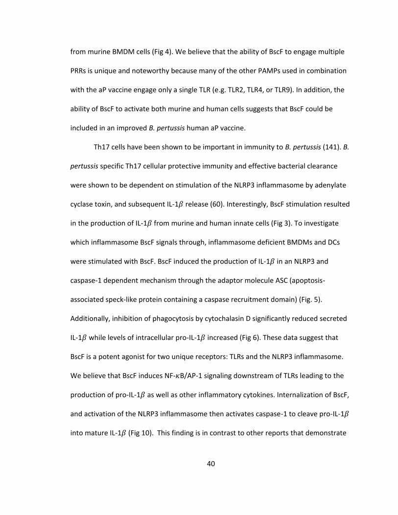
40
from murine BMDM cells (Fig 4). We believe that the ability of BscF to engage multiple
PRRs is unique and noteworthy because many of the other PAMPs used in combination
with the aP vaccine engage only a single TLR (e.g. TLR2, TLR4, or TLR9). In addition, the
ability of BscF to activate both murine and human cells suggests that BscF could be
included in an improved B. pertussis human aP vaccine.
Th17 cells have been shown to be important in immunity to B. pertussis (141). B.
pertussis specific Th17 cellular protective immunity and effective bacterial clearance
were shown to be dependent on stimulation of the NLRP3 inflammasome by adenylate
cyclase toxin, and subsequent IL-1𝛽 release (60). Interestingly, BscF stimulation resulted
in the production of IL-1𝛽 from murine and human innate cells (Fig 3). To investigate
which inflammasome BscF signals through, inflammasome deficient BMDMs and DCs
were stimulated with BscF. BscF induced the production of IL-1𝛽 in an NLRP3 and
caspase-1 dependent mechanism through the adaptor molecule ASC (apoptosis-
associated speck-like protein containing a caspase recruitment domain) (Fig. 5).
Additionally, inhibition of phagocytosis by cytochalasin D significantly reduced secreted
IL-1𝛽 while levels of intracellular pro-IL-1𝛽 increased (Fig 6). These data suggest that
BscF is a potent agonist for two unique receptors: TLRs and the NLRP3 inflammasome.
We believe that BscF induces NF-𝜅B/AP-1 signaling downstream of TLRs leading to the
production of pro-IL-1𝛽 as well as other inflammatory cytokines. Internalization of BscF,
and activation of the NLRP3 inflammasome then activates caspase-1 to cleave pro-IL-1𝛽
into mature IL-1𝛽 (Fig 10). This finding is in contrast to other reports that demonstrate
41
T3S system components and bacterial flagellin interact with the NLRC4 inflammasome
(153,154,155,156,157).
The previous studies (153,154,155,156,157) differ significantly in a number of
ways from the current study in terms of how the T3S system components or flagellin
were administered to the innate cells in vitro: 1. recombinant flagellin was fused to the
amino-terminal domain of anthrax lethal factor to target the protein to the cytosol
through an endocytosis mediated pathway (156) 2. the T3SS rod protein PrgJ and the
needle protein PrgI from Salmonella typhimurium were transfected directly into
macrophages following lipopolysaccharide (LPS) stimulation (155) 3. Cells were treated
with bacteria expressing intact T3S system to directly inject effector molecules into the
cytosol (157). In the current study, BscF was added to the culture medium, not directly
into the cell cytoplasm. It is possible that the differences in methodology among the
studies contributes to the discrepancies in inflammasome activation. In support of that
conclusion, bacterial amyloids produced in Escherichia coli and Salmonella typhimurium
produce IL-1𝛽 in vitro when administered in the culture medium of BMDM cells through
cooperation of TLR2 and the NLRP3 inflammasome (152). In addition to the Th1
polarizing cytokines produced by BscF stimulation, the ability of BscF to activate
intracellular inflammasomes and produce IL-1𝛽 suggests that BscF may provide B.
pertussis specific cellular immunity through Th17 cells.
In the current study we confirmed that BscF vaccination induced robust antibody
responses in mice (Figs 7A and B). Interestingly, BscF immunized serum effectively
opsonized B. pertussis compared to non-immunized serum (Fig 7C). One of the

42
insufficiencies of the aP vaccine is its inability to generate bactericidal antibodies
(158,159,160). Due to the extracellular localization of BscF on B. pertussis, we anticipate
that BscF might be a protective antigen, in part through the humoral response elicited
by BscF. It has been demonstrated that T3S system needle and translocon proteins act
as protective antigens, presumably through the generation of a robust antibody
response (145,132,146,147,148,149). Presently, it is unclear how BscF antibodies might
provide protection in vivo. We are currently addressing the ability of BscF-specific
antibodies to enhance complement mediated bacterial killing, inhibit bacterial
adherence, or augment opsonophagocytosis and subsequent bacterial uptake and
killing.
Despite widespread vaccination, whooping cough still remains as a global health
threat (88). This is due, in part, to the aP vaccine promoting robust Th2 responses
(114,142,117,143). Although the aP vaccine can control pertussis related symptoms, it
may not prevent bacterial colonization or transmission (113). An improved pertussis
vaccine should elicit strong Th1 responses, in addition to Th17 responses. Here, we
demonstrated that BscF immunization indirectly enhanced IFN𝛾 and IL-17A production
in splenocytes from immunized mice (Fig 8). In addition, immunization with BscF
absorbed in aluminum hydroxide gel provided modest protection in a sub lethal B.
pertussis challenge (Fig 9). Although bacterial burden is still relatively high, these results
demonstrate that BscF acts as a protective antigen; in addition to its innate stimulating
properties. Modest protection is also seen with YscF-induced protection against
Yersinia pestis (132). Taken together, these data demonstrate that BscF has the

43
immunomodulatory properties to be a beneficial vaccine candidate in an improved aP B.
pertussis vaccine. BscF acts as a strong innate agonist, induces robust inflammatory
cytokine release, and promotes protective cellular immunity critical to clear a B.
pertussis infection. BscF immunization produced robust humoral responses, and acted
as a modest protective antigen in a sub lethal B. pertussis infection. Studies are currently
ongoing to investigate the contribution of BscF when added to the aP vaccine.
Considering the shortcomings of the aP vaccine, BscF has significant potential to
enhance a next generation subunit pertussis vaccine.
44
Figure 1. Characterization of BscF protein preparation. (A) BscF or LPS were treated
with polymyxin B (PMB, 20 µg/ml) before addition to THP1-XBlue cells. (B) THP-1XBlue
cells were treated with 1 µg/ml of BscF expressed in BL21 (DE3) or BscF expressed in an
LPS free E. coli and activation of NF- 𝜅B and AP-1 was measured after 24 hours. (C) BscF
and Flagellin (FLA) were treated with proteinase K (40 µg/ml). Following treatment with
polymyxin B or proteinase K, THP-1XBlue cells were stimulated and NF- 𝜅B and AP-1
activation was measured by SEAP detection after 24 hours. Data are representative of
three experiments. ** Indicates p is between 0.01 and 0.001; *** Indicates p is between
0.001 and 0.0001.

45
Figure 2. BscF activates NF-𝜿B/AP-1 signaling in THP1-XBlue and HEK293 cells in a TLR2
and TLR4 dependent mechanism. (A) THP-1XBlue reporter cells were treated PBS, 1
µg/ml of BscF, or 1 µg/ml of LPS dissolved in PBS. (B) HEK-Blue hTLR2 and (C) HEK-Blue
hTLR4 reporter cells were treated with PBS, 1 µg/ml of BscF, 1 µg/ml of HKLM (Heat
Killed Listeria monocytogenes; TLR2 agonist), or 1 µg/ml of LPS (TLR4 agonist) dissolved
in PBS. SEAP levels were measured as an indirect measure of NF-𝜅B/AP-1 activation.
Data are presented as mean ± standard error of triplicate wells. Data are representative
of at least three experiments. **** Indicates p < 0.0001.

46
Figure 3. BscF stimulation induces robust inflammatory cytokine release from murine
and human innate cells. Murine Raw 264.7 cells were treated with 1 µg/ml of BscF, 1
µg/ml of LPS, 1 µg/ml of Pam3CSK4, or medium. After 24 h stimulation, levels of (A) TNF-
𝛼, (B) IL-6, and (C) IL-1𝛽 release were measured in the supernatant by ELISA. Human
THP-1 cells were treated with 1 µg/ml of BscF, 1 µg/ml of LPS, or 1 µg/ml of Pam3CSK4
for 24 h. Release of (D) IL-12p40, (E) TNF-𝛼, and (F) IL-1𝛽 was measured in the
supernatant by ELISA after 24 h stimulation. Data are presented as mean ± standard
error of triplicate wells. Data are representative of three experiments. * Indicates p is
between 0.05 and 0.01; *** Indicates p is between 0.001 and 0.0001; **** Indicates p <
0.0001.
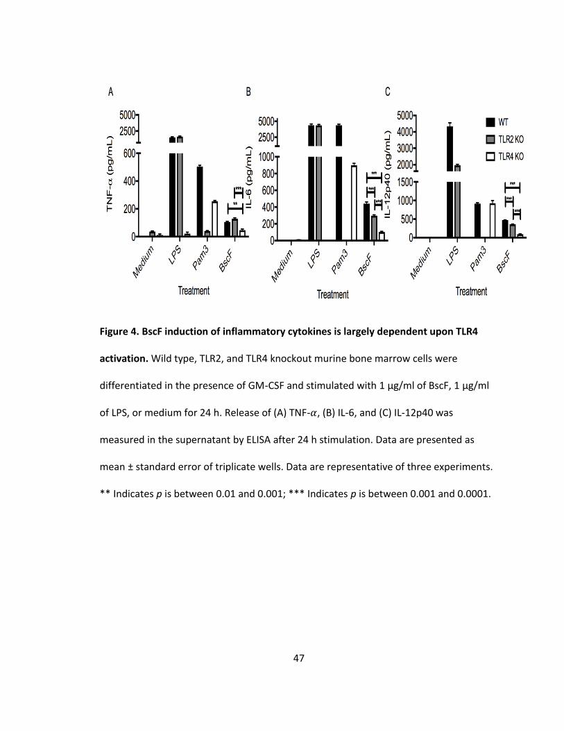
47
Figure 4. BscF induction of inflammatory cytokines is largely dependent upon TLR4
activation. Wild type, TLR2, and TLR4 knockout murine bone marrow cells were
differentiated in the presence of GM-CSF and stimulated with 1 µg/ml of BscF, 1 µg/ml
of LPS, or medium for 24 h. Release of (A) TNF-𝛼, (B) IL-6, and (C) IL-12p40 was
measured in the supernatant by ELISA after 24 h stimulation. Data are presented as
mean ± standard error of triplicate wells. Data are representative of three experiments.
** Indicates p is between 0.01 and 0.001; *** Indicates p is between 0.001 and 0.0001.
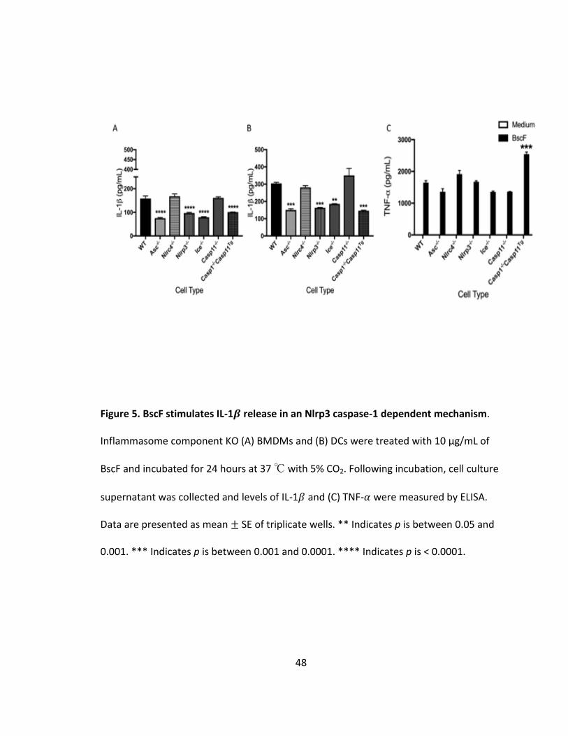
48
Figure 5. BscF stimulates IL-1𝜷 release in an Nlrp3 caspase-1 dependent mechanism.
Inflammasome component KO (A) BMDMs and (B) DCs were treated with 10 µg/mL of
BscF and incubated for 24 hours at 37 ℃ with 5% CO2. Following incubation, cell culture
supernatant was collected and levels of IL-1𝛽 and (C) TNF-𝛼 were measured by ELISA.
Data are presented as mean ± SE of triplicate wells. ** Indicates p is between 0.05 and
0.001. *** Indicates p is between 0.001 and 0.0001. **** Indicates p is < 0.0001.
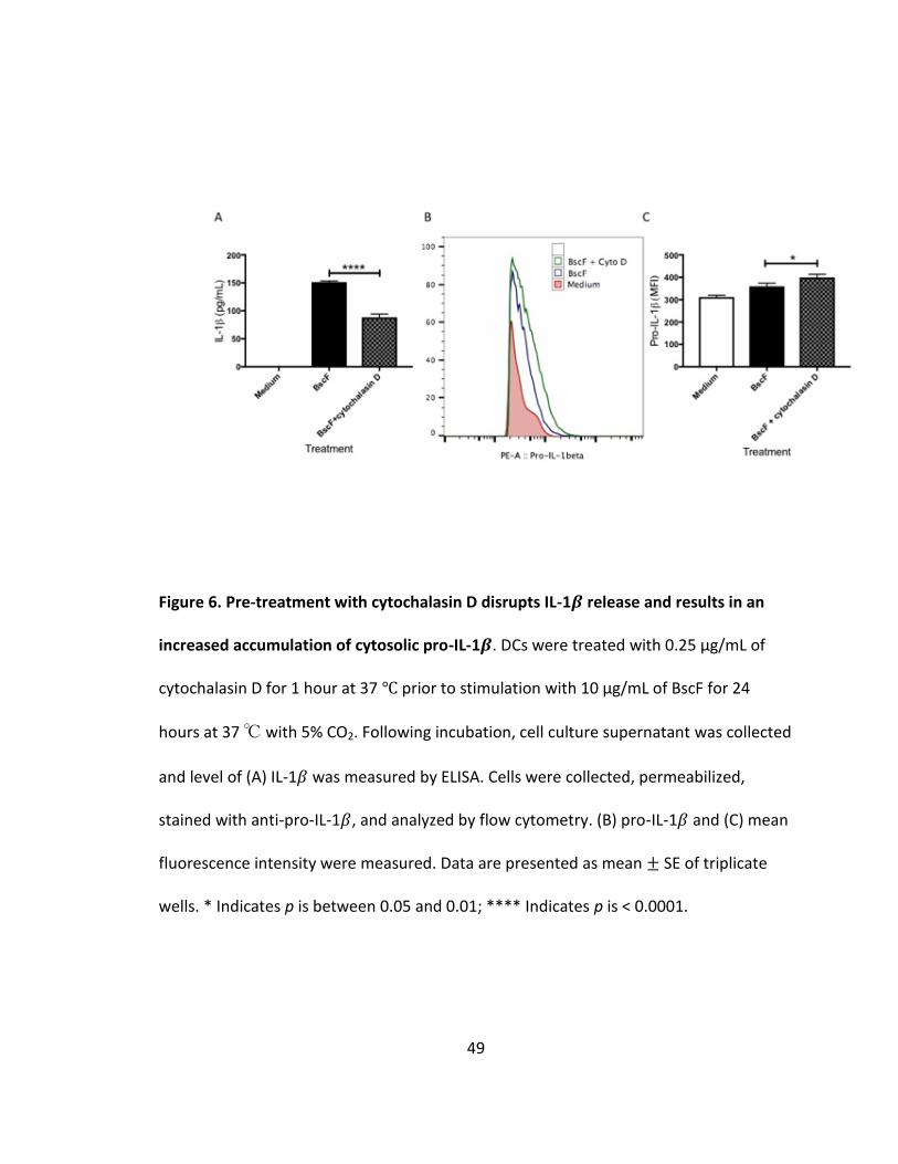
49
Figure 6. Pre-treatment with cytochalasin D disrupts IL-1𝜷 release and results in an
increased accumulation of cytosolic pro-IL-1𝜷. DCs were treated with 0.25 µg/mL of
cytochalasin D for 1 hour at 37 ℃ prior to stimulation with 10 µg/mL of BscF for 24
hours at 37 ℃ with 5% CO2. Following incubation, cell culture supernatant was collected
and level of (A) IL-1𝛽 was measured by ELISA. Cells were collected, permeabilized,
stained with anti-pro-IL-1𝛽, and analyzed by flow cytometry. (B) pro-IL-1𝛽 and (C) mean
fluorescence intensity were measured. Data are presented as mean ± SE of triplicate
wells. * Indicates p is between 0.05 and 0.01; **** Indicates p is < 0.0001.
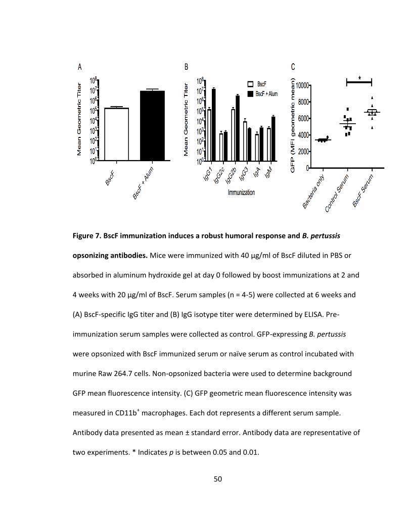
50
Figure 7. BscF immunization induces a robust humoral response and B. pertussis
opsonizing antibodies. Mice were immunized with 40 µg/ml of BscF diluted in PBS or
absorbed in aluminum hydroxide gel at day 0 followed by boost immunizations at 2 and
4 weeks with 20 µg/ml of BscF. Serum samples (n = 4-5) were collected at 6 weeks and
(A) BscF-specific IgG titer and (B) IgG isotype titer were determined by ELISA. Pre-
immunization serum samples were collected as control. GFP-expressing B. pertussis
were opsonized with BscF immunized serum or naïve serum as control incubated with
murine Raw 264.7 cells. Non-opsonized bacteria were used to determine background
GFP mean fluorescence intensity. (C) GFP geometric mean fluorescence intensity was
measured in CD11b+ macrophages. Each dot represents a different serum sample.
Antibody data presented as mean ± standard error. Antibody data are representative of
two experiments. * Indicates p is between 0.05 and 0.01.

51
Figure 8. BscF enhances activation of Th1 and Th17 cells, but not Th2 cells. Spleen cells
from BscF immunized mice were processed to a single cell suspension and stimulated
with 1 µg/ml of BscF (black bars) or medium (open bars) for 3 days. Supernatants were
tested for (A) IFN-𝛾, (B) IL-17A, and (C) IL-4 by ELISA. Data presented as mean ± standard
error; n = 4 mice/group. Data are representative of at least two experiments. * Indicates
p is between 0.05 and 0.01; *** Indicates p is between 0.001 and 0.0001.
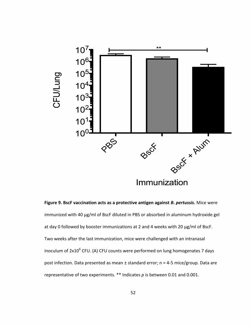
52
Figure 9. BscF vaccination acts as a protective antigen against B. pertussis. Mice were
immunized with 40 µg/ml of BscF diluted in PBS or absorbed in aluminum hydroxide gel
at day 0 followed by booster immunizations at 2 and 4 weeks with 20 µg/ml of BscF.
Two weeks after the last immunization, mice were challenged with an intranasal
inoculum of 2x10⁶ CFU. (A) CFU counts were performed on lung homogenates 7 days
post infection. Data presented as mean ± standard error; n = 4-5 mice/group. Data are
representative of two experiments. ** Indicates p is between 0.01 and 0.001.
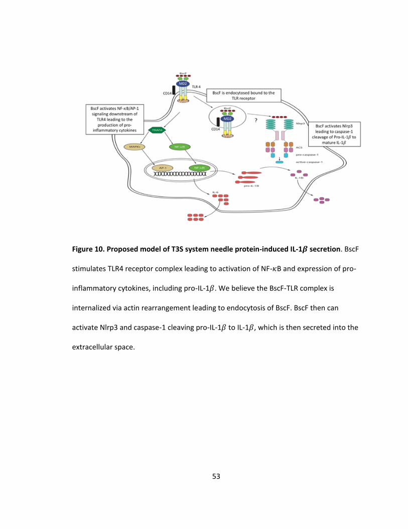
53
Figure 10. Proposed model of T3S system needle protein-induced IL-1𝜷 secretion. BscF
stimulates TLR4 receptor complex leading to activation of NF-𝜅B and expression of pro-
inflammatory cytokines, including pro-IL-1𝛽. We believe the BscF-TLR complex is
internalized via actin rearrangement leading to endocytosis of BscF. BscF then can
activate Nlrp3 and caspase-1 cleaving pro-IL-1𝛽 to IL-1𝛽, which is then secreted into the
extracellular space.
54
CHAPTER III
PURIFIED TYPE III SECRETION SYSTEM NEEDLE PROTEINS INDUCE CLATHRIN-DEPENDENT NF-𝜿B/AP-1 SIGNALING FROM ENDOSOMAL COMPARTMENTS
Introduction
The type III secretion (T3S) system is a protein secretion system that is conserved
among many gram-negative bacteria used to deliver virulence factors into eukaryotic
host cells to exploit the host by manipulating intracellular signaling (164,165,166,79). A
key component of the T3S system is a needle-like structure that extends from the
surface of the bacteria. This hollow structure is formed by the polymerization of single
proteins: YscF in Yersinia spp., PrgI and SsaG (Salmonella pathogenicity island 1 and
Salmonella pathogenicity island 2, respectively), PscF in Pseudomonas spp., MxiH in
Shigella flexneri, and BscF in Bordetella spp.
(166,167,168,169,170,171,172,173,174,175,176). Given the cellular localization of the
T3S system needle complex, this structure has the potential to interact with the innate
immune system.
During host-pathogen interactions, the innate immune system is the first to
respond to invading pathogens by recognizing conserved pathogen associated molecular
patterns (PAMPs). Toll-like receptors (TLRs) are a family of receptors that become
activated by microbial products to modulate innate and adaptive immune responses
(177). The cellular localization of these receptors either at the plasma membrane or

55
within intracellular endosomal compartments allows immune surveillance of both
extracellular and intracellular pathogens (178). Endocytosis of plasma membrane TLRs
has been shown to amplify ligand-induced TLR signaling from endosomal compartments
as well as activate distinct intracellular signaling pathways (179,180,181). One of the
key players in TLR activation, endocytosis, and ligand specificity is CD14. CD14 is a GPI-
linked or soluble protein expressed on many TLR4 expressing cells and was first
identified as a pattern recognition receptor (PRR) that binds directly to
lipopolysaccharide (LPS) (182). CD14 has also been implicated in TLR4 endocytosis upon
LPS stimulation (183). In addition to TLR4, CD14 has been shown to associate with TLR2
to enhance ligand specificity (184,185,186) and endosomal-dependent NF-𝜅B signaling
(187). The first TLR signaling event leading to early NF-𝜅B activation occurs at the
plasma membrane through the adaptor proteins MyD88/TIRAP leading to the up
regulation of inflammatory genes such as TNF-𝛼. A second signaling event occurs from
the endosomal compartment through the adaptor proteins TRAM/TRIF leading to late
NF-𝜅B signaling and type I interferon (IFN) production (188,189,190,191,192).
Recently, T3S system needle proteins have been identified as TLR2 and TLR4
ligands that induce NF-𝜅B/AP-1 activation and secretion of inflammatory cytokines in a
MyD88-dependent manner (130). In addition, we identified that the N terminus of T3S
system needle proteins is important for modulating TLR activation (131). Given the
importance of endosomal TLR signaling we hypothesized that T3S system needle
proteins may also exploit endosomal NF-𝜅B signaling to augment inflammatory cytokine
release. In this study, we examined the mechanism leading to T3S system needle

56
protein-induced NF-𝜅B/AP-1 activation downstream of TLR2 and TLR4 in human HEK
cells and monocytes. T3S system needle proteins: BscF, B. pertussis specific T3S system
needle protein, and YscF, Y. pestis specific T3S system needle protein, were found to
activate NF-𝜅B/AP-1 signaling from endosomal compartments mediated by a
clathrin/dynamin dependent endocytosis as clathrin-dependent endocytosis inhibition
significantly reduced NF-𝜅B/AP-1 signaling and inflammatory cytokine release. In
addition, NF-𝜅B/AP-1 activation by T3S system needle proteins downstream of both
TLR2 and TLR4 is mediated by CD14.
Materials and Methods
Bacterial strains and growth conditions
Escherichia coli Novablue (EMDMIllipore, Billerica MA), BL21 (DE3) star
(Invitrogen, Carlsbad, CA), and TOP10 (Invitrogen) were stored at -80°C in 25% glycerol
(vol/vol). E. coli strains were grown at 37°C in LB broth (BD, Franklin Lakes, NJ) or on
tryptose blood agar base (TBA, BD) plates, with kanamycin (50 µg/ml) as needed.
Expression and purification of His-tagged recombinant proteins
Template DNA for amplification was generated by using a DNeasy kit (Qiagen,
Valencia, CA) according to the manufacturer’s instructions. Oligonucleotide primers
(Eurofins MWG Operon, Inc. Huntsville, AL) were used to amplify BscF DNA from B.
pertussis Tohama I strain: BscF forward (5’-CAC CAT GGC CAT TAA CCT GGG AGG-3’) and
BscF reverse (5’-TCA ACT CGC CTT CTG TAT GAC GCC C-3’). Oligonucleotide primers
(Eurofins MWG Operon, Inc. Huntsville, AL) were used to amplify YscF DNA from Y.
pestis: YscF forward (5’- CGG GAT CCG ATG AGT AAC TTC TCT GGA TTT 3') and YscF
57
reverse (5’-CCG CTC GAG TGG GAA CTT CTG TAG GAT GCC-3’). PCR was performed using
Pfu Ultra polymerase (Agilent Technologies, Santa Clara, CA). The BscF amplified DNA
was cloned in frame with a N-terminal His-tag into pET200 by using a Champion TOPO
expression kit (Invitrogen). The YscF amplified DNA was cloned in frame with a C-
terminal His-tag into pET24b (Novagen, Madison WI). Plasmids for protein expression
were purified from E. coli TOP10 with a Qiaprep Miniprep kit (Qiagen). Purified plasmid
DNA was then transformed into chemically competent E. coli BL21 (DE3) Star
(Invitrogen). Plasmid constructs were verified by sequencing (Eurofins MWG Operon,
Inc.).
Protein purification was performed as previously described (130,131). Briefly, E.
coli BL21 (DE3) Star (Invitrogen) was grown overnight at 37°C in a shaking water bath in
non-inducing medium (50X M, 1 M MgSO4, 40% glucose, 5% aspartic acid (163))
supplemented with antibiotic. Bacteria were then inoculated into auto-inducing medium
(50X M, 1 M MgSO4, 50X 5052, NZ-amine S, yeast extract, distilled water (163))
supplemented with antibiotic and grown to an A620 of 0.6 to 0.8. Cells were harvested by
centrifugation at 4,000 x g for 10 min at 4°C and resuspended with wash buffer (50 mM
NaH2PO4, 300 mM NaCl, 10% glycerol (wt/vol)). The bacterial suspension was then
French pressed at 20,000 lb/in2 twice to lyse cells. The lysate was centrifuged at 10,000
x g for 20 min at 4°C. The supernatant was collected and diluted with 1,000 ml of wash
buffer before application to a pre-equilibrated TALON metal affinity resin (Clontech,
Mountain View, CA) column. The lysate was applied to the column twice followed by
washing the column with 15 bed volumes of wash buffer. Bound protein was eluted in
58
elution buffer (50 mM NaH2PO4, 200 mM NaCl,150 mM imidazole, and 20% glycerol
(wt/vol)). Purified protein was concentrated by centrifugation (Amicon Ultra centrifugal
filters, Millipore, Billerica, MA), and dialyzed against phosphate-buffered saline (PBS)
plus 10% glycerol (wt/vol) in Slide-A-Lyzer dialysis cassettes (Pierce, Thermo Fisher
Scientific, Rockford, IL). Protein concentration was determined by Bradford protein
assay (Pierce, Thermo Fisher Scientific), and purified protein was stored at -80°C for
future use.
Human HEK and THP-1 cell stimulation
HEK-Blue TLR2 and HEK-Blue TLR4 cells were suspended in DMEM and seeded at
5x10⁴ and 2.5x10⁴ cells/well, respectively in 96 well plates. T3S system needle proteins
were added to a final concentration of 1 μg/mL. 100 ng/ml of Pam3CSK4 (InvivoGen)
and 1 ng/ml of LPS (E. coli K12; InvivoGen) were used as positive controls for HEK-Blue
TLR2 and HEK-Blue TLR4 cells, respectively. Cells were stimulated at 37℃ in 5% CO2 for
24h. For inhibition experiments, cells were pre-treated with Chlorpromazine (CPZ),
Chloroquine (CHQ), Dynasore (Dyn), or blocking antibodies (10 𝜇g/ml; InvivoGen) for 1
hour at 37℃ in 5% CO2 prior to stimulation. The plates were then centrifuged at 1,200
rpm for 5 min at 4℃ and the supernatant was added to detection media and incubated
37℃ in 5% CO2 for 2h. Quantification of SEAP, the secreted embryonic alkaline
phosphatase reporter gene under the control of NF-κB and AP-1 in the HEK-Blue cells
was used as a surrogate to measure TLR activation and was measured by reading the
absorbance at 630 nm. THP-1 cells were suspended in RPMI and seeded at 1x10⁶

59
cells/well in 24 well plates and stimulated as indicated above. TNF-𝛼 was measured in
the cell culture supernatant at 24 hours by ELISA.
mRNA silencing
HEK-Blue TLR2 and HEK-Blue TLR4 cells were suspended in complete DMEM and
seeded at 5x10⁵ cells/well in 6 well plates or 1x10⁵ cells/well in 12 well plates 1 day prior
to transfection. The following day, the cells were washed with PBS and treated with 1
µM Accell siRNA against heavy chain clathrin 17 designed by supplier (GE Healthcare,
Dharmacon™ Accell™ siRNA) or with 1 µM of non-targeting siRNA. After 24 h, complete
DMEM was supplemented to the wells and transfection continued for a total of 72 h.
The cells were re-seeded at 5x10⁴ cells/well in 96 well plates and stimulated at 37℃ in
5% CO2 for 24h. The plates were then centrifuged at 1,200 rpm for 5 min at 4℃ and the
supernatant was added to detection media and incubated 37℃ in 5% CO2 for 2h.
Quantification of SEAP was measured by reading the absorbance at 630 nm.
Inhibitor cytotoxicity
HEK-Blue TLR2 and HEK-Blue TLR4 cells were treated with either Chlorpromazine
(CPZ), Chloroquine (CHQ), Dynasore (Dyn) as indicated above and cell toxicity was
measured by lactate dehydrogenase (LDH) release as indicated using the manufacturer’s
instructions (ThermoFisher Scientific).
Statistical analysis
Data were assembled into graphs using GraphPad Prism, version 5.0f (GraphPad
Software, La Jolla, CA). Data were analyzed using one-way analysis of variance (ANOVA)

60
followed by Bonferroni’s multiple comparison test. Differences were considered
statistically significant when p<0.05.
Results
T3S system needle proteins induce NF-𝜿B/AP-1 downstream of TLR2 and TLR4 by clathrin-dependent endocytosis
Recent data demonstrate the role of endosomal signaling downstream of TLR2
and TLR4 to augment plasma membrane TLR signaling (179,180,181,187). Clathrin-
mediated signaling events, in addition to the GTPase dynamin, have shown to be crucial
for localizing the TLR complex to the early endosome (180,187,193). Given the
localization of TLRs within the endosomal compartments, and how endosomal signaling
can augment NF-𝜅B activation and subsequent inflammatory cytokine production (187),
we determined the role that endosomal TLR signaling plays in T3S system needle protein
stimulated cells. To determine the role of TLR internalization in the activation of NF-
𝜅B/AP-1 we utilized HEK-Blue TLR2 that express TLR2 and CD14, and HEK-Blue TLR4 cells
which express TLR4, MD2, and CD14. These cells contain the secreted embryonic
alkaline phosphatase (SEAP) reporter gene under the control of NF-κB and AP-1 in the
HEK-Blue cells. HEK cells were treated with Chlorpromazine (CPZ), a clathrin-mediated
endocytosis inhibitor, Chloroquine (CHQ), endosomal maturation inhibitor, and
Dynasore (Dyn), the GTPase dynamin inhibitor, prior to T3S system needle protein
stimulation. As shown in Figure 11, CPZ, CHQ, and Dyn significantly reduced NF-𝜅B/AP-1
activation induced by both BscF and YscF in both HEK TLR2 as well as HEK TLR4 cells.
These data suggest that TLR2 and TLR4 internalization is important to augment T3S
61
system needle protein-induced NF-𝜅B/AP-1 signaling. In addition, TLR internalization is
mediated by clathrin and dynamin dependent mechanisms.
Clathrin-mediated endocytosis mediates T3S system needle protein induced TNF-𝜶 downstream of TLR2 and TLR4
Because HEK cells are engineered cells that assess NF-𝜅B/AP-1 signaling by SEAP
release, we wanted to examine the role of TLR endosomal signaling in a more realistic
innate cell context. To that end, we treated human THP-1 cells with the CPZ, CHQ, and
Dyn prior to T3S system needle protein stimulation and measured release of TNF-𝛼 in
the cell culture supernatant. As indicated in Figure 12, CPZ, CHQ, and Dyn significantly
reduced TNF-𝛼 release from T3S system needle protein stimulated THP-1 cells. These
data demonstrate that clathrin-mediated endocytosis augments NF-𝜅B/AP-1 signaling
and TNF-𝛼 expression downstream of TLR activation by T3S system needle proteins YscF
and BscF.
We further confirmed the role of clathrin in mediating NF-𝜅B/AP-1 signaling
downstream of TLR2 and TLR4 in the HEK cells. Interestingly, gene silencing of the
clathrin heavy chain in HEK TLR2 cells significantly reduced BscF induced NF-𝜅B/AP-1
signaling, while the reduction of NF-𝜅B/AP-1 signaling in YscF stimulated cells did not
reach significance (Fig 13A). Gene silencing of the clathrin heavy chain in HEK TLR4 cells
did not significantly reduce NF-𝜅B/AP-1 signaling induced by BscF or YscF (Fig 13B).
Brandt et al. demonstrated that targeting the clathrin heavy chain by gene silencing in
HEK-BLUE TLR4 cells did not significantly reduce NF-𝜅B activation induced by LPS (187),
and that the clathrin heavy chain was only partially inhibited by gene silencing. Our
results suggest that T3S system needle proteins activate NF-𝜅B/AP-1 signaling
62
downstream of TLR2 and TLR4, which is augmented, in part, by clathrin-mediated
endocytosis of the receptor complex.
CD14 facilitates T3S system needle protein activation of NF-𝜿B/AP-1
T3S system needle proteins from a number of gram-negative bacteria have been
identified as TLR2 and TLR4 ligands that induce NF-𝜅B/AP-1 signaling and secretion of
inflammatory cytokines in a MyD88-dependent manner (130,131). CD14 is a protein
expressed on innate cells that has been shown to enhance ligand binding, increase
ligand specificity, and facilitate endocytosis of TLR receptors (182,183,184,185,186,187).
Using HEK-Blue TLR2 that express TLR2 and CD14, and HEK-Blue TLR4 that express TLR4,
CD14, and MD2, we determined the contribution of CD14 to T3S system needle protein
induction of NF-𝜅B/AP-1 signaling. CD14 blocking antibody significantly reduced NF-
𝜅B/AP-1 signaling induced by YscF and BscF in HEK-Blue TLR2 cells, while isotype control
antibody had no affect (Fig 14A). For HEK-Blue TLR4 cells, blocking both CD14 and MD2
significantly decreased NF-𝜅B/AP-1 signaling induced by YscF and BscF (Fig 14B). Isotype
control antibody did not decrease signaling (Fig 4B). Neither CD14 or MD2 antibody
alone significantly reduced NF-𝜅B/AP-1 signaling induced by YscF and BscF in HEK-Blue
TLR4 cells (data not shown). These results suggest that CD14 enhanced T3S system
needle protein induced NF-𝜅B/AP-1 signaling in HEK-Blue cells.
LDH release from HEK-Blue cells treated with various pharmacological inhibitors
To ensure the reduction in NF-𝜅B/AP-1 signaling observed by pharmacological
inhibition was not due to direct cell toxicity, HEK-Blue TLR2 and HEK-Blue TLR4 cells
were pre-treated with either CPZ, CHQ, or Dyn, at the highest and lowest concentrations
63
and LDH was measured following treatment. To our surprise, CPZ significantly reduced
cell viability in HEK-Blue TLR2 cells at both the highest and lowest concentrations (Fig
15A). Lowest CPZ treatment did not reduce cell viability in the HEK-Blue TLR4 cells;
however, the highest CPZ concentration contributed to cell toxicity (Fig 15B). Neither
CHQ nor Dyn reduced cell viability at either the low or high concentrations (Fig 15).
Despite the cytotoxic affect observed by CPZ, our results are in line with previously
published data (187). Brandt et al., did not report any cytotoxic affects in human
monocytes induced by CPZ at the same concentrations used in the current study (187).
In addition, CPZ did not produce cytotoxic affects at the lowest concentration in HEK-
Blue TLR4 cells (Fig 15B), but still significantly reduced NF-𝜅B/AP-1 signaling (Fig 11A).
These results suggest that the reduced NF-𝜅B/AP-1 signaling observed with
pharmacological inhibition cannot solely be contributed to direct cytotoxic affects of the
inhibitors.
Discussion
The innate immune system is the first line of defense against invading
pathogens. TLRs are a group of innate receptors that are distinguished by ligand
specificity and cellular localization (178). The internalization of plasma membrane TLRs
has been shown to be a mechanism to enhance plasma membrane signaling or initiate
distinct signaling cascades originating from the endosomal compartments, despite
initially thinking that this process was a way to attenuate TLR signaling (180,181,190).
We have shown that T3S system needle proteins from many different gram-negative
bacteria act as TLR2 and TLR4 ligands, induce My-D88 dependent NF-𝜅B/AP-1 signaling,

64
and promote inflammatory cytokine release (130,131); however, the contribution of
endosomal NF-𝜅B/AP-1 signaling has not been addressed. In the current study, we
demonstrate that T3S system needle proteins, YscF and BscF, activate NF-𝜅B/AP-1
signaling from endosomal compartments. Our data indicate that clathrin-mediated
endocytosis mediates NF-𝜅B/AP-1 activation, and that CD14 controls, in part, TLR
activation.
Both TLR2 and TLR4 have been shown to be present within endosomal
compartments (179,190,194,195). In the case of TLR4, early My-D88/TIRAP-dependent
plasma membrane signaling leads to NF-𝜅B activation with a second endosomal-specific
signaling event through TRAM/TRIF leading to late NF-𝜅B activation and IFN production
(180,188,189,190,191,192). Like TLR4 signaling, endosomal-dependent signaling has
been described by TLR2 as well (186,187,189). While plasma membrane signaling is a
component in the generation of inflammatory cytokine production, data suggest that
the contribution of endosomal signaling is primarily responsible for inflammatory
cytokine production (180,196,197). Using our unique TLR ligands previously described to
bind TLR2 and TLR4 (130,131), we demonstrate that these ligands also exploit
clathrin/dynamin-mediated endosomal NF-𝜅B/AP-1 signaling to induce inflammatory
cytokine release (Figs 11 and 12). Pharmacological inhibition of clathrin-mediated
endocytosis with CPZ, endosomal maturation with CHQ, and dynamin with Dyn indicate
endocytosis of T3S system needle proteins are necessary to augment NF-𝜅B/AP-1
signaling. These data are consistent with reports demonstrating the requirement of
internalization of TLR ligands to induce NF-𝜅B-controlled inflammatory cytokine release
65
and ligand recognition (187,198,199), and demonstrate that endosomal localization and
TLR signaling augments T3S system needle protein-induced NF-𝜅B/AP-1 signaling and
inflammatory cytokine release from human THP-1 cells.
In the current study, the pharmacological inhibitor CPZ significantly decreased
HEK-Blue TLR2 and HEK-Blue TLR4 cell viability at the highest concentration used (Fig
15). In addition, the lowest CPZ concentration also reduced cell viability in HEK-Blue
TLR2 cells. This is surprising as it was reported by Brandt et al., (187) that no cell
cytotoxicity in human monocytes was reported in their study using the same CPZ
concentrations used in our study. No other cytotoxic affects were observed with any of
the other inhibitors (Fig 15). Despite the direct cytotoxic effects of CPZ, specifically with
the HEK-Blue TLR2 cells, CPZ reduced NF-𝜅B/AP-1 signaling in the HEK-Blue TLR4 cells in
the absence of direct cytotoxic affects. Additionally, siRNA targeting the clathrin heavy
chain in HEK-Blue TLR2 cells resulted in a significant inhibition of NF-𝜅B/AP-1 signaling
induced by BscF (Fig 13A). Although YscF activation was reduced, it did not reach
statistically significant levels (Fig 13A). No reduction in T3S system stimulated NF-𝜅B/AP-
1 signaling in HEK-Blue TLR4 cells treated with clathrin heavy chain siRNA was observed
(Fig 13B). These results are consistent with Brandt et al., (187), and could also be
attributed to only partial reduction in clathrin heavy chain expression by siRNA.
CD14 has been shown to be an important co-receptor for TLR4 binding of LPS
(182), TLR4 endocytosis (183), and for TLR2 by enhancing ligand specificity
(184,185,186) and endosomal NF-𝜅B signaling (187). CD14’s role in T3S system needle
protein TLR activation remains unclear. Here we show that blocking CD14 on HEK-Blue
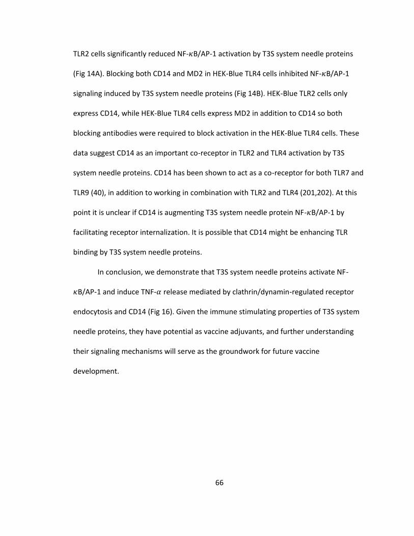
66
TLR2 cells significantly reduced NF-𝜅B/AP-1 activation by T3S system needle proteins
(Fig 14A). Blocking both CD14 and MD2 in HEK-Blue TLR4 cells inhibited NF-𝜅B/AP-1
signaling induced by T3S system needle proteins (Fig 14B). HEK-Blue TLR2 cells only
express CD14, while HEK-Blue TLR4 cells express MD2 in addition to CD14 so both
blocking antibodies were required to block activation in the HEK-Blue TLR4 cells. These
data suggest CD14 as an important co-receptor in TLR2 and TLR4 activation by T3S
system needle proteins. CD14 has been shown to act as a co-receptor for both TLR7 and
TLR9 (40), in addition to working in combination with TLR2 and TLR4 (201,202). At this
point it is unclear if CD14 is augmenting T3S system needle protein NF-𝜅B/AP-1 by
facilitating receptor internalization. It is possible that CD14 might be enhancing TLR
binding by T3S system needle proteins.
In conclusion, we demonstrate that T3S system needle proteins activate NF-
𝜅B/AP-1 and induce TNF-𝛼 release mediated by clathrin/dynamin-regulated receptor
endocytosis and CD14 (Fig 16). Given the immune stimulating properties of T3S system
needle proteins, they have potential as vaccine adjuvants, and further understanding
their signaling mechanisms will serve as the groundwork for future vaccine
development.
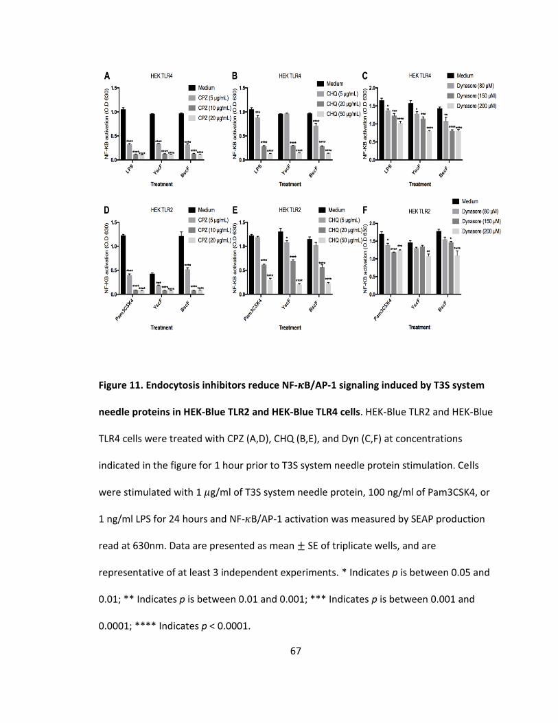
67
Figure 11. Endocytosis inhibitors reduce NF-𝜿B/AP-1 signaling induced by T3S system
needle proteins in HEK-Blue TLR2 and HEK-Blue TLR4 cells. HEK-Blue TLR2 and HEK-Blue
TLR4 cells were treated with CPZ (A,D), CHQ (B,E), and Dyn (C,F) at concentrations
indicated in the figure for 1 hour prior to T3S system needle protein stimulation. Cells
were stimulated with 1 𝜇g/ml of T3S system needle protein, 100 ng/ml of Pam3CSK4, or
1 ng/ml LPS for 24 hours and NF-𝜅B/AP-1 activation was measured by SEAP production
read at 630nm. Data are presented as mean ± SE of triplicate wells, and are
representative of at least 3 independent experiments. * Indicates p is between 0.05 and
0.01; ** Indicates p is between 0.01 and 0.001; *** Indicates p is between 0.001 and
0.0001; **** Indicates p < 0.0001.
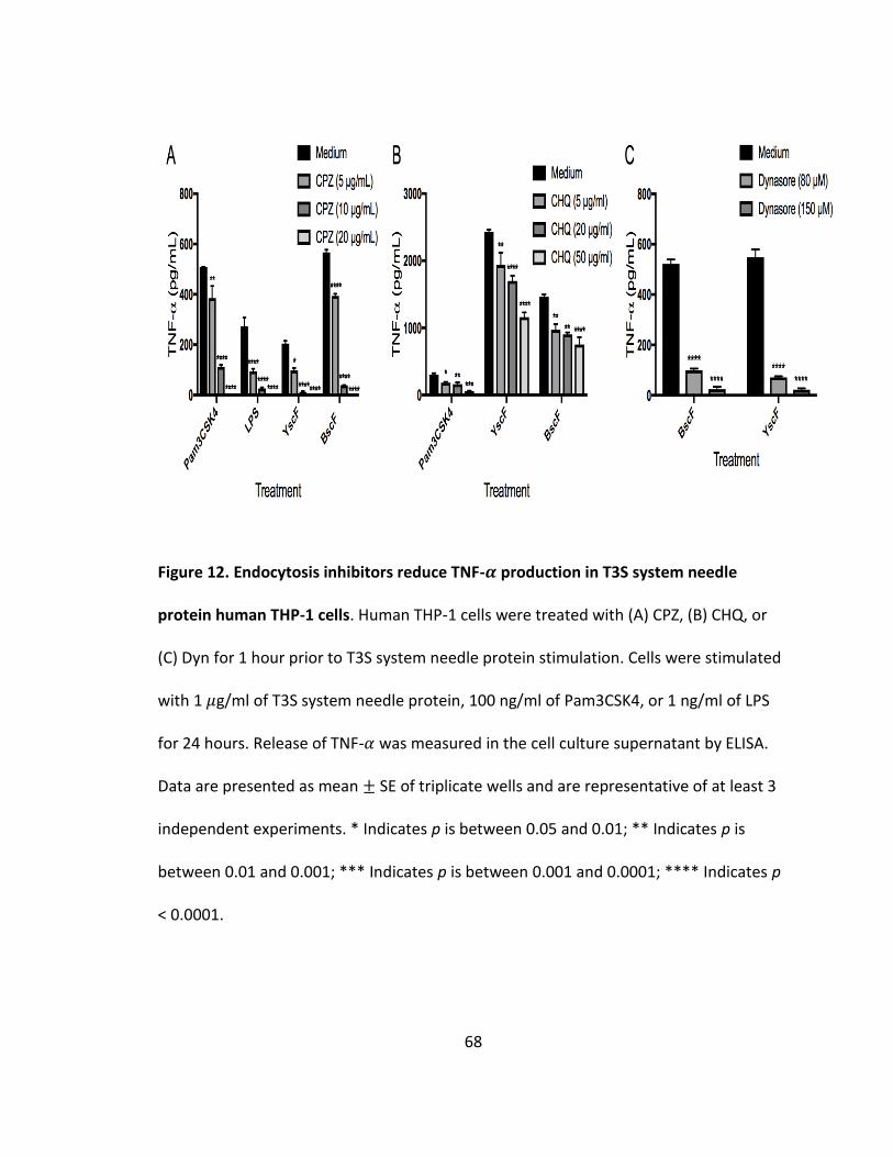
68
Figure 12. Endocytosis inhibitors reduce TNF-𝜶 production in T3S system needle
protein human THP-1 cells. Human THP-1 cells were treated with (A) CPZ, (B) CHQ, or
(C) Dyn for 1 hour prior to T3S system needle protein stimulation. Cells were stimulated
with 1 𝜇g/ml of T3S system needle protein, 100 ng/ml of Pam3CSK4, or 1 ng/ml of LPS
for 24 hours. Release of TNF-𝛼 was measured in the cell culture supernatant by ELISA.
Data are presented as mean ± SE of triplicate wells and are representative of at least 3
independent experiments. * Indicates p is between 0.05 and 0.01; ** Indicates p is
between 0.01 and 0.001; *** Indicates p is between 0.001 and 0.0001; **** Indicates p
< 0.0001.
69
Figure 13. siRNA gene knockdown of heavy chain clathrin reduced NF-𝜿B/AP-1
signaling induced by T3S system needle proteins in HEK-Blue TLR2 cells. (A) HEK-Blue
TLR2 and (B) HEK-Blue TLR4 cells treated with 1 µM Accell siRNA against heavy chain
clathrin 17 (grey bars) designed by supplier or with 1 µM of non-targeting siRNA (closed
bars). After 24 h, complete DMEM was supplemented to the wells and transfection
continued for a total of 72 h. Following transfection, cells were stimulated with 1 𝜇g/ml
of T3S system needle protein, 100 ng/ml Pam3CSK4, or LPS for 24 hours. NF-𝜅B/AP-1
activation was measured by quantification of SEAP by reading the absorbance at 630
nm. Data are presented as mean ± SE of triplicate wells and are representative of 2
independent experiments. * Indicates p is between 0.05 and 0.01; ** Indicates p is
between 0.01 and 0.001.
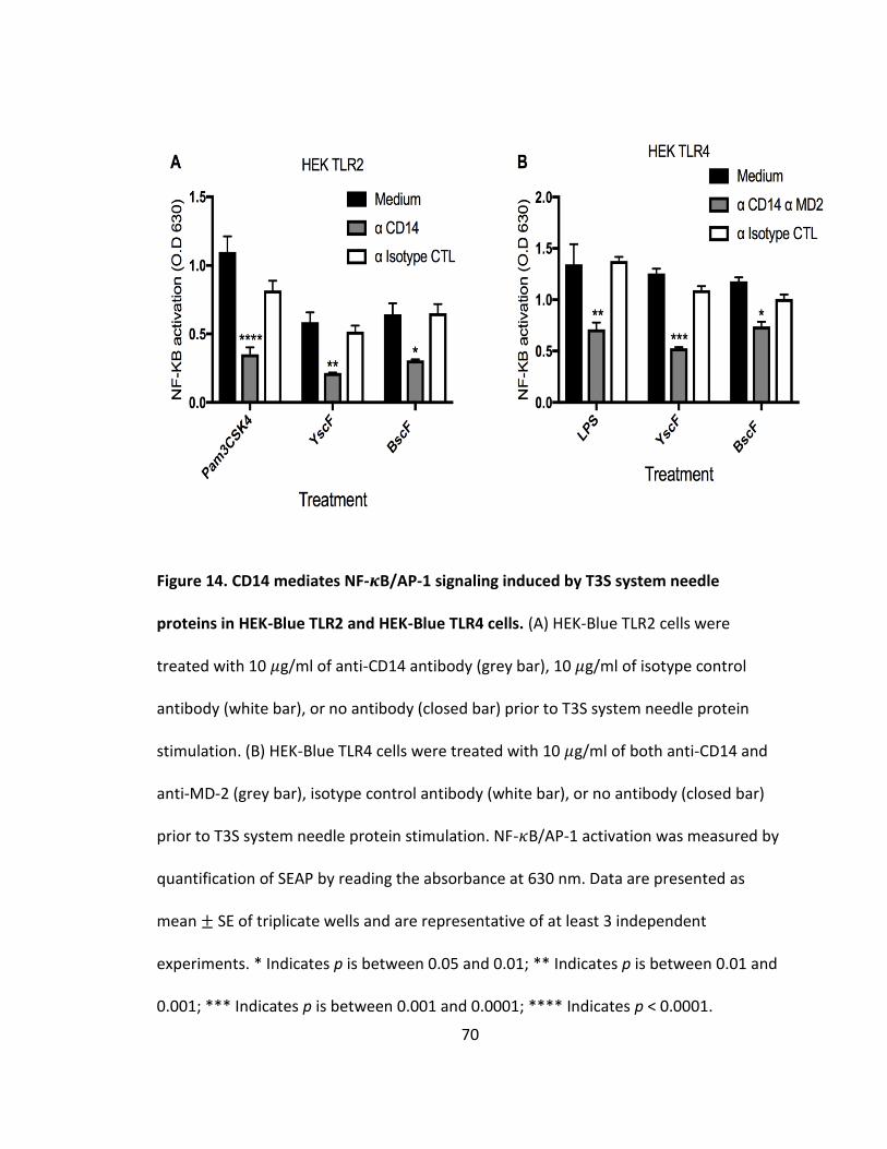
70
Figure 14. CD14 mediates NF-𝜿B/AP-1 signaling induced by T3S system needle
proteins in HEK-Blue TLR2 and HEK-Blue TLR4 cells. (A) HEK-Blue TLR2 cells were
treated with 10 𝜇g/ml of anti-CD14 antibody (grey bar), 10 𝜇g/ml of isotype control
antibody (white bar), or no antibody (closed bar) prior to T3S system needle protein
stimulation. (B) HEK-Blue TLR4 cells were treated with 10 𝜇g/ml of both anti-CD14 and
anti-MD-2 (grey bar), isotype control antibody (white bar), or no antibody (closed bar)
prior to T3S system needle protein stimulation. NF-𝜅B/AP-1 activation was measured by
quantification of SEAP by reading the absorbance at 630 nm. Data are presented as
mean ± SE of triplicate wells and are representative of at least 3 independent
experiments. * Indicates p is between 0.05 and 0.01; ** Indicates p is between 0.01 and
0.001; *** Indicates p is between 0.001 and 0.0001; **** Indicates p < 0.0001.
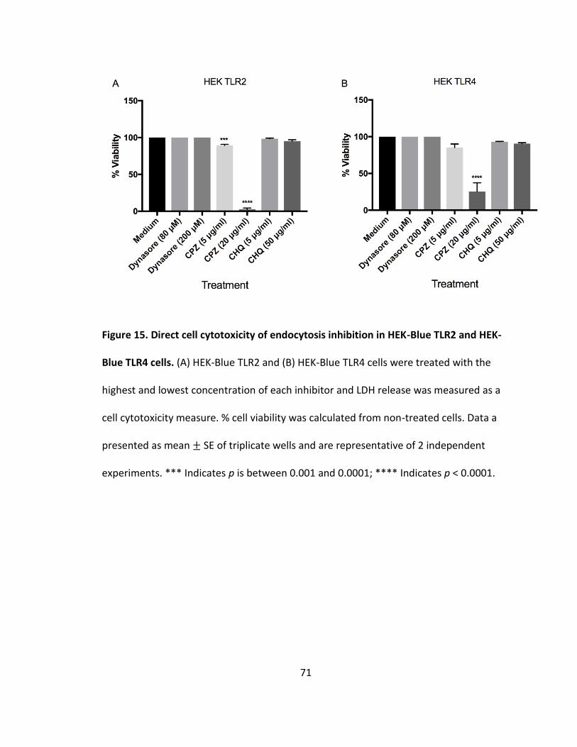
71
Figure 15. Direct cell cytotoxicity of endocytosis inhibition in HEK-Blue TLR2 and HEK-
Blue TLR4 cells. (A) HEK-Blue TLR2 and (B) HEK-Blue TLR4 cells were treated with the
highest and lowest concentration of each inhibitor and LDH release was measured as a
cell cytotoxicity measure. % cell viability was calculated from non-treated cells. Data a
presented as mean ± SE of triplicate wells and are representative of 2 independent
experiments. *** Indicates p is between 0.001 and 0.0001; **** Indicates p < 0.0001.
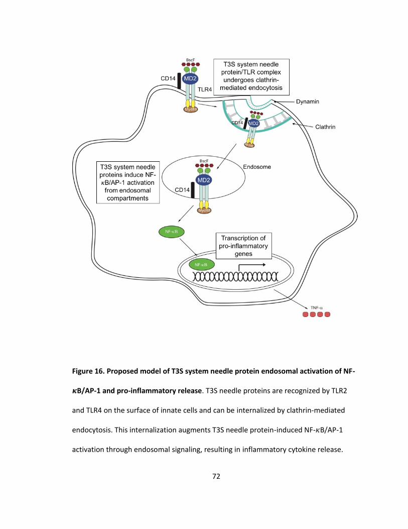
72
Figure 16. Proposed model of T3S system needle protein endosomal activation of NF-
𝜿B/AP-1 and pro-inflammatory release. T3S needle proteins are recognized by TLR2
and TLR4 on the surface of innate cells and can be internalized by clathrin-mediated
endocytosis. This internalization augments T3S needle protein-induced NF-𝜅B/AP-1
activation through endosomal signaling, resulting in inflammatory cytokine release.
73
CHAPTER IV
BSCF AS A VACCINE CANDIDATE FOR A NEXT GENERATION BORDETELLA PERTUSSIS ACELLULAR VACCINE
Introduction
Bordetella pertussis is a gram-negative bacterium and the causative agent of the
toxin-mediated disease whooping cough (pertussis). Despite widespread vaccination,
pertussis kills roughly 200,000 infants worldwide, mainly in developing countries.
Pertussis is characterized by airway inflammation and severe coughing episodes,
followed by the characteristic “whooping” sound. Pertussis cases are most commonly
found in infants and young children, however adolescents and adults can also be
infected. B. pertussis attaches to ciliated epithelial cells in the respiratory tract (51,203).
Although traditionally thought of as an extracellular pathogen, B. pertussis has been
shown to invade and reside within epithelial cells and alveolar macrophages (23,24).
With the advent of a whole cell pertussis (wP) vaccine in the 1940/1950s, cases of
pertussis were dramatically reduced. Effectiveness of the wP vaccine has been largely
attributed to its many antigens and PAMPs that bind and activate innate PRRs. The wP
vaccine proved to be too reactogenic and was replaced with a less reactogenic, subunit
acellular pertussis (aP) vaccine in the 1990s. The aP vaccine does not contain classical
PAMPs. The introduction of the aP vaccine was accompanied by a resurgence of
pertussis cases in both developed and developing countries (88). Given the contribution
74
of the many PAMPs to the effectiveness of the wP vaccine, we propose that the
addition of PRR stimulating agents could enhance the effectiveness of the current aP
vaccine.
There are a number of contributing factors that have lead to a resurgence of
pertussis cases attributed to deficiencies in the aP vaccine. First, many circulating
isolates of B. pertussis have been shown to be deficient in the protective antigens
included in the aP vaccine; namely pertussis toxin (PT), pertactin (Prn) and fimbriae
(Fim2 and Fim3) (103,134,135,76,136). In addition, a B. pertussis strain lacking Prn and
PT was recently isolated from an unvaccinated infant in the United States (136). Due to
the lack of these key antigens, it is possible that the aP vaccine is not providing robust
immune responses against many currently circulating pertussis strains. Second, many
studies have demonstrated that the aP vaccine does not elicit effective immunological
memory (137,138,139,140). This lack of long-term immunity is particularly concerning
because small infants that are most susceptible to severe symptoms of pertussis rely on
herd immunity. This incomplete immunity results in adults spreading pertussis to
infants, despite the adults receiving the full immunization schedule.
Finally, the current aP vaccine does not elicit strong immune responses that are
necessary for long-term protection (86,113). The aP vaccine has been shown to prevent
clinical pertussis symptoms, but the aP vaccine may not prevent bacterial colonization
or transmission (113). Requirements for the correct type of immune responses needed
to be effective against B. pertussis are evident in natural infections of B. pertussis.
Athymic nu/nu mice, which lack all T cells, are unable to clear a B. pertussis infection

75
(84,85)and adoptive transfer of total splenic T cells into athymic mice was able to
prevent chronic infection in nu/nu mice. Transfer of only B. pertussis specific CD8+ T
cells was not able to reverse chronic infection, indicating a critical role for CD4+ T cells in
controlling B. pertussis infections (85). CD4+ T cells collected from the draining lymph
nodes of B. pertussis infected mice produce high levels of interferon gamma (IFNy), a
prototypical T helper cell (Th) Th1-type cytokine, with little interleukin-4 (IL-4), a
prototypical Th2-type cytokine (85). In addition, IFNy mRNA levels in the lungs increased
sharply during the first week of infection (85), suggesting a protective role for IFNy and
CD4+ Th1 cells. The contribution for Th17 cells was demonstrated by persistent lung
colonization with B. pertussis in IL-17-/- mice (86). The lack of IL-17 and persistent
infection was correlated with a decrease in neutrophil recruitment into the lungs
(86,204,123). Both Th1 and Th17 cells have shown to be important in humans as well.
wP vaccines have been shown to induce predominately mixed Th1/Th17 type CD4+ T cell
responses in children, similar to natural infection (114,144,205) In terms of humoral
immunity, B cells appear to play an auxiliary role, potentially by activating the adaptive
immune response (206,207). These studies highlight the importance of both Th1 and
Th17 cells in protective immunity induced by a natural B. pertussis infection.
In contrast to a natural infection with B. pertussis or the wP vaccine, the aP
vaccine has been shown to have distinct immune stimulating properties, likely
contributing to the current resurgence of pertussis cases. In humans, aP vaccination has
been shown to induce a robust Th2 response, with minimal Th1/Th17 CD4+ T cell
responses (114,142,117). Mouse studies also identify a strong Th2 and a Th17 response
76
generated by the aP vaccine. aP vaccination is associated with CD4+ T cells that produce
IL-4, interleukin-5 (IL-5), and interleukin-17 (IL-17), but little IFNy (86,123). The
protective immunity generated by the aP vaccine was shown to be mediated through
Th17 cells, while the Th2 cells appear to be unnecessary (86). In addition, the ability of
the aP vaccine to provide protection for long periods of time has been called into
question. aP vaccination promoted differentiation of B. pertussis specific terminally
differentiated T cells (TTD) over central memory T cells (TCM) (208). In addition, two years
after vaccination, peripheral blood mononuclear cells (PBMCs) from children that
received the aP vaccine produced lower levels of B. pertussis specific IL-17 compared to
PBMCs from wP vaccinated children (117). It is clear that the aP vaccine elicits sub-
optimal immunity, and with the resurgence of pertussis cases, the need for a next-
generation pertussis vaccine is apparent.
As mentioned above, the numerous PAMPs associated with the wP vaccine
significantly enhanced its effectiveness. These endogenous PAMPs activate the innate
immune system through PRRs – specifically Toll-like receptors (TLRs), promote pro-
inflammatory cytokine release, and direct pertussis-specific adaptive immunity (118).
Because PAMPs appear to be an important aspect of the wP vaccine, it has been
suggested that the addition of TLR agonists to the aP vaccine could re-direct the
immune response generated by the aP vaccine to a more wP-like immune response. A
TLR2 agonist of B. pertussis when combined with the components of the aP vaccine
enhanced protection from an aerosol B. pertussis challenge, induced robust IgG2a
antibodies, and enhanced IL-17 and IFNy production from antigen stimulated
77
splenocytes ex vivo compared with the aP vaccine in alum (120). A TLR4 agonist,
monophosphoryl lipid A (MPL), when mixed with the aP vaccine increased protection
against B. pertussis challenge when compared with the aP vaccine in alum adjuvant,
while suppressing Th2 responses (121). A separate TLR agonist, LpxL1 (122), a
genetically engineered LPS from Neisseria meningitidis, enhanced antigen specific IFNy
and IL-17 CD4+ T cells and increased the number of specific memory CD4+ TCM cells when
added to the aP vaccine (123). Finally, CpG oligonucleotides from bacterial DNA that
activate TLR9 have been shown to induce antigen-specific IgG2a titers (124) and Th1 and
Th17 cells (125) when added to the aP vaccine (86). These studies show the feasibility
and effectiveness of incorporating novel TLR agonists into the aP vaccine to enhance
pertussis specific immunity that will not only promote the proper adaptive immune
responses, but could also augment the long-term efficacy of the aP vaccine.
We have recently identified that needle proteins from bacterial type III secretion
(T3S) systems are novel TLR agonists that activate TLR2 and TLR4, promote
inflammatory cytokine release, and can be modified to modulate TLR signaling
(130,131). In addition, we have also characterized the innate stimulating properties of
BscF, a purified T3S system needle protein from B. pertussis (unpublished results). In the
current study, we investigated the contribution of BscF when added to a laboratory
prepared aP vaccine. We demonstrate that BscF’s ability to engage TLRs has the
potential to enhance aP induced vaccine immunity to generate long lasting protective
immunity against B. pertussis.
78
Materials and Methods
Bacterial strains and growth conditions
E. coli Novablue (EMDMIllipore, Billerica MA), BL21 (DE3) star (Invitrogen,
Carlsbad, CA), TOP10 (Invitrogen), and Bordetella pertussis (Tohama I, ATCC BAA-589
and 12743, ATCC 5375[3865]) were stored at -80°C in 25% glycerol (vol/vol). E. coli
strains were grown at 37°C in LB broth (BD, Franklin Lakes, NJ) or on tryptose blood agar
base (TBA, BD) plates, with kanamycin (50 µg/ml) as needed. B. pertussis was
maintained as previously described (162). Briefly, B. pertussis was grown at 37°C on
Bordet-Gengou (BG) solid medium (RemelTM Thermo Fisher Scientific, Lenexa, KS)
supplemented with glycerol and 15% sterile sheep’s blood (Lampire Biological Labs,
Pipersville, PA). B. pertussis liquid cultures were grown in Stainer-Scholte broth
supplemented with heptakis (2,6-di-O-methyl-ß-cyclodextrin; Sigma-Aldrich, St. Louis,
MO) and Stainer-Scholte supplements at 37°C. In the case of the GFP-expressing
pertussis, both solid and broth medium were supplemented with kanamycin (50 µg/ml)
and gentamicin (30 µg/ml).
Expression and purification of His-tagged recombinant proteins
Template DNA for amplification was generated by using a DNeasy kit (Qiagen,
Valencia, CA) according to the manufacturer’s instructions. Oligonucleotide primers
(Eurofins MWG Operon, Inc. Huntsville, AL) were used to amplify BscF DNA from B.
pertussis Tohama I strain: BscF forward (5’-CAC CAT GGC CAT TAA CCT GGG AGG-3’) and
BscF reverse (5’-TCA ACT CGC CTT CTG TAT GAC GCC C-3’). PCR was performed using Pfu
79
Ultra polymerase (Agilent Technologies, Santa Clara, CA). The amplified DNA was cloned
in frame with a N-terminal His-tag into pET200 by using a Champion TOPO expression kit
(Invitrogen). Plasmid for protein expression was purified from E. coli TOP10 with a
Qiaprep Miniprep kit (Qiagen). Purified plasmid DNA was then transformed into
chemically competent E. coli BL21 (DE3) Star (Invitrogen). Plasmid construct was verified
by sequencing (Eurofins MWG Operon, Inc.).
Protein purification was performed as previously described (130,131). Briefly, E.
coli BL21 (DE3) Star (Invitrogen) was grown overnight at 37°C in a shaking water bath in
non-inducing medium (50X M, 1 M MgSO4, 40% glucose, 5% aspartic acid (163))
supplemented with antibiotic. Bacteria were then inoculated into auto-inducing medium
(50X M, 1 M MgSO4, 50X 5052, NZ-amine S, yeast extract, distilled water (163))
supplemented with antibiotic and grown to an A620 of 0.6 to 0.8. Cells were harvested by
centrifugation at 4,000 x g for 10 min at 4°C and resuspended with wash buffer (50 mM
NaH2PO4, 300 mM NaCl, 10% glycerol (wt/vol)). The bacterial suspension was then
French pressed at 20,000 lb/in2 twice to lyse cells. The lysate was centrifuged at 10,000
x g for 20 min at 4°C. The supernatant was collected and diluted with 1,000 ml of wash
buffer before application to a pre-equilibrated TALON metal affinity resin (Clontech,
Mountain View, CA) column. The lysate was applied to the column twice followed by
washing the column with 15 bed volumes of wash buffer. Bound protein was eluted in
elution buffer (50 mM NaH2PO4, 200 mM NaCl,150 mM imidazole, and 20% glycerol
(wt/vol)). Purified protein was concentrated by centrifugation (Amicon Ultra centrifugal
filters, Millipore, Billerica, MA), and dialyzed against phosphate-buffered saline (PBS)
80
plus 10% glycerol (wt/vol) in Slide-A-lyzer dialysis cassettes (Pierce, Thermo Fisher
Scientific, Rockford, IL). Protein concentration was determined by Bradford protein
assay (Pierce, Thermo Fisher Scientific), and purified protein was stored at -80°C for
future use.
Bone marrow derived cell isolation and differentiation
Bone marrow cells were collected from femurs of WT naïve mice. Femurs were
aseptically removed from each hind leg, briefly soaked in 70% ethanol, and placed in
fresh RPMI medium (10% heat inactivated FBS, 2 mM L-glutamine, 50 mM 2-
mercaptoethanol, and 50 µg/ml Pen-Strep). Both ends of the femur were cut and the
bone was flushed with 10 ml of RPMI and the cells were collected in a 50 ml conical
tube. The cell suspension was centrifuged at 400 x g for 10 min at 4°C. The cells were
resuspended in RPMI supplemented with 40 ng/ml granulocyte-macrophage colony-
stimulating factor (GMCSF; PeproTech, Rocky Hill, NJ), seeded at a density of 4 X 10⁶ in
20 ml of medium in a 150 x 20 mm round culture dish, and incubated at 37°C with 5%
CO2. On day 3, the cells were supplemented with 20 ml of fresh RPMI + 40 ng/ml GMCSF
and incubated for an additional 3 days. At day 6, the non-adherent cells were removed
(BMDCs), and were used for subsequent analysis.
Innate cytokine analysis and maturation assessment of BMDCs
BMDCs were seeded in triplicate at 1 X 10⁶ cells/ml into 24-well plates and
stimulated with 1 µg/ml of needle protein for 24 h at 37°C with 5% CO2. PBS, 1 µg/ml
LPS (E. coli K12; InvivoGen), and/or 1 µg/ml Pam3CSK4 (InvivoGen) were used as
negative and positive controls. Following stimulation, cells were centrifuged at 400 x g
81
for 5 min at 4°C and the cellular supernatant was removed and stored at -20°C for future
analysis. Numerous human and mouse innate cytokines were measured by DuoSet
enzyme-linked immunosorbent assay (ELISA) kits from R&D Systems (Minneapolis, MN)
or mouse inflammation panel cytometric bead analysis (CBA) kit (BioLegend, San Diego,
CA). CBA samples were collected by flow cytometer (LSR II, Becton Dickinson, San Jose,
CA), and analyzed by software provided by the manufacturer (LEGENDplex v7.0). For
BMDC maturation analysis, the stimulated cells were collected and surface stained for
antibodies specific for MHCII, CD80, and CD86. Surface expression and activation
markers were analyzed by flow cytometry (LSR II, Becton Dickinson, San Jose, CA).
Mouse immunization and intranasal B. pertussis challenge
6-8 week old C57BL/6 male mice were immunized intra peritoneally with 100 µL
of a laboratory prepared aP vaccine composed of 0.5 µg mutant PT (genetically
inactivated by site-directed mutagenesis (R9K) and (E129A) in the S1 subunit; List
Biological Laboratories, Campbell, CA), 1 µg FHA (List Biological Laboratories, Campbell,
CA) mixed with PBS (aP + PBS) or 40 µg purified BscF (aP + BscF). Mice were boosted at 4
weeks with the same components. Mice that received PBS injections served as controls.
Two weeks after the last immunization, mice were intra nasally challenged with 6 X 10⁶
CFU of B. pertussis 12743 in a 25 µl inoculum. 7 d post infection, lungs were aseptically
removed, and homogenized (Bullet blender, Next Advance, Averill Park, NY) in 1 ml of
sterile PBS. Lung homogenate was centrifuged at 130 x g for 1 min at 4°C, serial dilutions
were plated on 15% blood BG plates, and incubated at 37°C for 4-5 d. Lung bacterial

82
burden was determined by counting CFUs. All animal experiments were approved by
IACUC at the University of North Dakota.
ELISA assay of antibody levels in mouse serum
ELISA plates (Costar EIA/RIA, Corning) were coated with 100 µl of 1 µg/ml mPT or
FHA diluted in PBS and incubated overnight at 4°C. Plates were washed with wash buffer
(1X PBS, 0.05% Tween-20), and blocked with blocking buffer (1X PBS, 1% BSA, 0.05%
Tween-20) and incubated at room temperature for 1 h. Plates were again washed and
incubated with diluted mouse serum for 1 h at room temperature. Following incubation,
plates were washed, blocked for 10 min at room temperature with blocking buffer, and
incubated with rabbit anti-mouse IgG biotinylated (Invitrogen) antibody diluted
1:10,000 in blocking buffer for 1 h at room temperature. Both wash and blocking steps
were repeated as indicated above, and plates were incubated with streptavidin-HRP
(Invitrogen) diluted 1:2,000 in blocking buffer for 1 h at room temperature. For
measuring IgG isotypes, isotype specific goat anti-mouse IgG1, and IgG2c, (Sigma-
Aldrich) diluted 1:1,000 in blocking buffer was incubated following diluted serum for 30
min at room temperature. Plates were washed and blocked as indicated above, and
bound IgG was detected with biotinylated rabbit anti-goat IgG (Sigma-Aldrich) for 30
min at room temperature. Plates were again washed and blocked, and incubated with
streptavidin-HRP (Invitrogen) diluted 1:2,000 in blocking buffer for 30 min at room
temperature. Plates were washed with wash buffer and incubated with 3,3’,5,5’-
tetramethylbenzidine (TMB) substrate for 10 min at room temperature. The reaction
was stopped by adding 50 µl of 1 M H2SO4. Optical densities (OD) were measured at 450
83
nm with a microplate reader (Synergy HT, BioTek) and were analyzed with KC4 v3.3
software (BioTek). IgG was quantified by reading absorbance at 450 nm, correcting for
the background (day 0 serum).
T cell cytokine production
At 2 weeks post last immunization, spleens were collected from mice receiving
either PBS, aP+PBS, or aP+BscF, and processed to a single cell suspension. Following red
blood cell lysis, splenocytes were suspended in RPMI (10% heat inactivated FBS and 50
µg/ml of Pen-Strep) and seeded at 2 X 10⁶ cells/ml into 24 well plates. Splenocytes were
stimulated with 1 µg/ml purified mPT, FHA, BscF, or medium alone as negative control
for 72 h at 37°C with 5% CO2. Plates were centrifuged at 400 x g for 5 min at 4°C and
cellular supernatant was removed. IFN-y and L-17A production was determined by
DuoSet ELISA kits (R&D Systems).
Characterization of T cell response and T cell memory induced by aP vaccination
At 2 weeks post last immunization, spleens, inguinal lymph nodes, and blood
were collected. Splenocytes were prepared to single cell suspension as indicated above.
Lymph nodes were homogenized and prepared to a single cell suspension in complete
RPMI medium. 100 µl of blood from each mouse was used for staining and subsequent
analysis with the remaining blood being processed for serum and stored at -80°C. 1-
2x10⁶ splenocytes and lymphocytes as well as 100 µl of blood were stained with anti-
mouse CD3, CD4, CD8, CD44, and CD62L for 30 minutes at room temperature. Blood
samples were incubated with red blood cell lysis buffer for 10 minutes at room
temperature. The cells were washed 2 times and suspended in flow cytometry staining
84
buffer (2% FBS 1X PBS). Data were collected by flow cytometer (LSR II, Becton Dickinson,
San Jose, CA) and data were analyzed by FlowJo (FlowJo, LLC, Ashland, OR). The
following gating scheme was used to identify the populations of CD4+ and CD8+ T cells:
The first gate was on the cell population using SSC-A (cell complexity) vs FSC-A (cell size).
From there, singlets were identified by FSC-W (cell width), and the population of CD3+
cells was identified. Within the CD3+ population, a quadrant gate of CD4 and CD8 was
used to identify CD4+ and CD8+ cells. To identify naïve, memory (Tcm) and effector (Tem) T
cell subsets, cells and singlets were identified as indicated above. Within the singlets
population, a quadrant gate was used to identify either CD3+CD4+ or CD3+CD8+ T cells.
Within those populations a combination of CD62L and CD44 was used to identify naïve T
cells (CD62L+CD44low), Tcm (CD62LhighCD44int-high), and Tem (CD62LnegCD44high). Refer to
Figure 20 for a demonstration of the gating scheme.
Statistical analysis
Data were assembled into graphs using GraphPad Prism, version 5.0f (GraphPad
Software, La Jolla, CA). Data were analyzed using one-way analysis of variance (ANOVA)
followed by Tukey’s multiple comparison test. Differences were considered statistically
significant when p<0.05.
Results
BscF promotes DC maturation and inflammatory cytokine release
Since we have demonstrated that a number of T3S system needle proteins are
potent TLR agonists and induce inflammatory cytokine release from innate cells, we

85
examined the ability of BscF to activate mouse DCs in vitro. BscF stimulation for 24
hours promoted bone marrow derived DC maturation as measured by increased surface
expression of MHC class II, and the co-stimulatory molecules CD80, and CD86 (Fig 17A-
C). BscF stimulation also produced robust IL-6, TNF-𝛼, IL-1𝛽, and IL-12p40 release,
accompanied by a modest increase in IL-23 production (Fig 17D-F). These data indicate
that BscF is a TLR agonist and activates murine DC maturation and cytokine release in
vitro.
BscF is immunogenic in vivo and act as an adjuvant when added to a laboratory prepared aP vaccine
Having shown that BscF promotes inflammatory cytokine release by murine DCs
in vitro, we assessed BscF’s in vivo adjuvant capability when added to a laboratory
prepared aP vaccine. 6-8-week-old C57BL/6 male mice were immunized intra
peritoneally with 100 µL of a laboratory prepared aP vaccine composed of 0.5 µg mPT
and 1 µg FHA diluted in PBS (aP + PBS), or supplemented 40 µg purified BscF (aP + BscF).
Mice were boosted at 4 weeks with the same components. At 6 weeks post first
vaccination, antibody characterization was performed for both antigen specific IgG as
well as isotype antibody analysis. The addition of BscF (aP + BscF) to the aP vaccine
significantly increases FHA specific total IgG responses (Fig 18A). BscF did not
significantly enhance mPT specific total IgG responses (Fig 18D). FHA specific IgG1 as
well as IgG2c isotypes were significantly increased in the aP + BscF group when
compared to the aP + PBS (Fig 18B-C). mPT specific IgG1 and IgG2c isotypes were not
increased by the addition of BscF to the laboratory aP vaccine (Fig 18E-F). These data

86
demonstrate that BscF can enhance the aP vaccine specific antibody immune response
generated by a laboratory prepared aP vaccine.
BscF indirectly enhances IFN-𝜸 and IL-17 production by stimulated splenocytes in vitro
We have shown that BscF promotes inflammatory cytokine release by murine
DCs, including IL-6, IL-12, and IL-23. These cytokines are associated with the expansion
of Th1 and Th17 cells. To further characterize the immune modulating properties of
BscF when included in our laboratory prepared aP vaccine, splenocytes from mice
immunized with either aP + PBS, aP + BscF, or PBS were processed to a single cell
suspension and stimulated with each antigen, including BscF in vitro. The addition of
BscF to the aP vaccine significantly increased IFN-𝛾 from FHA stimulated splenocytes,
when compared to the aP + PBS (Fig 19A). Interestingly, IL-17 release was more robust
in the aP + PBS group when stimulated with FHA (Fig 19B). aP + BscF splenocytes
stimulated with mPT as well as BscF produced significantly more IFN-𝛾 when compared
to aP + PBS splenocytes (Fig 19A). IL-17 release did not differ between groups when
stimulated with mPT; however, BscF stimulated aP + BscF splenocytes produced robust
IL-17 release whereas no detectable levels were reported in the aP + PBS group (Fig
19B). These data suggest that BscF’s immune stimulating properties indirectly enhanced
our aP vaccine immune responses in vivo.
BscF modulates memory response generated by the aP vaccine
We next assessed the ability of BscF to influence the long-term immunity
generated by our laboratory aP vaccine. CD4+ and CD8+ T cells within the blood (data
not shown), spleen, and inguinal lymph nodes were investigated ex vivo based on CD62L

87
and CD44 expression within a population of CD3+CD4+ or CD3+CD8+ subsets following
vaccination. Within those populations a combination of CD62L and CD44 was used to
identify naïve T cells (CD62L+CD44low), Tcm (CD62LhighCD44int-high), and Tem
(CD62LnegCD44high) as indicated in Figure 20. Total CD4+ and CD8+ T cells were increased
in the lymph nodes in aP + BscF mice compared to aP + PBS mice (Fig 20A,E).
Interestingly, naïve CD8+ T cells, but not CD4+ T cells within the lymph nodes were
increased in the BscF group (Fig 20B,F). Tem cells were not increased in either
compartment by the addition of BscF (Fig 20C,G). aP + BscF significantly enhanced both
CD4+ and CD8+ Tcm cells within the lymph nodes when compared to the aP + PBS group
(Fig 20D,H). These results indicated that the addition of BscF to the aP vaccine induced
CD4+ and CD8+ T cells, but also enhanced a central memory T cell phenotype, potentially
indicating improved long term immunity generated by the aP vaccine with BscF.
The addition of BscF to our laboratory aP vaccine promotes protective immunity against a sub lethal B. pertussis challenge
Mice were immunized with either aP + PBS, aP + BscF, or PBS as indicated above.
At 6 weeks post vaccination mice were challenged intranasally with live B. pertussis and
lungs were harvested 7 days post infection, homogenized in 1 mL of sterile saline, and
plated to enumerate CFU in the lungs. PBS control mice were well colonized with B.
pertussis at 7 days post infection (Fig 21). Immunization with aP + PBS provided modest
protection when compared to non-immunized mice (Fig 21). In contrast, mice
immunized with aP + BscF demonstrated enhanced bacterial clearance when compared
to both non-immunized mice as well as aP + PBS mice (Fig 21). These data demonstrate
88
the adjuvant properties of BscF when included in our laboratory aP vaccine by increased
bacterial clearance in the aP + BscF mice.
Discussion In the current study, we characterized a novel TLR ligand from the T3S system
needle complex of B. pertussis that activates the innate immune system to enhance
protective immunity generated by our laboratory prepared aP vaccine. This protein,
BscF, has been shown to activate TLR2 and TLR4, induce intracellular NF-𝜅B/AP-1
signaling, and promote inflammatory cytokine release from innate cells in vitro [current
study and unpublished results]. The ability of BscF to engage multiple TLRs is in line with
other purified T3S system needle proteins that we have previously characterized
(130,131). BscF demonstrated potent immune modulating activities in vitro, driving
murine dendritic cell (DC) maturation and inflammatory cytokine production. The
addition of BscF to the laboratory prepared aP vaccine enhanced vaccine-specific IgG
responses, indirectly induced Th1 and Th17 responses in ex vivo stimulated splenocytes,
and provided enhanced protection against B. pertussis challenge.
The importance of generating cellular immunity, specifically Th1 and Th17 cells
has been highlighted in both natural and vaccine-induced immunity
(86,84,85,114,141,143). One of the inadequacies of the current aP vaccines is the lack of
innate immune stimulating properties necessary to effectively induce protective cellular
immunity. The wP vaccine was considered a strong Th1 producing vaccine due to its
numerous endogenous pathogen associated molecular patterns (PAMPs) that engaged
numerous pattern recognition receptors (PRRs), significantly enhancing its effectiveness
89
at generating protective immunity (118). We demonstrated that BscF has strong innate
immune stimulating properties and matures murine DCs as measured by inflammatory
cytokine release IL-12, IL-6, IL-23, and IL-1𝛽, and surface expression of prototypical
maturation markers. Expansion of Th1 and Th17 cells is influenced by the presence of
these innate cytokines (209). Due to the immune modulating properties of BscF, we
suggest that the addition of BscF to the aP vaccine may promote Th1 and Th17
responses and drive protective immunity.
Current literature has exploited a host of PRR stimulating agents in combination
with current aP vaccines to drive protective Th1 and Th17 responses that are generally
lacking with aP vaccination. Unlike natural B. pertussis infection or vaccine induced
immunity generated by the wP vaccine, the current aP vaccine in alum adjuvant
preferentially induces Th2-type responses (86,123,114,142,117). These stimulating
agents include TLR2 agonists (120), TLR4 agonists (123,121,122), and TLR9 agonists
(86,124,125). Interestingly, many of these agonists have proven effective at skewing the
aP vaccine-induced immunity to a more effective wP-like immune response by
increasing IgG2a antibody production, expanding Th1 and Th17 cellular immunity, and
providing enhanced protection from B. pertussis infections in mice. In the current study,
we report enhanced pertussis specific antibody titers, including the Th1 indicating IgG2c
isotype, when our novel T3S system needle protein, BscF, was included in our laboratory
prepared aP vaccine. Additionally, BscF indirectly enhanced Th1 and Th17 cytokine
production from ex vivo stimulated splenocytes.
90
Another limitation to the current aP vaccines absorbed in alum adjuvant is the
lack of long-lasting immunological memory (137,138,139,140). Waning immunity, in
addition to other factors, may be contributing to the rise in whooping cough cases
despite relatively high vaccination rates. Brummelman et al., demonstrated that the
addition of a TLR4 ligand to the aP vaccine resulted in an increased pertussis-specific Tcm
phenotype, suggesting enhanced long-term efficacy of the vaccine (123). The generation
of Tcm cells is a good predictor of long-term immunity (210,211,212,213). Here we report
that the addition of BscF to the aP vaccine increased CD4+ and CD8+ T cells within the
inguinal lymph nodes when compared to the aP vaccine in PBS. In addition, Total CD4+
Tcm and CD8+ were increased in the presence of BscF. These data provide evidence of
BscF’s ability to modulate vaccine induced immunity. It is worth noting that we did not
use any methods necessary to assess pertussis-specific memory T cells. This study differs
from Brummelman et al., who utilized MHC class II tetramer analysis to identify
pertussis-specific CD4+ T cells (123).
Although aP vaccine has been able to prevent clinical pertussis symptoms, it did
not reduce bacterial colonization of transmission in baboons (113). Here we
demonstrated the benefit of the immune modulating capacity of BscF to enhance
bacterial clearance of the aP vaccine. This benefit is likely the result of BscF promoting
protective cellular immunity through its innate stimulating properties. Taken together,
our results demonstrate that BscF, a purified T3S system needle protein from B.
pertussis has potential to improve aP vaccine-induced immunity by indirectly promoting
protective cellular immunity through its TLR stimulating properties. BscF has been
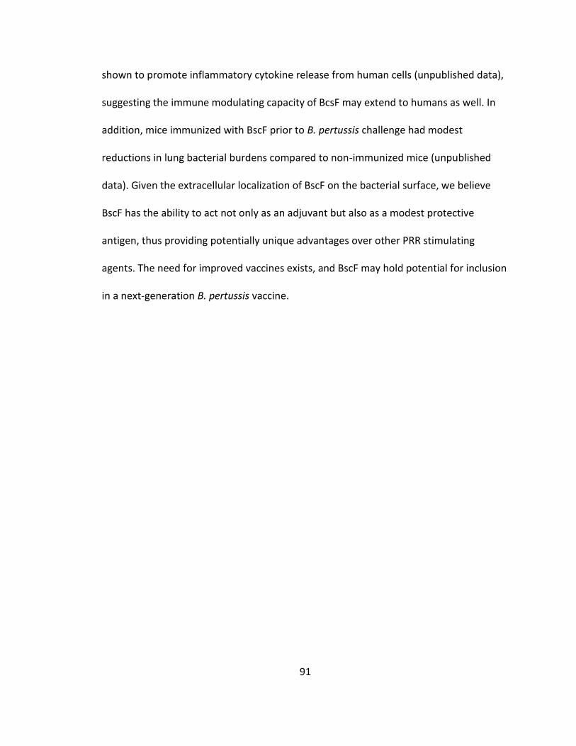
91
shown to promote inflammatory cytokine release from human cells (unpublished data),
suggesting the immune modulating capacity of BcsF may extend to humans as well. In
addition, mice immunized with BscF prior to B. pertussis challenge had modest
reductions in lung bacterial burdens compared to non-immunized mice (unpublished
data). Given the extracellular localization of BscF on the bacterial surface, we believe
BscF has the ability to act not only as an adjuvant but also as a modest protective
antigen, thus providing potentially unique advantages over other PRR stimulating
agents. The need for improved vaccines exists, and BscF may hold potential for inclusion
in a next-generation B. pertussis vaccine.
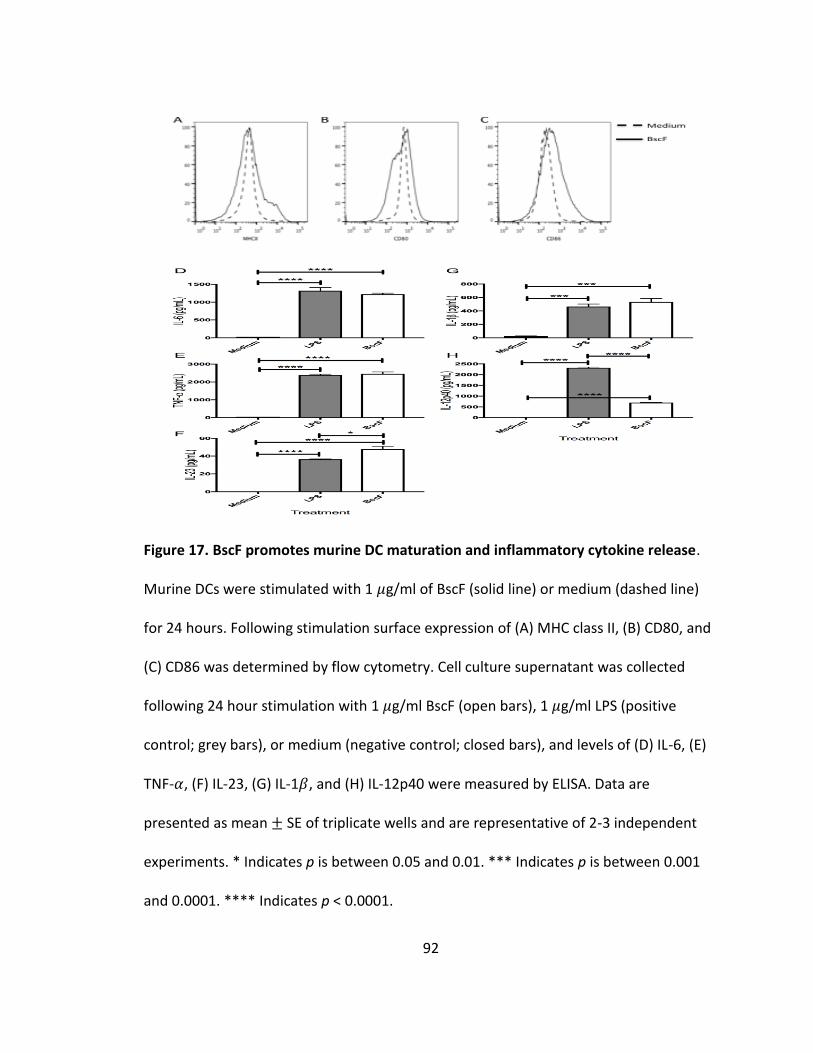
92
Figure 17. BscF promotes murine DC maturation and inflammatory cytokine release.
Murine DCs were stimulated with 1 𝜇g/ml of BscF (solid line) or medium (dashed line)
for 24 hours. Following stimulation surface expression of (A) MHC class II, (B) CD80, and
(C) CD86 was determined by flow cytometry. Cell culture supernatant was collected
following 24 hour stimulation with 1 𝜇g/ml BscF (open bars), 1 𝜇g/ml LPS (positive
control; grey bars), or medium (negative control; closed bars), and levels of (D) IL-6, (E)
TNF-𝛼, (F) IL-23, (G) IL-1𝛽, and (H) IL-12p40 were measured by ELISA. Data are
presented as mean ± SE of triplicate wells and are representative of 2-3 independent
experiments. * Indicates p is between 0.05 and 0.01. *** Indicates p is between 0.001
and 0.0001. **** Indicates p < 0.0001.
93
Figure 18. BscF acts as an adjuvant to enhance aP vaccine specific antibody responses.
Mice were immunized with aP + PBS or aP + BscF twice (0 and 4 weeks), and serum was
collected at 6 weeks. (A) Total FHA specific IgG or antibody isotype (B) IgG1 and (C)
IgG2c were measured in serum samples diluted 1:100. Serum mPT specific (D) total IgG,
(E) IgG1, and (F) IgG2c were determined in serum samples diluted 1:100. Data are
presented as mean ± SE of OD450nm absorbance readings corrected by non-immunized
serum, and are compiled from 4 independent experiments. aP + PBS n = 17 and aP +
BscF n = 18. ** Indicates p is between 0.01 and 0.001. *** Indicates p is between 0.001
and 0.0001. **** Indicates p < 0.0001.

94
Figure 19. BscF indirectly enhances IFN-𝜸 and IL-17 production from ex vivo stimulated
splenocytes. Mice were immunized with aP + PBS (grey bars) or aP + BscF (open bars)
twice (0 and 4 weeks). PBS immunized mice (closed bars) served a naïve control. At 6
weeks, spleens were harvested and stimulated ex vivo with 1 𝜇g/ml of either FHA, mPT,
or BscF, and release of (A) IFN-𝛾 and (B) IL-17 was measured in the cell culture
supernatant following 72 hour stimulation by ELISA. Data presented as mean ± SE of 4
mice per group, and are representative of 2 independent experiments. * Indicates p is
between 0.05 and 0.01. ** Indicates p is between 0.01 and 0.001. *** Indicates p is
between 0.001 and 0.0001.
95
Figure 20. The addition of BscF to the laboratory aP vaccine enhanced a central memory T cell phenotype. Mice were immunized with aP + PBS (closed bars) or aP + BscF (open bars) twice (0 and 4 weeks). PBS immunized mice (grey bars) served a naïve control. The following gating scheme was used to identify the populations of CD4+ and CD8+ T cells: The first gate was on the cell population using SSC-A vs FSC-A. From there, singlets were identified by FSC-W, and the population of CD3+ cells was identified. Within the CD3+ population, a quadrant gate of CD4 and CD8 was used to identify (A) CD4+ and (E) CD8+ cells. To identify naïve, memory (Tcm) and effector (Tem) T cell subsets, cells and singlets were identified as indicated above. Within the singlets population, a quadrant gate was used to identify either CD3+CD4+ or CD3+CD8+ T cells. Within those populations a combination of CD62L and CD44 was used to identify (B and F) naïve T cells (CD62L+CD44low), (C and G) Tem (CD62LnegCD44high) and (D and H) Tcm (CD62LhighCD44int-high). Data presented as mean ±SE of 4 mice per group, and are representative of 2 independent experiments. * Indicates p is between 0.05 and 0.01.
96
Figure 21. The addition of BscF to the laboratory aP vaccine enhanced protective
immunity against a sub lethal B. pertussis challenge. Mice were immunized with aP +
PBS (squares) or aP + BscF (triangles) twice (0 and 4 weeks). PBS immunized mice
(circles) served a naïve control. At 6 weeks, mice were challenged with an intranasal
inoculum of live B. pertussis. Lung homogenate CFU counts were recorded at 7 days
post infection. Data are presented as mean ± SE of 4-6 mice per group, and are
representative of 2 independent experiments. * Indicates p is between 0.05 and 0.01. **
Indicates p is between 0.01 and 0.001.

97
CHAPTER V
DISCUSSION
Bordetella pertussis, the causative agent of whooping cough, produces
significant morbidity and mortality worldwide. Because there are no known non-human
B. pertussis reservoirs, B. pertussis is truly a vaccine preventable disease. Current aP
vaccines have fallen short over the last 20-30 years or so of their use, partly contributing
to the reemergence of B. pertussis incidences in a number of developed and developing
countries. B. pertussis research has been disadvantaged mostly due to the lack of
suitable animal models; however, much of the work in these animal models on
understanding B. pertussis pathogenesis and the critical role both the innate and
adaptive arms of the immune system play in vaccine development have been validated
in a newly developed baboon model that exhibits clinical symptoms more similar to
humans. Moreover, human trials examining both the wP and aP vaccine safety and
immunogenicity have also been instrumental in moving the field forward. From both
animal and human studies, it is clear that a next-generation aP vaccine is necessary. In
the studies included here, we characterize the immune stimulating properties of a B.
pertussis specific protein called BscF, and assess its immune modulating capabilities
when added to an aP vaccine.
98
With the replacement of the wP vaccine with aP vaccines in the 1990’s cases of
B. pertussis have been on the rise in many countries. Waning and ineffective immunity
generated by the aP vaccine have been, in part, the cause of B. pertussis reemergence.
While there have been a number of unique approaches in the effort to develop better
vaccines, a large amount of work has been focused on the use of innate immune
agonists (specifically TLR agonists) to skew aP vaccine induced immunity from the
prototypical Th2 responses to protective Th1 and Th17 responses
(86,120,121,122,123,124,125). These studies have reported success in animal models
and provide evidence of the feasibility of adding other TLR ligands to a next-generation
aP vaccine. Recent work from our lab focusing on purified T3S system needle proteins
has identified a number of novel TLR agonists from a number of Gram-negative bacteria
(130,31). These proteins are of interest for a number of reasons. First, T3S system
needle proteins have been shown to be TLR2 and TLR4 agonists. Second, these proteins
activate MyD88 dependent NF-𝜅B/AP-1 signaling downstream of TLR activation, and
promote inflammatory cytokine release (130,131). Third, the strength of innate
activation and inflammatory cytokine release can be modulated by modifying the N-
terminus of the protein (131), allowing great flexibility if using these proteins and their
immune stimulating properties in newly developed vaccines.
In the current studies, we demonstrated that BscF, a purified T3S system needle
protein from B. pertussis acts as a strong TLR2 and TLR4 ligand. BscF activated NF-
𝜅B/AP-1 signaling and promoted inflammatory cytokine release from both mouse and
human cells in vitro. It is noteworthy that human cells respond similarly to BscF
99
stimulation when compared to mouse cells, indicating the possibility of the translational
application of BscF in human aP vaccines. While we have extensively characterized the
innate stimulating properties of T3S system needle proteins (130,131), to date, the
contribution of endosomal TLR NF-𝜅B/AP-1 signaling has not been addressed.
Additionally, clues to the mechanism of how T3S system needle proteins activate TLRs
warrants further investigation. Clathrin-mediated endocytosis of plasma membrane
TLRs (specifically TLR2 and TLR4) has been shown to amplify ligand-induced TLR
signaling from endosomal compartments as well as activate distinct intracellular
signaling pathways (179,180,181,187). One of the key players in TLR2 and TLR4
activation, endocytosis, and ligand specificity is CD14 (183,184,185,186,187). Our data
indicated that clathrin-mediated endocytosis mediated NF-𝜅B/AP-1 activation, and that
CD14 controls, in part, TLR2 and TLR4 activation. This is the first report demonstrating
endocytosis of the TLR augments NF-𝜅B/AP-1 signaling by T3S system needle proteins.
In addition, these data highlight the critical role of CD14 during T3S system needle
protein activation of TLR2 and TLR4. Understanding the signaling mechanisms leading to
TLR2 and TLR4 activation by T3S system needle proteins will facilitate their use in
vaccine development.
In addition to TLR activation, we demonstrated that BscF activates the NLRP3
inflammasome and requires internalization to process pro-IL-1𝛽 into mature IL-1𝛽. It is
thought that the wP vaccine was effective at driving CD4+ Th1 and Th17 cellular
immunity primarily through its immune stimulating properties. wP containing PAMPs
activate the innate immune system and promote inflammatory cytokine release and DC

100
maturation to induce Th1/Th17 cellular immunity (118). In addition, B. pertussis specific
Th17 cellular protective immunity and effective bacterial clearance were shown to be
dependent on stimulation of the NLRP3 inflammasome by adenylate cyclase toxin, and
subsequent IL-1𝛽 release (60). BscF stimulation of mouse DCs resulted in robust IL-12,
IL-6, IL-23, and IL-1𝛽 release. These cytokines have been shown to be effective at
polarizing naïve T cells to Th1 and Th17 subsets during antigen presentation.
Ex vivo BscF stimulated splenocytes from mice vaccinated with BscF produced
robust IFN-𝛾 and IL-17 release, indicating a strong Th1 and Th17 adaptive immune
response in mice immunized with BscF. Mouse and human studies have clearly
demonstrated the critical role for CD4+ Th1 and Th17 cells in protective immunity
elicited by natural infection or vaccination (84,85,86,123,204,114,144,205). In addition,
BscF immunization resulted in a modest, yet significant, reduction of bacterial burden in
the lungs 7 days post challenge. While BscF vaccinated mice were still highly colonized
with B. pertussis, these data indicate that BscF may also act as a protective antigen in
addition to its immune modulating capabilities in a next-generation aP vaccine. We
believe that the extracellular localization of BscF makes it an ideal therapeutic target.
Serum from BscF immunized mice was able to significantly enhance B. pertussis
opsonization and phagocytosis by mouse cells in vitro. It has been demonstrated that
T3S system needle and translocon proteins act as protective antigens, presumably
through the generation of a robust antibody response (145,132,146,147,148,149).
Further work is needed to identify the protective role of BscF specific antibodies during
a live B. pertussis mouse infection.
101
Although we saw modest protection in mice vaccinated with BscF alone, we do
not envision BscF as a stand-alone vaccine for B. pertussis. We next addressed the ability
of BscF to skew the immune response elicited by aP vaccination to a protective Th1/17
response by adding BscF to our laboratory prepared aP vaccine containing mPT and FHA.
Both mPT and FHA are included in any licensed aP vaccine, so our results may provide
insight for how BscF would work in future aP vaccines. aP vaccination has shown to
primarily induce Th2 type responses in both animal models and humans
(84,86,113,114,115). Our results demonstrate that the addition of BscF to the aP vaccine
induced significantly higher FHA specific antibody titers, and indirectly induced IFN-𝛾
and IL-17 from ex vivo stimulated splenocytes when compared to the aP vaccine in PBS.
In addition, bacterial clearance was enhanced by the addition of BscF to the aP vaccine 7
days post infection in mice compared to the aP vaccine alone. Our results are consistent
with other studies demonstrating the effectiveness of skewing aP induced immunity to a
protective Th1 and Th17 response by adding TLR ligands to aP vaccines
(86,120,121,122,123,124,125). One advantage of BscF compared to the other TLR
ligands is that BscF is B. pertussis specific, and could therefore be used as a protective
antigen as well as an adjuvant in next-generation aP vaccines. In addition, given the
prevalence of new circulating B. pertussis strains that have genetic modifications of
many common antigens included in the aP vaccines (103,104,105,77,108,109,110), the
use of new antigens that will likely not be lost by the bacteria would be advantageous.
B. pertussis mutants of the T3S system demonstrated reduced colonization and induced
102
exacerbated inflammation during infection (83), suggesting that the loss of the T3S
system due to vaccine pressure is highly unlikely.
This work has provided the foundation for future studies to further investigate
the immune mechanisms mediating BscF’s role during pertussis infections. For example,
characterizing the role that BscF specific antibodies play during B. pertussis infection
through passive transfer of BscF immune serum. Second, utilizing MHC class II tetramer
staining to facilitate our understanding of BscF and pertussis specific CD4+ T cell
responses during immunization. Third, investigating if BscF is able to provide any cross
protection to other Bordetella species (i.e. B. bronchiseptica and B. parapertussis).
103
REFERENCES
1. Clinical and Laboratory Standards Institute. 2008. Interpretive criteria for identification of bacteria and fungi by DNA target sequencing; approved guideline. Clinical and Laboratory Standards Institute, Wayne, PA.
2. McGowan KL, Garcia LS. 2007. Bordetella cultures, p 1–14. In Garcia LS (ed),
Clinical microbiology procedures handbook, 3rd ed. ASM Press, Washington, DC. 3. Leber AL. 2014. Pertussis: relevant species and diagnostic update. Clin Lab Med
34:237–255. http://dx.doi.org/10.1016/j.cll.2014.02.003. 4. Guiso N. 2009. Bordetella pertussis and pertussis vaccines. Clin Infect Dis
49:1565–1569. http://dx.doi.org/10.1086/644733. 5. Pearce-Duvet JM. 2006. The origin of human pathogens: evaluating the role of
agriculture and domestic animals in the evolution of human disease. Biol Rev Camb Philos Soc 81:369–382. http://dx.doi.org/10.1017/S1464793106007020.
6. Mooi FR, Bruisten S, Linde I, Reubsaet F, Heuvelman K, van der Lee S, King AJ.
2012. Characterization of Bordetella holmesii isolates from patients with pertussis-like illness in The Netherlands. FEMS Immunol Med Microbiol 64:289 –291. http://dx.doi.org/10.1111/j.1574-695X.2011.00911.x.
7. Njamkepo E, Bonacorsi S, Debruyne M, Gibaud SA, Guillot S, Guiso N. 2011.
Significant finding of Bordetella holmesii DNA in nasopharyngeal samples from French patients with suspected pertussis. J Clin Microbiol 49:4347–4348. http://dx.doi.org/10.1128/JCM.01272-11.
8. Cookson BT, Vandamme P, Carlson LC, Larson AM, Sheffield JV, Kersters K,
Spach DH. 1994. Bacteremia caused by a novel Bordetella species, “B. hinzii.” J Clin Microbiol 32:2569–2571.
9. Mazengia E, Silva EA, Peppe JA, Timperi R, George H. 2000. Recoveryof
Bordetella holmesii from patients with pertussis-like symptoms: use of pulsed-field gel electrophoresis to characterize circulating strains. J Clin Microbiol 38:2330–2333.
10. Guthrie JL, Robertson AV, Tang P, Jamieson F, Drews SJ. 2010. Novel duplex
real-time PCR assay detects Bordetella holmesii in specimens from patients with pertussis-like symptoms in Ontario, Canada. J Clin Microbiol 48:1435–1437. http://dx.doi.org/10.1128/JCM.02417-09.

104
11. Rodgers L, Martin SW, Cohn A, Budd J, Marcon M, Terranella A, Mandal S, Salamon D, Leber A, Tondella ML, Tatti K, Spicer K, Emanuel A, Koch E, McGlone L, Pawloski L, Lemaile-Williams M, Tucker N, Iyer R, Clark TA, Diorio M. 2013. Epidemiologic and laboratory features of a large outbreak of pertussis-like illnesses associated with cocirculating Bordetella holmesii and Bordetella pertussis—Ohio, 2010-2011. Clin Infect Dis 56:322–331. http://dx.doi.org/10.1093/cid/cis888.
12. Hadley K, Torres AM, Moran J, Schiller B. 2009. Bordetella bronchiseptica
peritonitis— beware of the dog! Perit Dial Int 29:670–671. 13. Speakman AJ, Binns SH, Dawson S, Hart CA, Gaskell RM. 1997. Antimicrobial
susceptibility of Bordetella bronchiseptica isolates from cats and a comparison of the agar dilution and E-test methods. Vet Microbiol 54:63–72. http://dx.doi.org/10.1016/S0378-1135(96)01256-4.
14. Matic NA, Bunce PE. 2015. Isolation of Bordetella bronchiseptica from blood and
a pancreatic abscess. J Clin Microbiol http://dx.doi.org/10.1128/JCM.00175-15. 15. Woolfrey BF, Moody JA. 1991. Human infections associated with Bordetella
bronchiseptica. Clin Microbiol Rev 4:243–255. 16. Belen O, Campos JM, Cogen PH, Jantausch BA. 2003. Postsurgical meningitis
caused by Bordetella bronchiseptica. Pediatr Infect Dis J 22:380–381. http://dx.doi.org/10.1097/01.inf.0000059766.51912.e8.
17. Chao Y. 610. Treatise on origin and symptoms of diseases. Sui Dynasty,Xian,
China. 18. Liang Y, Salim AM, Wu W, Kilgore PE. 2016. Chao Yuanfang: Imperial Physician
of the Sui Dynasty and an early pertussis observer? Open Forum Infect Dis http://dx.doi.org/10.1093/ofid/ofw017.
19. College of Physicians of Philadelphia. 2015. Guillaume De Baillou, the history of
vaccines. Sturgis Collections, Philadelphia, PA. 20. Aslanabadi A, Ghabili K, Shad K, Khalili M, Sajadi MM. 2015. Emergence of
whooping cough: notes from three early epidemics in Persia. Lancet Infect Dis http://dx.doi.org/10.1016/S1473-3099(15)00292-3.
21 Bordet J, Gengou O. 1906. Le microbe de la coqueluche. Ann Inst Pasteur (Paris)
20:731e41.

105
22. Locht C. 2016. Live pertussis vaccines: will they protect against carriage and spread of pertussis? Clinical Microbiology and Infection. 22:S96-S102.
23. Lamberti Y, Gorgojo J, Massillo C, et al. 2013. Bordetella pertussis entry into
respiratory epithelial cells and intracellular survival. Pathog Dis 69:194–204. 24. Lamberti YA, Hayes JA, Perez Vidakovics ML, et al. 2010. Intracellular trafficking
of Bordetella pertussis in human macrophages. Infect Immun 78:907–13. 25. Paddock CD, Sanden GN, Cherry JD, et al. 2008. Pathology and pathogenesis
of fatal Bordetella pertussis infection in infants. Clin Infect Dis 47:328–38. 26. de Gouw D, Diavatopoulos DA, Bootsma HJ, Hermans PW, Mooi FR. 2011.
Pertussis: a matter of immune modulation. FEMS Microbiol Rev 35:441–74. doi:10.1111/j.1574-6976.2010.00257.x
27. Higgs R, Higgins SC, Ross PJ, Mills KH. 2012. Immunity to the respiratory
pathogen Bordetella pertussis. Mucosal Immunol 5:485–500. doi:10.1038/mi.2012.54
28. Schellekens J, von König CH, Gardner P. 2005. Pertussis sources of infection and
routes of transmission in the vaccination era. Pediatr Infect Dis J 24:S19–24. doi:10.1097/01.inf.0000160909.24879.e6
29. Tan T, Trindade E, Skowronski D. 2005. Epidemiology of pertussis Pediatr Infect
Dis J 24:S10–8. doi:10.1097/01.inf.0000160708.43944.99 30. van der Zee A, Schellekens JF, Mooi FR. 2015. Laboratory diagnosis of pertussis.
Clin Microbiol Rev 28:1005–26. doi:10.1128/CMR.00031-15 31. Cherry JD, Grimprel E, Guiso N, Heininger U, Mertsola J. 2005. Defining pertussis
epidemiology: clinical, microbiologic and serologic perspectives. Pediatr Infect Dis J 24:S25–34. doi:10.1097/01.inf.0000160926.89577.3b
32. Mekalanos JJ. 1992. Environmental signals controlling expression of virulence
determinants in bacteria. J Bacteriol 174:1–7. 33. Dupre E, Herrou J, Lensink MF, Wintjens R, Vagin A, Lebedev A, Crosson S,
Villeret V, Locht C, Antoine R, Jacob-Dubuisson F. 2015. Virulence regulation with Venus flytrap domains: structure and function of the periplasmic moiety of the sensor-kinase BvgS. PLoS Pathog 11(3) e1004700. doi: 10.1371/journal.ppat.1004700.

106
34. Melton AR, Weiss AA. 1989. Environmental regulation of expression of virulence determinants in Bordetella pertussis. J Bacteriol 171:6206–6212.
35. Roy CR, Falkow S. 1991. Identification of Bordetella pertussis regulatory
sequences required for transcriptional activation of the fhaB gene and autoregulation of the bvgAS operon. J Bacteriol 173:2385–2392.
36. Scarlato V, Prugnola A, Arico B, Rappuoli R. 1990. Positive transcriptional
feedback at the bvg locus controls expression of virulence factors in Bordetella pertussis. Proc Natl Acad Sci U S A 87:10067.
37. Stibitz S, Yang MS. 1991. Subcellular localization and immunological detection of
proteins encoded by the vir locus of Bordetella pertussis. J Bacteriol 173:4288–4296.
38. Stein PE, Boodhoo A, Armstrong GD et al. 1994. The crystal structure of
pertussis toxin. Structure 2:45–57. 39. Stein PE, Boodhoo A, Armstrong GD, Heerze LD, Cockle SA, Klein MH, Read RJ.
1994. Structure of a pertussis toxin-sugar complex as a model for receptor binding. Nat Struct Biol 1:591–596. http://dx.doi.org/10.1038/nsb0994-591.
40. Plaut RD, Carbonetti NH. 2008. Retrograde transport of pertussis toxin in the
mammalian cell. Cell Microbiol 10:1130–1139. 41. Mangmool S, Kurose H. 2011. G(i/o) Protein-dependent and independent
actions of pertussis toxin (PTX). Toxins 3:884–899. 42. Mikelova LK, Halperin SA, Scheifele D et al. 2003. Predictors of death in infants
hospitalized with pertussis: a case control study of 16 pertussis deaths in Canada. J Pediatr 143:576–581.
43. Munoz JJ, Arai H, Bergman RK, et al. 1981. Biological activities of crystalline
pertussigen from Bordetella pertussis. Infect Immun 33:820–6. 44. Nasso M, Fedele G, Spensieri F et al. 2009. Genetically detoxified pertussis toxin
induces Th1/Th17 immune response through MAPKs and IL-10-dependent mechanisms. J Immunol 183:1892–1899.
45. Nishida M, Suda R, Nagamatsu Y et al. 2010. Pertussis toxin upregulates
angiotensin type 1 receptor through Toll-like receptor 4-mediated Rac activation. J Biol Chem 285:15268–15277.
107
46. Wang ZY, Yang D, Chen Q et al. 2006. Induction of dendritic cell maturation by pertussis toxin and its B subunit differentially initiate Toll-like receptor 4-dependent signal transduction pathways. Exp Hematol 34:1115–1124.
47. Carbonetti NH, Artamonova GV, Mays RM et al. 2003. Pertussis toxin plays an
early role in respiratory tract colonization by Bordetella pertussis. Infect Immun 71:6358–6366.
48. Carbonetti NH, Artamonova GV, Andreasen C, et al. 2005. Pertussis toxin and
adenylate cyclase toxin provide a one-two punch for establishment of Bordetella pertussis infection of the respiratory tract. Infect Immun 73:2698–703.
49. Kirimanjeswara GS, Agosto LM, Kennett MJ, et al. 2005. Pertussis toxin inhibits
neutrophil recruitment to delay antibody-mediated clearance of Bordetella pertussis. J Clin Invest 115:3594–601.
50. Andreasen C, Carbonetti NH. 2008. Pertussis toxin inhibits early chemokine
production to delay neutrophil recruitment in response to Bordetella pertussis respiratory tract infection in mice. Infect Immun 76:5139–48.
51. Carbonetti NH, Artamonova GV, Van Rooijen N, et al. 2007. Pertussis toxin
targets airway macrophages to promote Bordetella pertussis infection of the respiratory tract. Infect Immun 75:1713–20.
52. Carbonetti NH, Artamonova GV, Andreasen C, et al. 2004. Suppression of serum
antibody responses by pertussis toxin after respiratory tract colonization by Bordetella pertussis and identification of an immunodominant lipoprotein. Infect Immun 72:3350–8.
53. Mielcarek N, Riveau G, Remoue F, et al. 1998. Homologous and heterologous
protection after single intranasal administration of live attenuated recombinant Bordetella pertussis. Nat Biotechnol 16:454–7.
54. Connelly CE, Sun Y, Carbonetti NH. 2012. Pertussis toxin exacerbates and
prolongs airway inflammatory responses during Bordetella pertussis infection. Infect Immun 80:4317–32.
55. Gray MC, Donato GM, Jones FR et al. 2004. Newly secreted adenylate cyclase
toxin is responsible for intoxication of target cells by Bordetella pertussis. Mol Microbiol 53:1709–1719.
108
56. Hewlett EL, Donato GM, Gray MC. 2006. Macrophage cytotoxicity produced by adenylate cyclase toxin from Bordetella pertussis: more than just making cyclic AMP! Mol Microbiol 59:447–459.
57. Boyd AP, Ross PJ, Conroy H et al. 2005. Bordetella pertussis adenylate cyclase
toxin modulates innate and adaptive immune responses: distinct roles for acylation and enzymatic activity in immunomodulation and cell death. J Immunol 175:730–738.
58. Ross PJ, Lavelle EC, Mills KH et al. 2004. Adenylate cyclase toxin from Bordetella
pertussis synergizes with lipopolysaccharide to promote innate interleukin-10 production and enhances the induction of Th2 and regulatory T cells. Infect Immun 72:1568–1579.
59. Rossi Paccani S, Benagiano M, Capitani N et al. 2009. The adenylate cyclase
toxins of Bacillus anthracis and Bordetella pertussis promote Th2 cell development by shaping T cell antigen receptor signaling. PLoS Pathog 5:e1000325.
60. Dunne A, Ross PJ, Pospisilova E et al. 2010. Inflammasome activation by
adenylate cyclase toxin directs Th17 responses and protection against Bordetella pertussis. J Immunol 185:1711–1719.
61. Cookson BT, Tyler AN, Goldman WE. 1989. Primary structure of the
peptidoglycan-derived tracheal cytotoxin of Bordetella pertussis. Biochemistry 28:1744–1749. http://dx.doi.org/10.1021/bi00430a048.
62. Flak TA, Goldman WE. 1999. Signalling and cellular specificity of airway nitric
oxide production in pertussis. Cell Microbiol 1:51–60. http://dx.doi.org/10.1046/j.1462-5822.1999.00004.x.
63. Heiss LN, Moser SA, Unanue ER, Goldman WE. 1993. Interleukin-1 is linked to
the respiratory epithelial cytopathology of pertussis. Infect Immun 61:3123–3128.
64. Locht C, Berlin P, Menozzi FD et al. 1993. The filamentous haemagglutinin, a
multifaceted adhesin produced by virulent Bordetells spp. Mol Microbiol 9:653–660.
65. Jacob-Dubuisson F, El-Hamel C, Saint N et al. 1999. Channel formation by FhaC,
the outer membrane protein involved in the secretion of the Bordetella pertussis filamentous hemagglutinin. J Biol Chem 274:37731–37735.
109
66. Braat H, McGuirk P, Ten Kate FJ et al. 2007. Prevention of experimental colitis by parenteral administration of a pathogen derived immunomodulatory molecule. Gut 56:351–357.
67. Inatsuka CS, Julio SM, Cotter PA. 2005. Bordetella filamentous hemagglutinin
plays a critical role in immunomodulation, suggesting a mechanism for host specificity. Proc Natl Acad Sci 102:18578–18583.
68. Henderson MW, Inatsuka CS, Sheets AJ et al. 2012. Contribution of Bordetella
filamentous hemagglutinin and adenylate cyclase toxin to suppression and evasion of interleukin-17-mediated inflammation. Infect Immun 80:2061–2075.
69. Abramson T, Kedem H, Relman DA. 2001. Proinflammatory and proapoptotic
activities associated with Bordetella pertussis filamentous hemagglutinin. Infect Immun 69:2650–2658.
70. Dieterich C, Relman DA. 2011. Modulation of the host interferon response and
ISGylation pathway by B. pertussis filamentous hemagglutinin. PLoS One 6:e27535.
71. Ashworth LA, Irons LI, Dowsett AB. 1982. Antigenic relationship between
serotype-specific agglutinogen and fimbriae from Bordetella pertussis. Infect Immun 37:1278–1281.
72. Mattoo S, Miller JF, Cotter PA. 2000. Role of Bordetella bronchiseptica
fimbriae in tracheal colonization and development of a humoral immune response. Infect Immun 68:2024–2033
73. Brennan MJ, Li ZM, Cowell JL et al. 1988. Identification of a 69-kilodalton
nonfimbrial protein as an agglutinogen of Bordetella pertussis. Infect Immun 56:3189–3195.
74. Inatsuka CS, Xu Q, Vujkovic-Cvijin I et al. 2010. Pertactin is required for
Bordetella species to resist neutrophil-mediated clearance. Infect Immun 78:2901–2909
75. Hegerle N, Paris AS, Brun D, Dore G, Njamkepo E, Guillot S, et al. 2012.
Evolution of French Bordetella pertussis and Bordetella parapertussis isolates: increase of Bordetellae not expressing pertactin. Clin Microbiol Infect 18(9):E340–6. doi: http://dx.doi.org/10.1111/j.1469-0691.2012.03925.x.

110
76. Martin SW, Pawloski L, Williams M, Weening K, DeBolt C, Qin X, et al. 2015. Pertactin-negative Bordetella pertussis strains: evidence for a possible selective advantage. Clin Infect Dis 60(2):223–7. doi: http://dx.doi.org/10.1093/cid/ciu788.
77 Lam C, Octavia S, Ricafort L, Sintchenko V, Gilbert GL, Wood N, et al. 2014.
Rapid increase in pertactin-deficient Bordetella pertussis isolates, Australia. Emerg Infect Dis 20(4):626–33. doi: http://dx.doi.org/10.3201/eid2004.131478.
78. Safarchi A, Octavia S, Luu LD, Tay CY, Sintchenko V, Wood N, et al. 2015.
Pertactin negative Bordetella pertussis demonstrates higher fitness under vaccine selection pressure in a mixed infection model. Vaccine 33(46):6277–81. doi: http://dx.doi.org/10.1016/j.vaccine.2015.09.064.
79. Yuk, M. H., E. T. Harvill, P. A. Cotter, and J. F. Miller. 2000. Modulation of host
immune responses, induction of apoptosis and inhibition of NF-_B activation by the Bordetella type III secretion system. Mol. Microbiol. 35: 991–1004.
80. Skinner, J. A., M. R. Pilione, H. Shen, E. T. Harvill, and M. H. Yuk. 2005.
Bordetella type III secretion modulates dendritic cell migration resulting in immunosuppression and bacterial persistence. J. Immunol. 175:4647–4652.
81. Reissinger, A., J. A. Skinner, and M. H. Yuk. 2005. Downregulation of
mitogen-activated protein kinases by the Bordetella bronchiseptica type III secretion system leads to attenuated nonclassical macrophage activation. Infect. Immun. 73:308–316.
82. Skinner, J. A., A. Reissinger, H. Shen, and M. H. Yuk. 2004. Bordetella type
III secretion and adenylate cyclase toxin synergize to drive dendritic cells into a semimature state. J. Immunol. 173:1934–1940.
83. Fennelly NK, Sisti F, Higgins SC, Ross PJ, van der Heide H, Mooi FR, Boyd A, Mills
KH. 2008. Bordetella pertussis expresses a functional type III secretion system that subverts protective innate and adaptive immune responses. Infect Immun 76:1257–1266. http://dx.doi.org/10.1128/IAI.00836-07.
84. Gaillard ME, Bottero D, Castuma CE, Basile LA, Hozbor D. 2011. Laboratory
adaptation of Bordetella pertussis is associated with the loss of type three secretion system functionality. Infect Immun 79:3677–3682. http://dx.doi.org/10.1128/IAI.00136-11.
111
85. Jahnsen FL, Strickland DH, Thomas JA, et al. 2006. Accelerated antigen sampling and transport by airway mucosal dendritic cells following inhalation of a bacterial stimulus. J Immunol 177:5861–7.
86. Stumbles PA, Thomas JA, Pimm CL, et al. 1998. Resting respiratory tract
dendritic cells preferentially stimulate T helper cell type 2 (Th2) responses and require obligatory cytokine signals for induction of Th1 immunity. J Exp Med 188:2019–31.
87. Lambrecht BN. 2006. Alveolar macrophage in the driver’s seat. Immunity
24:366–8. 88. Zachariadis O, Cassidy JP, Brady J, et al. 2006. gammadelta T cells regulate the
early inflammatory response to Bordetella pertussis infection in the murine respiratory tract. Infect Immun 74:1837–45.
89. Misiak A, Wilk MM, Raverdeau M, Mills KHG. 2017. IL-17-producing innate and
pathogen-specific tissue resident memory 𝛾𝛿 T cells expand in the lungs of Bordetella pertussis-infected mice. J Immunol 198:363-374.
81. Andreasen C, Carbonetti NH. 2009. Role of neutrophils in response to Bordetella
pertussis infection in mice. Infect Immun 77:1182–8. 82. Eby JC, Gray MC, Hewlett EL. 2014. Cyclic AMP-mediated suppression of
neutrophil extracellular trap formation and apoptosis by the Bordetella pertussis adenylate cyclase toxin. Infect Immun 82:5256–69.
83. Byrne P,McGuirk P, Todryk S, et al. 2004. Depletion ofNK cells results in
disseminating lethal infection with Bordetella pertussis associated with a reduction of antigen-specific Th1 and enhancement of Th2, but not Tr1 cells. Eur J Immunol 34:2579–88.
84. Mills KH, Barnard A, Watkins J, et al. 1993. Cell-mediated immunity to
Bordetella pertussis: role of Th1 cells in bacterial clearance in a murine respiratory infection model. Infect Immun 61:399–410.
85. Barbic J, Leef MF, Burns DL, et al. 1997. Role of gamma interferon in natural
clearance of Bordetella pertussis infection. Infect Immun 65:4904–8. 86. Ross PJ, Sutton CE, Higgins S, et al. 2013. Relative contribution of Th1 and Th17
cells in adaptive immunity to Bordetella pertussis: towards the rational design of an improved acellular pertussis vaccine. PLoS Pathog 9:e1003264.
112
87. World Health Organization. 2014. SAGE Pertussis Working Group. Background paper. World Health Organization, Geneva, Switzerland. http://www.who.int/immunization/sage/meetings/2014/april/1_Pertussis_background_FINAL4_web.pdf.
88. Black RE, Cousens S, Johnson HL, Lawn JE, Rudan I, Bassani DG, Jha P, Campbell
H, Walker CF, Cibulskis R, Eisele T, Liu L, Mathers C, Child Health Epidemiology Reference Group of WHO and UNICEF. 2010. Global, regional, and national causes of child mortality in 2008: a systematic analysis. Lancet 375:1969–1987. http://dx.doi.org/10.1016/S0140-6736(10)60549-1.
89. World Health Organization. 2014. Immunization, vaccines and biologics
(pertussis). World Health Organization, Geneva, Switzerland. http://www.who.int/immunization/monitoring_surveillance/burden/vpd/surveillance_type/passive/pertussis/en/.
90. Minnesota Department of Health. 2012. Infectious Disease Epidemiology,
Prevention and Control Division 2012 annual report. Minnesota Department of Health, St. Paul, MN. http://www.health.state.mn.us/divs/idepc/2012.pdf.
91. Washington State Department of Health. 2015. Preliminary 2014 pertussis
summary for Washington State (confirmed and probable cases). Washington State Department of Health, Turnwater, WA. http://www.doh.wa.gov/Portals/1/Documents/Pubs/348-254-PertussisUpdate.pdf.
92. California Department of Public Health. 2015. Pertussis report. California
Department of Public Health, Sacramento, CA. http://www.cdph.ca.gov/programs/immunize/Documents/Pertussis_report_10-7-2014.pdf.
93. Lee GM, Lett S, Schauer S, LeBaron C, Murphy TV, Rusinak D, Lieu TA,
Massachusetts Pertussis Study Group. 2004. Societal costs and morbidity of pertussis in adolescents and adults. Clin Infect Dis 39:1572–1580. http://dx.doi.org/10.1086/425006.
94. Heininger U, Stehr K, Cherry JD. 1992. Serious pertussis overlooked in infants.
Eur J Pediatr 151:342–343. http://dx.doi.org/10.1007/BF02113254. 95. Rohani P, Drake JM. 2011. The decline and resurgence of pertussis in the US.
Epidemics 3:183–188. http://dx.doi.org/10.1016/j.epidem.2011.10.001.
113
96. Cody CL, Baraff LJ, Cherry JD, Marcy SM, Manclark CR. 1981. Nature and rates of adverse reactions associated with DTP and DT immunizations in infants and children. Pediatrics 68:650–660.
97. Jefferson T, Rudin M, DiPietrantonj C. 2003. Systematic review of the effects of
pertussis vaccines in children. Vaccine 21:2003–2014. http://dx.doi.org/10.1016/S0264-410X(02)00770-3.
98. Long SS, Deforest A, Smith DG, Lazaro C, Wassilak GF. 1990. Longitudinal study
of adverse reactions following diphtheria-tetanus pertussis vaccine in infancy. Pediatrics 85:294–302.
99. Stratton KR, Howe CJ, Johnston RB. 1990. DPT vaccine and chronic nervous
system dysfunction: a new analysis. National Academies, Washington, DC. 100. Sato Y, Kimura M, Fukumi H. 1984. Development of a pertussis component
vaccine in Japan. Lancet i:122–126. 101. Edwards KM, Meade BD, Decker MD, Reed GF, Rennels MB, Steinhoff MC,
Anderson EL, Englund JA, Pichichero ME, Deloria MA. 1995. Comparison of 13 acellular pertussis vaccines: overview and serologic response. Pediatrics 96:548–557.
102. Decker MD, Edwards KM, Steinhoff MC, Rennels MB, Pichichero ME, Englund
JA, Anderson EL, Deloria MA, Reed GF. 1995. Comparison of 13 acellular pertussis vaccines: adverse reactions. Pediatrics 96:557–566.
103. Bart MJ, Harris SR, Advani A, et al. 2014. Global population structure and
evolution of Bordetella pertussis and their relationship with vaccination. MBio. 5:e01074. [PubMed: 24757216].
104. Belcher T, Preston A. 2015. Bordetella pertussis evolution in the (functional)
genomics era. Pathog Dis. 73:ftv064. [PubMed: 26297914]. 105. van Gent M, Heuvelman CJ, van der Heide HG, et al. 2015. Analysis of Bordetella
pertussis clinical isolates circulating in European countries during the period 1998–2012. Eur J Clin Microbiol Infect Dis 34:821–830. [PubMed: 25527446].
106. Xu Y, Liu B, Grondahl-Yli-Hannuksila K, et al. 2015. Whole-genome sequencing
reveals the effect of vaccination on the evolution of Bordetella pertussis. Sci Rep. 5:12888. [PubMed: 26283022].
114
107. Octavia S, Maharjan RP, Sintchenko V, Stevenson G, Reeves PR, Gilbert GL, et al. 2011.Insight into evolution of Bordetella pertussis from comparative genomic analysis: evidence of vaccine-driven selection. Mol Biol Evol 28:707–15. doi: http://dx.doi.org/10.1093/molbev/msq245.
108. Pawloski LC, Queenan AM, Cassiday PK, et al. 2014. Prevalence and molecular
characterization of pertactin-deficient Bordetella pertussis in the United States. Clin Vaccine Immunol 21:119–125. [PubMed: 24256623].
109. Zeddeman A, van Gent M, Heuvelman CJ, et al. 2014. Investigations into the
emergence of pertactin-deficient Bordetella pertussis isolates in six European countries, 1996 to 2012. Euro Surveill. 19:20881. [PubMed: 25166348].
110. Bamberger E, Abu Raya B, Cohen L, et al. 2015. Pertussis resurgence associated
with pertactin-deficient and genetically divergent bordetella pertussis isolates in israel. Pediatr Infect Dis J 34:898–900. [PubMed: 25966915].
111. Pesco P, Bergero P, Fabricius G, Hozbor D. 2014. Modelling the effect of changes
in vaccine effectiveness and transmission contact rates on pertussis epidemiology. Epidemics. 7:13–21. doi:10.1016/j.epidem.2014.04.001. [PubMed: 24928665].
112. Mooi FR, van Loo IH, van Gent M, et al. 2009. Bordetella pertussis strains with
increased toxin production associated with pertussis resurgence. Emerg Infect Dis. 15:1206–1213. [PubMed: 19751581].
113. Warfel JM, Zimmerman LI, Merkel TJ. 2014. Acellular pertussis vaccines protect
against disease but fail to prevent infection and transmission in a nonhuman primate model. Proc Natl Acad Sci USA 111(2):787–92. doi: http://dx. doi.org/10.1073/pnas.1314688110.
114. Ryan M, Murphy G, Ryan E, Nilsson L, Shackley F, et al. 1998. Distinct T-cell
subtypes induced with whole cell and acellular pertussis vaccines in children. Immunology 93: 1–10.
115. Ausiello CM, Urbani F, la Sala A, Lande R, Cassone A. 1997. Vaccine- and
antigen-dependent type 1 and type 2 cytokine induction after primary vaccination of infants with whole-cell or acellular pertussis vaccines. Infect Immun 65: 2168–2174.
116. Schure RM, Hendrikx LH, de Rond LG, et al. 2012. T-cell responses before and
after the fifth consecutive acellular pertussis vaccination in 4-year-old Dutch children. Clin Vaccine Immunol 19:1879–86.
115
117. Schure RM, Hendrikx LH, de Rond LG, et al. 2013. Differential T- and B-cell
responses to pertussis in acellular vaccine-primed versus whole-cell vaccine-primed children 2 years after preschool acellular booster vaccination. Clin Vaccine Immunol 20:1388–95.
118. Fedele G, Nasso M, Spensieri F, et al. 2008. Lipopolysaccharides from Bordetella
pertussis and Bordetella parapertussis differently modulate human dendritic cell functions resulting in divergent prevalence of Th17-polarized responses. J Immunol 181:208–16.
119. Ryan M, McCarthy L, Rappuoli R, et al. 1998. Pertussis toxin potentiatesTh1 and
Th2 responses to co-injected antigen: adjuvant action is associated with enhanced regulatory cytokine production and expression of the co-stimulatory molecules B7–1, B7–2 and CD28. Int Immunol 10:651–662.
120. Dunne A, Mielke LA, Allen AC, Sutton CE, Higgs R, Cunningham CC, Higgins SC,
Mills KH. 2015. A novel TLR2 agonist from Bordetella pertussis is a potent adjuvant that promotes protective immunity with an acellular pertussis vaccine. Mucosal Immunol 8: 607-617.
121. Geurtsen J, Fransen F, Vandebriel RJ, Gremmer ER, de la Fonteyne-Blankestijn LJ, Kuipers B, Tommassen J, van der Ley P. 2008. Supplementation of whole-cell pertussis vaccines with lipopolysaccharide analogs: modification of vaccine-induced immune 596 responses. Vaccine 26: 899-906.
122 Zariri A, van der Ley P. 2015. Biosynthetically engineered lipopolysaccharide as vaccine adjuvant. Expert Rev Vaccines 14: 861-876.
123. Brummelman J, Helm K, Hamstra HJ, van der Ley P, Boog CJ, Han WG, van Els
CA. 2015. Modulation of the CD4(+) T cell response after acellular pertussis vaccination in the presence of TLR4 ligation. Vaccine 33: 1483-1491.
124. Sugai T, Mori M, Nakazawa M, Ichino M, Naruto T, Kobayashi N, Kobayashi Y, Minami M Yokota S. 2005. A CpG-containing oligodeoxynucleotide as an efficient adjuvant counterbalancing the Th1/Th2 immune response in diphtheria-tetanus-pertussis vaccine. Vaccine 23: 5450-5456.
125. Asokanathan C, Corbel M, Xing D. 2013. A CpG-containing oligodeoxynucleotide
adjuvant for acellular pertussis vaccine improves the protective response against Bordetella pertussis. Hum Vaccin Immunother 9: 325-331.
116
126. Mielcarek N, Debrie AS, Raze D, Bertout J, Rouanet C, Younes AB, et al. 2006. Live attenuated B. pertussis as a single-dose nasal vaccine against whooping cough. PLoS Pathog 2(7):e65.
127. Thorstensson R, Trollfors B, Al-Tawil N, Jahnmatz M, Bergström J, Ljungman M,
et al. 2014. A phase I clinical study of a live attenuated Bordetella pertussis vaccine–BPZE1; a single centre, double-blind, placebo-controlled, dose-escalating study of BPZE1 given intranasally to healthy adult male volunteers. PLoS ONE 9(1):e83449. doi: http://dx.doi.org/10.1371/journal.pone.0083449.
128. Feunou PF, Kammoun H, Debrie AS, et al. 2014. Heterologous prime boost
immunization with live attenuated B. pertussis BPZE1 followed by acellular pertussis vaccine in mice. Vaccine 32:4281–8.
129. Feunou PF, Bertout J, Locht C. 2010. T- and B-cell-mediated protection induced
by novel, live attenuated pertussis vaccine in mice. Cross protection against parapertussis. PLoS ONE 5(4):e10178. doi: http://dx.doi.org/10.1371/journal.pone.0010178.
130. Jessen DL, Osei-Owusu P, Toosky M, Roughead W, Bradley DS, Nilles ML. 2014.
Type III secretion needle proteins induce cell signaling and cytokine secretion via toll-like receptors. Infect Immun 82:2300-2309.
131. Osei-Owusu P, Jessen Condry DL, Toosky M, Roughead W, Bradley DS, Nilles
ML. 2015. The N terminus of type III secretion needle protein YscF from Yersinia pestis functions to modulate innate immune responses. Infect Immun 83:1507-1522.
132. Matson JS, Durick KA, Bradley DS, Nilles ML. 2005. Immunization of mice with YscF provides protection from Yersinia pestis infection. BMC Microbiology. 5:38 DOI: 10.1186/1471-2180-5-38
133. Koroleva EA, Kobets NV, Shcherbinin DN, Zigangirova NA, Shmarov MM, Tukhvatulin AI et al. 2017. Chlamydial Type III secretion system needle protein induces protective immunity against Chlamydia muridarum intravaginal infection. BioMed Research International. https://doi.org/10.1155/2017/3865802
134. Tsang RS, Lau AK, Sill ML, Halperin SA, Van Caeseele P, Jamieson F, Martin IE.
2004. Polymorphisms of the fimbria fim3 gene of Bordetella pertussis strains isolated in Canada. J. Clin. Microbiol. 42:5364-5367.
117
135. Barkoff AM, Mertsola J, Guillot S, Guiso N, Berbers G, He Q. 2012. Appearance of Bordetella pertussis strains not expressing the vaccine antigen pertactin in Finland. Clin. Vaccine Immunol. 19:1703-1704.
136. Williams MM, Sen KA, Weigand MR, Skoff TH, Cunningham VA, Halse TA, et al.
2016. Bordetella pertussis strain lacking Pertactin and pertussis toxin. Emerg. Infect. Dis. 22:319-322.
137. Klein NP, Bartlett J, Rowhani-Rahbar A, Fireman B, Baxter R. 2012. Waning
protection after fifth dose of acellular pertussis vaccine in children. N. Engl. J. Med. 367:1012-1019.
138. Klein NP, Bartlett J, Fireman B, Rowhani-Rahbar A, Baxter R. 2013. Comparative
effectiveness of acellular versus whole-cell pertussis vaccines in teenagers. Pediatrics. 131:1716-1722.
139. Liko J, Robison SG, Cieslak PR. 2013. Priming with whole-cell versus acellular
pertussis vaccine. N. Engl. J. Med. 368:581-582. 140. Witt MA, Arias L, Katz PH, Truong ET, Witt DJ. 2013. Reduced risk of pertussis
among persons ever vaccinated with whole cell pertussis vaccine compared to recipients of acellular pertussis vaccines in a large US cohort. Clin. Infect. Dis. 56:1248-1254.
141. Mahon BP, Brady MT, Mills KH. 2000. Protection against Bordetella pertussis in
mice in the absence of detectable circulating antibody: implications for long-term immunity in children. J. Infect. Dis. 181: 2087-2091.
142. Ausiello CM, Lande R, Urbani F, la Sala A, Stefanelli P, Salmaso S, et al. 1999.
Cell-mediated immune responses in four-year-old children after primary immunization with acellular pertussis vaccines. Infect. Immun. 67:4064-4071.
143. Redhead K, Watkins J, Barnard A, Mills KH. 1993. Effective immunization against
Bordetella pertussis respiratory infection in mice is dependent on induction of cell mediated immunity. Infect. Immun. 61:3190-3198.
144. Mascart F, Hainaut M, Peltier A, Verscheure V, Levy J, Locht C. 2007.
Modulation of the infant immune responses by the first pertussis vaccine administrations. Vaccine. 25:391-398.
145. Frank DW, Vallis A, Wiener-Kronish JP, Roy-Burman A, Spack EG, Mullaney BP,
et al. 2002. Generation and characterization of a protective monoclonal antibody to Pseudomonas aeruginosa PcrV. J. Infect. Dis. 186:64–73.
118
146. Hill J, Cpose C, Leary S, Stagg AJ, Williamson ED, Titball RW. 2003. Synergistic
protection of mice against plague with monoclonal antibodies specific for the F1 and V antigens of Yersinia pestis. Infect. Immun. 71:2234–2238.
147. Health DG, Anderson GW, Mauro JM, Welkos SL, Andrews GP, Adamovicz J, et
al. 1998. Protection against experimental bubonic and pneumonic plague by a recombinant capsular F1-V antigen fusion protein vaccine. Vaccine. 16:1131–1137.
148. Leary SEC, Williamson ED, Griffin KF, Russell P, Eley SM, Titball RW. 1995.
Active immunization with recombinant V antigen from Yersinia pestis protects mice against plague. Infect. Immun. 63:2854–2858.
149. Martinez-Becerra FJ, Kissmann JM, Diaz-McNair J, Choudhari P, Quick AM,
Mellado- Sanchez G, et al. 2012. Broadly protective Shigella vaccine based on type III secretion apparatus proteins. Infect. Immun. 80:1222–1231.
150. Higgins SC, Jarnicki AG, Lavelle EC, Mills KH. 2006. TLR4 mediates vaccine-
induced protective cellular immunity to Bordetella pertussis: role of IL-17-producing T cells. J. Immunol. 177:7980-7989.
151. Higgins SC, Lavelle EC, McCann C, Keogh B, McNeela E, Byrne P, et al. 2003. Toll-
like receptor 4-mediated innate IL-10 activates antigen-specific regulatory T cells and confers resistance to Bordetella pertussis by inhibiting inflammatory pathology. J. Immunol. 171:3119-3127.
152. Rapsinski GJ, Wynosky-Dolfi MA, Oppong GO, Tursi SA, Wilson RP, Brodsky IE,
Tukel C. 2015. Toll-like receptor 2 and NLRP3 cooperate to recognize a functional bacterial amyloid, curli. Infect. Immun. 83:693-701.
153. Miao EA, Alpuche-Aranda CM, Dors M, Clark AE, Bader MW, Miller SI, et al.
2006. Cytoplasmic flagellin activates caspase-1 and secretion of interleukin 1𝛽 via Ipaf. Nat. Immunol. 7:569-575.
154. Miao EA, Mao DP, Yudkovsky N, Bonneau R, Lorang CG, Warren SE, et al. 2010.
Innate immune detection of the type III secretion apparatus through the NLRC4 inflammasome. Proc. Natl. Acad. Sci. 107:3076-3080.
155. Rayamajhi M, Zak DE, Chavarria-Smith J, Vance RE, Miao EA. 2013. Mouse
NAIP1 detects type III secretion system needle protein. J. Immunol. 191:3986-3989.
119
156. Zhao Y, Yang J, Shi J, Gong Y, Lu Q, Xu H, Liu L, et al. 2011. The NLRC4 inflammasome receptors for bacterial flagellin and type III secretion apparatus. Nature. 477:596-602.
157. Suzuki S, Franchi L, He Y, Muñoz-Planillo R, Mimuro H, Suzuki T, et al. 2014.
Shigella type III secretion protein Mxil is recognized by naip2 to induce nlrc4 inflammasome activation independently of pkc𝛿. PLoS. Path. 10:e1003926.
158. Weingart CL, Keitel WA, Edwards KM, Weiss AA. 2000. Characterization of
bactericidal immune responses following vaccination with acellular pertussis vaccines in adults. Infect. Immun. 68:7175-7179.
159. Weiss AA, Mobberley PS, Fernandez RC, Mink CM. 1999. Characterization of
human bactericidal antibodies to Bordetella pertussis. Infect. Immun. 67:1424-1431.
160. Weiss AA, Patton AK, Millen SH, Chang S, Ward JI, Bernstein DI. 2004. Acellular
pertussis vaccines and complement killing of Bordetella pertussis. Infect. Immun. 72:7346-7351.
161. Cognet I, de Coignac AB, Magistrelli G, Jeannin P, Aubry JP, Maisnier-Patin K, et
al. 2003. Expression of recombinant proteins in a lipid A mutant of Escherichia coli BL21 with a strongly reduced capacity to induce dendritic cell activation and maturation. J. Immunol. Methods. 15: 199-210.
162. Hulbert RR, Cotter PA. 2009. Laboratory maintenance of Bordetella pertussis.
Current protocols in Microbiology. 4B:4B.1.1-4B.1.9. 163. Studier FW. 2005. Protein production by auto-induction in high density shaking
cultures. Protein Expr. Purif. 41:207-234. 164. Galán JE, Wolf-Watz H. 2006. Protein delivery into eukaryotic cells by type III
secretion machines. Nature 444:567–573. 165. Cornelis GR. 2006. The type III secretion injectisome. Nat. Rev. Microbiol.
4:811–825.
166. Raymond B, Young JC, Pallett M, Endres RG, Clements A, Frankel G. 2013. Subversion of trafficking, apoptosis, and innate immunity by type III secretion system effectors. Trends Microbiol. 21(8):430-441.
120
167. Tosi T, Pflug A, Discola KF, Neves D, Dessen A. 2013. Structural basis of eukaryotic cell targeting by type III secretion system (T3SS) effectors. Res. Microbiol. 164:605– 619.
168. Quinaud M, Chabert J, Faudry E, Neumann E, Lemaire D, Pastor A, Elsen S,
Dessen A, Attree I. 2005. The PscE-PscF-PscG complex controls type III secretion needle biogenesis in Pseudomonas aeruginosa. J. Biol. Chem. 280:36293–36300.
169. Quinaud M, Plé S, Job V, Contreras-Martel C, Simorre JP, Attree I, Dessen A.
2007. Structure of the heterotrimeric complex that regulates type III secretion needle formation. Proc. Natl. Acad. Sci. USA 104:7803–7808.
170. Sun P, Tropea JE, Austin BP, Cherry S, Waugh DS. 2008. Structural
characterization of the Yersinia pestis type III secretion system needle protein YscF in complex with its heterodimeric chaperone YscE/YscG. J. Mol. Biol. 377:819–830.
171. Hoiczyk E, Blobel G. 2001. Polymerization of a single protein of the pathogen
Yersinia enterocolitica into needles punctures eukaryotic cells. Proc. Natl. Acad. Sci. USA 98:4669–4674.
172. Deane JE, Cordes FS, Roversi P, Johnson S, Kenjale R, Picking WD, Picking WL,
Lea SM, Blocker A. 2006. Expression, purification, crystallization and preliminary crystallographic analysis of MxiH, a subunit of the Shigella flexneri type III secretion system needle. Acta. Crystallogr. Sect F. Struct. Biol. Cryst. Commun. 62:302–305.
173. Zhang L, Wang Y, Picking WL, Picking WD, De Guzman RN. 2006. Solution structure of monomeric BsaL, the type III secretion needle protein of Burkholderia pseudomallei. J. Mol. Biol. 359:322–330.
174. Wang Y, Ouellette AN, Egan CW, Rathinavelan T, Im W, De Guzman RN. 2007.
Differences in the electrostatic surfaces of the type III secretion needle proteins PrgI, BsaL, and MxiH. J. Mol. Biol. 371:1304–1314.
175. Blocker AJ, Deane JE, Veenendaal AK, Roversi P, Hodgkinson JL, Johnson S, Lea
SM. 2008. What’s the point of the type III secretion system needle? Proc. Natl. Acad. Sci. USA 105:6507–6513.
176. Cordes FS, Daniell S, Kenjale R, Saurya S, Picking WL, Picking WD, Booy F, Lea
SM, Blocker A. 2005. Helical packing of needles from functionally altered Shigella type III secretion systems. J. Mol. Biol. 354:206–211.

121
177. Akira S, Takeda K. 2004. Toll-like receptor signaling. Nat. Rev. Immunol. 4:499–511. [PubMed:15229469].
178. Barton GM, Kagan JC. 2009. A cell biological view of Toll-like receptor function:
regulation through compartmentalization. Nat. Rev. Immunol. 9:535–542. [PubMed: 19556980].
179. Husebye H, Halaas O, Stenmark H, Tunheim G, Sandanger O, Bogen B, Brech A,
Latz E, Espevik T. 2006. Endocytic pathways regulate Toll-like receptor 4 signaling and link innate and adaptive immunity. Embo. J. 25:683–692. [PubMed: 16467847].
180. Kagan JC, Su T, Horng T, Chow A, Akira S et al. 2008. TRAM couples endocytosis
of Toll- like receptor 4 to the induction of interferon-beta. Nat. Immunol. 9:361-368. doi:10.1038/ni1569. [PubMed: 18297073].
181. Sadowski L, Pilecka I, Miaczynska M. 2009. Signaling from endosomes: Location
makes a difference. Exp. Cell. Res. 315: 1601-1609. doi:10.1016/j.yexcr.2008.09.021. [PubMed: 18930045].
182. Wright SD, Ramos RA, Tobias PS, Ulevitch RJ, Mathison JC. 1990. CD14, a
receptor for complexes of lipopolysaccharide (LPS) and LPS binding protein. Science. 249:1431–1433. [PubMed:1698311].
183. Zanoni I, Ostuni R, Marek LR, Barresi S, Barbalat R et al. 2011. CD14 controls the
LPS-induced endocytosis of Toll-like receptor 4. Cell. 147: 868-880 doi:10.1016/j.cell.2011.09.051. [PubMed: 22078883].
184. Manukyan M, Triantafilou K, Triantafilou M, Mackie A, Nilsen N et al. 2005.
Binding of lipopeptide to CD14 induces physical proximity of CD14, TLR2 and TLR1. Eur. J. Immunol. 35:911-921. doi:10.1002/eji. 200425336. [PubMed: 15714590].
185. Nakata T, Yasuda M, Fujita M, Kataoka H, Kiura K et al. 2006. CD14 directly
binds to triacylated lipopeptides and facilitates recognition of the lipopeptides by the receptor complex of Toll-like receptors 2 and 1 without binding to the complex. Cell. Microbiol. 8: 1899-1909. doi: 10.1111/j.1462-5822.2006.00756.x. [PubMed: 16848791].
186. Nilsen NJ, Deininger S, Nonstad U, Skjeldal F, Husebye H et al. 2008. Cellular
trafficking of lipoteichoic acid and Toll-like receptor 2 in relation to signaling: role of CD14 and CD36. J. Leukoc. Biol. 84:280-291. doi: 10.1189/jlb.0907656. [PubMed: 18458151].

122
187. Brandt KJ, Fickentscher C, Kruithof EKO, de Moerloose P. 2013. TLR2 Ligands Induce NF-κB Activation from Endosomal Compartments of Human Monocytes. PLoS ONE 8(12): e80743. doi:10.1371/journal.pone.0080743
188. Kawai T, Akira S. 2011. Toll-like Receptors and Their Crosstalk with Other Innate
Receptors in Infection and Immunity. Immunity 34:637-650. doi:10.1016/j.immuni.2011.05.006. PubMed: 21616434.
189. Dietrich N, Lienenklaus S, Weiss S, Gekara NO. 2010. Murine tolllike receptor 2
activation induces type I interferon responses from endolysosomal compartments. PLoS One. 5:e10250. [PubMed: 20422028].
190. Barbalat R, Lau L, Locksley RM, Barton GM. 2009. Toll-like receptor 2 on
inflammatory monocytes induces type I interferon in response to viral but not bacterial ligands. Nat. Immunol. 10:1200–1207. [PubMed: 19801985].
191. Michaud F, Coulombe F, Gaudreault E, Kriz J, Gosselin J. 2010. Involvement of TLR2 in recognition of acute gammaherpesvirus-68 infection. PLoS One. 5:e13742. [PubMed: 21060793].
192. Stack J,Doyle SL, Connolly DJ, Reinert LS, O’Keeffe KM, McLoughlin RM,
Paludan SR, Bowie AG. 2014. TRAM is required for TLR2 endosomal signaling to type I IFN induction. J. Immunol. 193(12):6090-6102. doi:10.4049/jimmunol.1401605.
193. Shamsul HM, Hasebe A, Iyori M, Ohtani M, Kiura K et al. 2010. The Toll-like
receptor 2 (TLR2) ligand FSL-1 is internalized via the clathrin dependent endocytic pathway triggered by CD14 and CD36 but not by TLR2. Immunology 130:262-272. doi:10.1111/j. 1365-2567.2009.03232.x. PubMed: 20113368.
194. Underhill DM, Ozinsky A, Hajjar AM, Stevens A, Wilson CB et al. 1999. The Toll-
like receptor 2 is recruited to macrophage phagosomes and discriminates between pathogens. Nature 401:811-815. doi: 10.1038/44605. PubMed: 10548109.
195. Triantafilou M, Manukyan M, Mackie A, Morath S, Hartung T et al. 2004.
Lipoteichoic Acid and Toll-like Receptor 2 Internalization and Targeting to the Golgi Are Lipid Raft-dependent. J. Biol. Chem. 279:40882-40889. doi:10.1074/jbc.M400466200. PubMed: 15247273.
123
196. Yamamoto M, Sato S, Hemmi H, Uematsu S, Hoshino K et al. 2003. TRAM is specifically involved in the Toll-like receptor 4-mediated MyD88-independent signaling pathway. Nat. Immunol. 4:1144-1150. doi:10.1038/ni986. PubMed: 14556004.
197. Fitzgerald KA, Rowe DC, Barnes BJ, Caffrey DR, Visintin A et al. 2003. LPS-TLR4
Signaling to IRF-3/7 and NF-κB Involves the Toll Adapters TRAM and TRIF. J. Exp. Med. 198:1043-1055. doi:10.1084/jem.20031023. PubMed: 14517278.
198. Marre ML, Petnicki-Ocwieja T, DeFrancesco AS, Darcy CT, Hu LT. 2010. Human
Integrin a3B1 Regulates TLR2 Recognition of Lipopeptides from Endosomal Compartments. PLOS ONE 5:e12871. doi:10.1371/journal.pone.0012871. PubMed: 20877569.
199. Bunk S, Sigel S, Metzdorf D, Sharif O, Triantafilou K et al. 2010. Internalization
and Coreceptor Expression Are Critical for TLR2-Mediated Recognition of Lipoteichoic Acid in Human Peripheral Blood. J. Immunol. 185: 3708-3717. doi:10.4049/jimmunol.0901660. PubMed:20713893.
200. Baumann CL, Aspalter IM, Sharif O, Pichlmair A, Bluml S et al. 2010. CD14 is a
coreceptor of Toll-like receptors 7 and 9. J. Exp. Med. 207:2689-2701. doi:10.1084/jem.20101111. PubMed: 21078886.
201. Reed-Geaghan EG, Savage JC, Hise AG, Landreth GE. 2009. CD14 and Toll-like
receptors 2 and 4 are required for fibrillar A𝛽-stimulated microglial activation. J. Neurosci. 29(38): 11982-11992.
202. Flo TH, Ryan L, Latz E, Takeuchi O, Monks BG, Lein E, Halaas Ø, Akira S, Skjåk-
bræK G, Golenbock DT, Espevik T. 2002. Involvement of Toll-like Receptor (TLR) 2 and TLR4 in Cell Activation by Mannuronic Acid Polymers. J. Biol. Chem. 277(38): 35489-34595.
203. Coutte L, Alonso S, Reveneau N, Willery E, Quatannens B, Locht C, Jacob-
Dubuisson F. 2003. Role of adhesin release for mucosal colonization by a bacterial pathogen. J Exp Med 197: 735-742.
204. Raeven RH, Brummelman J, Pennings JL, Nijst OE, Kuipers B, Blok LE, Helm K,
van Riet E, Jiskoot W, van Els CA, Han WG, Kersten GF, Metz B. 2014. Molecular signatures of the evolving immune response in mice following a Bordetella pertussis infection. PloS one 9: e104548.
124
205. Mascart F, Verscheure V, Malfroot A, Hainaut M, Pierard D, Temerman S, Peltier A, Debrie AS, Levy J, Del Giudice G, Locht C. 2003. Bordetella pertussis infection in 2-month old infants promotes type 1 T cell responses. J Immunol 170: 1504-1509.
206. Leef M, Elkins KL, Barbic J, Shahin RD. 2000. Protective immunity to Bordetella
pertussis requires both B cells and CD4(+) T cells for key functions other than specific antibody production. J Exp Med 191: 1841-1852.
207. Linton PJ, Harbertson J, Bradley LM. 2000. A critical role for B cells in the
development of memory CD4 cells. J Immunol 165: 5558-5565. 208. de Rond L, Schure RM, Ozturk K, Berbers G, Sanders E, van Twiller I, Carollo M,
Mascart F, Ausiello CM, van Els CA, Smits K, Buisman AM. 2015. Identification of pertussis-specific effector memory T cells in preschool children. Clin Vaccine Immunol 22: 561-569.
209. Walsh KP, Mills KH. 2013. Dendritic cells and other innate determinants of T
helper cell polarisation. Trends Immunol 34: 521-530. 210. Sallusto F, Geginat J, Lanzavecchia A. 2004. Central memory and effector
memory T cell subsets: function, generation, and maintenance. Annu Rev Immunol 22: 745-63.
211. Sallusto F, Lenig D, Forster R, Lipp M, Lanzavecchia A. 1999. Two subsets of
memory T lymphocytes with distinct homing potentials and effector functions. Nature 401: 708-12.
212. Kaech SM, Wherry EJ, Ahmed R. 2002. Effector and memory T-cell
differentiation: implications for vaccine development. Nat Rev Immunol 2: 251-62.
213. Seder RA, Ahmed R. 2003. Similarities and differences in CD4+ and CD8+ effector
and memory T cell generation. Nat Immunol 4: 835-42.