Embed Size (px)
Citation preview
Chiari Type I Malformation Caused by Craniometaphyseal Dysplasia
Masato Tanaka*, Shinya Arataki, Yoshihisa Sugimoto, Tomoyuki Takigawa, Tomoko Tetsunaga, and Toshifumi Ozaki
Department of Orthopaedic Surgery, Okayama University Hospital, Okayama 700-8558, Japan
Craniometaphyseal dysplasia is a rare genetic condition characterized by progressive thickening of bones in the skull and metaphyseal abnormalities in the long bones. This disorder often causes pro-gressively symptomatic cranial nerve compression, but in rare cases foramen magnum stenosis may lead to quadriplegia. Chiari I malformation with craniometaphyseal dysplasia is extremely rare. The authors report on a 25-year-old woman with myelopathy due to Chiari I malformation along with craniometaphyseal dysplasia. There are only four previous case reports of this condition. The authors present here the fifth case report of this rare condition and summarize its characteristics.
Key words: craniometaphyseal dysplasia, Chiari malformation, cervicomedullary compression
raniometaphyseal dysplasia (CMD) is a rare systemic bone disorder first reported by
Jackson et al. in 1954 [1]. CMD is characterized by skull sclerosis presenting during childhood. Clinically, patients exhibit facial distortion, hampered cognition due to cranial nerve compression, and headaches from increased intracranial pressure [1]. Patients with craniometaphyseal dysplasia usually have a normal life span except in severe cases. However, in rare cases, foramen magnum compression contributes to medulla dysfunction, which is thought to cause fatal problems [2, 3]. Chiari I malformation with craniometaphy-seal dysplasia is an extremely rare condition, first reported by Day et al. in 1997 [3], with only 3 other published case reports [4-6]. We present here the fifth case of Chiari I malformation with CMD and summarize this conditionʼs characteristics.
Case Report
Patient history. A 25-year-old woman pre-sented to our department with progressive quadriple-gia over a 2-year period. She had had a normal delivery with a birthweight of 3,000g. Skeletal abnor-malities were noted at birth; however, further diag-nostics were not performed at that time. By 3 years of age, she had strabismus and amblyopia and was diagnosed with CMD at a national hospital. She expe-rienced hearing loss by age 7, vertigo and tinnitus by age 13 and had surgery for facial nerve palsy when she was 21 years old. Gradually, she began suffering from neck pain, headache, nausea, and numbness of the hands, so she visited our hospital. She had no significant perinatal or family history. Physical examination. On examination, she was able to walk without any support and had mild muscle weakness (MMT 4/5). She also had some sensory disturbance of her hands. All her extremities showed spasticity and hyperreflexia. The physical
C
Acta Med. Okayama, 2013Vol. 67, No. 6, pp. 385ン389CopyrightⒸ 2013 by Okayama University Medical School.
Case Report http ://escholarship.lib.okayama-u.ac.jp/amo/
Received May 7, 2013 ; accepted July 1, 2013.*Corresponding author. Phone : +81ン86ン235ン7151; Fax : +81ン86ン235ン1111E-mail : [email protected] (M. Tanaka)
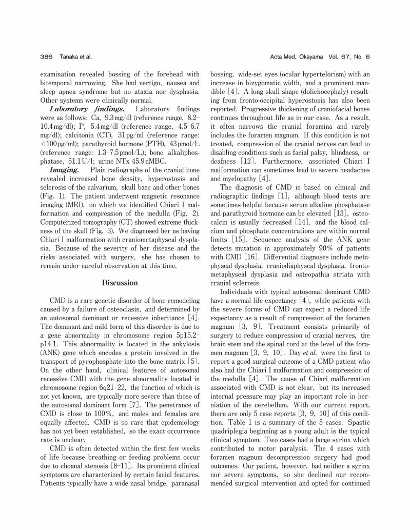
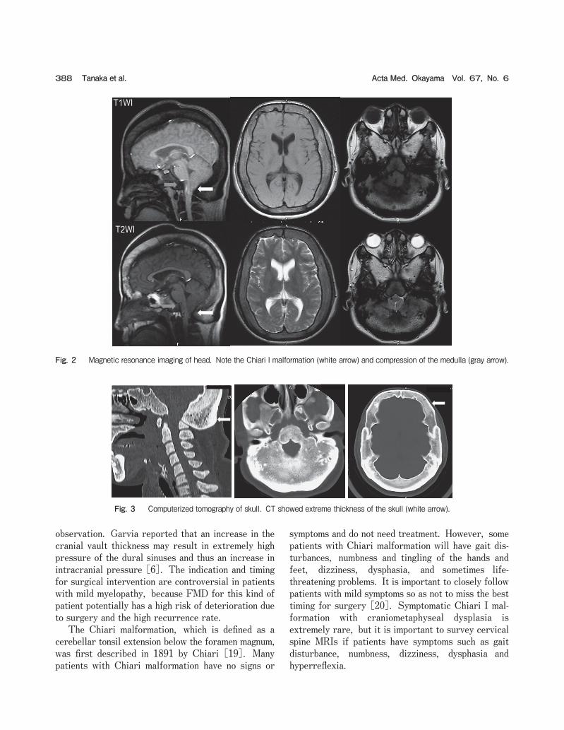
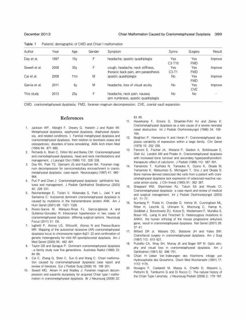
examination revealed bossing of the forehead with bitemporal narrowing. She had vertigo, nausea and sleep apnea syndrome but no ataxia nor dysphasia. Other systems were clinically normal. Laboratory findings. Laboratory findings were as follows: Ca, 9.3mg/dl (reference range, 8.2-10.4mg/dl); P, 5.4mg/dl (reference range, 4.5-6.7 mg/dl); calcitonin (CT), 31pg/ml (reference range:<100pg/ml); parathyroid hormone (PTH), 43pmol/L (reference range: 1.3-7.5pmol/L); bone alkaliphos-phatase, 51.1U/l; urine NTx 45.9nMBC. Imaging. Plain radiographs of the cranial bone revealed increased bone density, hyperostosis and sclerosis of the calvarium, skull base and other bones (Fig. 1). The patient underwent magnetic resonance imaging (MRI), on which we identified Chiari I mal-formation and compression of the medulla (Fig. 2). Computerized tomography (CT) showed extreme thick-ness of the skull (Fig. 3). We diagnosed her as having Chiari I malformation with craniometaphyseal dyspla-sia. Because of the severity of her disease and the risks associated with surgery, she has chosen to remain under careful observation at this time.
Discussion
CMD is a rare genetic disorder of bone remodeling caused by a failure of osteoclasis, and determined by an autosomal dominant or recessive inheritance [4]. The dominant and mild form of this disorder is due to a gene abnormality in chromosome region 5p15.2-p14.1. This abnormality is located in the ankylosis (ANK) gene which encodes a protein involved in the transport of pyrophosphate into the bone matrix [5]. On the other hand, clinical features of autosomal recessive CMD with the gene abnormality located in chromosome region 6q21-22, the function of which is not yet known, are typically more severe than those of the autosomal dominant form [7]. The penetrance of CMD is close to 100オ, and males and females are equally affected. CMD is so rare that epidemiology has not yet been established, so the exact occurrence rate is unclear. CMD is often detected within the first few weeks of life because breathing or feeding problems occur due to choanal stenosis [8-11]. Its prominent clinical symptoms are characterized by certain facial features. Patients typically have a wide nasal bridge, paranasal
bossing, wide-set eyes (ocular hypertelorism) with an increase in bizygomatic width, and a prominent man-dible [4]. A long skull shape (dolichocephaly) result-ing from fronto-occipital hyperostosis has also been reported. Progressive thickening of craniofacial bones continues throughout life as in our case. As a result, it often narrows the cranial foramina and rarely includes the foramen magnum. If this condition is not treated, compression of the cranial nerves can lead to disabling conditions such as facial palsy, blindness, or deafness [12]. Furthermore, associated Chiari I malformation can sometimes lead to severe headaches and myelopathy [4]. The diagnosis of CMD is based on clinical and radiographic findings [1], although blood tests are sometimes helpful because serum alkaline phosphatase and parathyroid hormone can be elevated [13], osteo-calcin is usually decreased [14], and the blood cal-cium and phosphate concentrations are within normal limits [15]. Sequence analysis of the ANK gene detects mutation in approximately 90オ of patients with CMD [16]. Differential diagnoses include meta-physeal dysplasia, craniodiaphyseal dysplasia, fronto-metaphyseal dysplasia and osteopathia striata with cranial sclerosis. Individuals with typical autosomal dominant CMD have a normal life expectancy [4], while patients with the severe forms of CMD can expect a reduced life expectancy as a result of compression of the foramen magnum [3, 9]. Treatment consists primarily of surgery to reduce compression of cranial nerves, the brain stem and the spinal cord at the level of the fora-men magnum [3, 9, 10]. Day et al. were the first to report a good surgical outcome of a CMD patient who also had the Chiari I malformation and compression of the medulla [4]. The cause of Chiari malformation associated with CMD is not clear, but its increased internal pressure may play an important role in her-niation of the cerebellum. With our current report, there are only 5 case reports [3, 9, 10] of this condi-tion. Table 1 is a summary of the 5 cases. Spastic quadriplegia beginning as a young adult is the typical clinical symptom. Two cases had a large syrinx which contributed to motor paralysis. The 4 cases with foramen magnum decompression surgery had good outcomes. Our patient, however, had neither a syrinx nor severe symptoms, so she declined our recom-mended surgical intervention and opted for continued
386 Acta Med. Okayama Vol. 67, No. 6Tanaka et al.
387Chiari Malformation Caused by Craniometaphyseal DysplasiaDecember 2013
R L A PA
B
C
Fig. 1 Plain skeletal radiographs. A, plain radiographs of the cranial bone revealed increased bone density (white arrow); B, spinal bone density of spine was increased; C, bony deformities and increased bone density were observed.
observation. Garvia reported that an increase in the cranial vault thickness may result in extremely high pressure of the dural sinuses and thus an increase in intracranial pressure [6]. The indication and timing for surgical intervention are controversial in patients with mild myelopathy, because FMD for this kind of patient potentially has a high risk of deterioration due to surgery and the high recurrence rate. The Chiari malformation, which is defined as a cerebellar tonsil extension below the foramen magnum, was first described in 1891 by Chiari [19]. Many patients with Chiari malformation have no signs or
symptoms and do not need treatment. However, some patients with Chiari malformation will have gait dis-turbances, numbness and tingling of the hands and feet, dizziness, dysphasia, and sometimes life-threatening problems. It is important to closely follow patients with mild symptoms so as not to miss the best timing for surgery [20]. Symptomatic Chiari I mal-formation with craniometaphyseal dysplasia is extremely rare, but it is important to survey cervical spine MRIs if patients have symptoms such as gait disturbance, numbness, dizziness, dysphasia and hyperreflexia.
388 Acta Med. Okayama Vol. 67, No. 6Tanaka et al.
T1WI
T2WI
Fig. 2 Magnetic resonance imaging of head. Note the Chiari I malformation (white arrow) and compression of the medulla (gray arrow).
Fig. 3 Computerized tomography of skull. CT showed extreme thickness of the skull (white arrow).
References
1. Jackson WP, Albright F, Drewry G, Hanelin J and Rubin MI: Metaphyseal dysplasia, epiphyseal dysplasia, diaphyseal dyspla-sia, and related conditions. I. Familial metaphyseal dysplasia and craniometaphyseal dysplasia; their relation to leontiasis ossea and osteopetrosis; disorders of bone remodeling. AMA Arch Intern Med (1954) 94: 871-885.
2. Richards A, Brain C, Dillon MJ and Bailey CM: Craniometaphyseal and craniodiaphyseal dysplasia, head and neck manifestations and management. J Laryngol Otol (1996) 110: 328-338.
3. Day RA, Park TS, Ojemann JG and Kaufman BA: Foramen mag-num decompression for cervicomedullary encroachment in cranio-metaphyseal dysplasia: case report. Neurosurgery (1997) 41: 960-964.
4. Puri P and Chan J: Craniometaphyseal dysplasia: ophthalmic fea-tures and management. J Pediatr Ophthalmol Strabismus (2003) 40: 228-231.
5. Reichenberger E, Tiziani V, Watanabe S, Park L, Ueki Y and Santanna C: Autosomal dominant craniometaphyseal dysplasia is caused by mutations in the transmembrane protein ANK. Am J Hum Genet (2001) 68: 1321-1326.
6. Rivero-Garvía M, Márquez-Rivas FJ, García-Iglesias A and Gutiérrez-González R: Intracranial hypertension in two cases of craniometaphyseal dysplasia: differing surgical options. Neurosurg Focus (2011) 31: E6.
7. Iughetti P, Alonso LG, WilcoxW, Alonso N and Passos-Bueno MR: Mapping of the autosomal recessive (AR) craniometaphyseal dysplasia locus to chromosome region 6q21-22 and confirmation of genetic heterogeneity for mild AR spondylocostal dysplasia. Am J Med Genet (2000) 95: 482-491.
8. Taylor DB and Sprague P: Dominant craniometaphyseal dysplasia --a family study over five generations. Australas Radiol (1989) 33: 84-89.
9. Cai C, Zhang Q, Shen C, Sun G and Wang C: Chiari malforma-tion caused by craniometaphyseal dysplasia: case report and review of literature. Eur J Pediatr Surg (2008) 18: 198-201.
10. Sewell MD, Akram H and Wadley J: Foramen magnum decom-pression and expanile duroplasty for acquired Chiari type I malfor-mation in craniometaphyseal dysplasia. Br J Neurosurg (2008) 22:
83-85.11. Haverkamp F, Emons D, Straehler-Pohl HJ and Zerres K:
Craniometaphyseal dysplasia as a rare cause of a severe neonatal nasal obstruction. Int J Pediatr Otorhinolaryngol (1996) 34: 159-164.
12. Beighton P, Hamersma H and Horan F: Craniometaphyseal dys-plasia--variability of expression within a large family. Clin Genet (1979) 15: 252-258.
13. Fanconi S, Fischer JA, Wieland P, Giedion A, Boltshauser E, Olah AJ, Landolt AM and Prader A: Craniometaphyseal dysplasia with increased bone turnover and secondary hyperparathyroidism: therapeutic effect of calcitonin. J Pediatr (1988) 112: 587-591.
14. Yamamoto T, Kurihara N, Yamaoka K, Ozono K, Okada M, Yamamoto K, Matsumoto S, Michigami T, Ono J and Okada S: Bone marrow-derived osteoclast-like cells from a patient with crani-ometaphyseal dysplasia lack expression of osteoclast-reactive vac-uolar proton pump. J Clin Invest (1993) 91: 362-367.
15. Sheppard WM, Shprintzen RJ, Tatum SA and Woods CI: Craniometaphyseal dysplasia: a case report and review of medical and surgical management. Int J Pediatr Otorhinolaryngol (2003) 67: 71-77.
16. Nurnberg P, Thiele H, Chandler D, Hohne W, Cunningham ML, Ritter H, Leschik G, Uhlmann K, Mischung C, Harrop K, Goldblatt J, Borochowitz ZU, Kotzot D, Westermann F, Mundlos S, Braun HS, Laing N and Tinschert S: Heterozygous mutations in ANKH, the human ortholog of the mouse progressive ankylosis gene, result in craniometaphyseal dysplasia. Nat Genet (2001) 28: 37-41.
17. Millard DR Jr, Maisels DO, Batstone JH and Yates BW: Craniofacial surgery in craniometaphyseal dysplasia. Am J Surg (1967) 113: 615-621.
18. Puliafito CA, Wray SH, Murray JE and Boger WP III: Optic atro-phy and visual loss in craniometaphyseal dysplasia. Am J Ophthalmol (1981) 92: 696-701.
19. Chiari H: Ueber Ver/inderungen des Kleinhirns infolge yon Hydrocephalie des Grosshirns. Dtsch Med Wochensehr (1891) 17: 1172-1175.
20. Novegno F, Caldarelli M, Massa A, Chieffo D, Massimi L, Pettorini B, Tamburrini G and Di Rocco C: The natural history of the Chiari Type I anomaly. J Neurosurg Pediatr (2008) 2: 179-187.
389Chiari Malformation Caused by Craniometaphyseal DysplasiaDecember 2013
Table 1 Patientsʼ demographic of CMD and Chiari I malformation
Author Year Age Gender Symptom Syrinx Surgery Result
Day et al. 1997 15y F headache, spastic quadriplegia YesC3-T10
YesFMD
Improve
Sewell et al. 2008 35y F cough, headache, neck stiffness,thoracic back pain, arm paraesthesis
YesC3-T1
YesFMD
Improve
Cai et al. 2008 11m M spastic quadriplegia No YesFMD
Improve
Garvia et al. 2011 6y M headache, loss of visual acuity No YesCVE
Improve
This study 2013 25y F headache, neck pain, nausea, arm numbness, spastic quadriplegia
No No -
CMD, craniometaphyseal dysplasia; FMD, foramen magnum decompression; CVE, cranial vault expansion.


![Rx161 Arnold-Chiari Malformationfinalcopy0048502.netsolhost.com/.../pdfs/RXforms/Arnold_Chiari_Malformation.pdfArnold-Chiari malformation [Chiari malformation (CM)] is a congenital](https://img.pdfslide.net/doc/110x75/5ab9a8f17f8b9ac60e8e5491/rx161-arnold-chiari-malforma-malformation-chiari-malformation-cm-is-a-congenital.jpg)