Embed Size (px)
Citation preview

Vol. 1, 473-48 1, October 1990 Cell Growth & Differentiation 473
5 The abbreviations used are: cDNA, complementary DNA; bp, basepair(s); SSC, standard saline citrate; poly(A), polyadenylated.
Chicken Cardiac Tropomyosin and a Low-Molecular-Weight Nonmuscle Tropomyosin Are Related byAlternative Splicing’
Suzanne Forry-Schaudies,2 Christian E. Gruber,3 andStephen H. Hughes4
ABL-Basic Research Program, NCI-Frederick Cancer Research andDevelopment Center, Frederick, M.i’yland 21701
Abstract
We have isolated and characterized complementaryDNAs (cDNAs) encoding chicken cardiac muscletropomyosin and a low-molecular-weight nonmuscletropomyosin. The cardiac muscle cDNA (pCHT-4)encodes a 284-amino acid protein that differs fromchicken skeletal muscle a- and �-tropomyosinsthroughout its length. The nonmuscle cDNA (pFT-C)encodes a 248-amino acid protein that is most similar(93-94%) to the tropomyosin class including ratfibroblast TM-4, equine platelet tropomyosin, andhuman fibroblast TM3O�. The nucleotide sequences ofthe cardiac and nonmuscle cDNAs are identical fromthe position encoding cardiac amino acid 81(nonmuscle amino acid 45) through cardiac amino acid257 (nonmuscle amino acid 221). The sequences differboth 5’ and 3’ of this region of identity. Thesecomparisons suggest that the chicken cardiactropomyosin and low-molecular-weight “platelet-like”tropomyosin are derived from the same genomic locusby alternative splicing. Si analysis suggests that thislocus encodes at least one other tropomyosin isoform
IntroductionThe tropomyosins are a family of ubiquitous, highly con-served structural proteins. In muscle and nonmusclecells, tropomyosins form dimeric a-helical coiled coils.In skeletal and cardiac muscle, these coiled structuresalign head-to-tail and lie in the grooves of F-actin, wherethey mediate Ca2�-regulated actomyosin contraction inassociation with the troponins (1-3). The smooth muscleand nonmuscle tropomyosins are structurally similar tothe muscle forms; however, their function(s) are less welldefined. Tropomyosins associate with microfilaments inthese cells and are thought to regulate contraction (4)
Received 5/29/90.
1 Research sponsored by the National Cancer Institute, Department of
Health & Human Services, under Contract N01-CO-74101 with ABL. Thecontents of this publication do not necessarily reflect the views or policiesof the Department of Health & Human Services, nor does mention oftrade names, commercial products, or organizations imply endorsementby the U.S. Government.2 5 Forry-Schaudies was supported by a Postdoctoral Fellowship from
the Muscular Dystrophy Association.3 Present address: Bethesda Research Laboratories, 1 81 7 Grovemont
Circle, Gaithersburg, MD 20877.4 To whom requests for reprints should be addressed, at ABL-Basic
Research Program, NCI-Frederick Cancer Research and Development
Center, P.O. Box B, Frederick, MD 21701.
and to stabilize F-actin (5, 6). The functionally differentinteractions of tropomyosins appear to require differentprotein isoforms.
Multiple forms of tropomyosin proteins ranging fromMr 30,000 to Mr 46,000 have been separated on two-dimensional gels. Skeletal muscle, cardiac muscle, andsmooth muscle tropomyosins are 284 amino acids inlength. In mammalian and avian species, fast and slowskeletal muscle have distinct a forms (Mr 34,000) andidentical � forms (M, 36,000). Chicken cardiac musclecontains one a-tropomyosin isoform that migrates in aposition distinct from skeletal muscle isoforms on two-dimensional gels (7). In contrast, the tropomyosin foundin mammalian cardiac muscle is identical to the mam-malian skeletal muscle a-tropomyosin (8, 9). The tropo-myosins in smooth muscle and nonmuscle cells are either284 or 248 amino acids in length. Chicken embryo fibro-blasts have as many as seven different tropomyosins thatare the products of different mRNAs (10-12).
Numerous tropomyosin cDNAs5 have been isolated ina variety of species. Although there are differences be-tween mammals and ayes, sequence comparisons haveindicated that vertebrates generate the mRNAs corre-sponding to these cDNAs from as many as four distinctgenomic loci by alternative splicing of primary RNA tran-scripts. The “a fast” locus gives rise to the a-tropomyosinin fast skeletal muscle, a smooth muscle isoform, several284- and 248-amino acid fibroblast isoforms, and threebrain isoforms (13-22). The “a slow” locus encodes thea-tropomyosin in slow skeletal muscle and a low-molec-ular-weight (248 amino acids) fibroblast tropomyosin (23,24). The “j3” locus gives rise to skeletal muscle $-tropo-myosin, a smooth muscle tropomyosin, and, in chickens,a low-molecular-weight (248 amino acids) fibroblast iso-form (25-28). A fourth locus gives rise to a 248-aminoacid “platelet” isoform (29-32). Neither the genomiclocus nor a cDNA encoding avian cardiac muscle tropo-myosin has been isolated.
In this paper, we describe the isolation and sequenceanalysis of the chicken cardiac muscle cDNA and ach icken fi broblast low-molecular-weight tropomyosincDNA that encodes a protein having 93% homology toequine platelet tropomyosin (30). Amino acid sequencecomparisons indicate that chicken cardiac tropomyosinis distinct from chicken skeletal muscle a- or fl-tropo-myosin. The cardiac muscle and fibroblast cDNAs areidentical from the position encoding amino acid 81 ofthe cardiac muscle clone (amino acid 45 of the fibroblastclone) through amino acid 257 of the cardiac clone(amino acid 221 of the fibroblast clone). These datasuggest that a low-molecular-weight “platelet-like” tro-
pomyosin isoform and the cardiac muscle tropomyosinare encoded by a single genomic locus in chickens.
Results
cDNA Isolation and Sequence Analysis. The chickenembryonic heart library was screened with a 32P-labeledDNA probe made from pSMT-10, a chicken smoothmuscle tropomyosin cDNA (33). Over 100 positiveclones were isolated, and the plasmids were tested todetermine the size of the inserts. Approximately 30 ofthe plasmids having the largest inserts were characterizedby nucleic acid sequence analysis. The plasmid havingthe largest insert (approximately 1050 bp) was namedpCHT-7a; it included a major portion of the coding region(amino acids 6-284), the entire 3’-untranslated region(149 bp), and a poly(A) tract.
Various studies have reported the simultaneousexpression of cardiac and skeletal muscle-specific iso-forms of troponin C, troponin 1, and actin within devel-oping skeletal muscle (34-37). Based on this information,we screened a cDNA library made from the body wall of10-day-old embryos in an attempt to isolate cDNA clonescontaining the 5’-most sequences of the cardiac tropo-myosin. A 3’ end probe extending from the Hindlll siteat amino acid 232 through the poly(A) tract of pCHT-7awas used to screen this library. Positive clones weresecondarily screened with a synthetic oligonucleotide (5’end labeled) extending from amino acid 16 to amino acid31 of pCHT-7a. Six positive clones were isolated; all ofthe clones contained sequences identical to pCHT-7a.pCHT-4 had a 1 150-bp cDNA insert that included 60 bpof the 5’-untranslated region, the entire coding region,and the entire 3’-untranslated region (Fig. 1).
Because clones pCHT-7a and pCHT-4 were from acardiac muscle and a body wall library, respectively, itwas necessary to confirm that the 5’-untranslated region
and amino acids 1 -6 of the body wall cDNA pCHT-4 areidentical to those found in cardiac muscle RNA. A 480-base uniformly labeled probe including 370 bases ofpCHT-4 from the 5’-untranslated region through the Apalsite at amino acid 102 was hybridized against 10- or 18-day chicken embryo cardiac muscle RNA and exposedto Si nuclease (see “Materials and Methods”). Fig. 2shows that a 370-base band was protected in both car-diac muscle samples, indicating that they contain RNAswith 5’ nucleotide sequences identical to those of pCHT-4.
All tropomyosin genomic loci studied to date generateat least two isoforms by alternatively splicing primaryRNA transcripts (22, 23, 25, 27). The 10-day chickenembryo body wall library was rescreened with a nick-translated pCHT-4 probe to isolate clones closely relatedto pCHT-4. Twenty clones were isolated; several clonesdiffering in the length of their 5’-untranslated regionswere analyzed. The longest clone, pFT-C, had 40 bases
COT
FT-C
COT GCCITICt601y A)
474 Chicken Cardiac Tropomyosin
FT C AAACGGTIICICGCIpo1y A)
oil cccliiTciciicAGAACiiiiIiiiGiliiCGiGACAAACICAGCCTA1CC1IIAGCAAAA
I 10 20
�t Gi.iAi� lietys Lys Lyo�t Gi.Met Ie.iys LeuApLy�Gi.AsnAia ii. AspCIII AlA �A (Li SIC SAG MA 541 AlA CAG AIG JIG 0*6 CIG � 446 ‘� Ut (LA All W
30 40ArqAidGiuGinAiaGiuihrAspLystysAtaAi4GiuAspiysCysiysGinVaiGiu
CHI A&0 i�A GAG CIA r�i GAO ACG 6*1 040 MG C�A (�1 GAG SAC MA TGC 00* COG GIC GAG
U -C CCCAGCACGiGCA�AGCiAGCGCCCiiACCGCCGCGCCCAiG 6CC GCG CCG�t Ala Ala Pr.
so 00Asp IOu t.u Vai AL. eu Si. Lys i.ys te. Lys Sly 15. Alu Asp Giu Let. Asp iys lyr
CAl GAl “ dO 61* rzi cis rAG SAG AM 116 AM OGA AC! GM GAl �- CIA GAl MA lAG
11 C AGC ICC CIG GAO AGG GIG SAG *00 MG AIC CM lAG CIG CAll CAG CAll GCG GAC GAG CLGSer Ser ies Giu Ala Oai iys Org Lys I le Gin Cys Leo Gin Gin Gin Ala Asp 61, Ala
10 20
10 80SerGiuAiaLeuLysAspAiaGinGi.Lyste,�GinGinAiaGiatystysAiaihrAsp
CIII ICC SAG AGI dO MA GAl i�C CAG GM MG CTG GAG CAG GO GAA MG MG 6CC AGO GAl
I I -C GAG r.Al CAG 0CC CAG GIC CIG CAG COG GAG CIG GAG C1G (.08 CGG GAC Clii CGG GAG AMGiu Asp Arq Ala Gin Vjl I,. Gin Arg 01. 1.., Asp Len Gi. Arg Asp Len Org Glu Lys
30 40
90 100Ala GiaGiyGia Vai Ala Ala L.uAsnArg Org ileilin Len ValGi.Gl,Gi, i.e. Asp
COT AGAGAA00lAM6l6GCGAG6ClCAAG�CGCAlCCAGCl6Gl6AMGAGAMClGGAl
FTC GCAMAAGIMGGIGAGGGCGCICAAGAGACAGAICCAGCT6GIGCOAGAGGAGCIGGAIAla IOu Sly Glu Val Ala Ala tea Asn Arg Arq lie 61. i.e. VaT Glu Glu IOu Len Asp
so 60
110 120ArgAiaGlnGlaArgLe.AlalPnA1aLe.GinLy.LeuGia6laAlaGluLysAlaAla
COT COG AGC GAG MG COG CIG OCC ACA 0CC CIII CAll MG CIG MA MG AGC MA MA f�G GCG
11 -C CM AGC GAG AM CM CIG 6CC ACA 0CC fIG C�AG AM CIG MA GAG 6CC MA AM GCG AGOArgAlaGln6l.Argte.AlalhrAlai.e.GlnLysie.GlaGlaAiaGiui.ysAlaAla
10 80
130 140Asp Gla Set Glu Org Gly �t Lys Val I 1. 01. A.. Org Ala Met Lys Asp Si. 01. Lys
CHI 601 GAG AOl GAG wa Gc.O AIG AM GTC All GAG MC AM AGO 016 AM GAl MA GM AM
FT.C MT GAG Ml AM AM r.C.A AIG AM 61C All GAG MC AGA OCA *16 AM MT MA GM AMAsp Gla Ser Gia Arg Oly �t Lys VaT I in 61. Asn Org Ala Net Lys Asp 61. 01, Lys
90 100
ISO 160�t Gia lie 61. 61. �t Gin i.e. Lys 01. Ala Lys H,. lie Ala Glu IOu Ala Asp Arg
COT ATGGMATICAGGAAAIGCAGCIGAAGGAGAGCAAGCACAICAGIGAGGAGAGCGACCAG
FT -C A16 00.4 All CAG MA AIG CM CIG MG MG 6CC MG GAG AIC AGT AM AM AGC MC CfL�t Gla 1 le Gin 61. �t Gin Ins Ly. Glu Ala Ly. His I in Ala Glu IOu Ala Asp Org
110 120
170 180
Lys lyr Glu 61. Vai Ala Arg Lys i.e. VaT lie i.e. 61. Giy 61. Len Glu Org Ala 61.COT AM TAG MA AM 611 AGC COt 0.06 116 616 All 016 AM 666 AM COG GAl AM AGT MA
FT-C AM TAG MA GAG 6TT AGC Cr� AM 116 616 ATT ITO GAG AGaG MA CIG MA AM AGT MALyslyroi,Gl,VaiAlaArgtysLe.Vaill.Le.6i,GiyGi,LnsGlaArqAlaGia
130 lAo
190 200Glu Arg Ala 61, Val Set Gb Hal Lys Cys Ser Asp i.e. 61. 61. 61. i.e. Lys Asn Val
COT GAG CAG GCA GAG GIG ICC MA GIl AM TAG AOl ‘� CIT MA GAG SAG TIG MG UT GIC
FT -C GAG GAG �LA GAG GIG ICC MA 611 AM TAG AG! MC CIT MA GAG GAG TlG SAG Ml GICGluArgAlaGiuVaiSer6luValLysCysSerAspLe.6l,61,Gl,Le.i.ysAsnVai
ISO 60
210 270Thr Asn As. i.e. Lys GAr i.e. Glu Ala 6ln Ser 61. Lys Tyr Set Glu tys Gl*i Asp Lys
COT AGO MC UT CIG O.AG ICT TIG MA AGI CM ICI MA AlL TAG 1C6 MA 806 MA Ml HAG
Fl-C ACA AAC OAT CIG MG ICI TIG MA OCT CAl ICI MA AM lAG ICG MA AM MA GAl HAGThrAsnAsnLe.i.ysSeri.e.GluAlaGlnSerGluIysTyrSerGluLysGl,Aspi.ys
hO 180
230 240lyr Giu iOu Glu I Ic i.ys I in i.e. Ser Asp Lys i.e. Lys Glu Ala Glu lhr Arg Ala Glu
COT TAT MA GAG MA AIC MG *11 CIC ICT � MG CII AM MA AGI MA ACI CGI OCT MG
F I-C TAI MA GAG MA ATC HAG All CIC ICI MC MG CII AM MA OCT MA ACT COT AGI GAGlyr Glu Glu 61. ll� Lys Il. i.e. Ser Asp Lys i.e. Lys Glu Ala Glu lhr Org Ala Glu
90 200
250 260Pp� Ala 61. Arg lhr Val Ala Lys Le.Gl, Lys Set Ii. AupAsp LeuGluAspGlu Leu
COT ITT AGI MG AM MA GTT AGC AM CTG MA HAG ICC ATT Ml MT CIG MA MT GAG CIG
F I -C ITT OCT GAG AGO ACA GIl AGC AM CTG MA HAG ICC All Ml MT CIG MA MG AM CIIPp� Ala 61. Arg Tin’ Hal Ala Lys i.e. Gl, i.ys Ser lleAspAn.pLeu 61. Asp Lys i.e.
210 220
270 280lyrAlaGlni.ysLnslyslyri.ysAlalleSerGlu6lui.e.AspOisAlaLe.AsnAsp
COT TAG Gd GAG HAG CTG MG TAC AM AGA AIC MI. �6G GAG dC MC CAC AGO CIC MT GAl
FT C Gd CM AGA HAG MG MG MC CIG AGO CIG MG GAG ACG CIG MC CAG PiG CIA MC MAAla Gln Ala tys Glu Glu Asn i.e. Gly i.e. Ass Gin Tin i.e. Asp Gln IOn i.e. As. Glu
230 240
COT
Fl-C
�t TSr Ser i.e.AIG 0CC ICC CIG TAGICICGACCTCCTG6060IAGTCCAT IIGCTGACCTCTTATAGICTCCTGGICIGTMTTC
CIG MC TGT AlA IGACCCCGAGCGGAACCCCAGGGCCGC.MAAGGTCAGCCGCCCTCAGCGCATCACCGCCCCCCi.e. Asn Cys lie
lCCClGCAGlCCMG&hT�GCTCG&AGC60lTClGlACCCCClACAGCCAG.ClCCCCCGCGGCCGCCAGGCGCAll
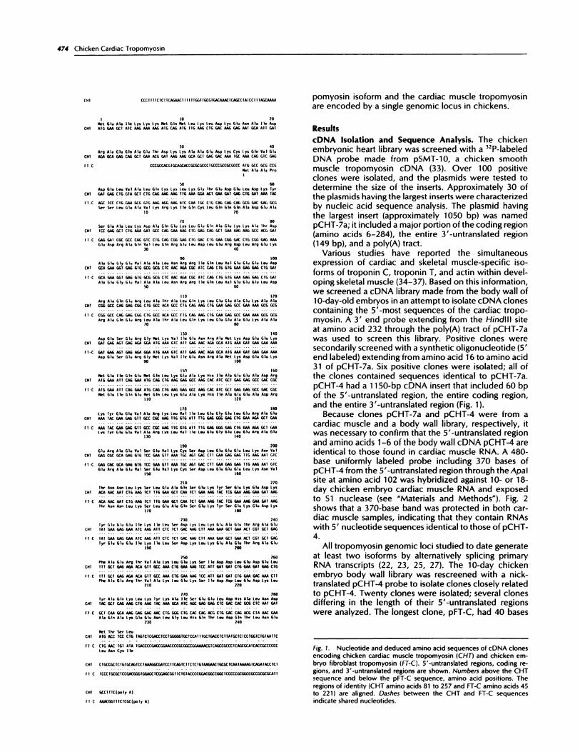
Fig. 1. Nucleotide and deduced amino acid sequences of cDNA clones
encoding chicken cardiac muscle tropomyosin (CHT) and chicken em-bryo fibroblast tropomyosin (FT-C). 5’-untranslated regions, coding re-gions, and 3’-untranslated regions are shown. Numbers above the CHTsequence and below the pFT-C sequence, amino acid positions. The
regions of identity (CHT amino acids 81 to 257 and FT-C amino acids 45to 221) are aligned. Dashes between the CHT and FT-C sequences
indicate shared nucleotides.

(I)C(5
- -
as.
Wit)
Cell Growth & Differentiation 475
6 � E. Gruber, unpublished data.
12
I 81 258284Cardiac (CHT) � %�x� MA
I 45 222 248Fibroblast (FT-C) � ‘
527- . . �.II- Probe
404 -
309 -
240- P.
110-
0-CVIt� ‘-
<(5
� I 480 base probe
�i 370 base product
Fig. 2. 51 analysis demonstrating the presence in cardiac muscle ofmRNAs corresponding to the body wall pCHT-4 cDNA. A uniformly
labeled, single-stranded 480-base probe that includes the 370-base 5’
EcoRl-Apal fragment from pCHT-4 was used (see diagram at bottom).Top, 51-resistant fragments from 10-day chicken embryo cardiac muscle(Lane 1) and 18-day chicken embryo cardiac muscle (Lane 2). Numbers
at left, sizes in bases as determined from pBR MspI-digested DNA markers.The position of the undigested 480-base undigested probe is indicated.
of 5’-untranslated region, an open reading frame encod-ing 248 amino acids, a 155-bp 3’-untranslated region,and a poly(A) tail.
The nucleotide and derived amino acid sequences ofcardiac muscle cDNA pCHT-4 and body wall cDNA pFT-C are compared in Fig. 1. The two cDNAs are colinearfrom the position encoding pCHT-4 amino acid 81 (pFT-C amino acid 45) through the position encoding pCHT-4 amino acid 257 (pFT-C amino acid 221) but differ 5’ ofthe position encoding pCHT-4 amino acid 81 and 3’ ofthe position encoding pCHT-4 amino acid 257. Thenucleotide sequence comparison of pCHT-4 and pFT-Cis diagrammed in Fig. 3. The pattern of nucleotide se-quence identity shown in Figs. 1 and 3 suggests that the
Fig. 3. Schematic representation of the nucleotide sequences of cardiac(CHT) and fibroblast (FT-C) tropomyosin cDNAs. The numbers aboveeach cDNA, amino acid position. The regions encoding CHT amino acid81 through 257 and FT-C amino acid 45 through 221 are identical and
are aligned. #{149},regions of identity between the two cDNAs; 0, fibroblast
sequence; G, cardiac muscle sequence.
mRNAs corresponding to cDNAs pCHT-4 and pFT-C aregenerated from a single genomic locus by alternativesplicing.
Characterization of Protein Products. The proteinsencoded by mRNAs corresponding to cDNAs pCHT-4and pFT-C were identified by in vitro transcription andtranslation in the presence of [35S]methionine, followedby two-dimensional gel electrophoresis (Fig. 4). Radiola-
beled translation products were co-run with cold proteinstandards from 18-day cardiac muscle or chicken embryofibroblasts. RNA derived from the cardiac muscle pCHT-4 cDNA directed the synthesis of the unphosphorylatedcardiac tropomyosin (Fig. 4, A and B). RNA derived fromthe pFT-C cDNA directed the synthesis of fibroblasttropomyosin 3b (1 1), the more acidic of the two chickenlow-molecular-weight tropomyosins (Fig. 4, C and D).
Amino Acid Sequence Comparisons. Rabbit cardiactropomyosin has been reported to be identical to rabbitskeletal muscle a-tropomyosin at the amino acid level(9). Accordingly, we compared the derived amino acid
sequence of chicken cardiac tropomyosin (pCHT-4) tothe amino acid sequences of chicken skeletal muscle a-
and fl-tropomyosins (Fig. 5). All three forms are 284amino acids long and have a cysteine residue at aminoacid 190. Skeletal muscle a-tropomyosin differs from thecardiac form at 29 amino acid positions (90% similarity),and skeletal muscle /3- and cardiac tropomyosins differat 34 amino acid positions (88% similarity). Both a- andfl-skeletal muscle tropomyosins have 19 conservativeamino acid changes when compared to the cardiac form.The a-skeletal form has 10 nonconservative amino acidchanges, and the fl-skeletal muscle tropomyosin has 15.The nucleotide sequences of the 5’- and 3’-untranslatedregions of chicken cardiac, skeletal muscle a-, and skel-etal muscle f3-tropomyosins were also compared andshowed no homology.6
Fig. 6 compares the derived amino acid sequence of
the 248-amino acid pFT-C to five other 248-amino acidtropomyosins (24, 30-32, 38). Amino acid comparison ofthe five known low-molecular-weight tropomyosins in-dicates that rat fibroblast TM-4 (32), equine platelet tro-pomyosin (30), and human TM30�1 (31) are probably thesame isoform since they are 97-98% similar. HumanTM3Onm and chicken FT-$ share only 80% similarity witheach other and only 75 or 82% similarity with the TM-4-equine platelet-TM30�1 class of tropomyosins. Therefore,the known low-molecular-weight tropomyosins probablyrepresent three distinct isoforms: (a) TM-4-equine plate-let-TM30�t; (b) TM3Onm; and (c) FT-f.�.

A
>1#{149}��
/
IEF- �‘acidic
B
/
- U)
. D U)
�3b.
Fig. 4. Two-dimensional gel electrophoresis of the in vitro translated cardiac pCHT-4 transcript and the fibroblast pFT-C transcript. A, Coomassie blue-
stained gel of 18-day embryo chicken heart proteins mixed with the [‘55]methionine-labeled in vitro translated product of pCHT-4; B, the fluorographcorresponding to the gel in A. Arrows in A and B, position of the unphosphorylated cardiac tropomyosin. C. Coomassie blue-stained gel of chickenembryo fibroblast tropomyosin standard mixed with the [‘55]methionine-labeled in vitro translated product of pFT-C. The proteins are labeled c, 1, 2, 3a,
and 3h as described in Ref. 1 1 . D, the fluorograph corresponding to the stained gel in C. Arrows in C and D, the co-localization of tropomyosin 3b andthe in vitro translated product of pFT-C. The M, markers indicated by arrowheads from top to bottom are: 94,000 (phosphorylase b), 68,000 (bovine
serum albumin), 43,000 (ovalbumin), 30,000 (carbonic anhydrase), and 21,000 (soybean trypsin inhibitor). 1FF, isoelectric focusing; SOS, sodium dodecylsulfate.
476 Chicken Cardiac Tropomyosin
>
>-
>
>
The six low-molecular-weight tropomyosins shown inFig. 6 are most divergent in three regions: amino acids1-44, 152-177, and 222-248. When pFT-C was com-pared to the TM-4-equine platelet-TM30�, class of tro-pomyosins, it was found that most nonconservativeamino acid changes were clustered in the first 41 aminoacids. pFT-C shows a switch from polar to nonpolaramino acids at positions 3, 30, and 37 and from nonpolarto polar at position 16. pFT-C and the TM-4-equineplatelet-TM3O�1 class of tropomyosins are 92-93% simi-lar; they are from 95-96% similar 3’ of amino acid 44.pFT-C is 76% similar to human TM3O2m and 79% similarto chicken FT-fl. However, these differences are slightlyexaggerated because amino acids 153-177 are encodedby alternate exons. Both FT-fl and TM3Onm use the up-stream “fibroblast” exon (23-25, 27), whereas pFT-C usesthe same exon as cardiac muscle pCHT-4. If the regionfrom amino acids 153-177 is not included, pFT-C is 80%
similar to TM3Onm and 83% similar to FT-fl.The amino acid comparisons in Figs. 5 and 6 indicate
that pCHT-4 and pFT-C are encoded by a genomic locusthat is distinct from the a fast, a slow, or fl-tropomyosinloci. The locus that generates the pCHT-4 and pFT-Ctropomyosins in chickens is likely to correspond to theloci giving rise to TM-4 in rats (29, 32), platelet tropo-myosin in horses (30), and TM30�1 in humans (31).
Expression in Different Tissues. The expression of themRNAs corresponding to cDNAs pCHT-4 and pFT-C invarious chicken embryonic tissues was determined byNorthern transfer analysis (Fig. 7). The mRNA corre-sponding to cDNA pCHT-4 is most abundant in cardiacmuscle and migrates at a position expected for a RNA ofapproximately 1.15 kilobases. A 1.15-kilobase mRNA isalso present in small amounts in leg muscle, breast mus-cle, and gizzard. Northern analysis does not reveal a1.15-kilobase band in body wall RNA presumably be-cause of the low amounts of RNA corresponding to thepCHT-4 cDNA. However, this mRNA is present in bodywall and can be detected by 51 analysis (data not shown).Chicken embryo fibroblasts (Lane 7) express a shorter,

1 10 20CHT Met Glu Ala tie Lys Cys lys Met Gin Met Leu Lys Leu Asp Lys Glu Asn Ala lie Asp
sKi-n Asp leu
Ski-s
30 40CAT Arg Aia Giu Gin Ala Giu Thr Asp Lys Lys Ala Ala Giu Asp lys Cys lys Gin Vai GiuSKT-, Ala Glu Arg Ser leuSKi-5 Ala Gin Arg Leo
so 60COT Asp Giu Leu Vai Ala i.eu tin tys i.ys leu i.ys Gly Thr Giu Asp Glu Leu Asp i.ys iyr
SKT -.
SKT-$ Giu Gin Gln Giy Vai Giu
70 80
COT 5cr Giu Ala Leo lys Asp Aia Gin Giu Lys Leu Glu Gin Ala Sb Lys Lys Aia The AspSKi-s Ser Leo AspSKT-5 5cr VaT Giu
90 100CAT Aia Giu Giy tlu Vai Aia Aia Leu Asn Arg Arg Ile Gin i.eu Vai Giu Giu tiu teu AspSKI-a Ser Ser
SKI-s Aia Ser
110 120CHT Arg Aia Gin Giu Arg Leu Ala The Ala leu Gin Lys Leu Giu Gb Ala Giu Lys Aia AiaSKI..Ski-s
130 140CAT Asp Giu 5cr tiu Arg tiy Met i.ys Vai lie Giu Asn Arg Aia Met Lys Asp Giu Giu LysSKI-s Gin
SKI-S
150 160CAT Met Giu lie Gin Glu Met Gin Ieu Lys Gb Ala lys His Ile Ala Glu Giu Aia Asp Arg
SKT-o lie
SKT-$ Leu
170 180COT Lys Tyr Giu Giu Val Ala Arg Lys Leu VaT lie Leu Gb Sly Gb Leu Giu Arg Ala GiuSki-s Aia lie Asp
SKT-$ Vai 5cr
190 200CAT Giu Arg Aia Giu Oat 5cr Giu Au Lys Cys Ser Asp Leu Giu Giu GTu Leu Lys Asn VaiSKi-s Leu 5cr Ala Glu Thr
SKi.$ Aia 5cr Giy lie
2)0 220CHT Thr Asn Ass Leu Lys 5cr leu Giu Ala Gin Ser Giu Lys Tyr Ser Giu Lys Giu Asp i.ysSKi-s Aia GinSKT-$ Aia Asp Thr
230 240CHI Tyr Giu Giu Giu lie lys lie i.eu 5cr Asp Lys Leu Lys Giu Aia Giu Thr Org Ala Giu
SKI-s Aai Thr
SKT-#{216} Leu Giy Gb
250 260CAT Phe Aia Giu Arg Thr Vai Aba i.ys Leu Gb Lys Per lie Asp Asp i.eu Giu Asp Giu LeuSKI-. 5cr TheSKT.fl 5cr TSr Val
270 280CHT Tyr Aba Gin Lys eu Lys Tyr i.ys Ala lie See Giu Giu Leu Asp His Ala Leu Asn Asp
SKT-sSKT-$ Met As,
Fl-CIN-A
(PL
3Opl30...FT-fl
Ft-CTN-A
(Pi.30pl
30,..FT-fl
FT-CiN-4(Pi.3Opl30,..
FT-fl
Fl-CTN-A[FL3OpT30100FT-fl
90 100Glu Lys Ala Ala Asp Glu See Glu Aeg Gly Net Lys VaT Tie Glu As. Aeg Ala Net Lys
1)0 120
Asp Glu Giu Lys Net Glu Ile Gin Giu Net Gin Leo Lys Glu Ala Lys His Ile Ala Giu
Leo lieLeo
Hal
FT-CTN-I(FL
30p130...FT-fl
TheLeo Gly 61.
FT-CTN-I(Pt3�T30..FT-fl
Cell Growth & Differentiation 477
CAT let Thr Ser LeuSAT-u IleSKIt tie
Fig. S. Comparison of the derived amino acid sequence of chickencardiac tropomyosin (CHT) and those of chicken skeletal muscle a (SKT-a) and fi (SKT-fi) tropomyosins. Only the chicken cardiac tropomyosin
sequence is shown where the amino acids are identical; divergent aminoacids are given.
approximately 1.0-kilobase mRNA corresponding tocDNA pFT-C. Chicken embryo body wall, from whichthe pFT-C cDNA was isolated, expresses a 1 .0-kilobasemRNA that hybridizes to the pCHT-4 probe. 1 .0-kilobasemRNAs can be seen in gizzard and 10-day heart samplesin small amounts, probably because of the presence offibroblasts in these organs. We believe that the 1.6-kilobase bands in leg and breast muscle samples are dueto cross-hybridization with the 1.6-kilobase skeletal mus-cle fl-tropomyosin that is very abundant in these tissues(38).
Most tropomyosin genes include alternate exons givingrise to amino acids 189-213 (or 152-177 for 248-aminoacid isoforms) (22, 23, 25, 27). We performed Si nucleaseanalysis to determine whether the locus that gives rise topCHT-4 and pFT-C also gives rise to RNAs differing inthis region. A probe extending from the NcoI site atamino acid 1 through the Hindlll site at the positionencoding amino acid 232 of the fibroblast pFT-C cDNA
10 20Ft-C Net Ala Ala Pro Ser Ser eu Glu Ala Hal lys Arg lys lIe Gin Cys i.e. Gin Gin GIn16�1 Gly Leo A.. Ala
(Pt Gly Len Mn Ala3Opl GTy ten As. Ala30m Gly lie Thr The ile ValFl-fl Gly lie Ile Asp Lys See VaT
30 40Fl�C Ala Asp Glu Ala 61. Asp Avg Ala Gin VaT Leo Gin Arg 61. Len Asp Leo Glu Arg Asp
iM�4 Asp Gly Gly Glu
(P1 Gly Gly GTu
3Opl Gly Gly Glu30,a Asp Gi. Glu Arg Hal Glu Gly Arg
FT�$ Glu IOu His Ala Ala Gin
50 60Fl-C Leo Arg Glu Lys Ala 61. Gly 6iu VaT Ala Ala Len As. Arg Arg fTc GIn ten Hal Glu10-4 Avg Asp Ala
(Pt Arq Asp3Opi Arq Asp30r� Ala Gin Ala SerFT., Ala Arg Ala Ser
70 80Glu6luLeuAspArgAla6ln6luArgleuAlathrAlal.euGintysle.jGluoluAla
130 140
OTu Ala Asp Arg I.ys Tyn’ Giu Giu Vai Ala Arg Lys Leo Hal Tie Leo Glu Gly Giu Leo
lie Asp
150 160Fl-C Glu Avg Ala Gb Glu Arg Ala Glu Vai Ser 61u Hal Lb’s C��s Set Asp Leo GTu Glu GluiN-A Leo See(Pi. Leo Giy
3Opl Leo Gly
30,_ Thr Lou Aia See Arg Arg Giu Net Asp Gln
FT-s Ser Ala Ser Arg Hal Arg Gin Leo
170 180Fl-C LeuLysAsnValThrAsnAsnLeuLysserLeu6luAiaoinserGiulystyrserGiu
iM-4 Ala(P1 At.
3Opl Ala3090 1 le Arg Leo Met Asp Gin Cys See Ala Giu GinFt., Arg Thr Net Asp Gln See lie Ser Glu GT, ihr
190 200i.ys G)u Asp Lys Tyr Giu Giu Giu lie Lys lIe Leo Ser Asp Lys Leo Lys Giu Aia Giu
210 220Ft-C The Arg Ala Glu Pie Ala Glu Arg Thr Val Ala Lys Leo Glu Lys Ser I Ic Asp Asp Leo10-4 Ser The(PL TSr30p1 The30r� See TheFT-$ See The
230 240FT-C Glu Asp Lys Leo Ala Gin Ala Lys Glu Giu Asn Leo Sly Leo His GTn The Leo Asp Gin
16-4 Giu VaTEPL Glu Hal
30p1 Glu Hal3Onn Lys Cys The His Cs’s The Gin Arg MetFT-$ 61. See Ser Hal Ile Vai
The Leo Asn Glu Leo Asn Cys lie
Asp
Leo Asp Giu NetLeo Asp Leo
Fig. 6. Comparison of the primary structures of 248-amino acid tropo-myosins. The derived amino acid sequence of the chicken embryofibroblast pFT-C cDNA is compared to those of rat embryonic fibroblastTM-4 (32), equine platelet (EPL) (30), human fibroblast hTM30� (31),
human fibroblast TM3Oe.. (24), and chicken embryo fibroblast FT-�9 (38).The entire FT-C deduced amino acid sequence is shown. Sequences of
the other tropomyosins are shown only where they differ from FT-C.

89
1.6-
1.1-1.0-
I 2345
622-
527- � �
404-��� -450
309- ‘ -.4
-N
240 -
217-
201- 5
190- 1
Vcxi r�. �It)‘-1--I
A
B
Fig. 8. Si analysisdemonstratingthe expression of RNAs having alternate
sequences at the position encoding amino acid 152. A 620-bp, 3’-end-labeled probe including the 584-bp Ncol -Hindlll fragment from fibroblast
clone pFT-C was hybridized to 20 �g of RNA from various chicken
embryo tissues. A, Si-resistant fragments from Lane 1, 10-day body wall;Lane 2, 18-day leg muscle; Lane 3, 18-day breast muscle; Lane 4, 18-daygizzard; Lane 5, IRNA control. Numbers at left, sizes in bases as deter-
mined from pBR Mspl-digested DNA markers. Numbers at right, positionsof the 620-base undigested probe, the 584-base protected product, and
the 450-base protected product. The length and sequence organizationof the 620-bp probe and the Si-resistant products are diagrammed in B.
0C.)z
* -
478 Chicken Cardiac Tropomyosin
1 234567
� C S
fig. 7. Northern transfer showing expression of RNAs hybridizing to anick-translated pCHT-4 (cardiac) probe. Twenty �ag of total RNA from
various chicken embryo tissues or cells were tested. Lane 1, 10-day bodywall; Lane 2, 18-day leg muscle; Lane 3, 18-day breast muscle; Lane 4,
18-day gizzard; Lane 5, 10-day heart; Lane 6, 18-day heart; Lane 7, chicken
embryo fibroblasts; Lane 8, 18-day brain; Lane 9, 18-day liver.
was labeled at the 3’ end and hybridized to total RNAfrom several embryonic chicken tissues (Fig. 8). As ex-pected, RNA that hybridizes to the probe throughout itslength (584 bases) is present in 10-day chicken embryobody wall, demonstrating the presence of the mRNAcorresponding to the pFT-C cDNA. Faint bands of thissize are also present in leg muscle and breast muscle.The 450-base band in body wall, leg muscle, and breastmuscle samples indicates the presence of RNAs differingfrom pFT-C at or near the position encoding amino acid152.
Discussion
Tropomyosin RNAs generated from a single locus byalternative splicing contain regions of nucleotide se-quence identity. We have isolated a chicken fibroblastcDNA (pFT-C) that shares a region of identity with thechicken cardiac cDNA. Amino acid sequence compari-sons and migration on two-dimensional gels suggest thatthe protein encoded by pFT-C is homologous to thegroup of low-molecular-weight tropomyosins includingrat TM-4 (29, 32), equine platelet tropomyosin (30), andhuman hTM3O, (3 1). Amino acid comparisons show thatthe 248-amino acid fibroblast tropomyosin encoded bythe pFT-C cDNA is more homologous to these proteinsthan to the other known low-molecular-weight forms(human TM3Onm and chicken FT-fl). In addition, the pat-tern of nonpolar amino acids thought to stabilize the a-helical coiled coil structure of equine platelet tropomy-osin (30, 39) has been highly conserved in pFT-C. Thepattern of amino acids differs only at amino acid 3, wherea nonpolar alanine residue in the protein encoded bypFT-C replaces a polar glycine residue in equine platelet
tropomyosin. This substitution of a nonpolar for a polarresidue should theoretically introduce more stability intothe pFT-C protein helix. Two-dimensional gel analysisindicates that the in vitro translated product of pFT-Cmigrates in the acidic, M, 30,000 position, as do rat TM-
620 bp
584 bases
450 bases
4 and human TM30�1 (30, 32). The differences betweenthe protein encoded by pFT-C and the three mammalianproteins are primarily in the first 41 amino acids and mayrepresent evolutionary differences between mammals
and ayes.The nucleotide sequence of pFT-C encodes a proline
residue at amino acid 4. Amino acid analysis as well asgel electrophoresis of [3H]proline-labeled fibroblast pro-

Cell Growth & Differentiation 479
teins have indicated that most tropomyosins lack prolineresidues (5, 8, 12, 40). However, amino acid analysis ofM� 29,000 and 31 ,000 rat ascites tumor cell microvillartropomyosin has indicated the presence of proline (41).The protein encoded by pFT-C migrates as chicken fibro-blast tropomyosin 3b on two-dimensional gels, the sameposition in which the protein encoded by the chickenFT-fl cDNA migrates (38). These two 248-amino acidtropomyosins carry the same charge and therefore wouldbe expected to migrate identically on two-dimensionalgels. Broschat and Burgess (42) have compared gel mi-gration as well as biochemical and physical properties of
chicken brain versus chicken intestinal epithelial tropo-myosins. Several of the tropomyosins from the two tis-sues comigrate on gels but have different biochemicaland physical properties. These results, coupled with ours,suggest that the low-molecular-weight tropomyosins area heterogeneous group of proteins that cannot always bedistinguished by electrophoretic migration.
The structures of several of the tropomyosin genomicloci have been determined (1 5, 1 7, 23, 25, 26, 43). Thechicken tropomyosin 1 gene (the “fi locus”) and thehuman hTMnm gene (the “a slow locus”) have similarstructures (23, 25). Each gene gives rise to a 284-aminoacid skeletal muscle tropomyosin and a 248-amino acidfibroblast tropomyosin. The chicken tropomyosin 1 genealso gives rise to a 284-amino acid smooth muscle tro-pomyosin. Each of these genes contains alternate firstexons, alternate internal exons coding for amino acids189-213 (amino acids 152-177 for 248-amino acid tro-pomyosins), and alternate 3’ terminal exons. Nucleotidesequence comparisons of the pCHT-4 cardiac cDNA andthe pFT-C fibroblast cDNA (Figs. 1 and 3) indicate thatthese two cDNAs could be derived from a genomic locuswith a structure similar to that of the chicken TM-i and
the human hTMnm genes. The two cDNAs differ in regionsgiving rise to the pCHT-4 5’-untranslated region throughamino acid 80 and the pFT-C 5’-untranslated regionthrough amino acid 44. We propose that these divergentregions are encoded by alternate 5’ exons. The cDNAsare identical in the region giving rise to pCHT-4 aminoacids 81-257 (pFT-C amino acids 45-221). We believethat this region derives from constitutive exons. Thedifferent regions 3’ of pCHT-4 amino acid 257 (pFT-Camino acid 221) would be encoded by alternate 3’ exons.51 analysis (Fig. 8) suggests that the locus giving rise tothe RNAs corresponding to cDNAs pCHT-4 and pFT-Cincludes alternate internal exons beginning at the posi-tion encoding pFT-C amino acid 1 52 and gives rise to atleast one additional mRNA.
The possibility that the protein encoded by the fibro-blast pFT-C cDNA represents the homologue of the ratTM-4, equine platelet, and human hTMnm tropomyosinsraises questions as to whether mammals generate addi-tional tropomyosin mRNAs from this gene, as do chick-ens. Since the tropomyosin loci are evolutionarily con-served, it seems likely that this locus makes more thanone tropomyosin in mammals. MacLeod et a!. (31) havepreliminary evidence that the human hTM�, gene gener-ates not only fibroblast isoform hTM30�1, but also a 284-amino acid muscle-type tropomyosin. Recently, how-ever, the genomic locus that encodes rat TM-4 has beenisolated and sequenced (29). This locus shows no evi-dence of alternative splicing. Nonfunctional remnants ofexons were found in the regions where skeletal muscle
exons 2b and 9a were expected. RNase protection assaysindicated the presence of only fibroblast tropomyosinTM-4 in a variety of tissues, including skeletal muscle andcardiac muscle. The results on the rat TM-4 gene areintriguing from an evolutionary standpoint: the homolo-gous loci in chickens (this report) and in humans (31)appear to generate not only the 248-amino acid non-muscle tropomyosin, but also striated muscle tropomy-osins by alternative splicing.
Figure 5 demonstrates that chicken cardiac tropomy-osin differs from the a- and fl-skeletal muscle tropomy-osins throughout its length. In contrast, the tropomyosinsexpressed in the cardiac muscle of humans, rabbits, andrats are identical to skeletal muscle a-tropomyosin (9, 22,43, 44). The region from tropomyosin amino acids 188to 213 is known to bind troponin T in order to regulateCa2-activated muscle contraction (45-50). Multipleforms of troponin T are specifically or simultaneouslyexpressed in different tissues, including cardiac muscle(34-36, 51-53). Therefore, the amino acid changes pres-ent in the region from amino acid 188 to 213 may allowcardiac tropomyosin to bind to cardiac forms of troponinT specifically and in this way may play an important rolein the specialized nature of cardiac muscle. The numberof amino acid changes between cardiac tropomyosin andthe skeletal forms indicates that they have distinct func-tions. The lack of a specific cardiac tropomyosin in mam-
mals raises questions about the significance of special-ized protein interactions in muscle contraction.
Materials and Methods
Screening of Chicken cDNA Libraries Two chickencDNA libraries, one made from 10-day-old embryonicchicken body wall and the other made from 14-day-oldembryonic chicken heart, were kindly provided by BjornVennstrom and Joseph Bisaha, respectively. The embry-onic chicken body wall library has been described pre-viously (54). The embryonic chicken heart library wasconstructed by the addition of EcoRl linkers to double-stranded cDNA and insertion of the cDNA into the EcoRlsite of phage XgtlO.
32P-Iabeled DNA probes were prepared by nick trans-lation (34). The chicken heart library was screened forinserts that hybridized to pSMT-10, a cDNA encodingchicken smooth muscle a-tropomyosin (33). pCHT-7a, acDNA clone having a 1050-bp insert, was isolated fromthe chicken heart library. A pCHT-7a probe extendingfrom the Hindlll site at the position coding for aminoacid 232 through the 3’ EcoRI site in the polylinker wasused to screen the chicken body wall library. Positiveclones were secondarily screened with a kinased syn-
thetic oligonucleotide extending from amino acid 16 toamino acid 31 of pCHT-7a. Several positive clones, in-cluding pCHT-4, were isolated. The chicken body walllibrary was also screened with a nick-translated proberepresenting the entire pCHT-4 insert, in order to isolateclones sharing identity with pCHT-4. The pFT-C cDNAclone was isolated in this screen.
Hybridization was at 42#{176}Covernight in 50% formam-ide, 50 mM 4-(2-hydroxyethyl)-1 -piperazineethanesul-fonic acid (pH 7.0), 1x Denhardt’s solution (0.2 mg/mIFicoll-0.2 mg/mI polyvinyl pyrrolidone-0.2 mg/mI bovineserum albumin), 3X SSC (lx SSC: 0.15 M NaCl-0.015 Msodium citrate, pH 7.0), and 160 �zg/ml carrier DNA. The

480 Chicken Cardiac Tropomyosin
filters were washed in 0.1 x SSC-0.1% sodium dodecylsulfate at 55#{176}Cand autoradiographed for 1-3 days withDuPont Cronex Lightning Plus X-ray intensifying screens.Positive clones were isolated, and phage DNA was pre-pared by standard techniques (55).
DNA Sequence Analysis. EcoRl inserts were isolatedfrom phage DNA preparations and subcloned into theEcoRI site of Bluescript KS� or KS (Stratagene CloningSystems), or the EcoRl site of M13mp18 or M13mp19.Subclones were generated using additional restrictionsites and ligating restriction fragments into Bluescript orMl 3. Double- and single-stranded dideoxy chain termi-nation sequencing (56) was performed using Sequenase(United States Biochemical Corp.). The sequence of bothDNA strands was determined.
In Vitro Transcription and Translation. The pCHT-4or pFT-C cDNA inserts that had been cloned into Blues-cript vectors were transcribed according to the protocoloutlined in the Stratagene mCAP mRNA capping kit.Briefly, linearized plasmids were transcribed using eitherT3 or T7 RNA polymerase in the presence of5’7meGppp5’G cap analogue. Approximately 5 �zg ofthe capped RNA transcripts were translated in vitro usinga nuclease-treated rabbit reticulocyte-lysate cell-free sys-tem (Promega) supplemented with [35S]methionine (1mCi/mI). Two-dimensional gel electrophoresis was car-ned out according to O’Farrell (57), using one-twenty-fifth of the translation product. To make the cardiacmuscle standard, an 18-day chicken embryo heart wasfrozen in liquid nitrogen and ground to powder. Thepowder was then suspended in isoelectric focusing lysisbuffer (57). Chicken embryo fibroblast tropomyosinstandards were prepared essentially as described (1 1).Gels were treated with Enlightning (NEN) and dried, andthe film was exposed for 3-5 days.
RNA Isolation and Northern Transfer. Total RNA wasprepared from embryonic chicken tissues according tothe RNAzoI procedure (Cinna/Biotecx Laboratories).Twenty �og of total RNA from chicken tissues were de-natured and fractionated on formaldehyde-agarose gels(55), transferred to nitrocellulose paper, and hybridizedwith 32P-labeled probes prepared by nick translation.Filters were washed in 0.1 x SSC containing 0.1% sodiumdodecyl sulfate at 55#{176}C,and Kodak XAR-5 film wasexposed overnight at -70#{176}Cwith intensifying screens.
Si Nuclease Analysis. The single-stranded, uniformlylabeled Si probe was made essentially as described byForry-Schaudies et a!. (25). The 370-bp EcoRl-ApaI frag-ment from pCHT-4 was subcloned into M13mp18. Ex-tension was carried out using the M13 (-20) sequencingprimer (New England Biolaboratories); the extendedproduct was digested with Sad to generate a 480-basefragment. To make the double-stranded Ncol-HindlIIprobe, the 584-bp EcoRl-Hindlll fragment from pFT-Cwas first subcloned into EcoRl-Hindlll cut Bluescript vec-tor. This subclone was digested with Ncol, end labeledwith the large (Klenow) fragment of Escherichia co!i DNApolymerase I, and digested with Asp718 (a Kpnl isoschi-zomer). This generated a 620-bp probe that included the584-bp Ncol-HindllI fragment of pFT-C. The 620-bp frag-ment was fractionated on a low-melting-point agarosegel, excised, and purified.
Si nuclease analysis was performed by standard pro-cedures (55) using 20 �zg of total RNA/sample. Approxi-mately 1 x i0� cpm of probe were used/sample. Hybrid-
izations were performed for 12-16 h at 55#{176}C.Hybridswere digested with approximately 120 units of 51 nu-clease, and the protected fragments were fractionatedon 5% polyacrylamide-urea gels.
Acknowledgments
We would like to thank C. J. Petropoulos for critical reading of themanuscript and H. Marusiodis for expert secretarial assistance.
References1 . Ebashi, S., Endo, M., and Ohtsuki, I. Control of muscle contractions.Quant. Rev. Biophys., 2: 351-384, 1969.
2. Ebashi, S., Maruyama, K., and Endo, M. Muscle Contraction: its Reg-ulatory Mechanisms. New York: Springer-Verlag, 1980.
3. Smillie, L. B. Structure and functions of tropomyosins from muscleand non-muscle sources. Trends Biochem. Sci., 4: 151-154, 1979.
4. Bretscher, A. Thin filament regulatory proteins of smooth and non-
muscle cells. Nature (Lond.), 321: 726-727, 1986.
5. Kobayashi, R., Nonomura, Y., Okano, A., and Tashima, Y. Purificationand some properties of porcine kidney tropomyosin. J. Biochem., 94:171-179, 1983.
6. Payne, M. R., and Rudnick, S. E. Tropomyosin as a modulator ofmicrofilaments. Trends Biochem. Sci., 9: 361-363, 1984.
7. Montarras, D., Fiszman, M. Y., and Gross, J. Characterization of thetropomyosin present in various chick embryo muscle types and in musclecells differentiated in vitro. J. Biol. Chem., 256: 4081-4086, 1981.
8. Giometti, C. S., and Anderson, N. L. Tropomyosin heterogeneity in
human cells. J. Biol. Chem., 259: 14113-14120, 1984.
9. Lewis, W. G., and Smillie, L. B. The amino acid sequence of rabbit
cardiac tropomyosin. J. Biol. Chem., 255: 6854-6859, 1980.
10. Hendricks, M., and Weintraub, H. Multiple tropomyosin polypep-tides in chicken embryo fibroblasts: differential repression of transcription
by Rous sarcoma virus transformation. Mol. Cell. Biol., 4: 1823-1833,1984.
1 1 . Lin, J. 1.-C., Helfman, D. M., Hughes, S. H., and Chou, C. S. Tropo-myosin isoforms in chicken embryo fibroblasts: purification, characteriza-tion and changes in Rous sarcoma virus-transformed cells. I. Cell Biol.,
100: 692-703, 1985.
12. Lin, J. J.-C., Matsumura, F., and Yamashiro-Matsumura, S. Tropomy-osin-enriched and alpha-actin-enriched microfilaments isolated from
chicken embryo fibroblasts by monoclonal antibodies. J. Cell Biol., 98:116-127, 1984.
1 3. Flach, J., Lindquester, C., Berish, S., Hickman, K., and Devlin, R.Analysis of tropomyosin cDNAs isolated from skeletal and smooth musclemRNA. Nucleic Acid Res., 14: 9193-921 1, 1986.
14. Hallauer, P. L., Hastings, K. E. M., Baldwin, A. S., Pearson-White, S.,
Merrifield, P. A., and Emerson, C. P. Closely related a-tropomyosinmRNAs in quail fibroblasts and skeletal muscle cells. J. Biol. Chem., 262:
3590-3596, 1987.
1 5. Lees-Miller, J. P., Goodwin, L. 0., and Helfman, D. M. Three novelbrain tropomyosin isoforms are expressed from the rat a-tropomyosin
gene through the use of alternative promoters and alternative RNAprocessing. Mol. Cell. Biol., 10: 1729-1742, 1990.
16. Lin, C-S., and Leavitt, J. Cloning and characterization of a cDNAencoding transformation-sensitive tropomyosin isoform from tumorigenichuman fibroblasts. Mol. Cell. Biol., 8: 160-168, 1988.
17. Lindquester, C. J., Flach, I. E., Fleenor, D. E., Hickman, K. H., andDevlin, R. B. Avian tropomyosin gene expression. Nucleic Acids Res., 17:
2099-2118, 1989.
18. MacLeod, A. R., and Gooding, C. Human hTM a gene: expression inmuscle and nonmuscle tissue. Mol. Cell. Biol., 8: 433-440, 1988.
19. Pearson-White, S. H., and Emerson, C. P., Jr. A novel hybrid a-
tropomyosin in fibroblasts is produced by alternative splicing of tran-scripts from the skeletal muscle a-tropomyosin gene. J. Biol. Chem., 262:15998-16010, 1987.
20. Ruiz-Opazo, N., Weinberger, J., and Nadal-Ginard, B. Comparisonof alpha-tropomyosin sequences from smooth and striated muscle. Na-ture (Lond.), 315: 67-70, 1985.
21. Takenaga, K., Nakamura, Y., Tokunaga, K., Kageyama, H., and Saki-
yama, S. Isolation and characterization of a cDNA that encodes mouse(ibroblast tropomyosin isoform 2. Mol. Cell. Biol., 8: 5561 -5565, 1988.
22. Wieczorek, D. F., Smith, C. W. J., and Nadal-Ginard, B. The rat a-

Cell Growth & Differentiation 481
57. O’Farrell, P. H. High resolution two-dimensional electrophoresis ofproteins. J. Biol. Chem., 250: 4007-4021, 1975.
tropomyosin gene generates a minimum of six different mRNAs coding
for striated, smooth and nonmuscle isoforms by alternative splicing. Mol.Cell. Biol., 8: 679-694, 1988.
23. Clayton, L., Reinach, F. C., Chumbley, C. M., and MacLeod, A. R.Organization of the hTM,.,. gene: implications for the evolution of muscle
and non-muscle tropomyosins. J. Mol. Biol., 201: 507-515, 1988.
24. MacLeod, A. R., Houlker, C., Reinach, F. C., and Talbot, K. ThemRNA and RNA-copy pseudogenes encoding TM3Onm, a human cyto-skeletal tropomyosin. Nucleic Acids Res., 14: 8413-8426, 1986.
25. Forry-Schaudies, S., Maihle, N. J., and Hughes, S. H. Generation of
skeletal, smooth and low molecular weight non-muscle tropomyosin
isoforms from the chicken tropomyosin 1 gene. J. Mol. Biol., 211: 321-
330, 1990.
26. Helfman, D. M., Cheley, S., Kuismanen, E., Finn, L. A., and Yamawaki-Kataoka, Y. Nonmuscle and muscle tropomyosin isoforms are expressedfrom a single gene by alternative RNA splicing and polyadenylation. Mol.
Cell. Biol., 6: 3582-3595, 1986.
27. Libri, D., Lemmonier, M., Meinnel, T., and Fiszman, M. Y. A singlegene codes for the $ subunits of smooth and skeletal muscle tropomyosin
in the chicken. J. Biol. Chem., 264: 2935-2944, 1989.
28. MacLeod, A. R., Houlker, C., Reinach, F. C., Smillie, L. B., Talbot, K.,Modi, G., and Walsh, F. S. A muscle-type tropomyosin in human fibro-blasts: evidence for expression by an alternative RNA splicing mechanism.Proc. NatI. Acad. Sci. USA, 82: 7835-7839, 1985.
29. Lees-Miller, J. P., Yan, A., and Helfman, D. M. Structure and complete
nucleotide sequence of the gene encoding rat fibroblast tropomyosin 4.
I. Mol. Biol., 213: 399-405, 1990.
30. Lewis, W. C., Cole, C. P., Mak, A. S., and Smillie, L. B. Amino acidsequence of equine platelet tropomyosin. Correlation with interactionproperties. FEBS Lett., 156: 269-273, 1983.
31 . MacLeod, A. R., Talbot, K., Smillie, L. B., and Houlker, C. Character-ization of a cDNA defining a gene family encoding TM30�, a human
fibroblast tropomyosin. J. Mol. Biol., 194: 1-10, 1987.
32. Yamawaki-Kataoka, Y., and Helfman, D. M. Isolation and character-ization of cDNA clones encoding a low-molecular-weight nonmuscletropomyosin isoform. J. Biol. Chem., 262: 10791-10800, 1987.
33. Helfman, D. M., Feramisco, I. R., Ricci, W. M., and Hughes, S. H.Isolation and sequence of a cDNA clone that contains the entire codingregion for chicken smooth-muscle a-tropomyosin. J. Biol. Chem., 259:14136-14143, 1984.
34. Bucher, E. A., Maisonpierre, P. C., Konieczny, S. F., and Emerson, C.
P. Expression of the troponin complex genes: transcriptional coactivation
during myoblast differentiation and independent control in heart andskeletal muscles. Mol. Cell. Biol., 8: 4134-4142, 1988.
35. Cooper, T. A., and Ordahl, C. P. A single troponin T gene regulatedby different programs in cardiac and skeletal muscle development. Sci-ence (Wash. DC), 226: 979-982, 1984.
36. Mar, J. H., Antin, P. B., Cooper, T. A., and Ordahl, C. P. Analysis ofthe upstream regions governing expression of the chicken cardiac tro-ponin T gene in embryonic cardiac and skeletal muscle cells. J. Cell. Biol.,107: 573-585, 1988.
37. Vandekerckhove, J., Bugaisky, G., and Buckingham, M. Simultaneous
expression of skeletal muscle and heart actin proteins in various striatedmuscle tissues and cells. J. Biol. Chem., 261: 1838-1843, 1986.
38. Bradac, J. A., Gruber, C. E., Forry-Schaudies, S., and Hughes, S. H.
Isolation and characterization of related cDNA clones encoding skeletal
muscle $-tropomyosin and a low-molecular-weight nonmuscle tropo-myosin isoform. Mol. Cell. Biol., 9: 185-192, 1989.
39. Stone, D., and Smillie, L. B. The amino acid sequence of rabbitskeletalmusclealpha-tropomyosin.J. Biol. Chem., 253: 1137-1 148, 1978.
40. Giometti, C. S., and Anderson, N. L. An electrophoretic variant of ahuman uibroblast protein with characteristics of smooth muscle tropo-myosin. I. Mol. Biol., 173: 109-123, 1984.
41. Liu, Y., Carothers Carraway, C. A., and Carraway, K. L. Nonmuccle
tropomyosin from ascites tumor cell microvilli. J. Biol. Chem., 261: 4568-
4573, 1986.
42. Broschat, K. 0., and Burgess, W. R. Low M, tropomyosin isoformsfrom chicken brain and intestinal epithelium have distinct actin-bindingproperties. J. Biol. Chem., 261: 13350-13359, 1986.
43. Ruiz-Opazo, N., and Nadal-Ginard, B. a-Tropomyosin gene organi-
zation. J. Biol. Chem., 262: 4755-4765, 1987.
44. Romero-Herrera, A. E., Nasser, S., and Lieska, N. G. Heterogeneityof adult human striated muscle tropomyosin. Muscle & Nerve, 5: 713-
718, 1982.
45. Hitchcock, S. E., Huxley, H. E., and Szent-Gyorgyi, A. G. Calciumsensitive binding of troponin to actin-tropomyosins: a two-site model for
troponin action. J. Mol. Biol., 80: 825-836, 1973.
46. Mak, A. S., and Smillie, L. B. Structural interpretation of the two-sitebinding of troponin in the muscle thin filament. J. Mol. Biol., 149: 541-550, 1981.
47. Ohtsuki, I. Molecular arrangement of the troponin-T in the thinfilament. J. Biochem., 86: 491-497, 1979.
48. Pearlstone, J. R, and Smillie, L. B. Identification of a second bindingregion of rabbit skeletal troponin-T for alpha-tropomyosin. FEBS Lett.,128: 119-122, 1981.
49. Pearlstone, J. R., and Smillie, L. B. Binding of troponin-T fragmentsto several types oftropomyosin. J. Biol. Chem., 257: 10587-10592, 1982.
50. Taylor, E. W. Mechanism of actomyosin ATPase and the problem ofmuscle contraction. CRC Crit. Rev. Biochem., 6: 103-164, 1979.
51. Cooper, T. A., and Ordahl, C. P. A single cardiac troponin T generegulates embryonic and adult isoforms via developmentally regulatedalternate splicing. J. Biol. Chem., 260: 1 1 140-1 1 148, 1985.
52. Saggin, L., Ausoni, S., Gorza, L., Sartore, S., and Schiaffino, S. Tro-
ponin T switching in the developing rat heart. J. Biol. Chem., 263: 18488-
18492, 1988.
53. Gahlman, R., Troutt, A. B., Wade, R. P., Gunning, P., and Kedes, L.Alternative splicing generates variants in important functional domains ofhuman slow skeletal troponin T. J. Biol. Chem., 262: 16122-16126, 1987.
54. Sap, J., Munoz, A., Damm, K., Goldberg, Y., Ghysdael, J., Leutz, A.,Beug, H., and Vennstrom, B. The c-erb-A protein is a high-affinity receptorfor thyroid hormone. Nature (Lond.), 324: 635-640, 1986.
55. Maniatis, T., Fritsch, E. F., and Sambrook, J. Molecular Cloning: ALaboratory Manual. Cold Spring Harbor, NY: Cold Spring Harbor Labo-ratory, 1982.
56. Sanger, F., Nicklen, S., and Coulson, A. R. DNA sequencing withchain-terminating inhibitors. Proc. NatI. Acad. Sci. USA, 74: 5463-5467,
1977.


![Alternative splicing of [ -tropomyosin pre-mRNA: cis-actinggenesdev.cshlp.org/content/5/11/2096.full.pdf · [Key Words: [3-Tropomyosin gene; alternative splicing; RNA processing]](https://img.pdfslide.net/doc/110x75/5f0253b47e708231d403b8ce/alternative-splicing-of-tropomyosin-pre-mrna-cis-key-words-3-tropomyosin.jpg)